(1) Universidade Federal de Santa Maria, Santa Maria, RS, Brasil.
(2) Associação Educacional Luterana Bom Jesus (IELUSC), Joinville, SC, Brasil.
Conlict of interest: non-existent
Speech therapy with phonation in
to
tubes in a patients with
vocal fold paralysis surgically medialized: a case study
Fonoterapia com fonação em tubos em paciente com paralisia de prega
vocal medializada cirurgicamente: estudo de caso
Joziane Padilha de Moraes Lima(1) Carla Aparecida Cielo(1) Mara Keli Christmann(2)
Received on: November 17, 2015 Accepted on: August 26, 2016
Mailing address:
Joziane Padilha de Moraes Lima Rua Araújo Viana, nº 545, apto 402, Centro Santa Maria – RS – Brasil
CEP: 97015-040
E-mail: [email protected]
ABSTRACT
The purpose of this study is to verify the perceptive-auditory and acoustic voice changes after speech the-rapy with phonation into tubes in a male subject with unilateral vocal fold paralysis, medialized surgically. The patient was a male, twenty nine years and one month, with otorhinolaryngological diagnosis of left vocal fold paralysis in abduction. The subject was referred to medialization surgery of the left vocal fold (Thyroplasty type I) and to speech therapy. Five collections of the vowel /a:/ occurred at different times, as follows: before surgery; ten days after surgery (before speech therapy); six days after the performance of the technique of phonation into latex tube immersed in water; six days after the performance of the technique of phonation into glass tube immersed in water; six days after the performance of the technique of phonation into tube of smaller diameter. There was acoustic analysis of glottal source, spectrographic analysis and perceptive-auditory analysis. Final comments: The speech therapy with three different exer-cises of phonation, for 18 days, provided, in the acoustic analysis, improvement of most frequency per-turbation measures and measures of voiceless or not sounded components; improvement of the intensity of the tracing color of the wide-band spectroscopy; improvement of the regularity of the spectrographic tracing and of the deinition of formants; in the perceptive-auditory analysis, there was reduction of ten-sion, roughness and breathiness.
Keywords: Voice; Voice Quality; Voice Training; Voice Disorders; Speech Therapy; Spectrography; Vocal
folds
RESUMO
O objetivo deste estudo foi veriicar as modiicações vocais perceptivo-auditivas e acústicas ocorridas após um método de fonoterapia breve usando três modalidades de fonação em tubos em um sujeito do gênero masculino com paralisia unilateral de prega vocal medializada cirurgicamente. Paciente do gênero masculino, 29 anos e um mês de idade, com diagnóstico otorrinolaringológico de paralisia de prega vocal esquerda em abdução e encaminhado para cirurgia de medialização da prega vocal esquerda (Tiroplastia tipo I) e fonoterapia. Foram realizadas cinco coletas da emissão da vogal /a:/ em momentos distintos, sendo eles: antes da cirurgia; após dez dias da cirurgia (antes da fonoterapia); após seis dias de execu-ção da técnica de fonaexecu-ção em tubo de látex imerso em água; após seis dias de execuexecu-ção de fonaexecu-ção em tubo de vidro imerso em água; após seis dias de execução da fonação em tubo de menor diâmetro. Foi realizada avaliação acústica de fonte glótica, espectrográica e perceptivo-auditiva. A fonoterapia com os três diferentes exercícios de fonação em tubo, durante 18 dias de tratamento condensado, proporcionou na análise acústica, melhora da maioria das medidas de perturbação de frequência e de componentes surdos ou não sonorizados; da intensidade da cor do traçado da espectrograia de banda larga, da regula-ridade dos traçados espectrográicos e da deinição dos formantes; na análise perceptivo-auditiva, houve
redução de tensão, aspereza e soprosidade.
Descritores: Voz; Qualidade da Voz; Treinamento da Voz; Distúrbios da Voz; Fonoterapia; Espectrograia;
INTRODUCTION
There are several etiologies for vocal folds paralysis. It can be caused by head, neck and thorax mechanic traumas, neoplasias, viral inlammatory processes or it metabolic, toxic or idiopathic origins 1-3. In unilateral vocal fold paralysis, there might be: dysphagia, reduced pneumophonoarticulatory coordination, vocal fatigue, decrease of loudness and of vocal projection, reduction of vocal range, resonant unbalance, and alteration of vocal quality (hoarseness, breathiness, roughness and asthenia). The degrees of alteration may vary according to the relative position of the paralyzed vocal fold, the time since the disease started and the skill of the contralateral vocal fold to compensate the glottic closure 1,4.
In cases of surgical medialization of the paralyzed vocal fold, speech therapy may disable improper compensations and it may promote proper vocalization to the new glottic coniguration after surgery 3. The semi-occluded vocal tract exercises (SOVTE) inluence the glottic source, through retrolex resonance. It changes the characteristics of vocal folds vibration, through increase of vocal tract impedance in the moment of emission 5-7. A SOVTE, type phonation into resonance tubes of different sizes, has been used in the treatment of different dysphonia diagnoses, either hypo- or hyperfunctional. Phonation into glass and latex tubes, immersed in water, enables increase of subglottic pressure and higher glottic adduction. Thus, these exercises aim at improving muscular strength, when the tubes are deeply immersed in water 6.
Currently, the scientiic literature presents a lot of information about surgical procedures in cases of vocal folds paralysis, but there is limited information related to speech therapy for these cases. Therefore, it is necessary to evaluate the beneits of speech therapy through different vocal techniques 1.
Bearing in mind the interest in research about the effects and effectiveness of phonation into tubes, the present case study aimed at verifying the perceptual auditory and acoustic voice changes after a brief speech therapy method, using three forms of phonation into tubes in a male subject with unilateral vocal fold paralysis, through surgical medialization.
CLINICAL CASE PRESENTATION
This is an original, longitudinal and quantitative case report, approved by the committee of ethics in research (n. 23081.016945/2010-76). The participant received
information about the study, accepted it and signed the free and clariied consent term (res.466 CONEP/2012).
The patient was a twenty nine -year-old male, Caucasian, with complaint of ‘vocal loss’ after a lu episode. He searched an otorhinolaryngologist (ORL) who requested videolaryngostroboscopy (VLS). A left vocal fold paralysis in abduction was detected. The ORL prescribed speech therapy in a private ofice, for approximately one year, one session per week. One year after the initial diagnosis, when there was possi-bility of reinnervation and return of paralysed vocal fold mobility 8, without improvements through speech therapy, the patient was referred for surgery of left vocal fold medialization (type I thyroplasty). Before the surgery, the ORL referred the patient for hearing, speech and language evaluation and therapy, in order to compare the effects of the surgery and the effects of the speech therapy.
Speech, Hearing and Language evaluation – data
collection
The Speech, Hearing and Language evaluation was performed in ive stages: before the surgery (M1); after ten days of post-surgery vocal rest and before speech therapy (M2); after six days performing the technique of phonation into latex tube (M3); after six days performing the technique of phonation into a glass tube (M4); and after six days performing the technique of phonation into a plastic tube of lower diameter (M5).
In the evaluation room, the noise level was lower than 50dB (veriied through sound pressure meter
Instrutherm, model Dec-480) 9,10. The collection of the vowel /a:/ was performed. The patient was in an ortho-static position, with usual pitch, loudness and vocal quality, in maximum phonation time (MPT), without using expiratory reserve 1,7.
The vocal emissions were recorded through microphone (stereo, unidirectional, 96kHz, 16bits, 50% of recording level of entrance signal) attached to the professional digital recorder Zoom H4n, ixed to a pedestal and positioned at a 90º angle from the subject’s mouth 1, at a distance of four centimeters from the microphone to the mouth 9.
Vocal Therapy
chosen, because of the beneits reported in the liter -ature, through SOVTEs 6,7,11.
After M2, the irst speech therapy session was performed, through the technique of phonation into a latex tube (35 cm in length, 0.9 cm in diameter and 0.2 cm in thickness), immersed in a 600 ml PET bottle (with 300 ml of water, at room temperature), 15 cm deep. The patient was instructed to perform the technique by putting the tube between the lips, emitting continuous sounded blowing. The patient remained seated during the technique performance and held the bottle at a comfortable height, fully upright, at a 90° angle from neck to chin, with the feet lat on the loor. There were three series of 15 repetitions of the technique in maximum phonation time (MPT), with passive rest of 30 seconds among the series 6,7,10. The same posture and the same number of series and repetitions were adopted in all the performed therapy sessions.
After the therapy session, the patient was instructed to perform the technique at home once a day (three series of 15 repetitions in MPT), ive days a week, with the same instructions in relation to posture, number of repetitions and passive rest among the series 10.
On the second week, there was another voice evaluation (M3), with the second session of speech therapy through the technique of phonation into a glass tube (27cm in length, 0.9cm in diameter and 0.1cm in thickness), with one extremity of the tube immersed 15 cm into a plastic recipient with water (12 cm in width, 12 cm in depth, 15 cm in length, with water to 9 cm height) 7. The technique was performed through three series of
15 repetitions in MPT and the patient was instructed to perform the mentioned series at home, for ive days.
On the third week and new voice collection (M4), the last speech therapy session was performed through the technique of phonation into a plastic tube of lower diameter (8,7cm in length and 1,5mm in diameter) 5,11. The technique was also performed through three series of 15 repetitions in MPT. The patient performed the technique at home in the same manner, once a day, for ive days. One week later, the last evaluation (M5) was performed.
Data analysis procedures
From the collected vocal samples, the perceptive-auditory voice evaluation and the acoustic spectrog-raphy were performed by three expert Speech, Hearing and Language therapists. The therapists were not this study’s authors and they did not know about the study objective, or about the patient’s disease. not
aware about the moments of voice samples collection. However, they knew about the subject’s age and sex 1,7.
The scale of perceptive-auditory evaluation, the RASATI scale (of hoarseness, roughness, breathiness, asthenia, tension and instability) (adapted GRBAS scale) was used. So, for every item of the scale, a value from zero to three (0 for normality, when no vocal disorder is perceived by the listener; 1 for discrete disorder, or in case of doubt about the presence of disorders; 2 for moderate, when the disorder is evident; and 3 for extreme vocal disorders) could be attributed 7,12,13. The experts were instructed to listen to the vocal
samples as much as necessary. They performed the evaluation individually, through headphones.
The spectrographic analysis was performed through the program Real Time Spectrogram by Kay Pentax®. The following parameters were analyzed, in
the wide-band spectrogram: irst formant tracing color intensity F1, F2, F3 and F4, tracing color intensity in the high frequencies and in all the vocal spectrum; deinition of F1, F2, F3 and F4; tracing regularity; and F band width 7,13.
In the narrow-band spectrogram, the following aspects were evaluated: color tracing intensity in the high frequencies and in all the vocal spectrum; presence of noise among the harmonics; deinition of harmonics; tracing regularity, number of harmonics and presence of sub-harmonics 7,13. The spectrograms were visually analyzed by the experts, who received the images without the subject’s voice presentations, so not to be inluenced by the sound signal presence 7.
They were asked to draw a line crossing an analogical visual scale, , a ten centimeters horizontal line with limits to the right and to the left. This line could better represent their perception about each evaluated item. The left extremity was characterized as ‘improper’ (values close to zero), except for the parameters related to noise presence in the spectrogram, which were considered as opposite 9.
For the acoustic analysis, the lowest time of vowel /a/ sustaining from all the emissions performed by the patient was used, excluding the vocal onset and the emission end, because of natural instability periods in these cases. So, the window for analysis was three seconds 7,11. this is a case study, the data could not be submitted to statistical analysis, and t were descrip-tively analyzed, as shown in Tables from 1 to 4.
RESULTS
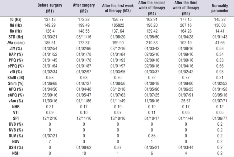
In the acoustic measures of MDVPA, an increase of most frequency perturbation measures after surgery and values reduction along the speech therapy sessions were veriied (Table 1).
amplitude perturbation quotient (sAPQ) (%);
noise-to-harmonics ratio (NHR); voice turbulence index (VTI); soft phonation index (SPI); number of sub-harmonic segments (NSH); degree of sub-harmonics (DSH) (%);
degree of voice breaks (DVB) (%); number of voice breaks (NVB) (%); degree of unvoiced segments (DUV)
(%); number of unvoiced segments (NUV); variation of amplitude (vAm) (%); variation of fundamental frequency (vf0) (%); fundamental frequency (f0) (Hz); f0 high (fhi) (Hz); f0 low (lo) (Hz); f0 standard deviation (STD) (Hz) (LIMA, 2013). Thus, the aperiodicity/noise level, the stability level and the vocal signal frequency
level were analyzed.
Table 1. Acoustic measures of the Multi Dimension Voice Program Advanced in the ive stages of vocal evaluations
Before surgery (M1)
After surgery (M2)
After the irst week of therapy (M3)
After the second week of therapy
(M4)
After the third week of therapy
(M5)
Normality parameter
f0 (Hz) 137.13 172.32 156.77 162.91 177.15 145.22
fhi (Hz) 149.29 199.49 185822 196.20 207.16 150.08
lo (Hz) 126.4 148.55 137. 64 139.42 164.28 14.41
STD (Hz) 01/03/21 05/11/16 01/06/20 01/05/50 01/04/28 01/01/43
Jita(usec) 185.51 172.37 199.90 210.33 102.10 41.66
Jitt(%) 01/02/54 01/02/96 03/12/16 01/03/42 01/08/16 0.58
RAP (%) 01/01/52 01/01/78 01/01/84 02/05/16 01/09/16 0.34
PPQ (%) 01/01/45 01/01/78 01/01/93 02/09/16 01/09/16 0.33
sPPQ (%) 01/01/64 01/01/87 01/01/97 02/08/16 01/04/16 0.56
vf0 (%) 01/02/34 01/02/97 01/03/95 01/03/37 01/02/42 0.93
ShdB (dB) 0.58 0.63 0.70 0.72 0.77 0.21
Shim (%) 01/06/68 01/07/27 01/08/56 01/08/18 01/09/00 01/02/52
APQ (%) 01/04/50 01/04/48 06/12/16 01/05/96 01/06/25 01/01/98
sAPQ (%) 05/09/16 01/05/47 01/07/63 01/07/25 01/07/91 03/05/16
vAm (%) 11/03/16 01/11/86 01/11/49 11/08/16 25.67 01/07/71
NHR 0.21 0.17 0.19 0.19 0.17 0.12
VTI 0.09 0.10 0.07 0.11 0.06 0.05
SPI 12/12/16 12/11/16 13/10/16 01/10/17 01/11/44 01/06/77
DVB (%) 0 0 0 0 0 0.2
NVB (%) 0 0 0 0 0 0.2
DUV (%) 01/07/21 0 0 0.86 0 0.2
NUV 7 0 0 1 0 0.2
DSH (%) 0 01/08/62 0.87 01/05/21 01/03/44 0.2
NSH 0 10 1 6 4 0.2
Legend: M:moment; Hz: hertz; dB: decibel; f0: fundamental frequency; fhi: f0 high; lo: f0 low; STD: standard deviation of f0; Jita: absolute Jitter; Jitt: percentual or
relative Jitter; RAP: relative average of perturbation of pitch; PPQ: pitch or frequency perturbation quotient; sPPQ: soft pitch or frequency perturbation quotient; vf0:
variation of f0; ShdB: absolute Shimmer or in dB; Shim: percentual or relative Shimmer; APQ: amplitude perturbation quotient; sAPQ: soft amplitude perturbation
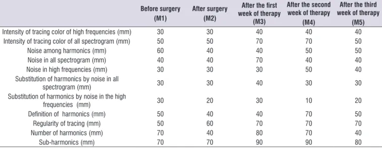
Table 2. Wide-band spectrographic evaluation in the ive stages of vocal evaluation
Before surgery (M1)
After surgery (M2)
After the irst week of therapy
(M3)
After the second week of therapy
(M4)
After the third week of therapy
(M5)
Intensity of tracing color of F1 (mm) 70 70 90 90 90
Intensity of tracing color of F2 (mm) 60 70 90 80 100
Intensity of tracing color of F3 (mm) 50 60 80 70 90
Intensity of tracing color of F4 (mm) 40 30 30 70 80
Intensity of tracing color of high frequencies (mm) 30 30 60 70 70 Intensity of tracing color of all spectrogram (mm) 50 50 70 70 80
Noise in all spectrogram (mm) 60 40 30 40 50
Noise in high frequencies (mm) 60 40 30 30 30
Deinition of F1 (mm) 80 50 80 80 80
Deinition of F2 (mm) 70 40 80 80 80
Deinition of F3 (mm) 50 60 80 70 90
Deinition of F4 (mm) 0 0 10 40 10
Tracing regularity (mm) 60 70 80 70 80
Legend: M: moment; F: formant; mm: millimeters
In the acoustic spectrographic evaluation, an improvement of most analyzed aspects along therapy; tracing regularity improvement, as in the narrow-band spectrogram, as in the wide-band spectrogram; and an increase of tracing F2 and F3 intensity, after surgery. F deinition worsening after surgery was observed, with gradual improvement during speech therapy (Tables 2 and 3).
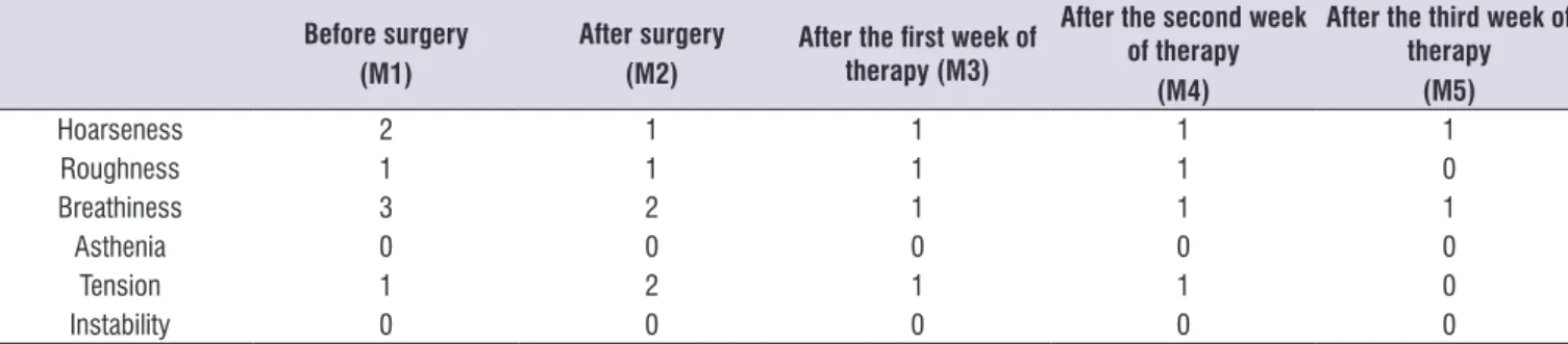
The perceptive-auditory vocal analysis showed hoarseness breathiness improvement, after the surgery; also after the speech therapy sessions, as well as tension worsening following the surgery, with gradual improvement along speech therapy (Table 4).
Table 3. Narrow-band spectrographic evaluation in the ive stages of vocal evaluation
Before surgery (M1)
After surgery (M2)
After the irst week of therapy
(M3)
After the second week of therapy
(M4)
After the third week of therapy
(M5)
Intensity of tracing color of high frequencies (mm) 30 30 40 40 40 Intensity of tracing color of all spectrogram (mm) 50 50 70 70 50
Noise among harmonics (mm) 60 40 40 50 50
Noise in all spectrogram (mm) 40 40 70 40 40
Noise in high frequencies (mm) 30 30 30 50 40
Substitution of harmonics by noise in all
spectrogram (mm) 30 30 40 30 30
Substitution of harmonics by noise in the high
frequencies (mm) 30 20 30 10 20
Deinition of harmonics (mm) 50 40 40 70 50
Regularity of tracing (mm) 50 60 70 70 70
Number of harmonics (mm) 70 40 80 70 40
Sub-harmonics (mm) 70 70 90 90 80
Table 4. Vocal perceptive-analysis in the ive stages of vocal evaluation
Before surgery (M1)
After surgery (M2)
After the irst week of therapy (M3)
After the second week of therapy
(M4)
After the third week of therapy
(M5)
Hoarseness 2 1 1 1 1
Roughness 1 1 1 1 0
Breathiness 3 2 1 1 1
Asthenia 0 0 0 0 0
Tension 1 2 1 1 0
Instability 0 0 0 0 0
Legend: M: moment
DISCUSSION
The approach of Hearing, Speech and Language therapists in cases of vocal fold paralysis, before and after surgery, is important, because speech therapy provides better balance control between the respi-ratory and the phonatory systems, it avoids ineffective phonatory compensation processes, gives more stability in the vocal folds mucosa vibration and accel-erates the patient’s rehabilitation 9.
The present case study reinforces the importance of speech therapy through systematic exercises performed in a short period (three weeks), because most frequency perturbation measures (Jitt, RAP, PPQ and sPPQ) increased immediately after the surgery (M2), but they started to decrease along the speech therapy sessions. This information conirms that this surgery promotes vocal folds approximation. However, individually, it does not ensure noise decrease and emission stability, which did not occur through systematic speech therapy after surgery (Table 1).
Thus, speech therapy through phonation into a tube provided more harmonic energy, noise decrease and higher emission stability. This type of therapy corroborates the improvement of most spectrographic evaluation aspects, such as tracing color intensity in wide-band spectrogram, after speech therapy (M5); tracing regularity, either in wide-band spectrogram or in narrow-band spectrogram improved after surgery and increased along speech therapy (Table 2). The F deinition got worse after the surgery, but, along the speech therapy sessions, it improved gradually (Table 2). These results suggest harmonic energy increase, aperiodic energy reduction and improvement of loudness and vocal projection 1,7. This information is conirmed by a research on the effects of the technique of phonation into a glass tube immersed in water in
women without complaints and with no laryngeal diseases 7.
The amplitude perturbation measures increased after the surgery (M2) and after the speech therapy sessions (M5). The presence of sub-harmonics, in the wide-band spectrogram, which did not present alterations after the surgery, worsened after the speech therapy sessions (Table 3). These results were contra-dictory when compared with the perceptive-auditory evaluation, which evidenced reduction of breathiness (Table 4), because, according to the literature, the measures of amplitude perturbation are more related to breathiness. However, it must be considered that the emissions of individuals with unilateral vocal fold paralysis are approached as aperiodic and that, even after surgery and speech therapy, there is permanent damage of the glottic source, mainly in relation to the closure of rima glottidis, either considering the closure completeness, or considering the irmness aspect. This fact may justify such acoustic indings, although they are not auditorily perceived. In these cases, the perceptive-auditory evaluation is extremely important 9.
The subharmonic segments measures (DSH and NSH) increased after the surgery (M2) and then decreased. The measures of the unvoiced segments (NUV and DUV) also decreased signiicantly after the surgery (Table 1). It probably happened due to better and greater glottic coaptation and because of mucosa mobilization. This inding corroborates the decrease of frequency perturbation measures and the reduction of breathiness. There was also noise reduction in the high frequencies and in all the spectrogram (Table 3) after the surgery and after the irst week of therapy. This improvement was maintained along the therapy sessions only in the high frequencies 1,4,9.
promotes activation of the thyroarytenoid, cricothyroid and lateral cricoarytenoid muscles 14. In the present case study, this action was inferred because of the positive results obtained through the speech therapy sessions, which suggests higher muscular conditioning of the healthy vocal fold.
Adopting the same view of the present study, a retro-spective research with 12 individuals with vocal fold unilateral paralysis evidenced, after speech treatment, signiicant improvement of breathing, followed by degree of dysphonia and asthenia. However, this study did not mention the vocal techniques used 1.
The results of the present investigation, in relation to vocal acoustic analysis, corroborate most of the literature about speech therapy in cases of unilateral vocal fold paralysis. The following beneits are expected: higher glottic closure, mucosa mobilization improvement, improvement of perceptive-auditory vocal alterations, reduction of acoustic measures jitter, shimmer, noise-to-harmonics ratio, f0 measures, vocal breaks, unvoiced segments and sub-harmonics 4.
In relation to the perceptive-auditory analysis, there was an increase in tension, after the surgery (M2), which reduced through the speech therapy sessions. There was also roughness improvement after the last speech therapy session (M5) (Table 4). Before the surgery (M1), the muscular hypertension was an expected compensatory mechanism of the patient, owing to the laryngeal gap, although the vocal tension perceptive-auditory aspect was classiied as discrete, because the extreme breathiness probably masked the tension in the perceptive-auditory analysis. After the surgery (M2), with higher glottic coaptation, it is probable that the patient maintained the compensatory mechanism. The tension was classiied as moderate, because it was more perceptible. Along speech therapy, the tension degree reduced and, at the end of the treatment, it was eliminated. Voice roughness is also justiied in the presence of the perceptive-auditory aspect of tension, and it was eliminated after the speech therapy sessions (M5) (Table 4) 12.
A study involving 24 men with unilateral paralysis of vocal fold (average age of 60.7 years) investigated the perceptive-auditory vocal characteristics through the RASATI scale and the paralyzed vocal fold position. Predominance of hoarseness, roughness and tension (higher frequency of moderate degree), breathiness (higher frequency of severe degree), asthenia and instability (higher frequency of mild degree) and general degree of high vocal disorder was veriied 2.
Another research, with the same sample, investigated the vocal perceptive-auditory characteristics after type I thyroplasty. There was a reduction of hoarseness, roughness and breathiness (higher frequency of mild degree); asthenia (higher frequency from mild degree to normal); tension and instability (higher frequency of normality). This inding agrees with the results of the present case study, except for breathiness, which was moderate after the surgery 15.
A patient diagnosed with right vocal fold paralysis, in paramedian position, was submitted to speech therapy, ive sessions of one hour each, once a week. The subject was instructed to perform the same techniques ive times a day at home. It must be highlighted that several techniques of different categories were used for treatment, such as laryngeal massage, yawn-sigh technique, reverse phonation, prolonged /b/, basal sound and nasal sounds, emission in maximum phonation time (MPT) and with frequencies variation. At the end of the therapy sessions, the type of voice was considered normal or adapted and the extensions of voice were spoken and sung. The vocal self-evaluation scores and the acoustic analysis data were normal, partially corroborating the present results 10.
According to the literature, the SOVTEs provide an increase of intraoral pressure and, thus, more contact of the vocal folds mucosa, with lower tension. Moreover, they increase the intrinsic laryngeal muscular activity, mucous wave and vibration stability 6,7,14. These aspects may justify the obtained and discussed results of the present study.
In this case study, there was also an increase in frequency measures, mainly after the surgery, which decreased after the irst speech therapy day and increased, signiicantly, at the end of the treatment (172Hz). Such result may show a compensation of the discrete abductor strength of the cricothyroid muscle, because of the balanced closure dificulty, from a permanently impaired glottic source 3,8,9.
diseases with more details, in relation to the perfor-mance of speech therapy techniques.
FINAL COMMENTS
The systematic speech therapy for 18 days, through three different techniques of phonation into a tube, in a male adult patient, presented with left vocal fold paralysis, surgically medialized, provided improve-ments of most frequency perturbation measures, voiceless or not sounded components, wide-band spectrogram tracing color intensity, spectrographic tracing regularity and formants deinition. Furthermore, in the perceptive-auditory analysis, there was tension, roughness and breathiness reduction. The main indings of this case study evidenced harmonic energy increase, aperiodic energy decrease, stability increase and vocal projection increase. Such results suggest that the type of speech therapy used in the present study, may be beneicial in cases of vocal fold unilateral paralysis.
REFERENCES
1. Gama CC, Faria AP, Bassi IB, Diniz SS. Alteração de mobilidade de prega vocal unilateral: avaliação subjetiva e objetiva da voz nos momentos pré e pós-fonoterapia. Rev. CEFAC. 2011;13(4):710-8.
2. Schwarz K, Cielo CA, Steffen N, Jotz GP, Becker J. Voz e posição de prega vocal em homens com paralisia unilateral de prega vocal. Braz J Otorhinolaryngol. 2011a;77(6):561-7.
3. Kašterović B, Veselinović M, Mitrović SM. Voice therapy and assistive techniques in voice disorders caused by unilateral vocal cord pareses. Med Pregl. 2014;67(3-4):91-6.
4. Cielo CA, Beber BC, Schwarz K. Evidências cientíicas de fonoterapia para as paralisias unilaterais de prega vocal – Revisão sistemática. In: Terapia fonoaudiológica baseada em evidências. Pró-fono. 2013. p. 421-40.
5. Costa CB, Costa LHC, Oliveira G, Behlau M. Efeitos imediatos do exercício de fonação em canudo. Rev Bras Otorrinolaringol. 2011;77(4):461-5.
6. Cielo CA, Lima JPM, Christmann MK, Brum R. Exercícios de trato vocal semiocluído: revisão de literatura. Rev. CEFAC. 2013;15(6):1679-89.
7. Lima JPM. Modiicações vocais e laríngeas imediatas em mulheres após a técnica de fonação em tubo de vidro imerso em água. [Dissertação] Santa Maria (RS): Universidade Federal de Santa Maria; 2013.
8. Young VVN, Smith LJ, Rosen C. Voice outcome following acute unilateral vocal fold paralysis. Ann Otol Rhinol Laryngol. 2013;122(3):197-204.
9. Behlau M. Voz: O livro do Especialista. Rio de Janeiro: Revinter, 2008.
10. Oliveira AG, Pinho MM. Amiotroia Nevrálgica Estendida: fonoterapia em um caso de paralisia de prega vocal. CoDAS. 2014;26(2):175-80.
11. Sampaio M, Oliveira G, Behlau M. Investigação de efeitos imediatos de dois exercícios de trato vocal semiocluído. Pro Fono R Atual Cient. 2008;20(5):261-6.
12. Pinho SEM, Pontes PAL. Escala de avaliação perceptiva da fonte glótica: RASAT. Vox Brasilis. 2002;3(1):11-3.
13. Valentim AF, Côrtes MG, Gama AC. Análise espectrográica da voz: efeito do treinamento visual na coniabilidade da avaliação. Rev Soc Bras Fonoaudiol. 2010;15(3):335-42.
14. Laukkanen AM, Titze IR, Hoffman HH, Finnegan E. Effects of a semioccluded vocal tract on laryngeal muscle activity and glottal adduction in a single female subject. Folia Phoniatr Logop. 2008;60(6):298-311.