Lung alterations in a rat model of diabetes mellitus:
effects of antioxidant therapy*
,**
Alterações pulmonares em um modelo de diabetes mellitus em ratos: o efeito da terapia antioxidante
Luiz Alberto Forgiarini Junior, Nélson Alexandre Kretzmann, Juliana Tieppo, Jaqueline Nascimento Picada, Alexandre Simões Dias, Norma Anair Possa Marroni
Abstract
Objective: To evaluate structural alterations of the lung in rats with diabetes mellitus (DM), by quantifying oxidative stress and DNA damage, as well as to determine the effects that exogenous superoxide dismutase (SOD) has on such alterations. Methods: A controlled experimental study involving 40 male Wistar rats, divided into four groups (10 animals each): control; SOD-only (without DM but treated with SOD); IDM-only (with streptozotocin-induced DM but untreated); and IDM+SOD (with streptozotocin-streptozotocin-induced DM, treated with SOD). The animals were evaluated over a 60-day period, day 0 being defined as the day on which the streptozotocin-injected animals presented glycemia > 250 mg/dL. The SOD was administered for the last 7 days of that period. At the end of the study period, samples of lung tissue were collected for histopathological analysis, evaluation of tissue oxidative stress, and assessment of DNA damage. Results: There were no significant differences among the groups regarding DNA damage. In the IDM-only group, there was a significant increase in the extracellular matrix and significantly greater hyperplasia of the capillary endothelium than in the SOD-only and control groups. In addition, there were significant changes in type II pneumocytes and macrophages, suggesting an inflammatory process, in the IDM-only group. However, in the IDM+SOD group, there was a reduction in the extracellular matrix, as well as normalization of the capillary endothelium and of the type II pneumocytes. Conclusions: Exogenous SOD can reverse changes in the lungs of animals with induced DM.
Keywords: Diabetes mellitus, experimental; Oxidative stress; Lung; DNA damage.
Resumo
Objetivo: Avaliar as alterações estruturais no pulmão de ratos com diabetes mellitus (DM) através da quantificação do estresse oxidativo e do dano ao DNA, assim como determinar os efeitos de superóxido dismutase (SOD) exógena nessas alterações. Métodos: Estudo experimental controlado com 40 ratos Wistar, divididos em quatro grupos (10 animais cada): grupo controle, grupo SOD (sem DM e tratados com SOD), grupo DM (com DM induzido por estreptozotocina), e grupo DM+SOD (com DM induzido por estreptozotocina e tratados com SOD). Os animais foram avaliados por um período de 60 dias, iniciado a partir do dia em que os animais com diabetes induzido por estreptozotocina apresentaram glicemia > 250 mg/dL. Nos últimos 7 dias do período, os animais nos grupos tratados receberam SOD. Ao final do tempo de estudo, amostras de tecido pulmonar foram coletadas para análise histopatológica e avaliação do estresse oxidativo e do dano ao DNA. Resultados: Não houve diferenças significativas entre os grupos em relação ao dano ao DNA. Houve um aumento significativo na matriz extracelular e hiperplasia do endotélio capilar no grupo DM quando comparado com os grupos controle e SOD. Também houve mudanças significativas em pneumócitos tipo II e macrófagos intravasculares, sugerindo um processo inflamatório no grupo DM. Entretanto, uma redução na matriz extracelular, endotélio capilar normal e pneumócitos tipo II normais foram encontrados no grupo com DM+SOD. Conclusões: A administração exógena de SOD pode reverter alterações nos pulmões de animais com DM induzido.
Descritores: Diabetes mellitus experimental; Estresse oxidativo; Pulmão; Dano ao DNA.
* Study carried out at the Federal University of Rio Grande do Sul, Porto Alegre, Brazil.
Correspondence to: Luiz Alberto Forgiarini Junior. Rua Wenceslau Escobar, 1086, apto. 916, Tristeza, CEP 91900-000, Porto Alegre, RS, Brasil.
Tel 55 51 3269-0663. E-mail: lafjunior@click21.com.br
Financial support: This study received financial support from the Fundo de Incentivo à Pesquisa (FIPE, Research Incentive Fund) of the Hospital de Clínicas de Porto Alegre.
Submitted: 22 April 2010. Accepted, after review: 24 May 2010.
Methods
This was a controlled study involving an experimental rat model of streptozotocin-induced DM and exogenous administration of SOD.
The study included 40 male Wistar rats (mean weight, 250 g) obtained from the animal facilities of the Federal University of Rio Grande do Sul, located in the city of Porto Alegre, Brazil. The rats were housed in propylene boxes, 5 animals per box, in a temperature-controlled environment (mean temperature, 24°C), and were maintained on a 12/12-h light/dark cycle, with free access to water and rat chow. All animals were treated in accordance with the World Health Organization Ethical Code for Animal Experimentation guidelines.
The animals were randomly assigned to four groups (10 per group): control; SOD-only (without DM but treated with SOD); IDM-only (with streptozotocin-induced DM but untreated); and IDM+SOD (with streptozotocin-induced DM and treated with SOD).
We induced DM with a single intraperitoneal injection of streptozotocin (70 mg/kg; Sigma Chemical, St. Louis, MO, USA) dissolved in a 10 mmol/L sodium citrate solution (pH = 4.5).
(11) Five days after the injection, glycemia was
measured. Animals presenting hyperglycemia (fasting glycemia > 250 mg/dL) were considered
to have developed DM.(12) Glycemia was
determined with a colorimetric enzymatic assay (Enzi-Color kit; Bio Diagnóstica, Pinhais, Brazil), in which a reagent is mixed with 20 mL of the plasma sample, and the product was subsequently read at a wavelength of 500 nm with a spectrophotometer (CARY 3E; Varian, Palo Alto, CA, USA).
The day on which the streptozotocin- injec ted animals presented glycemia > 250 mg/dL was considered day 0 of the 60-day study period. For the last 7 days of the experiment, SOD
(Ontosein®; Tedec Meiji Farma, Alcalá de Henares,
Spain) was administered subcutaneously at a
dose of 13 mg/kg of body weight.(13) On day 60,
the animals were anesthetized with ketamine (100 mg/kg) and xylazine (50 mg/kg) i.p., after which they were killed. Samples of lung tissue were collected for histopathological analysis and evaluation of tissue oxidative stress. The remaining material was collected, immediately
Introduction
Diabetes mellitus (DM) is an endocrine and metabolic disease that affects various organs, including the lungs. The main clinical manifestation is hyperglycemia, which triggers oxidative stress by increasing the production of mitochondrial superoxide anion and the non-enzymatic glycosylation of proteins, as well as by activating various cell transcription factors.(1)
The impact of DM on the respiratory tract is characterized by abnormalities in pulmonary function, such as a reduction in lung elastic recoil and in lung volumes, as well as diminished
diffusing capacity.(2,3) Experimental studies
have demonstrated that pulmonary venous resistance is elevated two weeks after the onset of the disease, and that hyperglycemia induces oxidative stress in lung tissue. Analysis of the lung tissue of animals with streptozotocin-induced diabetes has shown an increase in the thickness of the basement membrane of the lungs. In addition, animal models have shown that diabetic individuals present a deficit in the expression of proteins, including surfactant A, as
well as the hydrophobic surfactants B and C.(4)
Studies evaluating the use of antioxidant therapy in order to reduce pulmonary oxidative stress in animal models of diabetes have shown the potential effects of various antioxidants, such as aminoguanidine, simvastatin, and
N-acetylcysteine, in rats and mice.(5-7) Orgotein,
a superoxide dismutase (SOD) first described in 1969 by McCord & Fridovich, acts on the
inflammatory process and tissue lesion.(8) The
exogenous administration of this antioxidant eliminates the extracellular superoxide radical anions and maintains cell integrity. This reduces the number of acute inflammatory events and
influences late effects.(9) Orgotein also has other
effects that have yet to be well described in the literature, such as the role it plays in the process of cell aging, apoptosis, and viral replication, as well as its analgesic effects.(10)
incubated at 90°C for 30 min. Subsequently, 500 µL of a solution of 0.37% thiobarbituric acid were added to a 15% trichloroacetic acid solution and centrifuged at 2,000 rpm at 4°C for 15 min. Absorbance was determined
by spectrophotometry at 535 nm.(16) The
determination of SOD activity was performed in accordance with the method devised by Mirsa
& Fridovich, which is based on the inhibition of SOD in the formation of adrenochrome in the
autoxidation of epinephrine.(17) Catalase activity
was determined by means of a method that consists in the addition of 955 mL of phosphate
buffer to 25 mL of the lung tissue sample.(18)
Subsequently, 20 mL of hydrogen peroxide were added, and the sample was read at a wavelength of 240 nm by spectrophotometry. The results are expressed as pmol/g of protein.
The lung tissue samples were collected and immersed in a 10% formaldehyde solution for 12 h, after which they were transferred to a 70% alcohol solution. The samples were
then stained with picrosirius red and H&E.
The anatomopathological examination was performed by a pathologist of the Pathology
Laboratory of the Porto Alegre Hospital de
Clínicas who was blinded to the animal groups. The lung tissue was immediately fixed with 2% glutaraldehyde, and the samples were post-fixed with buffered 1% osmium tetroxide, followed by contrast enhancement in 2% uranyl acetate block. Subsequently, the tissue was dehydrated in a graded alcohol series. The material was pre-soaked in Epon 812 (Shell Chemical Co., New York, NY, USA) with pure acetone at decreasing proportions of 75%, 50%, and 25%. Samples were then soaked in pure Epon 812 for 24 h, after which the material was polymerized in an oven at 60°C for 72 h.
frozen in liquid nitrogen, and stored at −80°C
for subsequent biochemical assay.
Blood samples were obtained from the retro-orbital plexus,(14) via the orbital sinus,
and placed into a test tube with heparin
(Liquemine®; Roche,. Neuilly-sur-Seine, France)
in order to avoid coagulation. The material was then centrifuged at 4,000 rpm for 15 min. The precipitate was discarded, and the plasma was withdrawn with a pipette (Labsystems 4500, 200/100 mL; Labsystems, Helsinki, Finland) and
frozen at −80°C for subsequent determination
of glucose levels.
The lungs were dissected out and stored
at −80°C for the subsequent quantification of
thiobarbituric acid reactive substances (TBARS) and assessment of antioxidant (SOD and catalase) activity.
Lung fragments were immersed in saline solution at 2°C. Each fragment was then placed into a homogenization tube with PBS (KCl, 140 mmol/L; phosphate, 20 mmol/L; pH = 7.4; 9 mL per gram of tissue) and homogenized in an Ultra-Turrax homogenizer (IKA Labortechnik, Staufen, Germany) at 2°C for 30 s. The homogenate was centrifuged at 3,000 rpm for 10 min (Sorvall RC-5B refrigerated superspeed
centrifuge; DuPont, Wilmington, DE, USA).(14)
The supernatant was transferred to Eppendorf tubes, and the precipitate was discarded.
A solution of bovine albumin (Sigma Chemical) was used at a concentration of 1 mg/mL, in accordance with the method devised by Lowry et al.(15) The samples were read at a
wavelength of 625 nm in the spectrophotometer, and the results are expressed in mg/mL.
We quantified the lipid peroxidation product by determining the TBARS levels in 3 mg of proteins per sample analyzed. The samples were
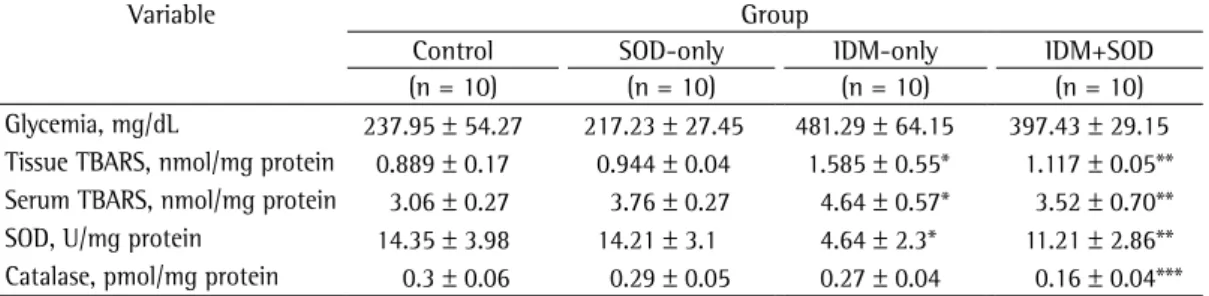
Table 1 - Oxidative stress and antioxidant enzyme activity, by group.
Variable Group
Control SOD-only IDM-only IDM+SOD
(n = 10) (n = 10) (n = 10) (n = 10)
10 mM Tris; pH = 10.0), with freshly added 1% Triton X-100 (Sigma) and 10% DMSO at 4°C for 48 h. The slides were subsequently incubated in freshly prepared alkaline buffer (300 mM NaOH and 1 mM EDTA; pH > 13) at 4°C for 20 min. An electric current of 300 mA and 25 V (0.90 V/cm) was applied for 15 min in order to perform DNA electrophoresis. The slides were then neutralized (0.4 M Tris; pH = 7.5), stained with silver, and analyzed in a microscope. Images of 100 randomly selected cells (50 cells from each of two replicate slides) were analyzed for each animal. Cells were also visually scored, by tail size, into five classes, ranging from undamaged
(0 points) to maximally damaged (4 points),(4)
resulting in a single DNA damage score for each animal and a mean score for each group. The damage index (DI) could range from 0 (completely undamaged; 100 cells × 0) to 400 (completely damaged; 100 cells × 4). The damage frequency (DF; in %) was calculated based on the number of tailed vs. tailless cells.(20)
Semi-fine sections (800 nm) were obtained with an ultramicrotome (Leica Ultracut UCT; Leica Microsystems Inc., Bannockburn, IL, USA) and stained with an aqueous solution of 1% toluidine blue and basic fuchsin. Ultrafine sections (70 nm) were also obtained. For contrast enhancement of the ultrafine sections, we used 2% uranyl acetate, followed by lead citrate. Those sections were then examined in an electron microscope (model EM208S; Philips, Eindhoven, The Netherlands) at high magnification (×10,000).
Alkaline single-cell gel electrophoresis (comet
assay) was carried out as previously described,(19)
with minor modifications.(20,21) Each lung
fragment was placed into a tube with 0.5 mL cold PBS and finely minced in order to obtain a cell suspension. Lung suspensions (5 µL) were embedded in 95 µL of a 0.75% low melting point agarose solution (Gibco-BRL, Rockville, MD, USA) and spread onto agarose-precoated slides. After solidification, slides were placed in lysis buffer (2.5 M NaCl, 100 mM EDTA, and
Results
The levels of TBARS were significantly higher in the lung tissue samples collected from animals in the IDM-only group than in those collected from animals in the control and SOD-only groups, whereas they were significantly lower in The oxidative stress data were analyzed with
the Statistical Package for the Social Sciences, version 13.0 (SPSS Inc., Chicago, IL, USA), with ANOVA followed by the Student-Newman-Keuls test. The comet assay data were analyzed with ANOVA followed by Tukey’s test. The level of significance was set at 5%.
a
b
c Control
Control
Control
SOD-only
SOD-only
SOD-only
IDM-only
IDM-only
IDM-only
IDM+SOD
IDM+SOD
IDM+SOD
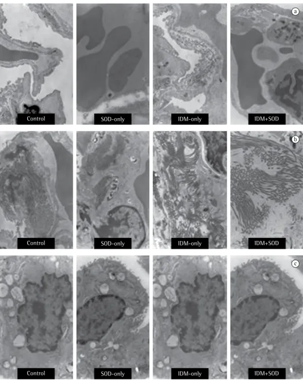
the control and SOD-only groups. When type II pneumocytes were analyzed, the animals in the IDM+SOD group presented lamellar bodies.
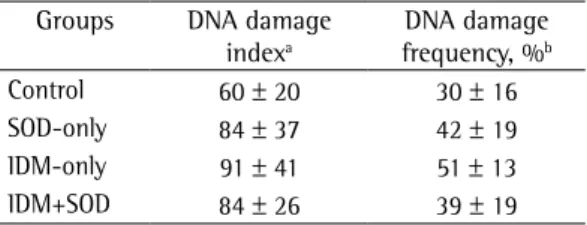
There were no significant differences in DI or DF between the control group and the IDM-only group or between the SOD-only group and the IDM+SOD group (Table 2).
Discussion
The present study investigated the effect of exogenous SOD in a rat model of DM. Although SOD did not decrease the plasma glucose levels in the rats with streptozotocin-induced DM, it did reduce lipid peroxidation, as evaluated by determining TBARS levels in the lung tissue and blood, and it also reduced the alterations to the lung structure in the rats with streptozotocin-induced DM. We observed no decrease in DNA damage in the lung tissue, there being no significant increases in ID or DF in the IDM-only group.
The antioxidant therapy employed here proved effective in reducing pulmonary oxidative stress as well as in activating the mechanisms of antioxidant enzymes (SOD and catalase). The changes in the mechanism of action of these enzymes seem to be crucial in the development of pulmonary alterations induced by DM. The SOD enzyme plays a crucial role in tissue protection because it acts as a scavenger of the superoxide anion, preventing the formation of other more potent oxidants, such as peroxynitrite
and the hydroxyl radical.(22) In a previous study,
antioxidant therapy was shown to decrease pulmonary oxidative stress in an experimental
rat model of hepatopulmonary syndrome.(23)
those collected from animals in the IDM+SOD group than in those collected from animals in the IDM-only group (Table 1).
The lipid peroxidation analysis of the blood samples showed that serum TBARS levels were significantly higher in the IDM-only group than in the control and SOD-only groups, whereas they were significantly lower in the blood samples collected from animals in the IDM+SOD group than in those collected from animals in the IDM-only group.
In the lung tissue samples, SOD levels were significantly lower in the IDM-only group than in the control and SOD-only groups. However, the SOD levels in the IDM+SOD group lung tissue samples were significantly higher than were those observed in the IDM-only group samples.
In terms of catalase activity, the IDM-only group did not differ significantly from the control and SOD-only groups. However, catalase activity was significantly lower in the IDM+SOD group than in any of the three other groups.
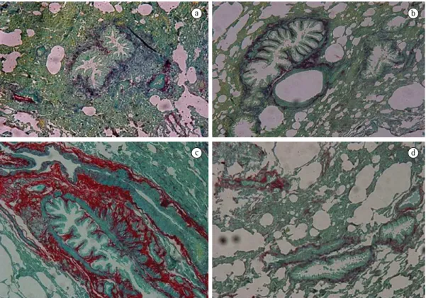
The analysis of the histological slides revealed intravascular macrophages, which suggests the presence of an inflammatory process in the tissue, in the IDM-only group but not in the control and SOD-only groups. In the IDM-only group, there was also an increase in the extracellular matrix, as evidenced by the presence of fibrosis, as well as an increase in the thickness of the alveolar-capillary membrane. However, in the lungs of animals in the IDM+SOD group, the pulmonary capillary endothelium was similar to that observed in the control and SOD-only group animals. In addition, the lungs of the IDM+SOD group animals showed a reduction in the extracellular matrix (less fibrosis) and in the number of intravascular macrophages (Figure 1).
The ultrastructural analysis of the lung tissue of the animals in the IDM-only group showed thickening of the basement membrane, fibrosis of the lung interstitium, disorganization of type II pneumocytes, and the absence of lamellar bodies, suggesting that surfactant production was reduced in the IDM-only group animals (Figure 2). These alterations were not significant in the control and SOD-only groups, whereas there was a reduction in the thickness of the basement membrane in the IDM+SOD group, which was similar to the results obtained for
Table 2 - DNA damage index and damage frequency, by group.
Groups DNA damage indexa
DNA damage frequency, %b
Control 60 ± 20 30 ± 16 SOD-only 84 ± 37 42 ± 19 IDM-only 91 ± 41 51 ± 13 IDM+SOD 84 ± 26 39 ± 19 SOD: superoxide dismutase; and IDM: induced diabetes mellitus. aRange, 0 (completely undamaged) to
400 (completely damaged). bCalculated based on number
administration of aminoguanidine, which plays a vasoregulatory role, there was an increase in the release of prostacyclin, with a mild effect on endothelium-dependent vasodilatation, followed by regulation of the pulmonary circulation
release of aminoguanidine.(5) However, in a
study examining the role that endothelial nitric oxide synthase (eNOS) plays in vascular remodeling in streptozotocin-induced diabetic mice, DM was found to accelerate the process of oxidative stress-related vascular remodeling and
to increase eNOS expression.(27)
Various studies have shown an increase in the oxidative damage to the DNA in the peripheral blood lymphocytes of diabetic patients or
of streptozotocin-induced diabetic rats.(28-30)
However, few studies have evaluated the DNA damage in other target tissues associated with
complications of DM.(29) In the present study,
there were no increases in the DNA damage in the lungs (Table 2) or in the peripheral blood (data not shown) of the rats with streptozotocin-induced DM. It is of note that DNA damage has been related to hyperglycemia in diabetic patients.(28,30) In the present study, the
administration of SOD did not decrease glycemia in rats. This could explain the fact that SOD treatment had no effect on the degree of DNA damage in the rats with streptozotocin-induced DM. In addition, the increase in oxidative stress was not sufficient to increase the degree of DNA damage in the lung tissue samples evaluated. Furthermore, some of the oxidative damage to the DNA was probably induced by DM. That damage was repaired, and no significant increase was observed.
The mechanism by which elevated glucose levels cause vascular injury and result in structural and functional alterations in various tissues can be multifactorial, the most prominent factors being the role of oxidative stress, the increase in the synthesis/accumulation of diacylglycerol, the activation of protein kinase C, the increased activation of the sorbitol pathway of glucose metabolism, the non-enzymatic glycosylation of proteins, and the relative or absolute alterations in the production of vasoactive substances, such as endothelin, prostaglandins, and nitric oxide byproducts.(4)
In our study, we found that pulmonary oxidative stress was significantly greater in the rats with streptozotocin-induced DM than in the In the present study, the administration of
SOD had no effect on plasma glucose levels, a finding that corroborates the results of another study, in which the use of mimetic SOD was evaluated in an experimental model of diabetes in rats.(24)
We observed an increase in pulmonary oxidative stress in the rats with streptozotocin-induced DM. This finding is probably attributable to the effects that free radicals have on the cell membrane structures in lung tissue, as previously described by another group of authors, who showed an increase in pulmonary oxidative stress in rabbits with alloxan-induced
diabetes.(25) In our study, this was evidenced by
the higher TBARS levels in blood and in lung tissue, suggesting that, in rats with DM, lipid peroxidation occurs within the first 60 days after the onset of the disease. However, after the administration of SOD, lipid peroxidation decreased, and, consequently, the lung tissue of rats with streptozotocin-induced DM showed increased SOD activity after treatment. This beneficial effect of the use of the exogenous antioxidant was described in a previous study
evaluating the antioxidant effect of α-lipoic
acid on oxidative stress and on the expression of inducible nitric oxide synthase in
streptozotocin-induced diabetic rats.(26)
In our study, we found that the rats with streptozotocin-induced DM showed alterations in the ultrastructure of the lung tissue, as assessed by electron microscopy, such as increased thickness of the basement membrane, increased pulmonary fibrosis, and alterations in type II pneumocytes. After those rats had been treated with SOD, the ultrastructure of the lung tissue reverted to near-basal conditions.
One group of authors investigated the effect that treatment with simvastatin has on pulmonary lipid peroxidation and on the activity of antioxidant enzymes, as well as evaluating the lung ultrastructure, in streptozotocin-induced diabetic rats. They showed that such rats develop alterations in the lung ultrastructure and an oxidant imbalance in the lungs, as well as that treatment with simvastatin can improve the antioxidant status and the organization of
the lung structure.(6) Another group of authors,
10. Dinçer Y, Akçay T, Ilkova H, Alademir Z, Ozbay G. DNA damage and antioxidant defense in peripheral leukocytes of patients with Type I diabetes mellitus. Mutat Res. 2003;527(1-2):49-55.
11. Maritim AC, Sanders RA, Watkins JB 3rd. Effects of alpha-lipoic acid on biomarkers of oxidative stress in streptozotocin-induced diabetic rats. J Nutr Biochem. 2003;14(5):288-94.
12. Seguí J, Gironella M, Sans M, Granell S, Gil F, Gimeno M, et al. Superoxide dismutase ameliorates TNBS-induced colitis by reducing oxidative stress, adhesion molecule expression, and leukocyte recruitment into the inflamed intestine. J Leukoc Biol. 2004;76(3):537-44. 13. Dias AS, Porawski M, Alonso M, Marroni N, Collado
PS, González-Gallego J. Quercetin decreases oxidative stress, NF-kappaB activation, and iNOS overexpression in liver of streptozotocin-induced diabetic rats. J Nutr. 2005;135(10):2299-304.
14. Llesuy SF, Milei J, Molina H, Boveris A, Milei S. Comparison of lipid peroxidation and myocardial damage induced by adriamycin and 4’-epiadriamycin in mice. Tumori. 1985;71(3):241-9.
15. Lowry OH, Rosebrough NJ, Farr AL, Randall RJ. Protein measurement with the Folin phenol reagent. J Biol Chem. 1951;193(1):265-75.
16. Buege JA, Aust SD. Microsomal lipid peroxidation. Methods Enzymol. 1978;52:302-10.
17. Misra HP, Fridovich I. The role of superoxide anion in the autoxidation of epinephrine and a simple assay for superoxide dismutase. J Biol Chem. 1972;247(10):3170-5.
18. Chance B, Machley AL. Assays of catalases and peroxidases. Methods Enzymol. 1995;2:764–75. 19. Tice RR, Agurell E, Anderson D, Burlinson B, Hartmann
A, Kobayashi H, et al. Single cell gel/comet assay: guidelines for in vitro and in vivo genetic toxicology testing. Environ Mol Mutagen. 2000;35(3):206-21. 20. Picada JN, Flores DG, Zettler CG, Marroni NP, Roesler R,
Henriques JA. DNA damage in brain cells of mice treated with an oxidized form of apomorphine. Brain Res Mol Brain Res. 2003;114(1):80-5.
21. Rodrigues CR, Dias JH, Semedo JG, da Silva J, Ferraz AB, Picada JN. Mutagenic and genotoxic effects of Baccharis dracunculifolia (D.C.). J Ethnopharmacol. 2009;124(2):321-4.
22. Ceriello A. Oxidative stress and glycemic regulation. Metabolism. 2000;49(2 Suppl 1):27-9.
23. Vercelino R, Tieppo J, Dias AS, Marroni CA, Garcia E, Meurer L, et al. N-acetylcysteine effects on genotoxic and oxidative stress parameters in cirrhotic rats with hepatopulmonary syndrome. Basic Clin Pharmacol Toxicol. 2008;102(4):370-6.
24. Peixoto EB, Pessoa BS, Biswas SK, Lopes de Faria JB. Antioxidant SOD mimetic prevents NADPH oxidase-induced oxidative stress and renal damage in the early stage of experimental diabetes and hypertension. Am J Nephrol. 2009;29(4):309-18.
25. Gumieniczek A, Hopkała H, Wójtowicz Z, Wysocka
M. Changes in antioxidant status of lung tissue in experimental diabetes in rabbits. Clin Biochem. 2002;35(2):147-9.
26. Hürdağ C, Uyaner I, Gürel E, Utkusavas A, Atukeren P, Demirci C. The effect of alpha-lipoic acid on NOS dispersion in the lung of streptozotocin-induced diabetic rats. J Diabetes Complications. 2008;22(1):56-61.
control animals. The rats with streptozotocin-induced DM showed greater lipid peroxidation, higher levels of glycemia, and less SOD activity. In those same rats, we observed alterations in the lung ultrastructure, as evidenced by the increased thickness of the basement membrane, together with pulmonary fibrosis and alterations in type II pneumocytes. We believe that those alterations are interrelated, because they were reduced after the administration of SOD. These results support the hypothesis that the oxidative stress seen in DM can cause alterations in the lung structure and that, after the administration of exogenous SOD, these parameters are restored to their basal conditions.
Our results indicate that treatment with exogenous SOD inhibits lipid peroxidation in streptozotocin-induced diabetic rats and minimizes structural alterations of the lung without causing further damage to the DNA. This suggests that oxidative stress is one of the pathways leading to such alterations and that exogenous antioxidants can be successfully used in the treatment of DM.
References
1. Brownlee M. Biochemistry and molecular cell biology of diabetic complications. Nature. 2001;414(6865):813-20.
2. Goldman MD. Lung dysfunction in diabetes. Diabetes Care. 2003;26(6):1915-8.
3. Walter RE, Beiser A, Givelber RJ, O’Connor GT, Gottlieb DJ. Association between glycemic state and lung function: the Framingham Heart Study. Am J Respir Crit Care Med. 2003;167(6):911-6.
4. Calles-Escandon J, Cipolla M. Diabetes and endothelial dysfunction: a clinical perspective. Endocr Rev. 2001;22(1):36-52.
5. Dewhurst M, Stevens EJ, Tomlinson DR. Effects of aminoguanidine and N(G)-nitro-L-arginine methyl ester on vascular responses of aortae and lungs from streptozotocin-diabetic rats. Prostaglandins Leukot Essent Fatty Acids. 1997;56(4):317-24.
6. Ozansoy G, Güven C, Ceylan A, Can B, Aktan F, Oz E, et al. Effects of simvastatin treatment on oxidant/antioxidant state and ultrastructure of streptozotocin-diabetic rat lung. Cell Biochem Funct. 2005;23(6):421-6.
7. Ohnishi T, Bandow K, Kakimoto K, Machigashira M, Matsuyama T, Matsuguchi T. Oxidative stress causes alveolar bone loss in metabolic syndrome model mice with type 2 diabetes. J Periodontal Res. 2009;44(1):43-51. 8. Esco R, Valencia J, Coronel P, Carceller JA, Gimeno M,
Bascón N. Efficacy of orgotein in prevention of late side effects of pelvic irradiation: a randomized study. Int J Radiat Oncol Biol Phys. 2004;60(4):1211-9.
29. Golubnitschaja O, Moenkemann H, Trog DB, Blom HJ, De Vriese AS. Activation of genes inducing cell-cycle arrest and of increased DNA repair in the hearts of rats with early streptozotocin-induced diabetes mellitus. Med Sci Monit. 2006;12(2):BR68-74.
30. Collins AR, Raslová K, Somorovská M, Petrovská H, Ondrusová A, Vohnout B, et al. DNA damage in diabetes: correlation with a clinical marker. Free Radic Biol Med. 1998;25(3):373-7.
27. Sasaki N, Yamashita T, Takaya T, Shinohara M, Shiraki R, Takeda M, et al. Augmentation of vascular remodeling by uncoupled endothelial nitric oxide synthase in a mouse model of diabetes mellitus. Arterioscler Thromb Vasc Biol. 2008;28(6):1068-76.
28. Lodovici M, Giovannelli L, Pitozzi V, Bigagli E, Bardini G, Rotella CM. Oxidative DNA damage and plasma antioxidant capacity in type 2 diabetic patients with good and poor glycaemic control. Mutat Res. 2008;638(1-2):98-102.
About the authors
Luiz Alberto Forgiarini Junior
Doctoral Student in Pulmonology. Federal University of Rio Grande do Sul, Porto Alegre, Brazil.
Nélson Alexandre Kretzmann
Doctoral Student in Hepatology. Federal University of Health Sciences of Porto Alegre, Porto Alegre, Brazil.
Juliana Tieppo
Researcher. Laboratory of Physiology and Experimental Hepatology, Research Center, Hospital de Clínicas de Porto Alegre, Federal University of Rio Grande do Sul School of Medicine, Porto Alegre, Brazil.
Jaqueline Nascimento Picada
Professor. Postgraduate Program in Genetics and Applied Toxicology, Lutheran University of Brazil, Canoas, Brazil.
Alexandre Simões Dias
Professor. Postgraduate Program in Rehabilitation and Inclusion, Methodist University Center, Porto Alegre Institute, Porto Alegre, Brazil.
Norma Anair Possa Marroni