O
WATER MOVEMENT ACROSS BONDED DENTIN
- TOO MUCH OF A GOOD THING
Franklin R. TAY
Pediatric Dentistry and Orthodontics, Faculty of Dentistry, The University of Hong Kong, China.
Ricardo M. CARVALHO
Department of Operative Dentistry, Endodontics and Dental Materials, Bauru School of Dentistry, University of São Paulo, Brazil.
David H. PASHLEY
Department of Oral Biology and Maxillofacial Pathology, Medical College of Georgia, Augusta, GA, USA.
Bonding of resinous materials to dental hard tissues began in the early 1950s in England by Dr. Oskar Hagger, who developed a monomer based on glycerophosphoric acid dimethacrylate that was chemically-cured with sulphinic acid1. His work led to
the development of Sevitron, an early commercial adhesive2. In the U.S., Dr. Michael Buonocore made
the second, and more important advance in adhesive dentistry, by demonstrating that acid-etching of enamel led to improved resin-enamel bonds using Sevitron-like resin formulations3. His rationale for acid-etching
enamel was that little adhesion was obtained on unetched enamel, which he correctly surmised lacked microscopic porosities for resin infiltration. He knew that concentrated (85 wt%) phosphoric acid was used in industry to pre-treat metal surfaces prior to painting or resin-coating. Thus, it was logical for him to use 85% phosphoric acid for 30 sec to etch enamel, followed by water rinsing. The results of his work were very controversial at the time. Many regarded Dr. Buonocore’s approach as unconventional and reckless because he advocated the use of dangerous, industrial-strength acids in the oral cavity.
Adhesive dentistry has advanced significantly since
the introduction of enamel bonding almost four decades ago. In a recent Buonocore Memorial Lecture, Van Meerbeek discussed that bonding to dental hard tissues can be effectuated through the use of three different approaches: etch&rinse (i.e. total-etch), self-etch or glass-ionomer approach4. The first two approaches
involve the use of dentin adhesives to bond to dentin via micromechanical retention. With the glass-ionomer approach, glass-ionomer cements (GICs) and resin-modified glass-ionomer cements (RMGICs) are applied to dentin after conditioning with polyalkenoic acids. Adhesion is achieved via a combination of micromechanical retention as well as chemical adhesion5. It is now known that hybrid layer formation
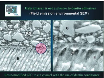
is not exclusive to the use of dentin adhesives, as a similar layer develops via demineralization and ion exchange when GICs and RMGICs are applied to enamel (Figure 1) and dentin6-8.
It is not the intention of this paper to provide an extensive review on bonding of dentin adhesives, RMGICs or GICs to dentin. Rather, evidence of water movement across material-dentin interfaces that are associated with the use of the dentin adhesives, RMGICs and GICs will be illustrated and discussed.
ver the last decade, the classic concept of 3-step bonding to dental tissues has developed rapidly to more user-friendly, simplified adhesive systems. These comprise the total-etch 2 step, etch 2step and the self-etch “all-in-one” adhesives. These adhesives carry along with simplicity some characteristics that are relevant to their efficacy in providing long-lasting bond stability. They share with the glass ionomer cements a class of materials that have high affinity for water. Such hydrophilicity renders such adhesives very permeable and denies their ability to hermetically seal dentin surfaces. Additionally, the water flux across simplified adhesives may compromise bonding in certain circumstances and their durability in the oral environment. This paper presents evidences of the water transport across simplified adhesive systems and glass ionomer cements and relates them with clinical implications of the phenomenon.
One-Step (Bisco), Prime&Bond NT (Dentsply DeTrey), Single Bond (3M ESPE) and Excite DSC (Vivadent) in which the first layer serves as a primer and the second layer serves as the adhesive10. When
manufacturers increased the concentration of acidic (ionic) monomers in their primer formulations they created self-etching primer adhesives. Self-etch adhesives eliminate the problem of incomplete resin infiltration within the hybrid layer that is seen when total-etch adhesives are applied to air-dried acid-etched dentin (Figure 3), as etching and bonding is performed simultaneously. Water is an integral component in these adhesives that allows the acidic monomers to be effectively ionized in order for them to demineralize the tooth substrates11. These primers combine two
steps (etching and priming) into a single step. These primed surfaces are subsequently covered with a more hydrophobic adhesive layer that is light-cured. Examples of such two-step self-etching primers are Clearfil SE Bond (Kuraray), AdheSE (Vivadent) and Tyrian SPE (Bisco). The latest advancement in adhesion technology is the all-in-one adhesives that simultaneously etch, prime and bond following a single application.11 Examples of these single-step self-etch
adhesives include Adper Prompt (3M ESPE), One-Up Bond F (Tokuyama), Xeno III (Dentsply DeTrey) and iBond (Heraeus Kulzer). These adhesives are extremely hydrophilic as they contain high concentrations of both ionic and hydrophilic monomers. Similar to the single-bottle total-etch adhesives, the single-step self-etch adhesives are directly coupled to resin composites without an additional coat of more hydrophobic bonding resin.
There is an ongoing trend to move away from classical multi-component bonding systems toward simplified adhesives that are more user-friendly. The combination of hydrophilic primers and the comparatively hydrophobic bonding resin components in contemporary two-step, total-etch systems and single-step, self-etch systems reflects the clinician-comsumer’s desire for operational efficiency and
time-do not bond well with most single-bottle total-etch adhesives (Figure 4) and all single-step self-etch adhesives (Figure 5). This is because the acidic monomers deactivate the more basic amines13,14. Both
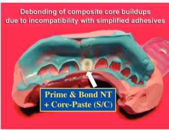
single-bottle total-etch adhesives and single-step, self-etch adhesives are utilized without an additional bonding resin layer. In these adhesives, the oxygen-inhibited layer contains acidic monomers that come into direct contact with the chemical-cured composite where they can titrate the basic amine accelerators and inactivate them (Figure 6). Clinically, this may result in the debonding of core buildups with self- or dual-cured composites during impression taking (Figure 7)15-18.This
problem, first reported in 1999, has largely been rectified in many single-bottle adhesives by the introduction of dual-cured versions that include an additional bottle of chemical co-initiator containing sodium benzene sulphinate14,20.Examples of these systems include
Prime&Bond NT Dual Cure (Dentsply DeTrey), Excite DSC (Vivadent), and OptiBond Solo Plus Dual Cure (Kerr). However, the use of a chemical co-initiator improves its tensile bond strength with self- or dual-cured composites only to a certain extent.21 A similar
situation is observed with single-step, self-etch adhesives. This is illustrated with Xeno CF Bond (Dentsply-Sankin), to which a BondLink (Den-Mat), a generic sodium benzene sulphinate solution was applied before coupling with the dual-cured composites22. The two pieces of work described
above21, 22 meticulously showed that adverse chemical
FIGURE 1- Formation of hybrid layers is not exclusive to
dentin adhesives. A hybrid layer is formed when Fuji II LC, a resin-modified glass-ionomer cement was applied to cut enamel that was pre-conditioned with polyacrylic acid
FIGURE 2- Classification of contemporary adhesives into
3-step total etch, 2-step total-etch, 2-step self-etch and the single-step self-etch systems. The 2-step total-etch and the single-step self-etch adhesives may be considered as simplified adhesives in which hydrophilic resins are employed without an additional coating of comparatively more hydrophobic resins
FIGURE 3- Incomplete resin infiltration results when an
acetone-based adhesive is applied to briefly air-dried, acid-etched dentin. The regions that are not infiltrated with resin can be seen by the deposition of metallic silver when resin-dentin interfaces are immersed in a silver nitrate tracer solution
FIGURE 4- Microtensile bond strengths of some common
FIGURE 5- A similar effect can be seen in all commercially
available single-step self-etch adhesives. Using the same dual-cured composite, microtensile bond strengths decrease substantially in the auto-cured mode when compared with the light-cured mode
FIGURE 6- Incompatibility between chemical-cured
composites and acidic adhesives bonded to dentin is partially caused by the neutralization of the basic tertiary amines in the chemical-cured composite by the residual acidic monomers from the oxygen inhibition layer of the adhesive
FIGURE 7 - Debonding of composite core buildups can
occur when a chemical-cured composite is coupled to an acidic simplified adhesive. The only exception is the single-bottle adhesive One-Step (Bisco, Inc,), that is the least acidic among the simplified adhesives
FIGURE 8- Some of the currently available single-step
Simplified adhesives behave as permeable membranes – in vitro evidence
It is often surprising to polymer chemists why dentin adhesives containing ternary catalytic systems bond remarkably well to chemical-cured composites, when resin composites, enamel or completely dehydrated dentin were used as bonding substrates25, and yet poor
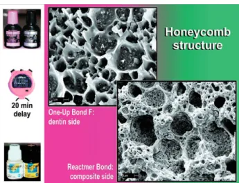
bonding resulted when normal non-dehydrated dentin was used as a bonding substrate (Figure 8). One simple experiment that can be used to demonstrate that simplified-step adhesives are permeable membranes when bonded to dentin is to place a light-cured adhesive resin on top of moist dentin; then place a light-cured resin composite over the cured adhesive and leave it in the dark for 10-20 minutes before light-activation23.
Light-cured composites utilize a completely different catalytic system that is generally less sensitive to acidic monomers26.Thus any adverse reaction observed in
light-cured composites cannot be attributed to adverse acid-base reactions. During this period, water will diffuse through the adhesive layer and is trapped in the form of water blisters along the adhesive-composite interface, with the hydrophobic resin composite taking an impression of these transudated water droplets23.
When these weak interfaces are fractured, the fracture surfaces demonstrate a honeycomb appearance that represents the resin partitions around these water droplets (Figure 9)24. Although the phenomenon of
water diffusion across a hydrophilic adhesive is novel in dentistry, such a process is commonly known to the resin-coating industry and is termed “osmotic blistering”27. It has also been suggested that the osmotic
gradient that is responsible for the induction of this type of water transport is derived from the dissolved ions that resided within the oxygen inhibition layer of these polymerized adhesives24.
Simplified adhesives are permeable membranes and do not provide a hermetic seal of vital deep dentin – in vivo evidence
An important goal in conservative dentistry is to restore the peripheral seal of dentin that originally exists prior to the removal of enamel28. As the smear layer
and smear plugs account for 86% of the total resistance to fluid movement in deep dentin29, the seal achieved by
contemporary total-etch or self-etch adhesives should not be worse than that achieved by the smear layer alone. Conventional thought is that a perfect seal of the resin-dentin interface can be established once the dentinal tubules and spaces within the demineralized collagen matrices are completely infiltrated by adhesive
resins. Such a notion is based on the assumption that polymerized resins used for bonding are nonporous and impermeable to fluid movement.
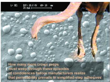
The increase in permeability of contemporary simplified adhesives (i.e. both the single-bottle total-etch adhesives and the single-step self-etch adhesives) to water can be readily seen when they are used for sealing crown preparations of vital deep dentin in vivo prior to impression taking for indirect restorations. After crown preparation was performed under local analgesia, we first took impressions of the smear layer-covered dentin using a low viscosity polyvinyl siloxane impression material. Then, we applied these adhesives to the vital crown preparations, removed the oxygen inhibited layer, and took impressions of these “sealed” crown preparations. These impressions were poured up in epoxy resins to produce replicas of the crown preparations for examination using scanning electron microscopy. The result of a single-step self-etch adhesive employed for sealing vital crown preparations, versus the result of the corresponding smear layer-covered dentin are shown in Figure 10. Coalescence of these dentinal fluid droplets can sometimes result in large pools of fluid on the surface of the cured adhesive (Figure 11). For the total-etch adhesives, we went one step further, and took additional impressions of the sealed crown preparations 7-10 days after insertion of the provisional crowns. There was a continuous transudation of dentinal fluid even at the period (Figure 12), with the fluid transudation being more severe for patients who opted for fitting their permanent crowns in the absence of local analgesia (Figure 13). The seal achieved by both the simplified self-etch and total-etch adhesives in areas that were close to the pulp horns were worse than those achieved with the unbonded smear layer.
From a clinical perspective, as the diffusion of dentinal fluid across the adhesives occurs relatively slowly, it is unlikely to result in severe post-operative sensitivity. However, if water and small ions can move across adhesive-sealed dentin, one wonders what the molecular cut-off will be, to prevent the permeation of noxious substances from the outside through the adhesive and dentin into the pulp (Figure 14). In a recent report in which the electrical resistance across total-etch and self-etch adhesive layers was measured before and after bonding, the electrical resistance increased 3-15 fold after bonding compared with the pre-coated smear layer-covered dentin.30 The electrical resistance was
further increased by 5-185 times after the application of a second adhesive coating30. In the same study, the
FIGURE 11- SEM micrographs of epoxy resin replicas taken
from a vital crown preparation that was sealed similarly with Adper Prompt prior to impression taking. Transudation of dentinal fluid was profuse. Coalescence of individual fluid droplets resulted in the formation of large pools of fluid beneath the impression material
FIGURE 12- Transudation of dentinal fluid from vital crown
preparations that were sealed with representative single-bottle total-etch adhesives. The SEM micrographs represented impressions that were taken immediately after bonding and removal of the oxygen inhibition layers
FIGURE 9- When interfaces such as those in Figure 8 are
fractured and examined with SEM, the fractured surface revealed the morphology of the fractured water blisters. The water was completely evaporated, leaving behind the resin partitions around the blisters, forming an intricate honeycomb pattern
FIGURE 10- SEM micrographs of epoxy resin replicas,
low viscosity bonding resin. These results provided the rationale for the use of the “resin coating technique”31
when indirect restorations were luted with Panavia F (Kuraray), a resin cement that employs a simplified self-etching primer (ED primer) for bonding to hydrated dentin. In this technique, the dentin is first sealed with a two-step self-etch adhesive (Clearfil SE Bond, Kuraray) and a light-cured, low viscosity microfilled resin prior to impression taking. Indirect restorations were subsequently luted to this resin-coated tooth surface using Panavia F.
Morphologic evidence of adhesive permeability
We have recently demonstrated the existence of two modes of nanoleakage within all single-step self-etch adhesives (Figure 15) and some single-bottle total-etch adhesives (Figure 16) that provide a plausible explanation on the permeation of water through them after polymerization32,33. These silver staining patterns can
be readily recognized after the resin-dentin interfaces were immersed in ammoniacal silver nitrate. The more easily recognized pattern is in the form of fractal-like, water channels that originate from the surface of the hybrid layer, and extend through the adhesive layer to reach the adhesive-composite interface. These water channels have been given the term “water trees” by Tay and Pashley34, to reflect their similarities in
appearance with those water trees that are formed as a result of water sorption and deterioration of polymer insulation around underground electrical cables (Figure 17)35. Water trees, when present, provide the most direct
pathways for water movement across the polymerized adhesive layers. Although water trees were initially reported using more technically advanced transmission electron microscopy, they can be similarly observed when silver-stained resin-dentin interfaces are examined using the backscattered mode of a scanning electron microscope. This is illustrated very nicely by the myriad of “unreported” water trees with the resin-dentin interfaces in a recent nanoleakage paper36. As
chemical-cured composites polymerize more slowly than light-cured composites, this allows sufficient time for water to diffuse from hydrated dentin across the all-in-one adhesive to form water blisters along the adhesive-composite interface.
Although water trees could be seen in all single-step, self-etch adhesives and the ethanol-based single-bottle adhesives, they have not been seen, at least immediately after bonding, in acetone-based single-bottle adhesives36.
A closer look at the silver staining patterns revealed the presence of a second mode of nanoleakage pattern – the isolated silver grains (Figure 16) – that is universally present in all simplified adhesives. They probably
represent hydrophilic domains with the polymerized adhesive that have an increased affinity for water34-36.
It was initially difficult for us to understand how water movement could occur with the acetone-based, single-bottle adhesives in the absence of direct evidence of water-connecting channels. After studying the literature, we realized that the transport of ions and small molecules across an amorphous polymer matrix may occur in the absence of physically-detectable water channels – via a process known as jump diffusion or ion hopping37.
This kind of ion hopping mechanism has been commonly observed in polymer ionic conductors, in which ionic salts are incorporated in a polar polymer that possesses significant ionic conductivity38.
The molecular model depicted above describes the diffusion of penetrant molecules through a polymer matrix in which microcavities of different sizes are formed and destroyed almost continuously (note the atomic time scale in picoseconds)39 in the polymer. The
more hydrophilic the polymer, the greater is the ease of formation of these microcavities, because of the reduced energy required to disruption the adjacent polymer chains. Small, dissolved molecules trapped in these locations are able to move into the direction of a driving force by cooperative motion of adjacent polymer chains. In our two previously described examples, the driving force was derived either from an osmotic gradient within the adhesive that acted as a “hydrophilic solid” in the in vitro delayed light-activation experiment, or from a positive pulpal pressure, in the case of the sealing of crown preparations with simplified adhesives in vivo.
Water movement across RMGIC-bonded dentin
RMGICs were developed in 1988 by adding polymerizable hydrophilic resins to conventional glass-ionomer formulations40,41.These materials were created
to overcome some of the problems of moisture sensitivity and low early mechanical strengths associated with conventional GICs, while maintaining their clinical advantages42. The basic RMGIC consists of
ion-leachable glass, polyalkenoic acid or a modified polyalkenoic acid with a photocurable side chain grafted onto the polymer backbone, a photocurable monomer such as 2-hydroxyethyl methacrylate (HEMA) and water43.Unlike conventional GIC, RMGIC has a dual
FIGURE 13- Transudation of dentinal fluid continued after
the removal of the provisional crown seven days after sealing with another single-bottle adhesive. Fluid transudation was severe in the absence of local analgesia, as there was no vasoconstriction
FIGURE 14- Many manufacturers are reluctant to accept
that their simplified adhesives are permeable after polymerization. Although dentinal fluid transudation does not occur fast enough with the immediate activation of light-cured composites, the permeability of the adhesives may provide channels for ingress of noxious products, and expediting water sorption that leads to the degradation of resin-dentin bonds
FIGURE15- Water trees that are detected within single-step
self-etch adhesives using a silver nitrate tracer solution provide a plausible morphological explanation of the permeability of these adhesives. Direct connection of a water tree with a water blister along the adhesive-composite interface could be seen in the right TEM micrograph
FIGURE 16- TEM micrographs showing the presence of
FIGURE 19- SEM and TEM micrographs showing the
presence of solid spherical bodies within the matrices of resin-modified glass-ionomer cements that were fractured close to the bonded moist dentin. These spherical bodies were absent from the air-voids in resin matrices that were fractured further away from the bonded dentin
FIGURE 20- SEM micrographs of Fuji IX, a conventional
GIC, that was fractured adjacent to the bonded moist dentin. Egg shell-like spherical bodies could be seen within the air voids that were incorporated in the polyalkenoate matrix
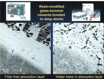
FIGURE 18- TEM micrographs showing the presence of a
resin-rich, non-particulate absorption layer on top of the hybridized dentin when a resin-modified glass-ionomer cement was applied to deep, polyacrylic acid-conditioned dentin. When these interfaces were immersed in ammoniacal silver nitrate tracer solution, water trees could be seen within the absorption layer
FIGURE 17- The term “water tree” was originated from the
by the HEMA, or the diffusion of HEMA from the resin matrices of the RMGICs into the water-rich dentin surface probably. The subsequent polymerization of the HEMA resulted in the form of a soft poly(HEMA) hydrogel layer. The RMGIC absorption layer has been thought to act as a stress-breaking layer47 and may
provide a similar function as a dentin adhesive layer in relieving polymerization shrinkage stresses.48
The use of the absorption layer as evidence of water movement across RMGIC-bonded dentin has been viewed upon with skepticism because of the limited resolution of the features of this layer, initially reported at an optical microscopical level. The existence of the absorption layer has been confirmed by Tay and Sidhu (unpublished results) using transmission electron microscopy. Such a layer was present in RMGICs that were bonded to moist dentin, but was absent when RMGICs were bonded to completely dehydrated dentin. Using a silver staining technique, water trees could also be identified within the absorption layer and in the resin matrices of RMGICS that were close to the bonded dentin. These water trees provided the channels of water movement for another novel feature that was never reported before in RMGIC/dentin interfaces (Figure 18). When RMGICs such as Fuji II LC (GC Corp.) and Photac Fil Quick (3M ESPE) bonded to moist dentin were fractured along the cement-dentin interfaces, spherical solid spherical bodies (Figure 19) could be identified with the use of conventional as well as environmental SEM in almost every air void that was close to the bonded dentin, or the absorption layer. Conversely, these spherical bodies were completely absent when fractures were performed at 3 mm away from the cement-dentin interfaces. Using EDX analyses of TEM sections, the internal core of these bodies were devoid of metallic ions. We speculated that the cores of these bodies were made up of a soft poly(HEMA) hydrogel that is similar to the absorption layer along the cement-dentin interface. The periphery of these bodies consisted of a thin crust with fairly high silicon and aluminum content. We believe that these spherical
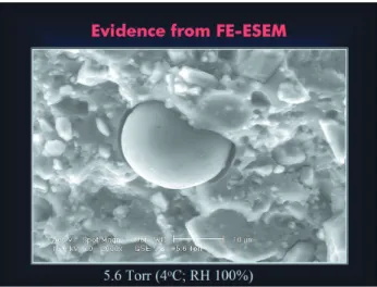
of water permeation across the GIC-dentin interface is unknown. Using a combination of conventional and environmental SEM, and TEM, we found that absorption layers are not observed in GICs as they do not contain polymerisable resin components. However, similar spherical bodies were seen within the air voids of GICs that were bonded close to the surface of the moist dentin (Figure 20). Unlike those identified from RMGICs, the spherical bodies observed in GICs are eggshell-like (Figure 21) and only consisted of a peripheral silicon-rich phase. These spherical bodies are not artifacts as they could be seen when fractured GICs were examined under wet conditions using field emission-environmental SEM (FE-ESEM) (Figure 22). They were also completely absent in sealed GICs that were fractured at 3 mm away from the bonded dentin (Figure 23). Similar to RMGICs, we believe that the spherical bodies near dentin surfaces result from a continuation of GI reaction, within the air voids of the original polyalkenoate matrix, that occur upon water diffusion from moist dentin.
Water movement across bonded dentin – simplified dentin adhesives vs. GI-based materials
We have seen that water movement across bonded dentin produced dramatically different results in dentin adhesives and in GI-based materials. The advent of dentin adhesives has no doubt improved the quality of preventive, restorative and prosthetic dentistry. However, in the attempt to simplify adhesives by reducing the number of steps, contemporary simplified adhesives are becoming increasingly hydrophilic and tend to be very permeable to water. While there may be no immediate concern when light-cured composites are placed on top of these adhesives and light-cured immediately, the results indicated that simplified adhesives, irrespective of whether they are self-etch or total-etch, do not provide a hermetic seal in the absence of an intermediate layer of more hydrophobic bonding resin49,50. These adhesives
FIGURE 21- SEM micrographs showing similar egg
shell-like spherical bodies in Fuji VII, another conventional GIC that contains iron pigment
FIGURE 22- Environmental SEM micrograph of an
unfractured spherical body in Fuji VII, showing that it is not a dehydration artifact. As the specimen was examined under wet condition, there was no cracking of the polyalkenoate matrix. The spherical body also was well adapted to the air void
FIGURE 23- SEM micrographs showing the presence of
has also been shown recently that both the spaces occupied by the isolated silver grains and water trees will increase in magnitude with the time of storage, and expedite the degradation of resin-dentin bonds52.
GICs and RMGICs bonded to deep dentin will continue to draw water from the underlying moist dentin for additional acid-base reaction; a process that does not occur with bonded enamel due to its lower water content. The spherical bodies are physical manifestations of the movement of water into pre-existing air voids within these materials. Unlike simplified adhesives in which the transudation of fluid droplets can result in substantial weakening of the adhesive-composite interface, the partial or complete filling up of the air-voids by the spherical bodies may be viewed upon as a self-toughening mechanism of GI-based materials. We hypothesized that these bodies may serve to deflect or blunt any cracks that attempt to propagate through the matrix, thereby toughening the material. The spherical bodies may play an adjunctive role by obliterating porosities in the resin matrix adjacent to the dentin and delay the growth of inherent cracks in this region under loading. The disadvantage is that the GICs and RMGICs may draw water from dentin fast enough to occasionally cause post-operative sensitivity that is associated with their uses. This also provides a rational explanation for the manufacturers’ recommendations that GI-based materials be used on slightly moist dentin.
ACKNOWLEDGMENTS
This work was supported by grant 20003755/90800/ 08004/400/01, Faculty of Dentistry, the University of Hong Kong, by grant DE014911 from the NIDCR, USA, and by grant 300481/95-0 from CNPq, Brazil. The authors are grateful to Zinnia Pang and Michelle Barnes for secretarial support.
Vijay P, Van Landuyt K, Lambrechts P, Vanherle G. Buonocore memorial lecture. Adhesion to enamel and dentin: current status and future challenges. Oper Dent 2003;28:215-35.
5- Glasspoole EA, Erickson RL, Davidson CL. Effect of surface treatments on the bond strength of glass ionomers to enamel. Dent Mater2002;18:454-62.
6- Ngo H, Mount GJ, Peters MC. A study of glass-ionomer cement and its interface with enamel and dentin using a low-temperature, high-resolution scanning electron microscopic technique. Quintessence Int 1997;28:63-9.
7- Ferrari M, Davidson CL. Interdiffuson of a traditional glass ionomer cement into conditioned dentin. Amer J Dent 1997;10:295-7.
8- Tay FR, Smales RJ, Ngo H, Wei SH, Pashley DH. Effect of different conditioning protocols on adhesion of a GIC to dentin. J Adhes Dent 2001;3:153-67.
9- Inoue S, Vargas MA, Abe Y, Yoshida Y, Lambrechts P, Vanherle G, Sano H, Van Meerbeek B. Microtensile bond strength of eleven contemporary adhesives to dentin. J Adhes Dent 2001;3:237-45.
10- Perdigão J. Dentin bonding as a function of dentin structure. Dent Clin N Amer 2002;46:277-301.
11- Tay FR, Pashley DH. Aggressiveness of contemporary self-etching systems. I: Depth of penetration beyond dentin smear layers. Dent Mater2001;17:296-308.
12- Degrange M. The cost of saving time. J Adhes Dent 2000;2:79-80.
13- Yamauchi J. Study of dental adhesive containing phosphoric acid methacrylate monomer. Jap J Dent Mater 1986;5:144-54.
14- Ikemura K, Endo T. Effect on adhesion of new polymerization initiator systems comprising 5-monosubstituted barbituric acids, aromatic sulphonate amides, and tert-butyl peroxymaleic acid in dental adhesive resin. J Appl Polym Sci 1999;72:1655-8.
16- Sanares AME, King NM, Itthagarun A, Tay FR, Pashley DH. Adverse surface interactions between one-bottle light-cured adhesives and chemical-cured composites. Dent Mater 2001;17:542-56.
17- Swift EJ Jr, Perdigão J, Combe EC, Simpson CH 3rd, Nunes MF. Effects of restorative and adhesive curing methods on dentin bond strengths. Amer J Dent2001;14:137-40.
18- O’Keefe KL, Powers JM. Adhesion of resin composite core materials to dentin. Int J Prosthod2001;14:451-6.
19- Dong CC, McComb D, Anderson JD, Tam LE. Effect of mode of polymerization of bonding agent on shear bond strength of autocured resin composite luting cements. J Canad Dent Assoc 2003;69:229-34.
20- Nyunt MM, Imai Y. Adhesion to dentin with resin using sulfinic acid initiator system. Dent Mater J1996;15:175-82.
21- Tay FR, Suh BI, Pashley DH, Prati C, Chuang S-F, Li F. Factors contributing to the incompatibility between simplified-step adhesives and chemical-cured or dual-cured composites. Part II. Single-bottle, total-etch adhesive. J Adhes Dent(in press).
22- Tay FR, Pashley DH, Yiu CKY, Sanares AME, Wei SHY. Factors contributing to the incompatibility between simplified-step adhesives and chemical-cured or dual-cured composites. Part I. Single-step, self-etch adhesive. J Adhes Dent 2003;5:27-40.
23- Tay FR, King NM, Suh BI, Pashley DH. Effect of delayed activation of light-cured resin composites on bonding of all-in-one adhesives. J Adhes Dent 2001;3:207-25.
24- Tay FR, Pashley DH, Suh BI, Carvalho RM, Itthagarun A. Single-step adhesives are permeable membranes. J Dent 2002; 30:371-82.
25- Tay FR, Pashley DR, Peters MC. Adhesive permeability affects composite coupling to dentin treated with a self-etch adhesive. Oper Dent (in press).
26- Suh BI, Feng L, Pashley DH, Tay FR. Factors contributing to the incompatibility between simplified-step adhesives and self-cured or dual-self-cured composites. Part III. Effect of acidic resin monomers. J Adhes Dent (in press).
27- Pommersheim JM, Nguyen T. Prediction of blistering in coating systems. In: Bierwagen GP, Editor. Proceedings of the American Chemical Society Symposium Series No. 689 - Organic coatings for corrosion control. Chapter 11. American Chemical Society publisher, Washington DC, 1998; 137-50.
28- Pashley DH, Pashley EL, Carvalho RM, Tay FR. The effects of dentin permeability on restorative dentistry. Dent Clin N Amer 2002; 46:211-45.
29- Pashley DH, Livingston MJ, Greenhill JD. Regional resistance to fluid flow in human dentine, in vitro. Arch Oral Biol 1978; 23:807-10.
30- Momoi Y, Akimoto N, Kida K, Yip KHK, Kohno A. Sealing ability of dentin coating using adhesive resin systems. Amer J Dent 2003; 16:105-11.
31- Jayasooriya PR, Pereira PN, Nikaido T, Tagami J. Efficacy of a resin coating on bond strengths of resin cement to dentin. Journal of Aesthetic and Restorative Dentistry 2003; 15:105-13.
32- Tay FR, Pashley DH, Yoshiyama M. Two modes of nanoleakage expression in single-step adhesives. J Dent Res 2002; 81:472-6.
33- Ferrari M, Tay FR. Technique sensitivity in bonding to vital acid-etched dentin. Oper Dent 2003;28:3-8.
34- Tay FR, Pashley DH. Water treeing – a potential mechanism for degradation of dentin adhesives. Amer J Dent 2003;16: 6-12.
35- Dissado LA, Fothergill JC. Electrical degradation and breakdown in polymers. Chapter 4. Water treeing degradation. IEE Material and Devices Series 9, London: Peregrinus, 1992; 75-116.
36- Li H, Burrow MF, Tyas MJ. The effect of concentration and pH of silver nitrate solution on nanoleakage. J Adhes Dent 2003;5:19-25.
37- Frisch FL, Stern SA. Diffusion of small molecules in polymers.
CRC Crit Rev Solid State Mater Sci 1983; 11:123-47.
38- Dürr O, Volz T, Dieterich W, Nitzan A. Dynamic percolation theory for particle diffusion in a polymer network. J Chem Phys 2002;117:441-7.
39- Müller-Plathesup F, Laaksonen L, van Gunsteren WF. Cooperative effects in the transport of small molecules through an amorphous polymer matrix. J Mol Graph 1993;11:118-36.
40- Wilson AD. Developments in glass-ionomer cements. Int J Prosthod 1989;2:438-46.
41- Mitra SB. Adhesion to dentin and physical properties of a light-cured glass-ionomer liner/base. J Dent Res 1991;70: 72-4.
42- Sidhu SK, Watson TF. Resin-modified glass ionomer materials. A status report for the American Journal of Dentistry. Amer J Dent 1995;8:59-67.
43- Nicholson JW, Anstice HM, McLean JW. A preliminary report on the effect of storage in water on the properties of commercial light-cured glass-ionomer cements. Brit Dent J 1992; 173:98-101.
44- Wilson AD. Resin-modified glass-ionomer cements. Int J Prosthod 1990; 3:425-9.
45- Watson TF, Sidhu SK, Griffiths BM. Ionomers vs. composites at the tooth interface. Proceedings of the Second International Symposium on Glass Ionomer Cements (ed. Peter Hunt). International Symposia in Dentistry, Philadelphia. 1994; 123-30.
46- Sidhu SK, Watson TF. Interfacial characteristics of resin-modified glass-monomer materials: a study on fluid permeability using confocal fluorescence microscopy. J Dent Res 1998;77: 1749-59.
pressure on adhesion of resin composite to dentin: bovine serum versus saline. Quintessence Int 1995; 26:221-6.
52. Tay FR, Hashimoto M, Pashley DH, Peters MC, Lai SCN, Yiu CKY, Cheong C. Aging affects two modes of nanoleakage expression in bonded dentin. J Dent Res 2003;82:537-41.