Cateter Peridural Deslocado:
U m a C a u s a d e F a l h a d e
Analgesia. Relato de Caso
Senhor Editor,
Interessante o relato de um caso de falha de analgesia devi-do ao cateter peridural deslocadevi-do 1. Na realidade objeti-vou-se descrever um caso de migração de cateter peridural para o interior do músculo psoas maior direito.
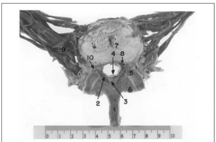
Para analisar e discutir detalhes da técnica utilizada julgo ne-cessário fazer algumas considerações anatômicas e depois formular perguntas para tentar esclarecer dúvidas. A parede posterior do canal vertebral lombar é formada pe-las lâminas vertebrais unidas pelos retangulares ligamentos amarelos (LA). Em L2-L3o LA tem 1,2 a 2 cm de altura e 1,2 a
2,2 cm de largura, tendo de importante o fato de sua borda la-teral fazer parte da parede posterior do forâmen interverte-bral reforçando, neste local, a cápsula dos processos articu-lares2. A borda interna do LA se une e continua com a borda interna do LA vizinho, formando um ângulo igual ou inferior a 90 graus e de abertura para o espaço peridural. Do vértice deste ângulo até o saco dural há uma distância que varia de 3 a 5 mm.
Com estes conhecimentos anatômicos e pela análise do for-mato do espaço peridural 3, apresentado na figura, con-clui-se que o cateter não deve ser introduzido no espaço peri-dural por mais de 2 cm pelo fato do formato do mesmo favore-cer a progressão lateral do cateter, na direção das raízes ner-vosas do forâmen intervertebral, provocando dores ou pa-restesias. Nestes casos, é obrigatório interromper a técnica. Qual foi a razão para a introdução do cateter por 5 a 10 cm, mesmo após a constatação de parestesias? Como explicar a penetração do cateter no músculo psoas maior após atra-vessar o forâmen intervertebral?
Após constatada falha de analgesia e inadequada coloca-ção, o cateter foi substituído por outro com bons resultados. Pergunto se foi fácil a nova identificação do espaço peridural e quantos cm de cateter foram introduzidos?
Na discussão, os autores afirmam que a analgesia controla-da pela paciente provavelmente cursaria com bom resultado e o mal posicionamento do cateter não seria investigado. Não se pode concordar com esta afirmação porque, na pior das hipóteses, haveria um bloqueio parcial do plexo lombar resultando em analgesia unilateral e insatisfatória. Concluindo: para realizar um bloqueio peridural é importante o conhecimento detalhado da anatomia da região e ter habili-dade manual para reconhecer pelo tato as estruturas atra-vessadas pela agulha de punção.
Atenciosamente.
Dr. Edmundo Zarzur Rua Ziembinsky, 314 05086-020 São Paulo, SP
Displaced Epidural Catheter: A
Reason for Analgesia Failure.
Case Report
Mr. Editor,
The report of an analgesia failure due to epidural catheter dis-placement was very interesting1. In fact, the aim was to des-cribe a case of epidural catheter migration to the right major psoas muscle.
In my opinion, some anatomic considerations in addition to questions in an attempt to answer open questions are nee-ded to analyze and discuss the described technique. The posterior wall of the lumbar vertebral canal is made up of vertebral blades connected by rectangular ligamentum fla-vum (LF). At L2-L3, LF is 1.2 to 2 cm high and 1.2 to 2.2 cm wide
with the major fact that its lateral border is part of the interver-tebral foramen posterior wall, thus reinforcing articular pro-cess capsules at this site2. LF internal border connects and proceeds with the neighbor LF internal border forming an an-gle equal to or smaller than 90 degrees, which opens to the epidural space. The distance from the vertex of this angle to the dural sac varies from 3 to 5 mm.
With this anatomic knowledge and considering the epidural space shape3shown below, the conclusion is that the cathe-ter should not be introduced in the epidural space for more than 2 cm because its shape favors the lateral progression of the catheter toward intervertebral foramen nervous roots ca-using pain or paresthesias. In those cases, it is mandatory to interrupt the technique.
Revista Brasileira de Anestesiologia 251
Vol. 52, Nº 2, Março - Abril, 2002
Rev Bras Anestesiol 2002; 52: 2: 251 - 254
CARTAS AO EDITOR
LETTERS TO THE EDITOR
Figura - Seção transversal da terceira vértebra lombar, através das bordas inferiores de dois LA vizinhos, mostrando o formato tri-angular do canal vertebral
Which was the reason for introducing the catheter 5 to 10 cm, even after confirmed paresthesias? How to explain the cat-heter escaping to the major psoas muscle, even after cros-sing the intervertebral foramen?
After confirming analgesia failure and misplacement, the cat-heter was replaced with good results. My question is: how easy it was to identify the epidural space and how many centi-meters of the catheter were introduced?
In their discussion, the authors state that patient controlled analgesia would probably have a good result and that the cat-heter malpositioning would not have been investigated. One cannot accept this statement because, at the worst, there would be a partial lumbar plexus block resulting in unsatisfac-tory unilateral analgesia.
In conclusion: to induce an epidural block it is important to have a thorough anatomic knowledge of the region and ma-nual skills to recognize by touch the structures crossed by the puncture needle.
Yours truly.
Edmundo Zarzur, M.D. Rua Ziembinsky, 314 05086-020 São Paulo, SP
REFERÊNCIAS-REFERENCES
01. Sudbrack G, Geier KO - Cateter peridural deslocado: uma causa de falha de analgesia. Relato de caso. Rev Bras Anestesiol, 2002;52:55-61
02. Zarzur E - Anatomic studies of the human lumbar ligamentum flavum. Anesth Analg, 1984;63:499-502
03. Zarzur E - The shape of the humans lumbar vertebral canal. Arq Neuropsiquiatr, 1996;54:451-454
252 Revista Brasileira de Anestesiologia
Vol. 52, Nº 2, Março - Abril, 2002 CARTAS AO EDITOR / LETTERS TO THE EDITOR
Figure - Cross-section of the third lumbar vertebra, through the lo-wer borders of two neighbor ALs, showing the triangular shape of the vertebral canal.
1. Spinous process; 2. LF; 3. Posterior epidural compartment; 4. Dura; 5. Upper articular process; 6. Lower articular process; 7. Intervertebral disk; 8. Vein; 9. Major psoas muscle; 10. Ante-rolateral compartment.