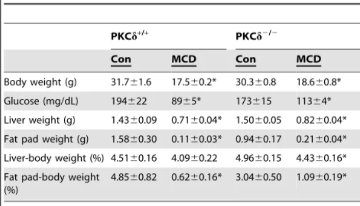
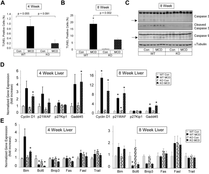
Lipid metabolism, oxidative stress and cell death are regulated by PKC delta in a dietary model of nonalcoholic steatohepatitis.
Texto
Imagem

Documentos relacionados
Nesse sentido, Chifamba (2014) considera que as mulheres têm significativo conhecimento acerca dos recursos hídricos (localização, qualidade, armazenamento), e são mais
SHRIMP U-Pb and Sm-Nd data for the Araxá Group and associated rocks: Constraints for the age of sedimentation and geodynamic context of the southern Brasília Belt, central
These results indicate that the COCO diet, high in saturated fatty acids, alters the lipid metabolism and AA turnover of peritoneal macrophages in female mice and also produces
Thus, this study was designed to develop an experimental model of NASH using a diet deicient in methionine and choline based on Brazilian-made animal food, and to evaluate the
Objective - The aim of this study was to determine the prevalence of simple steatosis and nonalcoholic steatohepatitis in patients with morbid obesity, subjected to bariatric
Background: The use of combined oral contraceptive (COC) has been related to changes in glycemic, lipid metabolism, increased oxidative stress, and systemic blood pressure,
From the histopathologic analysis, it was possible to observe that the liver of animals from the control group maintained the normal architecture (Figure 1A), while the
