TRIM5
a
-Imposed Genetic Barrier to Rhesus Macaque
Tropism
Steven J. Soll1,2,3, Sam J. Wilson1,2,3¤, Sebla B. Kutluay1,2, Theodora Hatziioannou2, Paul D. Bieniasz1,2,3*
1Laboratory of Retrovirology, The Rockefeller University, New York, New York, United States of America,2Aaron Diamond AIDS Research Center, The Rockefeller University, New York, New York, United States of America,3Howard Hughes Medical Institute, The Rockefeller University, New York, New York, United States of America
Abstract
Diversification of antiretroviral factors during host evolution has erected formidable barriers to cross-species retrovirus transmission. This phenomenon likely protects humans from infection by many modern retroviruses, but it has also impaired the development of primate models of HIV-1 infection. Indeed, rhesus macaques are resistant to HIV-1, in part due to restriction imposed by the TRIM5aprotein (rhTRIM5a). Initially, we attempted to derive rhTRIM5a-resistant HIV-1 strains using two strategies. First, HIV-1 was passaged in engineered human cells expressing rhTRIM5a. Second, a library of randomly mutagenized capsid protein (CA) sequences was screened for mutations that reduced rhTRIM5asensitivity. Both approaches identified several individual mutations in CA that reduced rhTRIM5a sensitivity. However, neither approach yielded mutants that were fully resistant, perhaps because the locations of the mutations suggested that TRIM5arecognizes multiple determinants on the capsid surface. Moreover, even though additive effects of various CA mutations on HIV-1 resistance to rhTRIM5awere observed, combinations that gave full resistance were highly detrimental to fitness. Therefore, we employed an ‘assisted evolution’ approach in which individual CA mutations that reduced rhTRIM5asensitivity without fitness penalties were randomly assorted in a library of viral clones containing synthetic CA sequences. Subsequent passage of the viral library in rhTRIM5a-expressing cells resulted in the selection of individual viral species that were fully fit and resistant to rhTRIM5a. These viruses encoded combinations of five mutations in CA that conferred complete or near complete resistance to the disruptive effects of rhTRIM5aon incoming viral cores, by abolishing recognition of the viral capsid. Importantly, HIV-1 variants encoding these CA substitutions and SIVmac239Vif replicated efficiently in primary rhesus macaque lymphocytes. These findings demonstrate that rhTRIM5ais difficult to but not impossible to evade, and doing so should facilitate the development of primate models of HIV-1 infection.
Citation:Soll SJ, Wilson SJ, Kutluay SB, Hatziioannou T, Bieniasz PD (2013) Assisted Evolution Enables HIV-1 to Overcome a High TRIM5a-Imposed Genetic Barrier to Rhesus Macaque Tropism. PLoS Pathog 9(9): e1003667. doi:10.1371/journal.ppat.1003667
Editor:Jeremy Luban, University of Massachusetts Medical School, United States of America
ReceivedMay 10, 2013;AcceptedAugust 13, 2013;PublishedSeptember 26, 2013
Copyright:ß2013 Soll et al. This is an open-access article distributed under the terms of the Creative Commons Attribution License, which permits unrestricted use, distribution, and reproduction in any medium, provided the original author and source are credited.
Funding:This work was supported by Grants from National Institutes of Health (R37AI64003 to PDB and R01AI078788 to TH) and the Howard Hughes Medical Institute. The funders had no role in study design, data collection and analysis, decision to publish, or preparation of the manuscript.
Competing Interests:The authors have declared that no competing interests exist. * E-mail: [email protected]
¤ Current address: MRC - University of Glasgow Centre for Virus Research, Glasgow, Scotland, United Kingdom.
Introduction
The narrow species tropism of HIV-1 is, in part, caused by species-specific variation in restriction factors that inhibit retroviral infection. This fact has important corollaries, one of which is that humans are likely protected from infection by many retroviruses. Conversely, many animal species commonly used in biomedical research cannot be infected by HIV-1, imposing severe limitations on the development of non-human primate models of HIV-1 infection and pathogenesis [1]. One antiretroviral protein that limits HIV-1 tropism is TRIM5a, a restriction factor that was initially identified in a screen of rhesus macaque (rh,M. mulatta) genes for inhibitors of HIV-1 infection [2], but in fact can have broad antiretroviral activity [3–8]. As a member of a large family of tri-partite motif (TRIM) containing proteins, TRIM5acontains N-terminal RING, B-box, and coiled-coil domains [9]. TRIM5a, like other restriction factors such as tetherin and APOBEC3 proteins, inhibits HIV-1 in a species-specific manner [10–17]. In particular, HIV-1 is vulnerable to restriction by rhTRIM5abut is
resistant to restriction by human TRIM5a due to sequence differences in their C-terminal PRY/SPRY domains [18–23]. TRIM5afunctions by targeting the incoming viral capsid within minutes of viral entry into the cell cytoplasm [24]. Viral capsids are likely directly bound by the PRY/SPRY domain during restriction [25–28] leading to inactivation of the viral core, followed by dissolution and, in some cases, degradation of viral core components [27,29,30].
degradation [10,11,13–15,32–38]. In contrast, TRIM5aproteins are unusual among the known antiretroviral restriction factors, because they bind to a specific viral protein (CA). While some TRIM5aproteins, particular those from old world monkeys have broad antiretroviral activity, and can inhibit retroviruses with widely divergent CA sequences [3,6–8], the requirement that a specific protein be recognized affords viruses the opportunity to evade restriction by evolving a CA protein that cannot be recognized. Indeed, transplantation of SIVmac239 CA in the
context of a chimeric HIV-1 confers rhTRIM5a resistance [8,39]. Similarly, sensitivity or resistance of murine leukemia virus to human TRIM5a can be acquired by mutations in CA [5,6,40,41]. Amino acids in CA that confer TRIM5asensitivity are generally exposed on the surface of the viral core [42,43] and are therefore accessible for binding to TRIM5a following viral entry into the cell.
While it should be possible for HIV-1 to evade rhTRIM5avia changes in CA sequence, it has proven difficult to isolate mutants of HIV-1 that have this property. A few studies have described mutations in HIV-1 CA that confer modestly reduced sensitivity to macaque (M. mulatta or M.fascicularis) TRIM5a proteins [44,45]. Recently one study showed that replacing the entire predicted surface of HIV-1 CA with SIVmac239sequences (twenty five amino
acid changes) conferred insensitivity to rhTRIM5a, but at the cost of an,15-fold reduction in single-cycle replication fitness [46]. As yet, no study has reported the identification of mutants that give full or nearly full resistance to rhTRIM5a while retaining viral fitness. A possible reason for this is that some monkey TRIM5a
proteins (including rhTRIM5a) are capable of recognizing retroviral CA proteins (e.g. HIV-1 and MLV) of very different sequence [3–8]. The molecular basis for this broad antiretroviral specificity, and consequently the difficulty with which rhTRIM5a
is evaded, may be that rhTRIM5a interacts with multiple determinants on the 3-dimensional HIV-1 CA protein structure [46,47]. It is also noteworthy that HIV-1 CA is the most genetically fragile (intolerant of amino acid substitution) protein for which robustness/fragility has been quantitatively measured [48]. Together, these factors may impose a high barrier to the evolution of fit, rhTRIM5a-resistant HIV-1 strains, an in a
broader sense may be why TRIM5aproteins have been selected as antiviral proteins during host evolution. If it were straightfor-ward for retroviruses to acquire resistance to TRIM5a, then little selective advantage would be conferred on hosts that employ TRIM5aas an antiretroviral protein.
An appreciation of how and why it is difficult for HIV-1 to acquire resistance to rhTRIM5a is important for a complete understanding of factors limiting transmission of primate lentivi-ruses among divergent primate species. Additionally, overcoming restriction by rhTRIM5ais required to enable HIV-1 replication in rhesus macaque cells and, thus, for the generation of an animal model that is based on HIV-1 infection of rhesus macaques [39]. Successful simian tropic (st) HIV-1 infection of pig-tailed macaques (M. nemestrina) [49], which express a TRIM5-cyclophilin fusion (TRIM-Cyp) that is unable to restrict HIV-1 [50–53], demonstrates that the HIV-1 host-range can include macaques when APOBEC3- and TRIM5-imposed restrictions are absent.
In the work presented here, we employed various strategies to identify a number of point mutations in HIV-1 CA that can confer partial resistance to restriction by rhTRIM5a. By combining assortments of these mutations, in an assisted evolution approach, and selecting for viruses that could replicate well in cells expressing rhTRIM5a, we generated mutant HIV-1 capsids that exhibit near-complete resistance to rhTRIM5a and retain fitness. Mutations that conferred resistance to rhTRIM5awere distribut-ed in several different locations over the exterior surface of the HIV-1 capsid, suggesting that rhTRIM5a recognizes several different determinants on HIV-1 CA. The mutations that caused resistance also abolished the ability of rhTRIM5a to cause disintegration of HIV-1 core components, as well as the ability of HIV-1 to saturate rhTRIM5a, suggesting that they exert their effect by preventing rhTRIM5a binding to CA. Notably, when incorporated into an stHIV-1 construct that encodes SIVmac239
Vif, rhTRIM5a-resistant CA sequences enabled efficient stHIV-1 replication in primary rhesus macaque lymphocytes.
Results
CA evolution during HIV-1 replication in human T-cell lines expressing rhTRIM5a
To derive HIV-1 CA proteins that confer resistance to rhTRIM5a we first employed an adaptation approach in which HIV-1 was passaged in human cells expressing rhTRIM5a. Initially, a cloned recombinant virus, termed NHG, that contains portions of HIV-1NL4-3 and HIV-1HXB2, and encodes GFP in
place of the Nef gene, was grown in MT2 cells to generate a virus stock with genetic diversity. After fifteen days of replication, cell-free supernatant was used to infect four different clonal cell lines stably expressing rhTRIM5a(two derived from MT2 cells and two derived from MT4 cells) or cells transduced with an empty vector (Figure 1 A–D). The rhTRIM5avariant used belongs to a class of rhTRIM5avariants that encode TFP at amino acids 239–241 and restrict HIV-1 more potently than other variants [54,55]. The first cycle of HIV-1 replication in these four rhTRIM5a-expressing lines was restricted by 50- to 100-fold relative to that in the empty vector control cells, confirming the potent anti-HIV-1 activity of rhTRIM5a therein (Figure 1 A–D). Infection was monitored by visual inspection of cytopathic effects and by measuring the fraction of GFP-positive cells in each culture at 1 to 3 day intervals. Because MT2 and MT4 cells are exceptionally permissive to HIV-1 replication (they were selected for this experiment for that reason), HIV-1 was able to spread through the four cultures and infect most cells after ,8 days, despite potent inhibition by rhTRIM5a (Figure 1 A–D). After the majority of cells became
Author Summary
GFP-positive and cytopathic effects had become abundant in the cultures, cell free supernatants were used to inoculate fresh rhTRIM5a-expressing cells, and this process was repeated for 10 passages in each of the four cell lines. The time taken for the majority of the cells to become GFP-positive during each passage appeared to decrease as the number of passages increased (Figure 1 A–D), implying adaptation of the virus to the rhTRIM5a
expressing cells. At each passage, we measured the infectious HIV-1 titer in the four culture supernatants on both rhTRIM5a
expressing and empty vector expressing cell lines. These analyses indicated that there was a progressive decrease in the sensitivity of the virus population to rhTRIM5aas the number of passages on rhTRIM5a expressing cells increased (Figure 1E–H). However,
even the passaged virus stock retained at least some degree of sensitivity to rhTRIM5a, and each of the four adapted viruses was restricted by,5- to 10-fold after 6 to 8 passages. Sensitivity to rhTRIM5a did not appear to decrease with further passage (Figure 1E–H).
PCR amplification and bulk sequencing of viral DNA present in the four rhTRIM5aexpressing cell cultures after various numbers of passages revealed that four CA mutations (V86E, I91N, I91T, and G116E) were present at a sufficiently high frequency to be detected in bulk sequences (Table 1). These mutations occurred at three different codons, all of which specified amino acids that are exposed on the presumptive outer face of the viral core (Figure 2A). Specifically, amino acids V86 and I91 are both within the
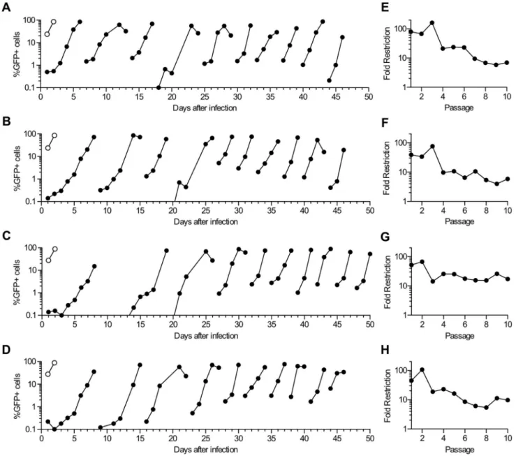
Figure 1. HIV-1 adaptation in human cell lines expressing rhTRIM5a.(A–D) MT2 and MT4 cells expressing either rhTRIM5a(filled symbols) or
an empty vector (open symbols) were infected with HIV-1NL4-3/HIV-1HXB2/GFP (NHG). Aliquots of cells from each culture were withdrawn and fixed for FACS analysis at the indicated times and the percentage of cells that were positive for GFP is plotted versus days after the infection of the first culture. Discontinuities indicate points at which cell-free supernatant was harvested and used to initiate the next passage in TRIM5a-expressing cell lines. NHG was passaged in four different cell lines expressing rhTRIM5a: MT2-rhTRIM5a#8 (A), MT2-rhTRIM5a#15 (B), rhTRIM5a#29 (C), and MT4-rhTRIM5a#32 (D). (E–H) Viral sensitivity to TRIM5a(Fold restriction) was calculated as the ratio of infectious titers present in culture supernatants at the end of each passage shown on the left, measured on MT2-vector and MT2-rhTRIM5a#8 cells.
cyclophilin-binding loop and G116 is on an outer turn of helix six (Figure 2A). Strikingly, each of the four mutations occurred in each of the four parallel virus cultures. In some cases, the mutant codons were present as mixtures with the WT codons, while in others WT codons became undetectable. In three virus cultures, all four amino acid changes were present in viral populations sampled after ten passages, at which point the WT I91 codon was no longer detected. Conversely, in one viral lineage, mutations at I91 were detected at passage 4, but then disappeared from the culture, and were undetectable after the tenth passage, at which point only V86E and G116E mutations were present as mixtures with the WT codon at both positions. Notably, amino acid substitutions V86M (V86E is described here) and G116E have previously been shown to decrease sensitivity to rhTRIM5aand crab-eating macaque TRIM5a, respectively [44,45].
To determine whether and how mutations that arose in CA during passage decreased sensitivity to rhTRIM5a, PCR products amplified from viral cultures were cloned and inserted into the parental pNHG proviral plasmid. The resulting clones included four single CA mutants; V86E, I91N, I91T and G116E, as well as double-mutants I91N/G116E and I91T/G116E. Certain combi-nations of mutations were not recovered as individual species from the four viral cultures, and so site-directed mutagenesis was used to construct all other possible double and triple mutant combinations of V86E, I91T, I91N and G116E (see below). To test the resulting viruses for sensitivity to rhTRIM5a, single-cycle infection assays were employed, using the MT2 cell lines expressing either rhTRIM5aor an empty vector as targets (Figure 2B).
All four of the single amino acid mutants (V86E, I91N, I91T and G116E) exhibited decreased rhTRIM5a sensitivity. In particular, substitutions V86E and I91T each reduced rhTRIM5a
sensitivity from,60-fold inhibition to,15-fold (Figure 2B). The I91N/G116E and I91T/G116E double mutants exhibited even lower rhTRIM5a sensitivity and were inhibited by less than 10-fold. Notably, the aforementioned mutants all occurred in individual species that were present in the viral culture experiment and exhibited little or no decrease in infectivity on the vector control cell line, suggesting that these individual and combined mutations did not incur major fitness costs. The data obtained with these reconstructed viral clones correlated quite well with the results obtained with each uncloned viral stock harvested after 10 passages in MT2-rhTRIM5a (Figure ’1E–H). Specifically, virus populations harvested from MT4-rhTRIM5a#29 carried V86E and G116E, but not I91T or I91N, and this stock was restricted by 16-fold by rhTRIM5a. Conversely the other three uncloned, adapted viruses all carried I91T, I91N, and G116E and were restricted by only 5–10 fold.
We next reconstructed the viral mutants that contained combinations of the aforementioned substitutions that were not detected as individual species in the passage experiment, namely
the V86E/I91N, V86E/I91T and V86E/G116E, double mutants and the V86E/I91N/G116E and V86E/I91T/G116E triple mutants. Strikingly, some of these mutants appeared completely, or nearly completely resistant to rhTRIM5a restriction in MT2 cells (Figure 2B). However, those mutants also exhibited severely reduced titers on the control vector expressing MT2 cell line, indicating that large fitness costs were imposed by combinations of mutations that gave rhTRIM5aresistance. The V86E/I91N and V86E/I91T double mutants also had apparent fitness defects that accompanied improvements in resistance to rhTRIM5a. These fitness costs were also observed in the context of spreading replication assays (Figure S1). Specifically, while the partially rhTRIM5a-resistant I91N/G116E and I91T/G116E double mutants were able to replicate as rapidly as WT on control vector expressing MT2 cells, the other combinations of mutations, particularly the V86E/I91N/G116E and V86E/I91T/G116E triple mutants, resulted in attenuated replication (Figure S1). Surprisingly, replication of these triple mutants was inhibited by rhTRIM5aduring spreading replication assays (Figure S1), even though they appeared nearly completely resistant in single-cycle infection assays (Figure 2), suggesting the possibility that poor fitness might amplify the effect of residual restriction by rhTRIM5a during spreading replication assays. Notably, the attenuated double and triple mutants exhibited low titer in hamster pgsA cells, but were not sensitive to restriction by human TRIM5aexpressed therein (Figure S2). This finding indicates that the apparent fitness defects observed in human cell lines were not due to acquisition of sensitivity to endogenous human TRIM5a, but instead were TRIM5a-independent deficits that coincided with decreases in sensitivity to rhTRIM5a. It therefore appeared that, in spite of the potential for HIV-1 CA to acquire near complete rhTRIM5a resistance via two or three amino acid substitutions, deleterious effects on fitness prevented some combinations of mutations from emerging as individual species during adaptation on rhTRIM5aexpressing cell lines.
All four of the capsid amino acid changes that arose during evolution in the rhTRIM5a cell lines were the result of single nucleotide substitutions. We considered the possibility that other amino acid substitutions at the same positions in CA would have conferred a greater degree of rhTRIM5a resistance, but simply did not arise because of their requirement for two or three nucleotide changes in a single codon. To address this possibility, we conducted a vertical mutagenesis experiment, in which we made every possible amino acid substitution at positions 86, 91, and 116 in CA. Thereafter, we screened each of the 57 single amino acid substitutions for rhTRIM5asensitivity (Figure S3A, B, C). A variety of mutations at positions 86, 91, and 116 decreased TRIM5a sensitivity. For example, at position 91 there were six different amino acid substitutions (I91A, I91P, I91Q, I91Y, I91D, and I91E) that were phenotypically approximately equivalent to
Table 1.Amino acid changes arising during HIV-1 passage in rhTRIM5aexpressing cell lines.
Passage Number Cell Line
MT2#8 MT2#15 MT4#29 MT4#32
P5 (MT2) or P4 (MT4) V86E I91N,T G116E V86E I91N G116E V86E I91N,T G116E V86E I91N,T G116E
P7 (MT2) or P6 (MT4) V86E I91N,T G116E I91N G116E V86E I91N,TG116E
P10 V86EI91N,TG116E V86EI91N,T G116E V86E G116E V86EI91N,TG116E
Amino acids symbols areboldwhere the WT codon was not detected in the sequence chromatogram. In other cases, the mutant was present as a mixture with the WT codon.
I91N and I91T. There were also several amino acid changes at position 91 that conferred a greater degree of TRIM5aresistance than I91N or I91T, but all of them caused decreases in viral titer. For example, the I91G substitution gave near complete rhTRIM5a resistance, but this mutation caused a 70-fold loss in viral titer on empty vector expressing cells (Figure S3B). Several different substitutions at positions 86 and 116 also conferred partial resistance to rhTRIM5a, but often these effects were accompanied by reduced viral titer. At position 86, most substitutions caused reduced rhTRIM5a sensitivity and a gluta-mine substitution caused the largest increase in viral titer on MT2-rhTRIM5a (Figure S3A). However, when V86Q was combined with substitutions at I91 and/or G116 it did not exhibit higher viral titers on MT2- rhTRIM5acells than the double mutants that emerged during the viral evolution experiments described in Figures 1 and 2 (unpublished observations). At position 116, none of the mutants exhibited higher titer on MT2-rhTRIM5athan did G116E (Figure S3C). Thus it appeared that the adaptation experiment selected near-optimal amino acids at positions 86, 91 and 116, but was not capable of selecting CA mutants with combinations of mutations that conferred the desired property.
Because our selection experiments were done in human cells, it was possible that endogenous human TRIM5acould have limited the spectrum of rhTRIM5a-resistance mutations that arose at these positions. Indeed, mutations at positions 86, 91, and 116 have been associated with sensitivity to human TRIM5a in the context of cytotoxic T lymphocyte escape [56]. Therefore, we screened the vertical mutant collection for sensitivity to human TRIM5a(Figure S4 A, B, C). Although some sensitivity to human TRIM5awas observed in some mutants, these effects were minor: while N-MLV was restricted by 160-fold by huTRIM5a, the degree to which some HIV-1 CA mutants were restricted was usually not more than 2- to 3-fold. One mutant, V86W, was restricted 6-fold by huTRIM5a. Therefore, it seems unlikely that endogenous TRIM5awould have profoundly limited evolution at these three positions during the adaptation experiment.
A screen of a randomly mutated CA library for substitutions conferring rhTRIM5aresistance
In a second independent strategy to identify mutations in HIV-1 CA that might confer rhTRIM5a resistance, we screened 91 infectious mutants from a library of NHG clones encoding PCR-mutagenized CA proteins (see Materials and Methods). MT2 cells expressing either the empty vector or rhTRIM5awere infected in a single cycle assay with a single dose of each clonal mutant virus and the ‘fold restriction’ by rhTRIM5a was calculated for those CA mutants that yielded measurable infectivity (.0.1% infected cells) on MT2-rhTRIM5a cells (Figure 3A). The parental virus, NHG was restricted by over 100-fold, as expected, while 22 CA mutant clones were restricted by less than 50-fold. The infectious titer of these 22 mutants was measured on vector expressing and rhTRIM5aexpressing cells to confirm their decreased rhTRIM5a
sensitivity (Figure 3B). Although there were a variety of mutations that decreased apparent rhTRIM5a sensitivity, many also
decreased viral titer on vector-expressing cells. For example, N57S was restricted by only 8-fold by rhTRIM5a, but its infectious titer on vector control cells was reduced 30-fold as compared to the parental NHG virus. Therefore, we selected only mutants that conferred increased titer on rhTRIM5a-expressing cells as candidates that had the potential to contribute to a CA that was both fit and resistant to rhTRIM5a. The infectivity of these mutants on control vector expressing cells was equivalent to or only marginally decreased compared to the parental virus.
Interestingly, two such mutations, V86E and I91T, had also emerged in the viral evolution experiments presented in Figures 1 and 2. Additionally, we selected M10L, M10V, V83M, H87Q, R100S, and A105T as candidate mutations for further analysis because they also caused an increase in infectivity on MT2-rhTRIM5a cells. Of note, each of these amino acid substitutions occurred at positions that are predicted to be exposed to the cytoplasm after viral entry (Figure 3C).
Fit, rhTRIM5a-resistant viruses, evolved from an assorted pool of CA mutants
While the above experiments demonstrated that a variety of mutations could confer partial resistance to rhTRIM5a, none of the aforementioned single amino acid CA mutants generated fully resistant viruses that did not incur a large fitness cost. However, the I91N/G116E and I91T/G116E double mutants demonstrated the potential for the additive effect of multiple mutations in the acquisition of TRIM5a resistance, without fitness penalties (Figure 2). Conversely, for other double mutants, particularly the combination of V86E and G116E, mutations that conferred partial rhTRIM5a resistance and had little fitness cost on their own, combined to generate rhTRIM5a resistant viruses at the expense of large fitness costs (Figure 2). Thus, it was possible that combinations of mutations that we had identified might generate fully rhTRIM5a-resistant viruses, but it was unpredictable as to whether and which combinations of mutations would allow the maintenance of high viral fitness.
Therefore, in an attempt to derive combination mutant CA proteins that were both fully fit and rhTRIM5a resistant, we adopted an ‘assisted evolution’ approach and generated a viral population that contained all possible combinations of the aforementioned non-deleterious mutations that conferred partial TRIM5aresistance (Figure 4A). Overlapping oligonucleotides that contained degenerate nucleotides encoding mixtures of WT and mutant amino acids were used to assemble a synthetic CA library pool containing random assortments of the mutations M10L/V, L83M, V86E, H87Q, I91N/T, R100S, A105T, and G116E. The synthetic CA assortment was used to generate a proviral plasmid library whose theoretical complexity was 576. Proviral plasmid DNA was isolated from 2.56103 pooled bacterial colonies and
transfected as a mixture, yielding a viral population that should contain every possible combination of the above mutations.
This viral population was evolved in MT2-rhTRIM5athrough a series of 15 passages (Figure 4B). Additionally, the final four passages were also carried out in CEM-rhTRIM5a cells. The
Figure 2. HIV-1 CA mutations selected during passage in rhTRIM5a-expressing cells decrease sensitivity to rhTRIM5a.(A) Positions of
amino acid residues at which mutations arose during replication in cells expressing rhTRIM5aare indicated on the HIV-1NL4-3capsid hexameric structure [43]. V86E and I91N/T are in the cyclophilin-binding loop and G116E is in helix six. The capsid hexamer shown on the left represents the exterior of the viral core, viewed from the cytoplasm of an infected cell. The hexamer on the right is oriented with the cytoplasmic face toward the top of the picture and the residues that face toward the interior of the core on the bottom. Images were generated using MacPyMOL. (B) NHG (WT) or NHG derivatives carrying the indicated mutations were titrated on MT2-vector (open symbols) or MT2-rhTRIM5a#8 (filled symbols) cells. Single-cycle infectivity was determined by FACS analysis. Mutant CA sequences that were found in the passaged viral population are marked with a green asterisk while mutants that were not found in the passaged viral population but were constructed using site-directed mutagenesis are marked with a red asterisk.
rationale for this was that CEM cells are less permissive than MT2, and we reasoned that this might impose a more stringent requirement for rhTRIM5aresistance and high replicative fitness on the viral population. Unfortunately, beginning at about passage 10, the maximum number of GFP-positive cells began to decrease with each subsequent passage (Figure 4B), presumably as a result of inactivating mutations in the GFP reporter gene in NHG. Nonetheless, visual inspection of cytopathic effects suggested that virus replication accelerated during the serial passage experiment. We conducted PCR amplification and bulk sequencing of viral DNA from infected cells at passage 4, 9, 11 and 15 (Table 2). This analysis revealed that some mutations were purged from the viral population at varying rates while others apparently became fixed. Additionally, new mutations arose during passage. Specifically, V86E, R100S, and A105T were rapidly purged (by passage 4) while L83M and H87Q were detected at passage 4, 9,and 11, but
lost by passage 15. Conversely, mutations M10L, I91N, and G116E became dominant in the viral population, with I91N and G116E rising to dominance rapidly and M10L becoming dominant in MT2 cells (but not CEM cells) between passages 11 and 15. Two additional mutations, namely A92E and M96I, that were not deliberately included in the starting assortment, arose spontaneously and became dominant in the viral population in MT2-rhTRIM5acells. Curiously, these cyclophilin-binding loop mutations arose at about the same time as two other cyclophilin-binding loop mutations (L83M and H87Q) were lost from the population, suggesting the possibility that these mutations were not compatible with each other. It is also possible that A92E and M96I have the same biological effect as L83M or H87Q but at a lower fitness cost. The apparent lack of heterogeneity at the aforemen-tioned positions in the bulk PCR product suggested that a single CA species containing five mutations (M10L, I91N, A92E, M96I
Figure 3. Screen of random HIV-1 CA mutants for rhTRIM5aresistance.(A) NHG clones containing CA substitutions generated by random
mutagenesis were used to challenge MT2 cells expressing an empty vector or rhTRIM5ausing a single dose of each virus. Fold-restriction by rhTRIM5a(the ratio of the numbers of infected cells in each cell line) is plotted. Only data from viruses that gave measurable infectivity (.0.1% infected cells) in MT2-rhTRIM5acells are plotted. The WT NHG is plotted as a red symbol and viruses restricted by less than 50-fold are represented by open symbols. (B) NHG mutants carrying CA mutations that decreased TRIM5asensitivity were retested and infectious titers in MT2-vector (open symbols) and MT2-rhTRIM5a#8 (filled symbols, referencing the left Y-axis) cells, as well as the ratio of these titers (fold restriction red triangles, referencing the right Y-axis) are plotted. (C) Two views of the NL4-3 capsid hexamer [43] are shown, as in Figure 2B. The sites of amino acid substitutions found in (B) to increase infectious titer on MT2-rhTRIM5acells (M10L, V83M, V86E, H87Q, I91T, R100S and A105T) regardless of effects on titer in vector control cells are indicated.
and G116E) had come to dominate the population in MT2-rhTRIM5a cells (Table 2). In CEM-rhTRIM5acells, the results were slightly different in that a single species did not dominate the population at passage 15. Rather M10L and M96I were present as
mixtures with the WT codons, and a third spontaneous mutation, L52I, was identified, again as a mixture with the WT codon.
Sequences encoding CA were cloned from the viral DNA present in both MT2-TRIM5a and CEM-TRIM5a cell lines at
Figure 4. An assisted evolution strategy to derive fit, rhTRIM5a-resistant HIV-1 strains.(A) Schematic representation of the experimental
design used to select fit, rhTRIM5aresistant viruses. CA mutations shown to decrease TRIM5a-mediated restriction, without major fitness penalties were incorporated into a synthetic CA amplicon to generate a virus population containing random assortments of the mutations. (B) Serial passages of the virus population described in (A) in MT2-rhTRIM5acells (filled symbols). Infection was monitored by measuring the proportion of cells expressing GFP over time, as described in the legend to Figure 1. In parallel, the last four of fifteen serial passages were also carried out in CEM-rhTRIM5acells (open symbols).
doi:10.1371/journal.ppat.1003667.g004
Table 2.Amino acid changes present during passage of HIV-1 encoding an assortment of CA mutations in a rhTRIM5aexpressing cell line.
Passage Number Amino Acid residue:
M10 L83 V86 H87 I91 R100 A105 G116 A92 M96 L52
P4 L M Q N, T E
P9 L M Q N E E
P11 L M Q N E E I
P15 (MT2) L N E E I
P15 (CEM) L N E E I I
Amino acids symbols areboldwhere the WT codon was not detected in the sequence chromatogram. In other cases, the indicated mutation was present as a mixture with the WT codon.
passage 15. One clone, containing the substitutions M10L/I91N/ A92E/M96I/G116E (LNEIE), corresponded to the dominant species in MT2-rhTRIM5a cells and the other, L52I/I91N/ A92E/M96I/G116E (INEIE), corresponded to a species that was co-dominant with LNEIE in CEM-rhTRIM5a cells. With the exception of L52I, all of the aforementioned mutations that became fixed, or arose during the passage experiment, were at residues that are predicted to be exposed on the surface of the HIV-1 capsid (Figure 5A).
We next compared the properties of the species that arose via assisted evolution from the assorted CA mutant pool (LNEIE and INEIE, Figure 5A), with the best performing virus species (in terms of fitness and rhTRIM5a resistance) that arose spontaneously during the initial passage in rhTRIM5a expressing cell lines, namely I91N/G116E (NE) (Figure 5B). Additionally, because L83M was among the best performing mutations in the random mutagenesis screen (Figure 3B) and may not have had the opportunity to coexist with the late appearing and proximal A92E and M96I mutations, we also used site-directed mutagenesis to introduce L83M into LNEIE to generate a virus carrying six mutations M10L/L83M/I91N/A92E/M96I/G116E (LMNEIE, Figure 5B).
While the I91N/G116E (NE) double-mutant was restricted in MT2-rhTRIM5aby,5-fold, LNEIE, INEIE and LMNEIE were completely or nearly completely resistant to inhibition by rhTRIM5a in single cycle infection assays, similar to a virus expressing the SIVmac239CA (Figure 5C). Moreover in spreading
replication assays, LNEIE, LMNEIE, and INEIE replicated with equivalent kinetics in rhTRIM5a-expressing MT2 cells and empty vector expressing MT2 cells (Figure S5). Moreover, the titers and replication kinetics of these viruses on non-restricting MT2 cells were indistinguishable from that of NHG (Figures 5C and S5). Thus, an assisted evolution approach, in which mutations that individually had modest effects on rhTRIM5a were randomly combined and subjected to selection in cell culture, enabled the acquisition of near complete rhTRIM5aresistance, with apparent retention of viral fitness in this cell line.
In some human cells lines, the A92E mutation, which was present in the LNEIE, LMNEIE, and INEIE mutants, has been shown to cause HIV-1 infection to become inhibited by cyclophilin A (CypA). Consequently, infection by A92E CA mutant viruses is increased in the presence of cyclosporin A (CsA), a drug that disrupts the CA-CypA interaction [57–59]. To test whether the CsA-dependence observed in A92E mutants is also present in the context of LNEIE, LMNEIE, or INEIE, VSV-G pseudotyped WT and mutant viruses were titrated on HeLa cells in the presence or absence of CsA (Figure S6). As has previously been shown to be the case in HeLa cells [57], the presence of CsA increased infection by the A92E mutant by.10-fold. In contrast, the enhancing effect of CsA on infection was minor in the case of LNEIE (2-fold) and absent in the case of NHG, LMNEIE, and INEIE.
Lack of rhTRIM5ainduced core disruption in rhTRIM5a -resistant viruses
To further demonstrate resistance of the aforementioned CA mutant viruses to rhTRIM5a, we tested whether viruses carrying the CA mutations were able to avoid the disruptive effects of rhTRIM5a on incoming viral cores [27,29,30], and thereby complete reverse transcription. To accomplish this, we employed our recently described assay in which the integrity of a subviral complex containing CA, integrase (IN) and viral nucleic acids is monitored in infected cells shortly after infection [30]. Specifically, VSV-G pseudotyped viruses encoding either WT or mutant CA
proteins were generated, and equivalent amounts (Figure 6A) were applied to pgsA-745 or pgsA-rhTRIM5acells, in a synchronized infection protocol. Two hours later, cytoplasmic extracts were fractionated and the presence of CA and IN protein, as well as viral cDNA was measured in each fraction. As expected, in the case of WT HIV-1, a dense complex containing CA and IN as well as viral DNA was readily detected in infected pgsA cells (Figure 6B). This complex appeared largely absent in identically infected pgsA-rhTRIM5a cells, consistent with the notion that rhTRIM5adisrupts incoming HIV-1 cores. In contrast, the dense complex containing CA and IN, as well as viral DNA, was not disrupted by rhTRIM5a during infection with viruses encoding the LNEIE, LMNEIE, INEIE, or NE CA mutants (Figure 6C–F) and only minor differences in the levels of CA, IN, and viral DNA in the dense complex were observed. These data suggest that the subviral complexes generated by LNEIE, LMNEIE, INEIE, and NE CA mutant viruses are preserved in the presence of rhTRIM5a and thus that the cores of these viruses are largely resistant to the biochemical effects of rhTRIM5a. Although the NE CA mutant retained residual sensitivity to rhTRIM5a in MT2-rhTRIM5a cells (Figure 5C), all four CA mutants (NE, LNEIE, LMNEIE, and INEIE) were able to infect pgsA-rhTRIM5a cells at similar levels (Figure S7). The minor differences among the four mutants in pgsA-rhTRIM5a cells were, apparently, insufficient to be evident in the biochemical assay of rhTRIM5arestriction (Figure 6C–F).
CA mutations conferring rhTRIM5aresistance prevent recognition by rhTRIM5a
In principle, the above HIV-1 mutants may have acquired resistance to rhTRIM5a by avoiding recognition by rhTRIM5a
or, less likely, by acquiring the ability to infect cells despite recognition by rhTRIM5a. To distinguish between these possibil-ities we performed an ‘abrogation of restriction’ assay. TRIM5a
Figure 5. Selection of fit, rhTRIM5a-resistant viral clones from a synthetic CA library containing assortments of mutations
conferring partial rhTRIM5aresistance.(A) Two views of the HIV-1 capsid hexamer [43] are shown, as in Figure 2B. The sites of amino acid
Figure 6. RhTRIM5a-resistant mutants are immune to viral core disruption by rhTRIM5a.(A) Virions harvested from cells transfected with
plasmids expressing HIV-1NL4-3GagPol (WT or CA mutants), VSV-G, and an HIV-1 vector carrying a GFP reporter were pelleted through 20% sucrose and CA protein was detected by western blotting. (B–F) PgsA (none) and pgsA-rhTRIM5a(rhT5a) cells were infected with VSV-G pseudotyped HIV-1NL4-3(WT or CA mutants), carrying a GFP reporter. Infected cells were harvested at T = 2 hr. after infection and post-nuclear supernatants were fractionated on 10–50% (w/v) sucrose gradients as described in Materials and Methods. Ten fractions were collected from the gradients. Q-PCR analysis of reverse transcription products and western blot analysis of CA and integrase (IN) in each fraction is represented for HIV-1NL4-3with WT (B), LNEIE (C), LMNEIE (D), INEIE (E) and NE (F) CA sequences.
doi:10.1371/journal.ppat.1003667.g006
TRIMCyp (Figure 7B), indicating that they were intrinsically sensitive to a TRIM5 protein that is able to bind to these capsids.
HIV-1 strains encoding rhTRIM5a-resistant CA proteins and SIVmac Vif replicate in rhesus macaque lymphocytes
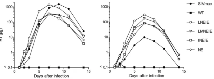
Along with APOBEC3 proteins, TRIM5a imposes a major block to HIV-1 replication in rhesus macaque cells [39]. To determine whether HIV-1 mutations that conferred resistance to rhTRIM5a expressed in human cell lines enabled replication in primary rhesus macaque cells, the TRIM5a-resistant CA sequences NE, LNEIE, INEIE and LMNEIE were inserted into a chimeric HIV-1 containing SIVmac239Vif, named stHIV-1 [49].
Virus stocks bearing either WT, NE, LNEIE, INEIE, LMNEIE or SIVmac239(stHIV-SCA) [39] CA sequences were normalized for
reverse transcriptase content and used to challenge peripheral blood mononuclear cells (PBMC) from two rhesus macaque donors (Figure 8). As expected, stHIV-1 carrying the WT HIV-1 CA sequence failed to replicate, while stHIV-SCA carrying the SIVmac239 CA initiated a spreading infection. Notably, all four
rhTRIM5a-resistant CA sequences enabled efficient stHIV-1 replication in rhesus macaque PBMC. In one donor, the rhTRIMa-resistant CA mutants replicated similarly to stHIV-SCA while in a second, apparently less permissive donor, all of the CA-mutant HIV-1 strains outperformed stHIV-SCA (Figure 8). The LNEIE mutant appeared to perform marginally better than the other CA mutants, and better than stHIV-SCA in PBMC from both donors. Genotyping revealed that both donors were heterozygous for rhTRIM5aalleles. The first donor carried alleles 4 and 5, while the second carried alleles 3 and 4 as (following the nomenclature described by Newmanet aland Wilsonet al[54,55]). Alleles 4 and 5 belong to a class that encodes a glutamine at residue 339 and allele 3 belongs to a class that encodes TFP at the same position. These two classes of rhTRIMavariants have been shown to differ in their restriction specificity and potency [54,55]. Importantly, the ability of the HIV-1 mutants described here to replicate in PBMC from both animals suggests that they have acquired resistance to restriction by both classes of rhTRIM5a
alleles.
Discussion
In this study, we identified a number of mutations in HIV-1 CA that individually could reduce the sensitivity of the incoming capsid to restriction by rhTRIM5a. When present in the right combination, collections of these mutations could confer near complete resistance to rhTRIM5a, sometimes without a fitness cost. Indeed, mutations in capsid were necessary and sufficient for HIV-1 to evade restriction by rhTRIM5a, consistent with the notion that the antiviral activity of TRIM5 depends on specific capsid recognition [5,8,39,40,47]. The rhTRIM5a resistant CA sequences abolished the ability of rhTRIM5ato disrupt incoming HIV-1 cores, enabling reverse transcription and the formation of a complex containing CA, IN and viral DNA, which would normally be blocked by rhTRIM5a [30]. Ultimately, these mutations enabled uninhibited infection of human cell lines stably expressing rhTRIM5a, which ordinarily exhibit.50-fold reduced susceptibility to WT HIV-1 infection.
With one exception, the amino acid substitutions that were found in the rhTRIM5a-resistant CA sequences encoded amino acids that are exposed on the presumptive exterior surface of the capsid lattice. However, L52I, (which occurred in only one of two cell lines in which the assorted CA mutant pool was evolved) is buried in the interior of the CA protein structure [43]. It is possible that L52I contributes to resistance by shifting the conformation of
Figure 7. RhTRIM5a-resistant HIV-1 capsids are not recognized
by rhTRIM5a.(A) The indicated CA sequences were tested for their
ability to generate viral capsids that could saturate rhTRIM5ain a rhesus macaque cell line. FRhK-4 cells were infected with VSV-G pseudotyped virions containing HIV-1NL4-3GagPol proteins encoding either wildtype CA, SIVmac239CA, or the indicated mutant CA. An HIV-1 based vector that encoded Tat was packaged into these virions and ‘abrogating virus dose’ is given in TZM infectious units (I.U.). Cells were tested for rhTRIM5aabrogation by simultaneous infection with a fixed amount of a VSV-G pseudotyped virions carrying a WT HIV-1NL4-3GagPol and a GFP-reporter gene. (B) VSV-G pseudotyped virions carrying the indicated WT or mutant CA-encoding HIV-1NL4-3 GagPol and a GFP-reporter gene were used to infect pgsA-745 cells or pgsA-745 cells stably expressing owl monkey TRIM-Cyp. Cells (16104) were infected
the capsid surface. Alternatively, it may marginally stabilize or destabilize the viral core, thereby affecting rhTRIM5aaction. It is also possible that L52I, or indeed other mutations described herein, arose as a compensatory mutation to maintain high viral fitness while not directly contributing to rhTRIM5a resistance. The contribution of the L52I mutation in the interior of the CA structure notwithstanding, the primary mechanism by which the HIV-1 CA mutants acquired resistance to rhTRIM5aappeared to be through loss of rhTRIM5arecognition, rather than resistance to the effects of TRIM5aafter recognition of the incoming capsid. This conclusion is based on the findings that (i) rhTRIM5a -resistant mutant capsids were unable to saturate rhTRIM5aand thereby facilitate WT HIV-1 infectivity in rhesus macaque cells, suggesting that they were not recognized by rhTRIM5a and (ii) the rhTRIM5a-resistant capsids retained full sensitivity to another TRIM5 protein (omkTRIMCyp) with a different CA binding specificity. Neither of these results would be expected if resistance were acquired via a mechanism in which the viral capsid retained rhTRIM5abinding, but acquired the ability to resist its antiviral effects.
Our initial failure to derive HIV-1 variants with complete resistance to rhTRIM5athrough replication in cell lines express-ing this inhibitor, or through random mutagenesis, underscores the difficulty in deriving HIV-1 strains with this property. Indeed, our eventual success required the combined application of the two different approaches, and then further evolution during selection from an assortment of mutations identified by each strategy. A comparison of the results obtained from the initial adaptation (Figure 2) and random mutagenesis (Figure 3) approaches suggests that each had distinct advantages and disadvantages. Despite the fact that four parallel cultures were initiated, the adaptation approach clearly did not produce a wide variety of solutions, and the same four mutations (V86E, I91N, I91T, and G116E) arose in four independent cultures. No combination of these four mutations gave fully fit rhTRIM5a-resistant CA sequences. It is possible that this result is defined by a potentially limited complexity of the viral population to which selection pressure was applied, and that rather than selection of several independent solutions, a small number of initially dominant genotypes persisted. However, another explanation might be that mutations that reduce HIV-1 sensitivity to rhTRIM5awithout a fitness cost
are few in number. Consistent with this latter interpretation is the fact that 2 of the 3 positions that we found to be mutated in rhTRIM5a-selected viruses have also been identified in similar, but completely independent, selection experiments in other laboratories, in different cell lines [44,45]. Those mutations that confer rhTRIM5a resistance but are marginally deleterious to virus replication might not be selected during the adaptation approach, even though they could contribute rhTRIM5aevasion if their accompanying fitness costs were alleviated by compensat-ing mutations. Screencompensat-ing a random library of virus clones might be a better way to identify such mutants. Indeed, the random mutagenesis screen identified a larger number of mutants that conferred reduced rhTRIM5a sensitivity, even though many of these did have associated fitness defects. Notably, V86E and I91T emerged from both adaptation and the random mutant library screening approaches. Interestingly, however, the random muta-genesis screen identified several mutations that did not arise during adaptation yet exhibited reduced sensitivity to rhTRIM5a
without an obvious fitness cost (e.g. V83M and M10L). Nevertheless, a drawback of random mutant screens is that only a limited number of mutants can be individually tested. Indeed, two amino acid substitutions emerged duringin vitroevolution that were not represented in the random mutant library (G116E and I91N). Additionally, because the experiments were done in human cells, a potential limitation of both the random mutant library screening and thein vitro evolution strategies was the possibility that some rhTRIM5a-resistant mutants could be missed if they simultaneously caused gain of sensitivity to endogenous human TRIM5a.
Overall, however, the application of both approaches and assortment of the resulting mutants in an assisted evolution approach led to derivation of fit, rhTRIM5a-resistant CA sequences. Even then, furtherde novo mutations of the assorted variant pool was required to generate the optimally resistant CA sequences. One possible reason for the eventual success of our approach is that the second round of selection was performed using a population of CA sequences that was highly enriched for mutations conferring partial TRIM5aresistance. This population contained individual mutant assortants that are highly unlikely to have occurred by chance through the standard approaches to viral evolution that were attempted initially.
Figure 8. HIV-1 replication in rhesus macaque PBMC.Simian tropic (st) HIV-1 viral stocks encoding SIVmacVif, and either SIVmac239CA, WT HIV-1NL4-3CA, or the indicated mutant HIV-1 CA proteins were normalized according to reverse transcriptase activity and used to infect rhesus macaque PBMC. Viral replication was monitored by measuring the level of reverse transcriptase in the culture supernatant at the indicated times. The two charts show replication assays conducted using PBMC from two different macaque donors.
Clearly, each individual mutation identified by either adapta-tion or random mutant screening approaches enabled only a partial evasion of rhTRIM5a(Figures 2 and 3). These mutations were distributed over several determinants on the surface of the HIV-1 capsid, as has been found in previous studies of retrovirus sensitivity to TRIM5a [40,41,44–47]. Indeed, although the cyclophilin-binding loop was featured prominently as a site at which mutations conferring decreased HIV-1 sensitivity to rhTRIM5aoccurred (positions L83, V86, I91, A92, M96), other determinants included the N-terminalb-hairpin (M10) and helix 6 (G116). One reasonable interpretation of these data is that several different sites on the capsid exterior contribute to the binding interaction with TRIM5a, and that mutations at any one of these sites, reduces, but does not eliminate interaction. Interestingly, a comparison of the distribution of mutations in MLV CA and HIV-1 CA that arose during the selection of rhTRIM5a-resistant viral variants [47] reveals striking similarity (Figure S9A and S9B). A key difference, however, is that single amino acid substitutions in MLV CA conferred near complete resistance to rhTRIM5a[47], while multiple substitutions were required in HIV-1 to achieve the same effect. Perhaps the viral challenges to which rhTRIM5ahas been subjected to during its evolutionary history have shaped it in such a way that it is a more robust inhibitor of lentiviruses than gammaretroviruses.
Because mutations in the cyclophilin-binding loop of HIV-1 CA have previously been shown to reduce the contribution of CypA to rhTRIM5a activity [61,62], it is possible that perturbation of CypA binding may have contributed to the acquisition rhTRIM5a
resistance described herein. Although retention of sensitivity to restriction by TRIM-Cyp suggests that CypA binding has been retained in the mutants described herein (Figure 7B), it remains possible that the role of CypA in rhTRIM5a activity was perturbed. Indeed previous work has suggested that a V86M CA mutation, while not preventing CypA binding, eliminates the contribution of CypA to the restriction of HIV-1 by huTRIM5a
mutants [63]. It therefore remains unclear whether cyclophilin-binding loop mutations in rhTRIM5a-resistant capsids have altered the involvement of CypA in restriction or simply decrease the binding of rhTRIM5ato HIV-1, independent of CypA.
Although HIV-1 CA mutations could be combined to give near complete escape from restriction, some combinations, particularly those derived from the first round of selection, were apparently deleterious to viral fitness. The need to alter multiple determi-nants, coupled with the inherent genetic fragility of HIV-1 CA [48] likely underlies the difficulty in generating combinations of CA mutations that confer rhTRIM5aresistance while maintaining high fitness. It is possible that the LNEIE and INEIE mutants, which were both fit and resistant to restriction by rhTRIM5a, include compensatory changes that alleviate fitness costs that accompanied escape from rhTRIM5arestriction. Our group has recently demonstrated that about 70% of randomly introduced single amino acid substitutions, and nearly all two-amino acid substitutions in CA are lethal to HIV-1 [48]. This lack of robustness in CA is also evident in MLV, as the N-terminal domain of MLV CA, unlike other regions of Gag, is intolerant of small insertions [64]. In the case of HIV-1, most lethal CA mutations were shown to disrupt normal particle formation [48]. However, interactions with multiple host factors, or structural requirements that dictate proper disassembly may also decrease the genetic robustness of CA. Thus, mutants containing combi-nations of changes required to both escape rhTRIM5aand retain fitness should be very rare, and this may be one reason why evolution has selected CA as a target for host antiviral proteins. Notably, the incomplete escape that was evident in viral
populations derived from initial selection experiments included a double amino acid mutant (NE), which was both fit and incompletely resistant to restriction in MT2-rhTRIM5a cells, as well as mixtures of viruses containing WT and mutant codons at V86 and G116. Although a V86E/G116E double mutant clearly had the opportunity to dominate in these cultures, it exhibited a clear loss of fitness, probably explaining why it did not. While it is formally possible that the fitness defect in V86E/G116E and other unfit rhTRIM5a-resistant mutants is species-specific and would not exist in monkey cells, we consider this unlikely, given that low infectiousness was also observed in hamster cells (Figure S2).
Importantly, mutations conferring resistance in rhTRIM5a -expressing cell lines allowed HIV-1 encoding SIVmac239 Vif to
replicate in PBMC from rhesus macaques. Indeed, an HIV-1 encoding the LNEIE mutant CA protein replicated as well as or better than a virus encoding an SIVmac239CA protein, suggesting
that TRIM5aactivity was entirely bypassed. Therefore, the HIV-1 CA mutations identified here enable HIV-HIV-1 to overcome the species-specific tropism barrier imposed by rhTRIM5a. Thus, although the barrier to the evolution of fit, rhTRIM5a-resistant HIV-1 CA proteins is apparently high, it is not insurmountable. Moreover, although the two amino acid NE mutant was inferior to the 5 and 6 amino acid CA mutants, in that it retained some degree of sensitivity to rhTRIM5a in human cell lines, the NE mutant replicated well in rhesus PBMC, and had little if any ability to saturate rhTRM5ain the FRhK cell line. Thus, one caveat that should be attached to the above discussion is that the level of rhTRIM5aexpression in the engineered human cell lines in which our selection experiments were performed likely exceeds the endogenous levels present in rhesus macaque cells. The number of mutations required to operationally escape TRIM5ainhibition in a natural setting may therefore be fewer than is suggested by our studies. In any case, the CA mutants identified herein should facilitate the development of rhesus macaque-based non-human primate models of HIV-1 infection.
Materials and Methods
Cell lines and viruses
Primate adherent cells (293T and FRhK-4) and suspension cells (MT2, MT4, CEM, and rhesus macaque PBMC) were cultured in DMEM and RPMI, respectively. CHO K1-derived pgsA-745 and the previously described pgsA-rhTRIM5aand pgsA-omkTRIM-Cyp cell lines [30] were maintained in Ham’s F12 media with 1 mM L-glutamine. All growth media was supplemented with 10% fetal bovine serum and gentamicin.
The replication competent HIV-1/GFP used in viral evolution experiments and for measurements of rhTRIM5a sensitivity was produced by transfection of an HIV-1NL4-3/HIV-1HXB2
recombi-nant proviral plasmid that encoded GFP in place of Nef (pNHG, GenBank accession number: JQ585717) [66]. Infectious titer was determined by inoculating MT2 cells (26104per well) in 96-well plates with 50mL of serially diluted supernatant. After allowing infection to proceed overnight, further replication was blocked by the addition of 100mg/mL dextran sulfate. Two days after infection, cells were fixed in 2% paraformaldehyde and the number of infected cells enumerated by FACS.
Viruses used to saturate rhTRIM5a in FRhK-4 cells were prepared by cotransfection of 293T cells with pCRV1-HIV-1NL4-3
GagPol (WT or rhTRIM5a-resistant mutant), the HIV-1 based vector pV1, which lacks a reporter gene, and a VSV-G expression plasmid at a 5:5:1 ratio, respectively [16]. Titers were determined using TZM-bl indicator cells, infected colonies were stained and counted using X-Gal two days post-infection. The reporter virus used to test for rhTRIM5asaturation in FRhK-4 cells, the viruses used for the biochemical analysis of core components, and the viruses used to infect pgsA-745 cell lines were produced in the same manner, except an HIV-1 based vector encoding GFP (pCSGW) [67] was used instead of pV1. VSV-G enveloped N-MLV was also produced in the same manner, except N-N-MLV GagPol expression plasmid and an MLV based vector encoding GFP (pCNCG) were used [68].
Virus evolution in rhTRIM5aexpressing cells
Prior to inoculation of MT2 and MT4 cells expressing rhTRIM5a, a cloned pNHG-derived virus that lacked Vpr was allowed to replicate in MT2 cells to generate sequence diversity. Specifically, 56105MT2 cells were infected with NHG at an MOI of 0.001. To maintain a viable cell population (the culture would have otherwise died due to cytopathic effects) and expand the viral population, uninfected MT2 cells were added as follows: 56105 cells at 4 dpi, 106cells at 6 dpi and 8 dpi, 36106cells at 10 dpi, and 46106 cells at 13 dpi. After 15 days of replication, cell-free
supernatant was harvested and titrated on MT2 cells. Thereafter, 1.256106 IU were used to infect two cultures of 56106 MT2-rhTRIM5acells, two cultures of MT4-rhTRIM5acells, and one culture each of MT2 and MT4 cells transduced with empty vector. Cells were diluted to 35 mL after an overnight infection in 5 mL. To measure the initial infection, dextran sulfate was added to a 100mL aliquot of cells one day after infection, which were then fixed the following day. To monitor the spreading infection, 100mL aliquots of cells were fixed periodically in 2% paraformaldehyde and the fraction of infected (GFP positive) cells determined by FACS analysis. After cytopathic effects became abundant in the culture, and/or the percentage of infected cells had peaked, 2 mL cell-free supernatant was used to infect 56106fresh cells as before. However, beginning with the fifth passage in MT2- rhTRIM5acell lines and the fourth passage in MT4- rhTRIM5acell lines 5 mL of supernatant was used to initiate new passages.
To recover CA sequences from viruses replicating in rhTRIM5aexpressing cells, DNA was isolated from infected cells using the Qiagen DNeasy Blood and Tissue Mini Kit and sequences encoding CA were amplified using PCR and a sense primer, 59-AAGGAAGCCTTAGATAAGATAGAGG-39 along with an antisense primer 59- TTGCCTTTCTGTATCATTA TGGTAG -39. Products were directly sequenced, without cloning, using the sense primer. To generate proviral clones containing adapted CA sequences, PCR amplification was done using primers 59-GCACAGCAAGCGGCCGCTGACACAGG-39and 59-GCC AAAACGCGTGCTTTATGGCCGGG-39 to add NotI and
MluI restriction sites for insertion into a pNHG derivative that was engineered (by silent mutagenesis) to contain NotI and MluI restriction sites flanking CA (GenBank accession number JQ686832). Additional combinations of mutations were intro-duced into CA by PCR-based mutagenesis methods as previously described [69]. For follow-up studies of CA mutants, virus stocks were generated by transfection of 293T cells with 1mg proviral plasmid/well in a 24-well plate. Reverse transcriptase in culture supernatants was quantified by a previously described assay based on real-time PCR [70], and infectious titers were determined as described above. Some CA mutant viruses were also used in spreading replication experiments, in which 26105 MT2-rhTRIM5a cells or empty vector transduced MT2 cells in 12-well plates were infected at an MOI of 0.002. Culture volumes were kept at 1 mL throughout the experiment. One day after infection, a 100mL aliquot of cells was treated with 100mg/mL dextran sulfate and fixed the following day to measure the initial infection. Beginning two days after infection, 100mL aliquots of cells were fixed daily for quantitation of GFP positive cells by FACS analysis.
Screen of random HIV-1 CA mutants for rhTRIM5a resistance
The construction and analysis of a library of pNHG clones encoding PCR-mutagenized CA sequences has been previously described [48]. Briefly, the mutagenized CA library was generated using the Genemorph II random mutagenesis kit (Agilent) using the following oligos 59-GTA AGA AAA AGG CAC AGC AAG CGG CCG CTG -39and 59- CTT GGC TCA TTG CTT CAG CCA AAA CGC GTG-39. The PCR template was a pNHG derivative (JQ686832) that contained silently mutated sequences to generate NotI and MluI sites flanking CA-encoding sequences (pNHGCapNM). The PCR product was cloned using a TOPO TA Cloning Kit and plasmid DNA was extracted from approx-imately,16104pooled, insert-positive colonies. After sequencing the amplicon from 20 clones to obtain a preliminary estimate of the mutagenesis frequency, this pooled plasmid DNA was digested using NotI and MluI and the CA-library insert subcloned into pNHGCapNM. Proviral plasmid DNA was extracted from individual cultures derived from 1056 colonies and subjected to sequencing and further analysis. Ninety-one of the resulting proviral plasmids that yielded infectious virions were screened for mutations that decrease sensitivity to restriction by rhTRIM5a. Specifically, viral stocks containing WT NHG or each of the 91 CA mutants were prepared by transfecting 2.56104293T cells/ well with 100 ng of pNHG and polyethylenimine in a 96-well plate. Thereafter, MT2-rhTRIM5acells or MT2 cells transduced with empty vector were infected with 2mL filtered virus in a 96-well plate at 26104cells/well. Virus replication was limited to a
single cycle by adding 100mg/mL dextran sulfate after an overnight infection. Two days after infection, cells were fixed in 2% paraformaldehyde and subjected to FACS analysis. Stocks of 22 mutants that infected at least 0.1% MT2-rhTRIM5acells and were restricted by less than 50-fold (see results) were prepared again by transfecting 26105293T cells with 1mg pNHG in a 24-well plate. Again, single-cycle infections were done in MT2-rhTRIM5acells or MT2 cells transduced with empty vector, using 50mL of serially diluted supernatant.
Construction and evolution of a library of synthetic CA sequences containing mutant assortments
I91N/T, R100S, A105T, and G116E), ten overlapping oligonu-cleotides, four containing degenerate nuoligonu-cleotides, were used in a PCR-based gene synthesis to produce a 400 base pair DNA encoding the 59 portion of CA that included all of the mutated positions. The heterogeneous gene synthesis product was designed to encode WT or mutant amino acids at a 1:1 ratio, or a 1:1:1 ratio in cases where two mutations were included at a single position. Specifically, a 50mL PCR reaction contained 3 pmol of each oligonucleotide and gene synthesis was carried out for 20 cycles using Phinzymes Phusion (1 min. 98uC, 1 min. 50uC, and 2 min. 72uC). Thereafter, 2mL of crude gene synthesis product was used as template in a 50mL PCR reaction with 200 nM external primers (1 min. 98uC, 1 min. 55uC, and 2 min. 72uC) and the resulting product was gel-purified. The 59end of the gene synthesis product was designed to contain a Not I restriction site while the 39end was designed to overlap with a second amplicon that was used to extend the synthetic CA sequence to include the 39portion of CA and an MluI restriction site. The two overlapping DNAs were used together as templates for a final PCR, which produced a DNA that was gel purified, and inserted into pNHG. Proviral plasmid DNA was purified from 2.56103pooled bacterial colonies and 5mg of this DNA was used to transfect 293T cells. The resulting virus was used to infect 56106 MT2-rhTRIM5a
cells at an MOI of 0.005. Then, 1 mL of cell-free virus was used in a second passage onto MT2-rhTRIM5a cells and 100mL was used in all subsequent passages. Following 11 passages in MT2-rhTRIM5acells, 2 mL of cell-free supernatant was used for serial passaging in CEM-rhTRIM5acells.
Biochemical analysis of retroviral cores
Biochemical analysis of retroviral cores in infected cells was done as previously described [30]. Briefly, pgsA745 cells or its derivative stably expressing rhTRIM5a were plated in 10-cm dishes one-day before infection. Cell culture supernatants containing VSV-G pseudotyped HIV-1NL4-3 GagPol/CSGW
viruses were filtered and treated with RNase free DNaseI (Roche) at a concentration of 1 unit/ml for 1 hour at 37uC in the presence of 6 mM MgCl2. Cells were washed with ice-cold
phosphate-buffered saline (PBS) and 7 ml of chilled virus (with 20 mM HEPES) was added to the cells at an MOI of ,0.01. After allowing virus to bind to cells for 30 minutes at 4uC, the inoculum was removed and cells were washed three times with PBS. Cells were then incubated at 37uC for 2 hours in complete cell culture media. Cells were collected in PBS-EDTA, pelleted, and resuspended in 1 ml of hypotonic buffer (10 mM Tris-Cl pH 8.0, 10 mM KCl, 1 mM EDTA) supplemented with complete protease inhibitors (Roche) and SuperaseIN (Life Technologies). After incubation on ice for 15 minutes, the cell suspension was dounce homogenized for 50 strokes, using pestle B. Nuclear material was pelleted by centrifugation at 10006gfor 5 minutes and post-nuclear supernatant was layered on top of a 10–50% (w/ v) linear sucrose gradient prepared in 1X STE buffer (100 mM NaCl, 10 mM Tris-Cl pH 8.0, 1 mM EDTA). Samples were ultracentrifuged using a SW50.1 rotor at 30000 rpm for 1 hour. Ten 500ml fractions from the top of the gradient were collected, and proteins and DNA in each fraction were analyzed as previously described [30].
Virus replication in rhesus macaque PBMC
Rhesus macaque PBMC were stimulated with 3mg/mL staphylococcus enterotoxin B (SEB) for three days and 5% IL-2 (Hemogen) throughout the experiment. Following SEB stimula-tion, PBMC were infected with st1, which is a chimeric HIV-1NL4-3 –derived virus in which the Env protein is derived from
SHIV/KB9 and the Vif protein is replaced by that of SIVmac239.
Additionally, stHIV-1/S-CA encodes an SIVmac239CA sequence
[39,49]. Mutant HIV-1NL4-3CA sequences were transferred from
NHG to stHIV-1, using BssHII and AgeI restriction sites. Virus stocks were normalized for reverse transcriptase activity and 16105
PBMC were infected in a 96-well V-bottom plate with inocula containing 20 pg reverse transcriptase. Following overnight infec-tion, cells were washed three times and supernatant from the third wash was frozen for RT quantitation (this value is plotted as 1 day after infection). After washing, cells were transferred to a 96-well U-bottom pate. At days 2, 3, 5, 7, 9, 11, and 14 after infection 80% of the cell supernatant was frozen for RT quantitation and replaced with fresh medium. For rhTRIMagenotyping, DNA was isolated from PBMC from both donors using the QIAGEN DNeasy Blood & Tissue Mini Kit. RhTRIMaexon 8 was amplified by PCR using sense primer 59- TTGATGTGACACTGGCTCCAAACAAC-39
and antisense primer 59-TGGGTAAAGCGGCCGCCAGAGC TTG-39. The PCR products were cloned using the Invitrogen Zero Blunt TOPO PCR Cloning Kit.
Supporting Information
Figure S1 Replication of HIV-1 carrying combinations of CA mutations that reduce sensitivity to rhTRIM5a.NHG
(WT) or NHG derivatives carrying the indicated mutations were used to infect MT2-vector (open symbols) or MT2-rhTRIM5a#8 (filled symbols) cells. All infections were done at an equivalent MOI. To monitor the spreading infections, aliquots of cells were fixed daily for FACS analysis. The percentage of cells expressing GFP is plotted against the number of days after infection.
(TIF)
Figure S2 Attenuated CA mutants did not acquire sensitivity to human TRIM5a.PgsA-745 cells (open symbols)
or pgsA-745 cells stably expressing human TRIM5a (filled symbols) were infected with VSV-G pseudotyped virions that carried a GFP-reporter gene and WT or mutant CA sequences in the context of HIV-1NL4-3 GagPol, as indicated. A VSV-G
enveloped virus carrying N-tropic MLV GagPol and a GFP-reporter gene was also included as a positive control for human TRIM5aactivity. The infectious titers on each cell line are plotted (circles, referencing the left Y-axis) and the ratio of the titers is shown as fold restriction (triangles, referencing the right Y axis). (TIF)
Figure S3 Vertical mutagenesis at positions where mutations arose during passage in rhTRIM5a
-express-ing cells. (A–C) MT2-vector (open symbols) or MT2-rhTRIM5a#8 (filled symbols) were infected with WT NHG or NHG encoding the amino acids indicated on the X axis at positions V86 (A), I91 (B) or G116 (C). Each mutant encoded a single amino acid substitution, and the panel covered each of the 19 possible amino acid substitutions at each position. Infectious titers (circles, referencing the left Y-axis) and the ratio of the titers (fold restriction, red triangles referencing the right Y-axis) are plotted. WT NHG is the leftmost data point on each chart. (TIF)
Figure S4 Some mutations at CA positions V86, I91 and G116 have modest effects on sensitivity to human TRIM5a. (A–C) PgsA-745 cells (open circles) or pgsA-745 cells
stably expressing human TRIM5a (filled circles) were infected with VSV-G pseudotyped virions that carried a GFP-reporter gene and HIV-1NL4-3 GagPol encoding WT or mutant CA