·Original article·
SD
-
OCT morphological changes in wet age
-
related
macular degeneration patients after bevacizumab
treatment
Manuel Noronha
1, Nuno Moura - Coelho
1, Teresa Gomes
1, Rita Flores
1,2, Marco Dutra
Medeiros
1,21Department of Ophthalmology, Central Lisbon Hospital Center, Lisbon 1169-050, Portugal
2NOVA Medical School, Universidade NOVA de Lisboa, Lisbon 1169-050, Portugal
Correspondence to: Marco Dutra Medeiros. Department of Ophthalmology, Central Lisbon Hospital Center, NOVA Medical School, Universidade NOVA de Lisboa, Lisbon 1169 -050, Portugal. marcodutramedeiros@ gmail. com
Received:2016-09-20摇 摇 Accepted:2016-12-08
贝伐单抗治疗湿性年龄相关黄斑变性患者
SD-OCT
形态学变化
Manuel Noronha1,Nuno Moura - Coelho1, Teresa Gomes1, Rita Flores1,2,Marco Dutra Medeiros1,2
(作者单位:1葡萄牙,里斯本 1169-050,中央里斯本医疗中心, 眼科;2葡萄牙,里斯本1169-050,新里斯本大学新医学院) 通讯作者:Marco Dutra Medeiros. marcodutramedeiros@ gmail. com
摘要
目的:研究频域光学相干断层成像(spectral domain optical
coherence tomography,SD-OCT)定量和定性检测指标在接 受玻璃体腔内注射贝伐单抗的年龄相关性黄斑变性(
age-related macular degeneration, AMD)患者人群中的变化,以 评估这些指标是否可以用于预测治疗后视力情况。 方法:回顾性分析66眼61例未进行过AMD相关治疗的 患者接受至少3mo玻璃体腔内注射贝伐单抗治疗的情
况。 治疗前后SD-OCT定量检测指标[中央视网膜厚度
(central foveal thickness, CFT),外 界 膜(external limiting
membrane,ELM)和椭圆区(ellipsoid zone,EZ)长度]和定 性检测指标进行分析和比较。 同时,分析这些指标和治疗 前后的视力的相关性。
结果:平均视力(Log MAR)、CFT、ELM和EZ长度治疗前 为0. 62依0. 41、419. 3依110. 0滋m、378. 2依377. 2滋m和156. 4依
253. 7滋m,治疗后为0. 53 依0. 44、325. 8 依117. 9滋m、547. 1 依
421. 5滋m和173. 1 依207. 1滋m。 治疗前视力和 CFT( rs=
0郾 27)、ELM长度(rs= -0. 30)及ELM断裂(rs= 0郾 43)有相 关性。 治疗后视力同样和治疗后ELM长度相关(rs=-0郾 40)。 治疗后视力和治疗前视力(rs=0. 66)、ELM长度(rs= -0. 35) 和ELM断裂(rs=0.46)相关。
结论:研究显示:治疗前视力、ELM长度和ELM断裂可以 用于预测治疗后视力。
关键词:光学相干断层扫描;外界膜;椭圆区;年龄相关性 黄斑变性
引用:Noronha M, Moura-Coelho N, Gomes T, Flores R, Dutra Medeiros M. 贝伐单抗治疗湿性年龄相关黄斑变性患者SD-OCT 形态学变化.国际眼科杂志2017;17(3):399-403
Abstract
誗AIM: To investigate the changes in spectral domain optical coherence tomography (SD-OCT) quantitative and qualitative parameters in a group of patients with age -related macular degeneration (AMD) that underwent bevacizumab intravitreal injections(IV). We assessed if one or more of these parameters can be used as prognostic factors of the post treatment visual acuity (VA).
誗METHODS:Totally66eyes of61patients,with treatment naive AMD, that were treated with at least 3 monthly bevacizumab IV, were retrospectively studied. SD-OCT quantitative [central foveal thickness (CFT), external limiting membrane (ELM) and ellipsoid zone (EZ) lengths] and qualitative parameters were studied and compared before and after IV. We also tried to establish correlation between these parameters and before / after treatment VA.
誗RESULTS:Mean VA(logMAR),CFT(滋m),ELM length (滋m) and EZ length(滋m) changed from pre-IV values of 0.62依0.41,419.3依110.0,378.2依 377.2and156.4依 253.7 to post-IV values of0.53 依 0.44,325.8 依 117.9,547.1 依 421.5 and173.1 依 207.1. There was correlation between pre-IV VA and pre-IV CFT (rs= 0.27),ELM length (rs= - 0.30) and ELM disruption (rs = 0. 43). There was also correlation between post-IV VA and post-IV ELM length (rs= -0.40). Post-IV VA showed correlation with pre-IV VA(rs= 0. 66), ELM length (rs = - 0. 35) and ELM disruption(rs= 0.46).
誗CONCLUSION: In our study group pre-IV VA, ELM length and ELM disruption can be used as post-IV VA prognostic factors.
誗KEYWORDS:optical coherence tomography; external limiting membrane;ellipsoid zone;age-related macular degeneration;visual acuity
DOI:10. 3980 / j. issn. 1672-5123. 2017. 3. 3
Citation:Noronha M, Moura - Coelho N, Gomes T, Flores R, Dutra Medeiros M. SD OCT morphological changes in wet age -related macular degeneration patients after bevacizumab treatment.
INTRODUCTION
A
ge-related macular degeneration (AMD) is an important chronic disease, being the major cause of important and irreversible vision loss, in developed countries, in individuals aged 50y or older[1-2]. The neovascular form of AMD, while accounting for only 20% of total AMD cases, is responsible for almost 90% of the serious vision loss associated with AMD[3]. Choroidal neovascularization ( CNV ) leading to hemorrhage, fluid and scar tissue formation is the cause for the vision loss associated with neovascular AMD.Intravitreal anti - vascular endothelial growth factor ( VEGF) injections are considered the first - line treatment option, slowing down and stabilizing most forms of neovascular AMD[4].
Spectral domain optical coherence tomography ( SD - OCT) allows the detailed study of the microstructural changes that occur in the retinal layers with neovascular AMD. Photoreceptor lesion and disruption can be seen in the high-resolution SD-OCT images as loss of integrity of the external retinal layers: external limiting membrane( ELM), ellipsoid zone(EZ) and interdigitation zone[5-7].
Several studies show that in neovascular AMD patients, treated with anti - VEGF injections or with photodynamic therapy, the integrity and length of the ELM and EZ correlate to a greater or lesser degree with before / after treatment visual acuity( VA)[8-9]. In neovascular AMD patients submitted to intravitreal anti-VEGF injections, the integrity and length of the ELM and EZ can also be used as prognostic factors for the post treatment VA[10-11].
The purpose of this study was to investigate the changes in SD-OCT quantitative and qualitative parameters in a group of patients with neovascular AMD that underwent bevacizumab intravitreal injections ( IV). We assessed if one or more of these parameters can be used as prognostic factors of the post treatment VA.
SUBJECTS AND METHODS
Retrospective study of 66 eyes of 61 patients with neovascular AMD diagnosis (treatment naive) that underwent 3 or more (4 maximum) consecutive, monthly bevacizumab IV ( Avastin 誖 , Genentech, South San Francisco, CA, USA) -1. 25mg per injection. The treatment of each patient was done in the Ophthalmology Department of Central Lisbon Hospital Center between Jan. 2013 and Jan. 2015. All patients were submitted to a complete ophthalmological evaluation ( including best corrected VA, intraocular pressure measurements and fundoscopy assessment ) and fluorescein angiography, SD-OCT exams before and after treatment with bevacizumab IV. Mean follow-up between treatment and SD-OCT was 6依2mo.
Neovascular AMD diagnosis was made with fundoscopy, fluorescein angiography and SD-OCT. The decision was made to exclude from the study eyes that had other retinal diseases (diabetic retinopathy,epiretinal membrane, myopic degeneration, venous and arterial occlusion), optic neuropathy (glaucomatous, ischemic, compressive) and eyes that had previous vitreo
-retinal surgery, IVs, laser photocoagulation or photodynamic therapy.
SD- OCT images were obtained with Spectralis ( 5. 1. 3. 0 version, Heidelberg, Germany) with Heidelberg Eye Explorer software(1. 7. 1. 0 version, Heidelberg, Germany). For each eye horizontal macular scans were made. Active eye tracking automatic software assured that scan positions were correct and that they didn蒺t change with treatment. Foveal center was determined manually. Eyes for which the SD - OCT images were poor in quality and / or quantity (poor fixation or medium opacity) were excluded from the study.
SD-OCT quantitative [central foveal thickness(CFT), ELM and EZ lengths] and qualitative[ELM and EZ disruption, sub-retinal fluid, intra - sub-retinal fluid, sub - sub-retinal fibrosis and retinal pigment epithelium ( RPE ) detachment ] parameters were studied and compared before and after bevacizumab IV. Taking into consideration that the foveal physiologic diameter is 1. 50mm[12], all of these parameters were studied and measured 1mm ( 1000滋m ) nasal and 1mm ( 1000滋m ) temporal to the foveal center, in an area including 2mm ( 2000滋m ) total diameter ( Figure 1 ). Mean CFT was obtained by SD-OCT automatic measurement.
ELM and EZ lengths were determined by manual measurement, with the caliper tool of the Heidelberg Eye Explorer software, of the intact, without disruption ELM and EZ zones. These measurements were made by two independent observers. The final ELM and EZ length values were obtained by the mean of these two measurements.
Disruption was considered to be present when more than 50% of ELM and EZ continuity was affected.
Best corrected VA was evaluated before and after treatment. It was measured with Snellen chart and converted to logarithm of the minimum angle of resolution ( logMAR) equivalent for statistical study.
Patient gender, age and CNV angiographic pattern (classic or occult) were also assessed.
Statistical analysis was made with IBM SPSS Statistics for Windows, version 23. 0. 0. 0. (IBM Corp. , Armonk, N. Y. , USA ) statistical software. Descriptive analysis of the quantitative results are expressed in mean 依 standard deviation and the qualitative results in percentages unless otherwise noted. Wilcoxon signed - rank test was used to compare the values of each variable before and after treatment. Correlation between the variables was made with Spearman rank - order correlationtest. All values ofPless than 0. 05 were considered to be statistically significant.
RESULTS
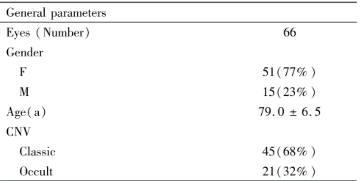
Mean age was 79. 0依6. 5y, gender was 77% female and 23% male (51 eyes female and 15 eyes male). CNV angiographic pattern was classified as classic in 68% (45 eyes) and as occult in 32% (21 eyes) of the eyes(Table 1).
Table1摇 General parameters General parameters
Eyes (Number) 66
Gender
摇 F 51(77% )
摇 M 15(23% )
Age(a) 79. 0 依 6. 5
CNV
摇 Classic 45(68% )
摇 Occult 21(32% )
CNV: Choroidal neovascularization.
173. 1依207. 1 with P values of 0. 02, <0. 01, < 0. 01 and 0郾 07 respectively ( Table 2 ). There was significant improvement of the best corrected VA with treatment. CFT was reduced significantly ( 22% reduction ) and ELM length recovered significantly (45% recovery) ( Figure 2 and 3). EZ length recovery was not statistically significant.
Qualitative parameter changes with treatment are shown in Table 3. There were significant reductions with treatment in ELM disruption, sub - retinal fluid, intra - retinal fluid and RPE detachment. Sub-retinal fibrosis increased significantly with treatment. There were no statistically significant changes with treatment in EZ disruption.
When studying the correlation results between VA and the pre and post treatment parameters there were correlations between pre-IV VA and pre-IV CFT (rs= 0. 27), ELM length (rs= -0. 30) and ELM disruption (rs= 0. 43). There was also correlation between post-IV VA and post-IV ELM length (rs= -0. 40) (Table 4 and Figure 4).
Post-IV VA showed correlation with pre-IV VA (rs= 0. 66), ELM length (rs= -0. 35) and ELM disruption (rs= 0. 46) (Table 5 and Figure 5).
Of the total of the eyes studied (66 eyes), 35 eyes improved, 15 eyes maintained and 16 eyes worsened the VA with treatment. In the group of the eyes that had improved VA (35 eyes) post-IV VA also showed correlation with pre-IV VA (rs= 0. 68), ELM length (rs= -0. 41) and ELM disruption (rs= 0. 56)(Table 6 and Figure 6).
DISCUSSION
In our study bevacizumab anti-VEGF treatment seems to lead to an increase in ELM length and decreased ELM disruption. There wasn蒺t significant EZ length recovery or reduced EZ disruption with treatment. These results suggest that in our patients ELM suffersstructural recovery and that this recovery does not occur in the EZ. Loss of the integrity of the external retinal layers occurring first in the EZ layer and only after in the ELM, as shown previously in retinitis pigmentosa[6,13], can be one possible explanation for these results. However, it has also been shown that after macular hole surgery anatomical recovery of these retinal layers follows an inverted order, with first ELM followed by EZ being reestablished[14-15] and that this process can take up to 12mo[14]. The EZ recovery also occurs only where there is intact ELM[16]. While obtained in
Figure 1 摇 SD-OCT macular study area摇 摇 All of the parameters were studied and measured 1mm (1000滋m) nasal and 1mm (1000滋m) temporal to the foveal center.
Figure2摇 CFT variation.
Figure3摇 ELM and EZ length variation. Table2摇 Quantitative parameters
Parameters Pre-treatment Post-treatment P
VA (logMAR) 0. 62依0. 41 0. 53依0. 44 0. 02 CFT (滋m) 419. 3依110. 0 325. 8依117. 9 <0. 01 ELM length (滋m) 378. 2依377. 2 547. 1依421. 5 <0. 01 EZ length (滋m) 156. 4依253. 7 173. 1依207. 1 0. 07 VA: Visual acuity; CFT: Central foveal thickness; ELM: External limiting membrane; EZ: Ellipsoid zone.
Table3摇 Qualitative parameters n(% ) Parameters Pre-treatment Post-treatment P
Figure4摇 Pre and post-treatment correlations摇 Dispersion graphs of the correlations between A) pre-treatment VA and pre-treatment CFT, B) pre-treatment VA and pre-treatment ELM length, C)pre-treatment VA and pre-treatment ELM Disruption, D: Post-treatment VA and post-treatment ELM Length. VA: Visual acuity; CFT: Central foveal thickness; ELM: External limiting membrane;rs: Correlation coefficient.
Figure5摇 Post-treatment VA with pre-treatment parameterscorrelations摇 Dispersion graphs of the correlations between A) post -treatment VA and pre--treatment VA, B)post--treatment VA and pre--treatment ELM length, C) post--treatment VA and pre--treatment ELM disruption. VA: Visual acuity; ELM: External limiting membrane;rs: Correlation coefficient.
Figure6摇 Correlations in the group with improved post AV摇 Dispersion graphs of the correlations in the group with improved AV between A) post-treatment VA and treatment VA, B) post-treatment VA and treatment ELM length, C) post-treatment VA and pre-treatment ELM disruption. VA: Visual acuity; ELM: External limiting membrane;rs: Correlation Coefficient.
Table4摇 Pre and Post-treatment correlations
Correlations rs P
Pre VA/ Pre CFT 0. 27 0. 03 Pre VA / Pre ELM length -0. 30 0. 01 Pre VA / Pre ELM disruption 0. 43 <0. 01 Post VA / Post ELM length -0. 40 0. 01 VA: Visual acuity; CFT: Central foveal thickness; ELM: Externallimiting membrane;rs: Correlation coefficient.
Table5摇 Post-treatment VA with pre-treatment parameters correlations
Correlations rs P
Post VA/ Pre VA 0. 66 0. 01 Post VA / Pre ELM length -0. 35 <0. 01 Post VA / Pre ELM disruption 0. 46 <0. 01 VA: Visual acuity; ELM: External limiting membrane; rs: Correlation coefficient.
different diseases these results may be one possible explanation for the differences in ELM and EZ structural recovery that we observed. It can be that in our patients the structural damage of the ELM wasn蒺t serious enough ( less
Table6摇 Correlations in the group with improved AV
Correlations Rs P
Post VA/ Pre VA 0. 68 <0. 01 Post VA / Pre ELM length -0. 41 0. 02 Post VA / Pre ELM disruption 0. 56 <0. 01 VA: Visual acuity; ELM: External limiting membrane; rs: Correlation coefficient.
likely if we consider the initial ELM mean length and disruption) or that the period of time following treatment wasn蒺t long enough to allow for sufficient EZ structural recovery. It can also be the case that in neovascular AMD (mainly in advanced disease), EZ damage is more serious that in other retinal diseases, not allowing for significant future structural recovery.
may signify greater before treatment ELM structural damage in our patients, with more recovery potential.
In our study pre-treatment VA was mildly correlated with CFT and moderately correlated with ELM length and ELM disruption. Post-treatment VA was moderately correlated with ELM length. Retinal external layer lesion or disruption is a sign of photoreceptor damage or dysfunction in several retinal diseases[17-19].
Disruption or integrity loss of the retinal external layers ( in our study ELM) seems to negatively influence pre and post-treatment VA. Our results seem to follow other previous studies where neovascular AMD patients VA was better or worse according to better or worse retinal external layer structural integrity[8,11,20].
In our study, post-treatment VA was strongly correlated with pre treatment VA and moderately correlated with pre -treatment MLE length and disruption. These correlation results were similar in the group of patients that had improved VA with treatment. As also seen in previous studies[11], the most important factor for visual prognosis was pre -treatment VA. However, ELM length and disruption ( although less consistently so) also seem to predict the after treatment VA recovery potential. These SD - OCT structural integrity parameters may eventually be used as VA recovery prognostic factors,but presently pre-treatment VA seems to be the most important factor in our study.
This study had several limitations:1) it was a retrospective study and not prospective, randomized or controlled; 2) SD-OCT and fluorescein angiography exams made after treatment were not at the same defined time for each patient; 3) SD-OCT precise ELM and EZ length measurements were difficult in some eyes that had too much macular distortion, abundant sub- retinal fluidor fibrosis and RPE detachment; 4) MLE and EZ length recovery in some cases may only reflect better visualization of these layers because of less intra / sub-retinal fluid and not real structural integrity recovery.
Our study suggests that in neovascular AMD patients submitted to bevacizumab treatment ELM has potential for structural integrity recovery. We also conclude that certain SD-OCT structural changes before starting treatment ( ELM length and disruption) can be used as predictors for the VA recovery that occurs with treatment. However, the most important prognostic factor seems to be pre - treatment VA. More studies are needed to clarify if there is a direct causal association between SD -OCT structural parameters and post bevacizumab treatment VA.
REFERENCES
1 Ratnapriya R, Chew EY. Age-related macular degeneration-clinical review and genetics update.Clin Genet2013;84(2):160-166
2 Ferris FL 3rd, Wilkinson CP, Bird A, Chakravarthy U, Chew E, Csaky K, Sadda SR. Clinical classification of age - related macular degeneration.Ophthalmology2013;120(4):844-851
3 American Academy of Ophthalmology Retina/ Vitreous Panel. Preferred Practice Pattern襆Guidelines. Age-Related Macular Degeneration. San
Francisco, CA: American Academy of Ophthalmology; 2015
4 Solom SD, Lindsley K, Vedula SS, Krzystolik MG, Hawkins BS.
Anti-vascular endothelial growth factor for neoAnti-vascular age - related macular degeneration.Cochrane Database Syst Rev2014;29(8):CD005139 5 Staurenghi G, Sadda S, Chakravarthy U, Spaide RF, International Nomenclature for Optical Coherence Tomography ( IN · OCT) Panel. Proposed lexicon for anatomic landmarks in normal posterior segment spectral-domain optical coherence tomography: the IN·OCT consensus.
Ophthalmology2014;121(8):1572-1578
6 Mitamura Y, Mitamura-Aizawa S, Katome T, Naito T, Hagiwara A, Kumagai K, Yamamoto S. Photoreceptor impairment and restoration on optical coherence tomographic image.J Ophthalmol2013;2013:518170 7 Spaide R F, Curcio C A. Anatomical correlates to the bands seen in the outer retina by optical coherence tomography. Retina2011;31(8): 1609-1619
8 Oishi A, Hata M, Shimozono M, Mandai M, Nishida A, Kurimoto Y. The significance of external limiting membrane status for visual acuity in age-related macular degeneration. Am J Ophthalmol 2010;150(1):27-32. e1
9 Sayanagi K, Sharma S, Kaiser PK. Photoreceptor status after antivascular endothelial growth factor therapy in exudative age - related macular degeneration. Br J Ophthalmol2009;93(5):622-626 10 Kwon YH, Lee DK, Kim HE, Kwon OW. Predictive findings of visual outcome in spectral domain optical coherence tomography after ranibizumab treatment in age - related macular degeneration. Korean J Ophthalmol2014;28(5):386-392
11 Oishi A, Shimozono M, Mandai M, Hata M, Nishida A, Kurimoto Y. Recovery of photoreceptor outer segments after anti - VEGF therapy for age related macular degeneration. Graefes Arch Clin Exp Ophthalmol
2013;251(2):435-40
12 Ross M, Pawlina W. Eye. Histology: A text and atlas: with correlated cell and molecular biology. 6th edition. USA: Lippincott Williams & Wilkins; 2011: 896-926
13 Mitamura Y, Mitamura-Aizawa S, Nagasawa T, Katome T, Eguchi H, Naito T. Diagnostic imaging in patients with retinitis pigmentosa. J Med Invest2012;59(1-2):1-11
14 Bottoni F, De Angelis S, Luccarelli S, Cigada M, Staurenghi G. The dynamic healing process of idiopathic macular holes after surgical repair: a spectral-domain optical coherence tomography study. Invest Ophthalmol Vis Sci2011;52(7):4439-4446
15 Wakabayashi T, Fujiwara M, Sakaguchi H, Kusaka S, Oshima Y. Foveal microstructure and visual acuity in surgically closed macular holes: spectral - domain optical coherence tomographic analysis.
Ophthalmology2010;117(9):1815-1824
16 Ooka E, Mitamura Y, Baba T, Kitahashi M, Oshitari T, Yamamoto S. Foveal microstructure on spectral - domain optical coherence tomographic images and visual function after macular hole surgery.Am J Ophthalmol2011;152(2):283-290. e1
17 Saxena S, Srivastav K, Cheung CM, Ng JY, Lai TY. Photoreceptor inner segment ellipsoid band integrity on spectral domain optical coherence tomography.Clin Ophthalmol2014;8:2507-2522
18 Boretsky A, Khan F, Burnett G, Hammer DX, Ferguson RD, van Kuijk F, Motamedi M. In vivo imaging of photoreceptor disruption associated with age-related macular degeneration: A pilot study. Lasers Surg Med2012;44(8):603-610
19 Lazow MA, Hood DC, Ramachandran R, Burke TR, Wang YZ, Greenstein VC, Birch DG. Transition zones between healthy and diseased retina in choroideremia ( CHM ) and Stargardt disease ( STGD ) as compared to retinitis pigmentosa (RP). Invest Ophthalmol Vis Sci2011; 52(13):9581-9590
20 Hayashi H, Yamashiro K, Tsujikawa A, Ota M, Otani A, Yoshimura N. Association between foveal photoreceptor integrity and visual outcome in neovascular age - related macular degeneration. Am J Ophthalmol