Gustavo Borges Moreno e Mello
Dissertation presented to obtain the Ph.D degree in Biology | Neuroscience
Instituto de Tecnologia Química e Biológica António Xavier | Universidade Nova de Lisboa
Oeiras,
Gustavo Borges Moreno e Mello
Dissertation presented to obtain the Ph.D degree in Biology | Neuroscience
Instituto de Tecnologia Química e Biológica António Xavier | Universidade Nova de LisboaOeiras, March, 2016
Neural and Behavioral
Mechanisms of Interval Timing in
the Striatum
Gustavo Borges Moreno e Mello
Dissertation presented to obtain the Ph.D degree in Biology | Neuroscience
Instituto de Tecnologia Química e Biológica António Xavier | Universidade Nova de Lisboa
Oeiras, December, 2015
Neural and Behavioral
Mechanisms of Interval Timing in
the Striatum
Neural and Behavioral Mechanisms of Interval
Timing in the Striatum.
Gustavo Borges Moreno e Mello
Dissertation presented to obtain the Ph.D degree in Biology | Neuroscience
Instituto de Tecnologia Química e Biológica António Xavier | Universidade Nova de Lisboa
The work presented in this dissertation was carried out under the International
Neuroscience Doctoral Programme (INDP, at the Champalimaud Neuroscience
Programme, Champalimaud Centre for the Unknown, Lisbon, Portugal) under
supervision of Dr. Joseph James Paton. Financial support was given by a
doctoral fellowship from the Portuguese Science and Technology Foundation
(FCT, Fundação para a Ciência e Tecnologia), with the reference SFRH/BD/
TABLE OF CONTENT
ABBREVIATIONS 9
ACKNOWLEDGEMENTS 11
RESUMO 13
ABSTRACT 15
CHAPTER 1: Introduction 17
Psychophysical studies of interval timing 19 Theoretical models of interval timing 21 Neurobiological systems involved in interval timing 26 Organization of the basal ganglia 29 Anatomy, physiology and histochemistry of striatal neurons 34 Decodings and decoders 37 Decoding information from ongoing behavior 39 Neural and behavioral dynamics code of interval timing 40 Structure of the thesis 41
REFERENCES 42
CHAPTER 2: A scalable population code for time in the striatum 57
SUMMARY 57
INTRODUCTION 57
RESULTS 60
Lever pressing start time under fixed interval reinforcement schedules is a behavioral measure of rats’ expectation of time until reward 60 Striatal neurons display temporal tuning 61 Striatal populations encode information about timing behavior 63 Striatal neurons multiplexed information about action and time. 66
DISCUSSION 68
METHODS 71
Behavior 71
Neurophysiology 71 Selection for cells with consistent relative response profiles 72
Scale factors 72
Latency and width of responses 73 Decoding methods 73 Muscimol infusions 74 Identification of pressing onset related neurons 74 Identification of press start time modulated neurons 75 AUTHOR CONTRIBUTIONS 75 ACKNOWLEDGEMENTS 76
REFERENCES 76
CHAPTER 3: Simulation of timing behavior from a multiplexing sequential
neural state based reinforcement learning model 87
SUMMARY 87
INTRODUCTION 88
RESULTS 91
Simulation’s strict conformity with scalar variance property of interval timing highlights experimental data’s violation to scalar variance property 98
DISCUSSION 100
METHODS 104
Model description 104 Model optimization 107 AUTHOR CONTRIBUTIONS 107 ACKNOWLEDGMENTS 107
REFERENCES 108
CHAPTER 4: Decoding time from ongoing behavior 113
SUMMARY 113
INTRODUCTION 114
RESULTS 118
Rats manifest typical behavior under serial fixed interval of reinforcement 118 Behavioral events estimates derived from video closely map the behavior observed by sensors in the operant chamber 118 Single trial decoding captures behavioral systematic temporal error across trials on block switches 120 Multimodal estimates of time within trial might suggest repetitive behavior within trials that lack sequential trajectory structure 123
DISCUSSION 123
METHODS 125
Behavioral set-up 126 Behavior and subjects 126 Video acquisition and tracking 126 Video maximum likelihood decoder 128 ACKNOWLEDGEMENTS 129
REFERENCES 130
CHAPTER 5: Conclusion and Discussion 135
Overview of the empirical findings 135 Mechanisms of interval timing and action selection 136 Relevance and mechanism of multiplexing action and time 142 Interactions between ongoing behavior and time perception 144 Future directions: the source of timing signal in the striatum 147
ABBREVIATIONS
2D Two dimensions or bi-dimensional
3D Three dimensions or three-dimensional
AP Anteroposterior
BeT Behavioral theory of timing
BF Beat frequency
BG Basal Ganglia
ChAT Choline acetyltransferase
CPu Caudate-putamen
COM Center of mass
CTX Cortex
CV Coefficient of variation
DA Dopamine
Dn Dopamine receptor of n type
DLPFC Dorsolateral prefrontal cortex
DV Dorsoventral
EP Entopenducular nucleus
FI Fixed interval
FS Fast spiking interneurons
fMRI Functional magnetic resonance imaging
GABA - aminobutyric acid
GPe Globus pallidus pars externa
GPi Globus pallidus pars interna
LED Light-emitting diode
LeT Learning-to-time
LFP Local field potential
LOFC Lateral orbitofrontal cortex
LTS Low-threshold spiking interneurons
M1 Primary motor cortex
MDpl Thalamic mediodorsal nucleus (lateral part)
ML Mediolateral
Ml Maximum likelihood
Nac Nucleus accumbens
OC Operant conditioning
PBS Phosphate-buffered-saline
PC Principal component
PCA Principal component analysis
PD Parkinson's disease
PFC Prefrontal cortex
PKP Parkinsonian patients
PI Peak interval
PPN Pendunculopontine nucleus
PRP Post reinforcement Pause
PST Pressing start time
PSTH Peri-stimulust time histogram
PV Parvalbumine
RL Reinforcement learning
SBF Striatal beat frequency
SDF Spike density function
SET Scalar expectancy theory
SFI Serial fixed interval
SN Substantia nigra
SNc Substantia nigra pars compacta
SNr Substantia nigra pars reticulata
STN Subthalamic nucleus
STR Striatum
TAFC Two-alternative forced choice task
TAN Tonically active neuron
VApc Thalamic ventral anterior nucleus (parvocelullar part)
VAmc Thalamic ventral anterior nucleus (magnocellular part)
VLcr Thalamic ventrolateral nucleus (rostral division of the
caudal part)
VLm Thalamic ventrolateral nucleus (medial part)
VLo Thalamic ventrolateral nucleus (pars oralis)
ACKNOWLEDGEMENTS
To my mother Antonia Borges Moreno, who made all this possible by teaching
me values and life lessons that have proven to be the best knowledge I ever
acquired. She is a woman of restless perseverance, greater sense of purpose
and kindness; a role model of character that I will carry dearly until my very last
day on earth;
To my girlfriend Natalia Ziolkowska, for her support and comprehension,
always making me feel loved and understood, especially during times when
work was frustrating or required long hours of solitude;
To my colleague (a.k.a. Partner in crime) Sofia Soares, whose hard work
and diligence were inspiring, for her support during the lab work, friendship and
conversations;
To the Learning lab members, especially Tiago Monteiro, Thiago Gouvêa
and Gonçalo Lopes whose great hearts and witty minds were always ready to
help a colleague;
To Alfonso Renart, member of my thesis committee, for the support and
critical comments that always allowed me to move forward;
To Masayoshi Murakami, Andreia Cruz, Rodrigo Oliveira, Nivaldo
Vasconcelos, Ekaterina Vinnik, Guillaume Dugué, Fatuel Tecuapetla, Lauren
McElvain, for sharing their knowledge, skills and opinions;
To the Champalimaud Neuroscience Programme, this outstanding group of
people that managed in few years to create a magnificent place with
outstanding science, and a culture that is warm and humane;
To Joseph James Paton (a.k.a. Joe), my thesis advisor, for being a mentor,
a role model and a friend. Also for being able to see beyond what was the
immediate situation, transforming every experience and experiment, good or
bad, into a life lessons that allowed me to go through the most inspiring,
"What are we doing when we measure silence, and say that this silence has lasted as long as that voice lasts? Do we not project our thought to the measure of a sound, as if it were then sounding, so that we can say something concerning the intervals of silence in a given span of time? For, even when both the voice and the tongue are still, we review -- in thought -- poems and verses, and discourse of various kinds or various measures of motions, and we specify their time spans -- how long this is in relation to that. "
RESUMO
Para orientar o comportamento e aprender a partir de suas consequências, o
cérebro precisa representar o tempo em múltiplas escalas, desde
milissegundos a dias. Ainda não se conhece que sinal neuronal é usado para
codificar o tempo na escala de segundos a minutos. O corpus striatum é a principal área de entrada de informação para os gânglios da base; ele
desempenha um papel importante na aprendizagem, controle motor e é
necessário para o comportamento normal de cronometragem na escala de
segundos a minutos. Nós investigamos como a atividade de neurônios no
corpo estriado pode codificar o tempo nesta escala. Para este fim, gravamos a
atividade elétrica de neurônios do estriado de ratos enquanto estes resolviam
uma tarefa temporal. Nesta tarefa os animais tiveram que estimar intervalos de
tempos fixos para obter uma recompensa, e estes intervalos mudavam
aleatoriamente ao longo da sessão em blocos de ensaios. Embora o tempo de
início de resposta tenha sido proporcional ao intervalo, a precisão deste tempo
não caiu linearmente com o tamanho do intervalo. O que sugere uma
estratégia paralela para otimizar a adaptação à mudanças de contingências
temporais e consequentemente melhorar a taxa de reforço ao longo da
sessão. Quanto à atividade neuronal, observamos que neurônios dispararam
em atrasos que se estenderam por dezenas de segundos e que este padrão
de resposta refletiu uma interação entre tempo e o estado sensório-motor dos
animais ao longo da sessão. Surpreendentemente, os neurônios re-escalaram
suas repostas no tempo em conformidade com as mudanças de intervalo, o
que indica que a população de neurônios do corpo estriado codifica tempo
relativo. Ademais, estimativas de tempo descodificadas a partir da atividade
predisseram ensaio-a-ensaio a estimativa temporal do animal a medida em
estes animais ajustavam aos novos intervalos, e perturbações no
funcionamento do corpo estriado, através de injeções locais de muscimol,
causaram decréscimo na competência de adaptar o comportamento às
demandas de tempo da tarefa. Diante da limitação prática em testar a
suficiência de um fenômeno em sistemas biológicos, nós corremos uma
simulação simples da tarefa. Nesta simulação, nós mostramos que respostas
produzir comportamentos adaptados no tempo. Finalmente, para testar a
hipótese de que os animais poderiam usar sequência de ações para
representar a passagem do tempo, nós geramos estimativas de tempo a partir
de vídeos em alta velocidade dos animais desempenhando a tarefa temporal.
Nós não conseguimos encontrar evidências que expliquem os processos
temporais exclusivamente a partir do comportamento corrente. Em conjunto,
estes resultados sugerem que a atividade dos neurônios no corpo estriado
constitui um código escalonável para o tempo, sendo portanto uma provável
fonte de informação temporal que animais podem usar para organizar suas
ABSTRACT
To guide behavior and learn from its consequences, the brain must represent
time over many scales. Yet, the neural signals used to encode time in the
seconds to minute range are not known. The striatum is the major input area of
the basal ganglia; it plays important roles in learning, motor function and
normal timing behavior in the range of seconds to minutes. We investigated
how striatal population activity might encode time. To do so, we recorded the
electrical activity from striatal neurons in rats performing the serial fixed interval
task, a dynamic version of the fixed Interval schedule of reinforcement. The
animals performed in conformity with proportional timing, but did not strictly
conform to scalar timing predictions, which might reflect a parallel strategy to
optimize the adaptation to changes in temporal contingencies and
consequently to improve reward rate over the session. Regarding the neural
activity, we found that neurons fired at delays spanning tens of seconds and
that this pattern of responding reflected the interaction between time and the
animals’ ongoing sensorimotor state. Surprisingly, cells rescaled responses in
time when intervals changed, indicating that striatal populations encoded
relative time. Moreover, time estimates decoded from activity predicted
trial-by-trial timing behavior as animals adjusted to new intervals, and disrupting
striatal function with local infusion of muscimol led to a decrease in timing
performance. Because of practical limitations in testing for sufficiency a
biological system, we ran a simple simulation of the task; we have shown that
neural responses similar to those we observe are conceptually sufficient to
produce temporally adaptive behavior. Furthermore, we attempted to explain
temporal processes on the basis of ongoing behavior by decoding temporal
estimates from high-speed videos of the animals performing the task; we could
not explain the temporal report solely on basis of ongoing behavior. These
results suggest that striatal activity forms a scalable population firing rate code
for time, providing timing signals that animals use to guide their actions.
Key-words: Basal Ganglia, Striatum, Interval Timing, Fixed Interval, Embodied
CHAPTER 1: Introduction
Animals live in an ever-changing and complex environment. To survive, they
must not only learn which actions to take, but also the spatial and the temporal
context in which these actions effectively produce the desired outcome. Hence,
identifying temporal regularities is extremely important for adaptive behavior.
Indeed, to guide their behavior, animals operate with temporal information from
different orders of magnitude, from microseconds to years. For instance, most
(if not all) organisms exhibit endogenous circadian rhythms in physiological [1],
metabolic [2,3] and behavioral [4] functions with periods close to 24 h. These
cycles are synchronized and entrained by external cycles of light and
darkness. In the opposite extreme, birds and some mammals (including
humans) are able to use microsecond differences between the times that a
sound reaches each ear to localize its source [5].
Relative to microsecond and circadian timing process, millisecond timing
and interval timing can be deployed flexibly under animals’ willful control; tasks
such as coordination of movement (e.g. reaching, walking, dancing, speaking,
music execution), perception (e.g. speech comprehension, music appreciation)
and some aspects of learning (e.g. classical conditioning; [6]) all require
millisecond timing to be properly executed.
Finally, interval timing (i.e. the ability to perceive, estimate and
discriminate intervals between events in the range of seconds to minutes to
hours) has been identified in organisms as diverse as insects [7], birds [8], fish
[9], rat pups [10] and adult rodents [11], primates [12], human infants [13] and
adults [14]. Interval timing is critical for important adaptive behaviors. In
foraging, animals use temporal estimates to estimate how much reward per
time (i.e., the rate of return) any given behavioral strategy or area can provide
[15,16]. Timing is also important for decision making [17]; animals can use time
as their decision criteria (e.g., decide when to act, or what to do depending on
how much time has elapsed) or they can take time into account when
comparing two outcomes that are expected to happen at different times.
Animals can also easily stretch and contract chains of behaviors. These
sequence, showing that temporal control is also relevant to co-ordinate
movements within a sequence [18]. Additionally, which is perhaps more
important, timing can provide most of the information needed to derive sense
of proximity, causality and order, which are necessary to implement operant
conditioning [19].
Uncovering the underpinning of interval timing might help to elucidate
many important cognitive processes in humans. For instance, it has been
suggested that interval timing requires rudimentary comparisons and
estimations of quantities. These operations could be the basis for high
cognitive faculties, such as arithmetics [20]. Moreover, interval timing is not an
isolated faculty. It interacts directly with processes such as attention [21,22],
memory [23], reward expectation [24] and arousal [25], so that variations in
these processes cause temporal delusions. Therefore, Interval timing is not
only a primitive ability that is useful to detect relevant patterns from the
ever-changing environment and generate anticipatory behaviors. But might underlie
most, if not all, high cognitive functions of the human brain. By understanding
the brain implements interval timing, we can gain insight into the processes
supported by it, and perhaps identify unifying principles of how the nervous
systems across species organize information about the environment and
behavior.
In contrast to other sensorial modalities (e.g., visual, tactile, etc), timing
has no sensorial organ. Hence, the sense of time either emerges from an
internal clock mechanism, which generates a trackable time varying signal, or
alternatively, arises from learning the temporal statistics of change in sensory
and/or motor signals, which vary naturally with time. In either case, the brain
must perform temporal estimations in the scale of seconds to minutes using
neuronal activity. The Basal Ganglia (BG), and especially the striatum, are
necessary for time estimation in the supra-second range [26]. Many models
have been proposed on how the brain might perform flexible estimations of
time over one second [27,28,29,30,31], some of them directly implicating the
BG and the striatum [28,32]. And although some experimental data can
the means by which the BG might perform temporal computations remains
elusive.
By combining behavioral, electrophysiological and computational
approaches, this work investigated how the rodent nervous system is able to
time at supra-second scale and how these estimations are used to generate
adaptive behavior.
Psychophysical studies of interval timing
Much of our knowledge about interval timing is derived from psychophysical
studies. These studies are based on retrospective and prospective timing
methodologies to collect time duration judgments from a subject [33].
Traditionally, the prospective study of time durations is based on the
estimation, production, and reproduction of time intervals [34]. Estimation
protocols require that a subject observes an interval and reports orally how
much time has elapsed. Production protocols, on contrary, inform the subject
about a temporal constraints (usually an interval between actions) he/she will
have in order to perform an action. Usually a symbolic cue is associated with a
particular interval, a spoken communication (e.g., “five seconds”). Because
estimation and production protocols require some verbal interaction, most of
timing research that uses animal models employ the reproduction protocols. In
this protocol the subject is presented to one interval with a given duration
criterion, then the subject has to reproduce this interval. Animals are usually
deprived of either food or water, so that they are motivated to perform an action
(e.g., pressing a lever, pushing a button) in a programmed schedule of time in
order to receive a reward (i.e., drop of water, food pellet).
The most often studied interval timing schedule is perhaps the fixed
interval (FI) schedule of reinforcement and its variations. During a FI schedule,
the behavior is reinforced for the first response (e.g., press of a lever) made
after elapse of a pre-determined interval since the previous reinforcement.
When the reinforcer is delivered, the cycle restarts. A wide range of animal
species (e.g., turtles, fish, cats, primates, humans and bees) trained on the FI
protocol exhibit very predictable temporally-regulated behavior that scales with
marks the end of the previous and the beginning of the subsequent FI).
Secondly, the subjects generally engage in grooming or exploratory behaviors.
Thirdly, the animals gradually orient their position and actions towards the
response site (e.g., lever). Finally, despite the absence of any external time
cues, the animals start to respond after a fixed proportion of the interval has
elapsed. The responses under FI schedule generally are manifested in two
characteristic patterns. The first one is the scallop performance. This pattern
describes an increase in the frequency of responses as the end of the FI
approaches. The second pattern, and more frequently observed in over-trained
subjects, is the break-and-run. In this pattern, response frequency is kept at a
fixed rate from the moment responding starts to the moment of the
reinforcement. This fixed rate of response varies together with the reward rate
(i.e., magnitude of reward per time in the FI; [36]), and the post reinforcement
pause (PRP) of responding is proportional to the length of the interval.
The PRP is the duration of the interval between the acquisition of the
reinforcer and the press start time (PST; i.e., the first response) to produce the
subsequent reinforcer, and because it is sensitive to the length of the estimated
interval, it is the standard metric of timing performance. It has two important
features relevant to the interval timing studies: the sensitivity of mean accuracy
to the FI and the scalar variance (scalar timing). The mean accuracy sensitivity
describes the observed phenomena that PSTs tend to occur in average around
one half [35] to two thirds of the length of FI. The scalar variance feature
characterizes that the dispersion of PST is a linear function of the average
PST. Consequently, it also predicts that the coefficient of variation (standard
deviation of PST divided by the average PST) is constant across all FIs. This
latter feature is considered to be a manifestation of Weber-Fechner’s Law in
the time domain, also known as Scalar Timing [37]. The Weber-Fechner’s law
is obeyed by many sensory modalities [38, 39, 40, 41]. It states that the
threshold to detect changes in magnitudes of stimuli (i.e., the just noticeable
difference) is proportional to the magnitude. In other words, the just noticeable
difference is a constant ratio between the measured magnitudes, for all
magnitudes. Scalar timing affects behavior and neuronal activation by making
The Serial Fixed Interval Timing (SFI) task is a variation of FI task used in
our lab to sample timing performance from a broad range of intervals. In the
SFI task the reward became available at
t
seconds after the previous reward,provided that the animal has responded. Each reward marked the end of one
trial and the start of the next one.
t
varied in a block-wise fashion over intervalsfrom 12 seconds to 1 minute. The performance in this task was consistent with
previous results in dynamic versions of FI task [34, 35, 43, 44, 45, 46] in three
aspects. Firstly, average PST is a function of the FI. Secondly, the response
rate is proportional to the reward rate. Finally, rodents seem to adopt strategies
that take into account for the whole distribution of FIs in the session. So that,
PST was relatively later in short FI trials than it was within trials with long FIs.
Because they adapted quickly to the new interval, taking less than 5 trials, this
violation to the scalar variance property could reflect a strategy to facilitate
exploration of intervals. The SFI task offers the possibility to analyze steady
and changing timing conditions, while providing statistical power to infer
parametric relationships between temporal demands, behavior and neural
activity.
Theoretical models of interval timing
Although the long history of psychophysical experiments of interval timing has
provided great insight into how temporal perception is manifested through
behavior, little is known about the nature of temporal information itself. As
stated before, unlike other sensorial modalities, there is no sensory organ for
time sensing. Hence, temporal information must be inferred from other sensory
modalities or produced by the organism. The idea that animals must produce
their temporal representation (instead of directly sensing it ) motivated a quest
for the internal clock.
In this quest, several models have been suggested to describe animals’
performance in timing tasks and explain the mechanisms of interval timing
[Franois cited in 47, 48]. These models diverge in how well and how generally
they can predict temporal performance, and what are the underlying
mechanisms supporting interval timing. Broadly, these models can be grouped
namely: information-processing models, beat-frequency models and
sequential-state models.
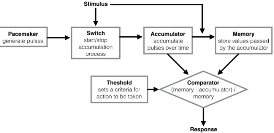
Information-processing models were first formulated by Treisman [49]
and later developed by Gibbon [50] with the scalar expectancy theory (SET)
model (Figure 1.1). SET assumes: an internal poisson-variable pacemaker that
generates pulses, an accumulator, a reference memory, a switch and a
comparator. When a time marker (cue or reward) is received, the switch allows
the pulses to be stored in the accumulator. These pulses were accumulated
until a short time after the reinforcement. The rate by which these pulses were
generated depended on many other psychological or behavioral variables,
such: as arousal, reinforcement magnitude, attention and mood [49]. By the
time of the reinforcement, the value stored in the accumulator is transferred to
the reference memory and the accumulator is reset to zero. The perceived
duration was a monotonic function of the total number of pulses transferred
into the accumulator. The behavioral response is dependent on the ratio
between the values stored in the accumulator and reference memory. For
instance, when the difference falls below a threshold (which may also vary) in a
FI task, responding at a steady rate begins. This feature explains that
steady-Pacemaker generate pulses Switch start/stop accumulation process Accumulator accumulate pulses over time
Memory
store values passed by the accumulator
Theshold
sets a criteria for action to be taken
Comparator
(memory - accumulator) / memory
Stimulus
Response
state measures of time discrimination, such as wait time (i.e., PST, break point)
on fixed interval schedules or peak-rate time (on peak procedure), are
proportional to the to-be-timed interval (i.e., proportional accuracy sensitivity to
the FI; [35]). Because SET posits that the error generated during the
accumulation of pulses is proportional to the duration criterion, it presents an
explanation for scalar variance sensitivity to the interval. More importantly, SET
incorporates two features that have been supported by experimental data.
Firstly, the current time estimate (encoding) and the memory for times
reinforced in the past (decoding) follow independent laws [27]; and secondly,
the behavior is driven by some sort of comparison between current and
remembered time of reinforcement [29].
In opposition to information processing models, sequential-state models
characterize orderly transitions between different states which can be used to
encode time [51,52,53]. The behavior theory of timing (BeT) formulated by
Killeen and Fetterman [54] and the learning-to-time (LeT) formulated by
Machado inspired by BeT [30] are the most prominent of the sequential state
models.
Figure 1.2 | Schematics of sequential state timing models. (A) Top: Frequency of occurrence of different activities of two rats over time since reinforcement on fixed interval schedule of 30 s. E = eating, D= drinking, W= in running wheel, L = contact the lever (adapted from Roper in Killeen & Fetterman [51]). Bottom: schematics of the fixed
interval schedule of reinforcement paradigm used to acquire the data in the top panel. T= time, R=responses (each subscript is one different response e.g. Eating, Drinking), S = reinforcer, and each vertical line is one occurrence of a response. (B) Schematics of Learn to Time model structure (adapted from Machado [30]. After a time marker, a set of states (top circles) is activated in series. The states may be coupled to various degrees (associative links) with one or more operant responses (bottom squares). The strength of each response is determined by the dot product between the vectors of state activation and coupling. Hence, in FI schedules multiple responses over time might be elicited with varying degrees of strength.
These models are based on the empirical observation that sequential
chains of behaviors emerge in tasks where reward delivery is contingent on
passage of time (e.g., FI, SFI; Figure 1.2A). For instance, in a FI task, behavior
would transit from consummatory, to post-consummatory, to exploration, to
reorientation to the source of reinforcer and finally to the reinforced behavior
across the interval. In BeT, each behavior is associated with a distinct
underlying state. Transitions between states occur probabilistically driven by a
poisson-variable pacemaker. The speed of this pacemaker depends on the rate
of reinforcement, so that increases in reinforcement rate lead to an increase of
the speed. The successive underlying states take on the role of a clock
process. Consequently, to perform at a temporal criteria, subjects would learn
to use their temporally organized behavioral states as discriminative stimuli.
Thus, instead of reading an internal clock, subjects are assumed to use their
current sensorimotor states to tell time. LeT extends BeT by positing that each
underlying state is associated with an operant response, and that association
strength varies through means of differential reinforcement in the context they
were learned (Figure 1.2B). Therefore, the strength of the operant response at
a given moment is the result of the combination between the predominantly
active state at that moment, and how strong is the association between this
state and the response.
Two major distinctions between information-processing and
sequential-state models may argue for the broader explanatory model of the latter models.
Firstly, in the former, the decision to respond is made only after the target time
interval has elapsed, while in BeT it is done in anticipation to that time interval
[53]. Experimental evidence points that accuracy of choice is higher under the
prospective conditions than under the retrospective condition. Indeed, under
retrospective condition, performance returned to baseline levels [55],
suggesting that animals' approach to timing is prospective. Secondly, previous
studies have reported contextual effects on timing [56]: having learned the
discriminations 1 s (red) vs. 4 s (green) and 4 s (blue) vs. 16 s (yellow),
preference for the green over blue key increases with the signal duration. In
Information-processing models the memory stores are independent, and
contextual effects. In contrast LeT is sensitive to the errors that occur during
the learning of the two discriminations; these errors weaken the connection
between the behavioral states and the associated operant responses.
Therefore, these errors would bias green keys to be perceived as long and
blue as short, regardless the fact that both cues relate to the exact same
interval.
The oscillation-based model uses a library of oscillatory pacemaker
neurons, which could be independently entrained in different rhythms, to
encode a temporal waveform by forming its Fourier series. Torras [cited in 57]
said that this combination could be done either by choosing pacemakers with
appropriate oscillation periods or through plastic changes to the period of
oscillation of each cell. The beat-frequency model (BF; Figure 1.3A) and its
updated and more biologically plausible version, the striatal beat-frequency
model (SBF; Figure 1.3B), uses “beats” (i.e., frequency at which cells spike
Figure 1.3 | Schematics of beat-frequency timing models. (A) Schematics of oscillation library from a set of units over time (adapted from Miall [57]). On meaningful event at t0, all oscillations are synchronized. After that, they start to oscillate freely. By picking a subset of oscillations and responding to when they synchronize (star symbol), the model can estimate how much time has elapsed. (B) Schematics of striatal beat-frequency model (SBF; adapted from Matell [28,58]). Loops involving the cortex (CTX), the basal ganglia (BG) and thalamus implement this mechanism. The striatum act as a coincidence detector of the oscillations provided by the CTX; dopamine signals from the substantia nigra pars compact (SNc) synchronize cortical and thalamic oscillations at every meaningful event (e.g., reward) and serve as the reinforcement signal. Once synchronized neurons oscillate at their inherent periods, allowing the patterns of activity to become meaningful. Striatal spiny neurons fire when a previously reinforced pattern of input is detected, consequently impinging to itself the current oscillatory inputs through the striato-thalamic-cortical loop.
o s c il la to ry u n it
time (arbitrary unit)
t0 t1
* 1 2 3 4 A B SNc
CTX - oscillating neurons of different rates
simultaneously) between pairs or groups of oscillatory cells to store time
intervals. After resetting the oscillations with a synchronizing event, a specific
time can be encoded by selectively weighting the activity of oscillatory cells
that are currently active at the time criterion. This process is equivalent to
multiplication (e.g., 3 Hz and 5 Hz will first synchronize at 15 Hz), thus
providing an efficient process to encode long intervals with neuronal
mechanisms which operate in much shorter timescale. The SBF posits that
loops involving the cortex (CTX), the basal ganglia and the thalamus
implement these mechanisms. More specifically, the striatum would act as a
coincidence detector; the DA signals would synchronize cortical and thalamic
oscillations at every meaningful event (e.g., reward), hyper-polarizing the
striatum membrane, and thereby resetting the integration mechanisms. DA
signals would also serve as reinforcement/teaching cues, strengthening the
cortico-striatal representation of a particular duration criterion. Once
synchronized, neurons oscillate at their inherent periods, allowing the patterns
of activity to become meaningful. Striatal spiny neurons fire when a previously
reinforced pattern of input is detected, consequently informing that the time
criterion was reached. The striatum can entrain itself in the current oscillatory
inputs through the striato-thalamic-cortical loop, allowing for alterations of time
perception.
Data from striatal neurons during the delay period before an anticipated
reward or movement [59,60,61] can equally support any of these models.
Hence, it is unclear which model best describes striatal function.
Neurobiological systems involved in interval timing
In our current understanding, interval timing is a complex and primitive function
of the brain which engages multiple areas of the brain depending on
environmental and behavioral demands. Data from functional magnetic
resonance imaging (fMRI) show that multiple areas have time dependent
activity which is also affected by task and context, suggesting that interval
timing is a distributed and complex process in the brain [62]. But, not all of
instance, the primary motor cortex (M1) processes signals correlated with time
[63]. Nonetheless, M1’s ablation or manipulations do not influence timing
reports [64]. The prefrontal cortex (PFC) also has time correlated activity[65].
The PFC seems to play a role modulating time perception. Xu et al. [63] demonstrated that time reports change when PFC activity is modulated with
cooling. An other study [66] suggests that lesions in the medial prefrontal
cortex disrupt the ability to discriminate intervals in the range of seconds. Time
correlated signals can be found all over the telencephalon from the PFC [65],
to the parietal cortex [67], and even in early sensory areas [68]. As we saw,
some models rely on the assumption that the cortex provides the temporal
basis for time estimation. But timing experiments done in decorticated animals
[64] call this hypothesis into question. These experiments showed that
decorticated animals are still able to perform in interval timing tasks.
There is a major consensus that subcortical areas are critical to interval
timing. Hence, much of the recent research has been focused on the
cerebellum, hippocampus and BG. Data from these researches depict interval
timing as a distributed process in which each subcortical area contributes to
interval timing in a different and contextually dependent manner. For instance,
the cerebellum seems to have a peculiar role in interval timing. Many studies
that attempted to affect interval timing through means of cerebellar lesions
have failed [69]. Nonetheless, data from stroke patients [70] with lesion in the
middle to superior lateral dentate nuclei, especially in the left hemisphere,
suggest that the cerebellum is necessary for proper interval timing in durations
lower than 12 seconds. Why and how cerebellum contributes to timing in this
range is still to be shown. It might coordinate learned actions at a fine
timescale [71], playing a mediating role between the sub-second timing [72]
and the supra-second timing. The cerebellum receives input from the PFC
through the pontine nuclei and connects to the PFC through two paths, both
starting at the dentate nuclei: a short (mediodorsal/ventrolateral thalamic
nuclei) and a long one (reticulotegmental nucleus, penduculopontine and
ventral tegmental). These pathways could be relevant for cerebellum’s role in
On the other hand, a recent growing body of evidence [73-78] highlights
the importance of the hippocampus, an area that is usually associated to
spatial learning [79] and explicit memory [80], for interval timing. Gradual
changes in hippocampal activity are strongly influenced by time and distance
[76]. Additionally, lesions in the dorsal or ventral hippocampus produce leftward
or rightward shifts in time estimation respectively [78]. Curiously, the effects
that hippocampal inactivations have on time estimation seem to be stronger
when the time scale estimated is over one minute [81], and the temporal
discrimination is difficult (i.e., intervals with similar durations). Yet, the same
group of studies could not provide the evidence that manipulations in the
hippocampus produce disruptions on timing of intervals above a second and
below one minute. Anatomically, the hippocampus is highly connected with
other areas relevant to interval timing such as the nucleus accumbens (Nac;
[82]) and the PFC [83]. This connectivity pattern strengthens the argument that
the hippocampus has a relevant role in interval timing.
Arguably, the study of interval timing mechanisms in the BG has proven
to be more prolific regarding unveiling the biological mechanisms of interval
timing. Evidence from multiples sources implicates the BG, and more
specifically the striatum, as a locus for the representation of
supra-second-below-one-minute timing. Firstly, activity in the striatum is modulated by timing
task as shown in studies using ensemble recording techniques in animals
[84,85], and regional increase in blood flow captured by fMRI in humans during
interval timing tasks [42,86]. Secondly, striatal lesions [26], diseases that affect
the BG such as Huntington’s [87] and Attention Deficit Disorder [88], all cause
interval timing dysfunctions.
Furthermore, patients with disorders that involve meso-striatal
dopaminergic pathways, such as schizophrenia [89,90,91] and Parkinson’s
disease (PD; [92,93]), display impaired performances during interval timing
tasks. PD is characterized by a progressive degeneration of nigrostriatal
dopaminergic projections, leading to low levels of dopamine (DA) in the
striatum. These low levels of DA cause interval timing deficits which can be
alleviated by L-dopa (L-3,4-dihydroxyphenylallanine; a precursor of dopamine)
over interval timing and the therapeutic effect of L-dopa to segregate storage
from retrieval dysfunction in the temporal memory in PD patients. Malapani’s
data suggest that DA has the power to increase discrimination between
intervals on retrieval, to control the speed and the extension of internal
representation of time during encoding.
How directly DA affects time perception might be a difficult question to
answer, because DA is involved in multiple processes other than timing. For
instance, although genetic manipulations that affect the DA system in the BG
[95] cause interval timing dysfunctions, a different source of evidence [96]
suggests that DA dependent timing deficits might be a cofound of
manipulations which affect directly animals’ motivation.
Altogether, the multiple areas involved in interval timing seem to
constitute not one but multiple “internal clocks”, which use diverse sources of
information to implement aspects of interval timing. These areas appear to be
mutually influenced by each other to generate congruent temporal estimations
and subsequent adaptive behavior. A very clear example of such coordination
of multiple clocks derives from the interaction among timing mechanisms of
different time scales. For instance, the cerebellum exerts an influence in time
estimation in the second to sub-second range. It is possible that the cerebellum
exerts its influence to the BG either through modulation of thalamic input or
through projections to VTA and PFC [97-100]. Conversely, circadian timing
mechanisms can affect the interval timing indirectly by regulating DA [101].
Finally, the hippocampus might have a direct effect on the computations done
in the striatum, especially in long intervals, in which animals are more likely to
move (so distance can be an extra source of information about the rate of
change of the environment), and when information about sequence is relevant
[74,76].
Organization of the basal ganglia
As it should be clear by now, the BG play a central role in interval timing, and
part of this role is derived from its anatomical position and connectivity. The BG
are a group of subcortical nuclei localized in the core of the forebrain, ventral to
thalamus [102,103]. The BG are in a strategic position to receive input from
most of the areas of the brain and influence both motor and associative
processing. A variety of processes including reinforcement learning [104-106],
motor control [107-111], limbic [112,115] and associative functions [116-119]
depend on the BG. Although it is not yet clear how the BG integrate and
modulate information from multiple sources, it is agreed that BG’s internal
connectivity plays a major role in it.
The BG's internal connectivity is complex as it involves many overlaid
pathways through which information passes and is processed across the
multiple nuclei of the BG. These nuclei differ drastically from each other in
anatomical and histological characteristics. These differences are important
because they establish constraints and possibilities for the timing mechanisms
we are interested to explain. The classic anatomical description of the BG in
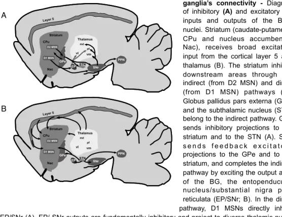
Figure 1.4 | Diagram basal ganglia’s connectivity - Diagram of inhibitory (A) and excitatory (B)
inputs and outputs of the BG’s nuclei.Striatum (caudate-putamen - CPu and nucleus accumbent - Nac), receives broad excitatory input from the cortical layer 5 and thalamus (B). The striatum inhibits downstream areas through the indirect (from D2 MSN) and direct (from D1 MSN) pathways (A). Globus pallidus pars externa (GPe) and the subthalamic nucleus (STN) belong to the indirect pathway. GPe sends inhibitory projections to the striatum and to the STN (A). STN s e n d s f e e d b a c k e x c i t a t o r y projections to the GPe and to the striatum, and completes the indirect pathway by exciting the output area of the BG, the entopenducular nucleus/substantial nigra pars reticulata (EP/SNr; B). In the direct pathway, D1 MSNs directly inhibit EP/SNr (A). EP/ SNr outputs are fundamentally inhibitory and project to diverse thalamic nuclei (e.g. md, vl, pf, vm) depending on whether the input to the striatum was associative, limbic or motor. Cortex, thalamus and striatum are connected in loop.
PPN
GPe
EP STN SNr
Thalamus vl vm pf md CPu Nac Striatum Layer5 D2 MSN D1 MSN PPN GPe
EP STN SNr
rats is organized as follows (Figure 1.4): rostrodorsally to the other BG nuclei
and most close to the CTX lies the largest division of the BG, the striatum (or
caudate-putamen); located ventro-medio-posteriorly to the striatum is the
globus pallidus (GP, external segment of the globus pallidus in primates) and
the entopeduncular nucleus (EP, internal segment of globus pallidus in
primates, GPi); the subthalamic nucleus (STN) is located ventroposteriorly to
the GP and ventrally to the thalamus; and posterior to all structures is the
substantia nigra (SN). The SN is further divided into two main parts, the dorsal
pars compacta (SNc) in which the dopaminergic nigrostriatal neurons are
located, and the more ventral pars reticulata (SNr). In addition to these
structures which are linked to motor and associative functions, there is a
ventral division of the BG associated with limbic functions. This limbic division
is composed by the ventral striatum or nucleus accumbens (Nac), ventral
pallidum and ventral tegmental area (VTA).
As the main input to the BG, the striatum receives glutamatergic input
from neurons of layers 2 and 5 from nearly the entire CTX [120-122], strong
glutamatergic afferent projections from the thalamus [124,125], dopaminergic
input from SNc [126,127] and dense GABAergic input from the GPe [128,129].
Cortical inputs to the striatum are topographically segregated according to
associative, motor, oculomotor and limbic functions. This topographical
segregation is repeated all over downstream areas of the BG [102], suggesting
some sort of parallel processing starting at the striatum. Regarding its outputs,
the striatum sends efferent inhibitory projections to the GPe and to SNr/
EP(GPi; [102,103]).
In the other extreme of the BG, the EP (GPi in primates) together with the
SNr constitute the main output of the BG. Both areas receive direct inhibitory
projections from the striatum (direct or striato-nigral pathway) and from the
GPe, in conjunction with glutamatergic excitatory input from the STN. Efferent
projections of the EP are inhibitory and target mainly the ventral thalamus.
The GPe is a nucleus of great clinical importance. It is the site where
lesions (pallidotomy) and deep-brain stimulation procedures have been applied
to alleviate PD symptoms [130]. When it comes to the connectivity, the GPe
projections to the STN and receives glutamatergic excitatory feedback
projections [128,130]. This sort of connectivity has been argued to promote an
oscillatory activity [130]. It was thought that the BG modulates cortical activity
indirectly through inhibitory projections to the thalamus, but a recent study from
Saunders et al. [131] has demonstrated that the GPe also projects GABAergic/ cholinergic projections to the frontal cortex. The existence of these projections
suggests that GPe can directly influence the cortex.
The STN is the next nucleus in the canonical indirect pathway of the BG
which has a great clinical relevance. Nowadays it is the favored site for
deep-brain stimulation in the treatment of the PD. It receives excitatory projections
from the CTX and the thalamus, as well as inhibitory projections from the
striatum and the GPe. Additionally, the STN receives cholinergic projections
from the pendunculopontine nucleus, a region involved in the control of arousal
and alertness. All the output projections that arise from the STN are
glutamatergic and excitatory. The STN projects to EP (or GPi in primates),
GPe, SNr and to the pendunculopontine nucleus. Finally, the STN also feeds
back into the striatum with sparse glutamatergic projections that are poorly
branched [132,133], and provides en passant excitatory influence over striatal cells [103].
Other output nuclei of the BG is the SN and it is subdivided in SNc and
SNr. The SNc sends broadly dopaminergic projections to the whole BG and to
other parts of the brain. It has a major modulatory role over the entire brain
processing, mostly related to the apparent encoding of errors in reward
prediction, value and saliency of events [134,135]. As discussed before, SNr
together with EP constitute the main output of the BG. SNr sends inhibitory
projections to many different regions in the brain, among which are the
pendunculopontine nucleus (
PPN), the superior colliculus and the thalamus[136]. Since there is an anatomical segregation of information in all nuclei of
the BG, depending on the cortical area from where the signal arises, the
thalamic nuclei targeted by these projections differ [137]. For instance, the
motor (lateral) circuit, that carries information from motor cortex, outputs to the
ventrolateral nucleus pars oralis (VLo), the medial part of the ventrolateral
where projections are sent back to motor areas of the cortex. In associative
(medial) circuit, that carries information from higher order cortices, the BG
projects to the VApc, the magnocellular part of the ventral anterior nucleus of
the thalamus (VAmc), the rostral division of the caudal part of the ventrolateral
nucleus (VLcr) and finally, to the lateral part of the mediodorsal nucleus of the
thalamus (MDpl). The thalamo-glutamatergic projections from these regions
target the lateral orbitofrontal cortex (LOFC) and the dorsolateral prefrontal
cortex (DLPFC), the same areas that provide input to this BG-thalamocortical
circuit.
Functionally, it has been hypothesized that the BG receive inputs from
other areas of the brain and act upon these inputs as a filter. According to this
hypothesis, the BG would select information derived from cortical and thalamic
activity and send the resulting information back to the cortical source of the
information. In parallel, it would also send copy of the same information to
other systems of the brain to implement behavior. Since DA modulates the gain
of cortico-striatal synapses [138-141], and because reinforcement-based
plasticity occurs in the BG, it is thought that this plasticity might influence the
input selection process based on previous experience [142]. The striatum and
DA are considered to be key components to this filtering process.
The way in which information travels through the multiple nuclei of the
BG might offer clues about their functional role. In the canonical perspective of
the BG, information from the striatum can pass through the nuclei of the BG by
two different parallel circuits: the direct and the indirect pathways (Figure 1.4).
Neurons within the striatum can project directly to the EP/SNr that constitute
the output nuclei of the BG (direct pathway). Or instead, striatal neurons can
project first to intermediate nuclei, namely GPe and STN, and then to the
output nuclei EP/SNr (indirect pathway; [143]).
These signaling pathways are regulated by DA in the striatum, and they
have been the subject of intense study. It is known that driving activity in the
direct pathway increases motor output (i.e., locomotion; [138,141,123]). On the
other hand, stimulation of the indirect pathway seems to inhibit behavior [123].
For long it has been hypothesized that the direct pathway encodes the set of
the direct pathway would make these motor plans stronger, and consequently
increase the motor output. Complementarily to this hypothesis, the indirect
pathway would map the set of competing behaviors which must be suppressed
so that the selected behavioral plan can occur with less interference. In this
perspective, the BG would be responsible for filtering motor, associative and
limbic information using a center-surround-like receptive field in the pertinent
space (e.g., behavioral, cognitive) [19].
Anatomy, physiology and histochemistry of striatal neurons
Closer inspection of neuronal population and its characteristics reveals an
astonishing complexity that has substantial implications to how the striatum
might implement interval timing and other related processes. Most of the
neurons with cell bodies within the striatum releases γ-aminobutyric acid
(GABA). Since they are inhibitory neurons, it becomes clear that activity inside
the striatum is either spontaneous or driven from extrinsic excitatory inputs.
Neurons in the striatum have been characterized anatomically,
histochemically and physiologically [144,145]. Regarding their anatomic
characteristics, striatal neurons can be either medium spiny projection neurons
(MSNs) or aspiny interneurons. MSNs are the most abundant cell type of the
striatum, constituting ~95% of striatal neurons [146]. MSNs are driven by
extrinsic excitatory input projections from CTX, thalamus and STN and
inhibited by interneurons and extrinsic input projections from GPe.
The MSNs are the only output neurons of the striatum [144,147], and
they can be further classified into two subpopulations according to their axonal
projection targets, the expression of genes for certain peptides, and the
expression of DA receptors. These two subpopulations have approximately the
same number of cells and they bring about the canonical direct and indirect
pathways [143], previously discussed. The first population, striatonigral
neurons, give rise to the direct pathway of the BG by sending projections
directly to the output neurons of the BG in the EP and SNr. The second
population, striatopallidal neurons, are the starting point of the indirect pathway
by connecting striatum with the EP/SNr indirectly, through the GPe and STN
activation of the indirect pathway inhibits movement [143,148]. These
functional properties of these two pathways have been tested by Kravitz [149]
and colleagues. They excited bilaterally the striatopallidal MSNs in transgenic
mice using optogenetic methods and observed that this protocol induced a
parkinsonian state (identified by increased freezing, decreased locomotor
initiations and bradykinesia). Conversely, activation of striatonigral MSNs
caused a decrease in freezing and an increase in locomotion.
MSNs show selective expression of certain peptides and receptors for
DA, depending on whether they belong to the direct or indirect pathway.
Studies using in situ hybridization histochemistry combined with retrograde
labeling of striatonigral neurons suggest that striatonigral MSNs express
substance-P, dynorphin and the DA type 1 (D1, D2) receptor, and therefore they
are also referred to as D1MSNs. The striatopallidal neurons express enkephalin
and the D2 type receptor (D2, D3, D4), and for that reason they are also known
as D2 MSNs [142].
The remaining 3-5% of striatal neurons are anatomically defined as
aspiny interneurons and include among them cholinergic interneurons and
several types of GABA-releasing interneurons [150]. Cholinergic interneurons
constitute 1-2% of the neurons in the striatum [151]; these neurons are
characterized by a large soma, often >50 µm long. They stain positively for
choline acetyltransferase (ChAT) and express both D2 and D5 receptors as
revealed by immunohistochemical analysis [152,153]. The GABAergic
interneurons appear to express mainly D5 receptors [152] and can be divided
into at least three groups based on their distinct histochemical and
physiological properties [150,154]. Histochemically, striatal GABAergic
interneurons can be subdivided into: parvalbumin (PV)-positive;
somatostatin-positive, neuropeptide Y-somatostatin-positive, and nitric oxide synthase-positive; and
calretinin-positive [154].
The anatomical and histochemical differences discussed so far have
physiological consequences in how striatal neurons behave. These three
groups of striatal GABAergic interneurons can be further divided into at least
two different types based on the firing patterns that they exhibit [154]. While
injection, somatostatin-positive interneurons display lower firing rates and
plateau potentials. Consequently, PV-positive neurons are also known as fast
spiking (FS) interneurons and somatostatin-positive interneurons are known as
low-threshold spiking (LTS) interneurons. Calretinin-positive interneurons
appear to share some characteristics of LTS interneurons, but this similarity
requires confirmation [155]. Albeit numerically scarce, striatal GABAergic
interneurons play a major role regulating spike timing in the MSNs, mainly
through feedforward inhibition [155]. Like MSNs, striatal interneurons receive
glutamatergic input from cortex, thalamus and STN; and also get GABAergic
input from the GPe. But on the contrary to MSNs, interneurons’ output is
directed primarily to MSNs and other interneurons inside the striatum. This sort
of connectivity can strategically grant to GABAergic interneurons a
disproportionately strong power to modulate striatal output despite their
numerical minority.
Physiologically, the cholinergic interneurons, also known as tonically
active neurons (TANs), display almost constant spontaneous activity. TANs are
considered to be key mediators of dopamine-dependent striatal plasticity [156]
and learning [157]. They exhibit significant hyperpolarization-activated currents,
nonetheless TANs display a pause in their tonic firing in the presence of salient
cues, including reward [144,158] which are usually followed by a rebound in
the activity. Finally, a recent study [159] has found that TANs display
fluctuations in their activity that follow changes in behavioral task conditions
(e.g., context, task rules). Moreover, these fluctuations are inflexible to
particular events (e.g., stimuli or behavior). These facts lead to the idea that
TANs encode context-dependent information [159]. Whether these TANs are
sensitive to temporal context has yet to be shown.
MSNs' average firing rates vary from 1 to 5 Hz depending on whether the
animal is sleeping or awake [160]. Some of MSNs are known to produce spike
bursts that are locked to behavior or to action sequence initiation [161]. These
characteristics are especially relevant to the study of time, as it might be
Because MSNs are the most abundant neurons in the striatum, easy to
characterize electrophysiologically and the only output neurons of the striatum,
our study focused on the activity of the MSNs in the SFI task.
Decodings and decoders
The brain faces the same elementary problem of communication when it
operates on sensorial information and controls its effectors to produce adaptive
behaviors. In communication, information from an original source must be
transformed into a code that allows this information to be stored and
transmitted over space and time in an effective way. This encoded information
can be later translated back into a format in which it is meaningful for usage.
Advances in communication technology developed our understanding of how
to translate information into different codes. We perform these translations
across different codes through the usage of encoding and decoding algorithms.
We can use these algorithms to understand how neuronal activity might
represent environmental events and how animals might use the same
information encoded in the neuronal activity to produce behavior. In the
particular scope of this thesis, we can use these algorithms to learn about how
animals implement interval timing behavior. We can achieve that either by
observing how neural time signals might emerge in face of task demands, or
by observing how animals might use these temporal signals to produce
behavior.
We assume that animals use encoding and decoding processes
themselves. Encoding is the process by which animals generate
representations of sensorial and behavioral events through the connectivity
and activity of neurons [162]; this process can be thought as a mapping
between stimuli and neuronal response. To profit from the encoded information,
the brain has to be able to infer what is happening in the real world based on
the activity dynamics of neurons. This process is called decoding and can
either refer to the process of mapping neural activity back to the original stimuli
or to behavior [162].
We can use mathematical techniques to exploit responses of one or
![Figure 1.3 | Schematics of beat-frequency timing models. (A) Schematics of oscillation library from a set of units over time (adapted from Miall [57])](https://thumb-eu.123doks.com/thumbv2/123dok_br/15766083.640510/26.748.122.598.114.341/figure-schematics-frequency-timing-schematics-oscillation-library-adapted.webp)