online | memorias.ioc.fiocruz.br
Leishmania
amazonensis
DNA in wild females of
Lutzomyia cruzi
(Diptera: Psychodidae) in the state of Mato Grosso do Sul, Brazil
Everton Falcão de Oliveira1/+, Aline Etelvina Casaril2, Nathália Lopes Fontoura Mateus2, Paula Guerra Murat2, Wagner Souza Fernandes2, Elisa Teruya Oshiro2,
Alessandra Gutierrez de Oliveira2, Eunice Aparecida Bianchi Galati1,3
1Universidade de São Paulo, Faculdade de Saúde Pública, Programa de Pós-Graduação em Saúde Pública, São Paulo, SP, Brasil 2Universidade Federal de Mato Grosso do Sul, Centro de Ciências Biológicas e da Saúde, Campo Grande, MS, Brasil
3Universidade de São Paulo, Faculdade de Saúde Pública, Departamento de Epidemiologia, São Paulo, SP, Brasil
Studies on natural infection by Leishmania spp of sandflies collected in endemic and nonendemic areas can pro-vide important information on the distribution and intensity of the transmission of these parasites. This study sought to investigate the natural infection by Leishmania in wild female sandflies. The specimens were caught in the city of Corumbá, state of Mato Grosso do Sul (Brazil) between October 2012-March 2014, and dissected to investigate flag-ellates and/or submitted to molecular analysis to detect Leishmania DNA. A total of 1,164 females (77.56% of which were Lutzomyia cruzi) representing 11 species were investigated using molecular analysis; 126 specimens of Lu. cruzi were dissected and also submitted to molecular analysis. The infection rate based on the presence of Leishma-nia DNA considering all the sandfly species analysed was 0.69%; only LeishmaLeishma-nia (LeishmaLeishma-nia) amazonensis was identified in Lu. cruzi by the molecular analysis. The dissections were negative for flagellates. This is the first record of the presence of L. (L.) amazonensis DNA in Lu. cruzi, and the first record of this parasite in this area. These find-ings point to the need for further investigation into the possible role of this sandfly as vector of this parasite.
Key words: natural infection - Lutzomyia cruzi - Leishmania amazonensis - molecular biology - polymerase chain reaction - dissection
doi: 10.1590/0074-02760150317
Financial support: FAPESP (2011/23414-0), FUNDECT/DECIT-MS/ CNPq/SES 04/2013 - PPSUS-MS - 23/200.537/2013
+ Corresponding author: efalcao.oliveira@usp.br Received 23 August 2015
Accepted 20 October 2015
The urbanisation of the human population and the transformation of the eminently rural cycle of leishmani-asis into a concomitantly urban and periurban phenom-enon with the adaptation of some species of sandflies to urban environments have contributed to an increase in the incidence of the disease in Brazil in recent decades (Gontijo & Melo 2004, Lainson & Rangel 2005, Tauil 2006). According to the Information System on Notifi-able Diseases (SINAN), 3,245 cases of visceral leish-maniasis (VL) and 3,188 cases of cutaneous leishmani-asis (CL) in humans were confirmed in the state of Mato Grosso do Sul (MS) between January 1999-December 2014 (dtr2004.saude.gov.br/sinanweb). In the city of Co-rumbá (MS), one of the oldest urban VL foci registered in Brazil, 104 human cases of VL and 21 of CL were confirmed between 2001-2013 (dtr2004.saude.gov.br/ sinanweb). However, there are no studies on genotyp-ing of Leishmania species in this city, where Lutzomyia cruzi (Mangabeira, 1938) has been suspected as a vec-tor of Leishmania (Leishmania) infantum [senior syn. of Leishmania (Leishmania) infantum chagasi (Cunha &
Chagas, 1937)]. This suspicion was based on the absence of Lutzomyia longipalpis (Lutz & Neiva, 1912), the main vector of this parasite in the Americas, together with ecological and epidemiological evidence (Galati et al. 1997), the observation of flagellates in dissected females and their identification as L. (L.) infantum by monoclo-nal antibodies (Santos et al. 1998), and the detection of the kDNA of this parasite followed by hybridisation (Pi-ta-Pereira et al. 2008). Additionally, the vectorial com-petence of Lu. cruzi for L. (L.) infantum and Leishmania (Leishmania) amazonensis Lainson & Shaw, 1972 was demonstrated experimentally when this sandfly bite and transmitted these parasites to hamsters (Oliveira 2015). Thus, the participation of this sandfly in the transmis-sion of Leishmania spp should be more investigated.
Studies investigating the natural infection of vector insects are useful to detect the intensity of the transmis-sion of Leishmania Ross, 1903 and to understand the eco-epidemiology of leishmaniasis. Such studies are essential to local health authorities in their attempts to establish prevention measures and evaluate the effectiveness of pro-grams aimed at controlling the transmission of leishmani-asis (Michalsky et al. 2002, Martín-Sánchez et al. 2006).
al. 1994, da Silva & Grunewald 1999, Aransay et al. 2000, Paiva et al. 2007), offers considerable sensitivity and spec-ificity in the detection and identification of Leishmania species (Schönian et al. 2003).
The aim of the present study was to investigate the natural infection by Leishmania in wild female sandflies caught in Corumbá through dissection to investigate flagellates and/or detection of the Leishmania DNA.
MATERIALS AND METHODS
Study area - The specimens used in the present in-vestigation were caught between October 2012-March 2014 in the urban perimeter of Corumbá (19º00’33”S 57º39’12”W; 118 m above sea level), which is located in the northeastern portion of MS (Central-West Brazil). The municipality has an area of 64,962.8 km2, which
represents 18.19% of the total area of the state, and is located 415 km from the state capital (Campo Grande) in the Pantanal wetland region on the border with Bolivia.
Five collection sites (convenience sampling) were de-termined in neighbourhoods with records of human cases of VL in the year prior to the beginning of the study: four residential areas in the peripheral region and one in the commercial district of the city. Table I displays a brief de-scription of the characteristics of each collection site.
Collections and acquisition of sandflies for molecu-lar analysis - Automatic light traps were installed week-ly in the peridomicile area of the five residences select-ed. For the identification of females, the genitalia were dissected on slides containing a drop of saline solution, whereas males were clarified and mounted on slides in balsam. The identification of both sexes was performed as described by Galati (2014). Engorged females and those whose entire bodies were clarified for the identifi-cation of the species were not included in the study.
In the first six months of analysis (October 2012-March 2013), females were grouped in pools of up to 10 insects of the same species, location and collection date, and placed individually in 1.5 mL microtubes with isopropyl alcohol and stored at -20ºC for subsequent PCR. The remaining specimens were placed individu-ally in 1.5 mL microtubes.
Collections and acquisition of sandflies for dissec-tion - A sample of females was dissected to investi-gate the presence of flagellates in accordance with the method described by Johnson et al. (1963). The speci-mens were caught with an aspirator in a chicken coop (the same collection site in the neighbourhood of Maria Leite used for the light trap collection) between 07:00 pm-09:00 pm on three different days (1 day in Septem-ber 2013, 1 in NovemSeptem-ber 2013, and the last in DecemSeptem-ber 2013). After exposing the gut and spermathecae of the females for the investigation of flagellates and species identification, respectively, the contents on the slide (gut, thorax, and head) were transferred to 1.5 mL mi-crotubes with isopropyl alcohol and stored at -20ºC for subsequent PCR. The specimens negative in the direct exam for flagellates were grouped in pools of up to ten.
PCR and restriction fragment length polymorphism analyses - For DNA extraction, the specimens were ground with the aid of a plastic pestle in 1.5 mL tubes
with 300 μL of 5% Chelex® resin solution (Bio-Rad,
USA). The solution was mixed in a vortex for 15 s, cen-trifuged at 13,000 rpm for 60 s and placed in a water bath at 80ºC for 30 min. The vortex and centrifugation pro-cedures were repeated and the supernatant was removed and transferred to a different sterile Eppendorf tube. The extraction product was stored at -20ºC.
PCR was performed targeting a region of the internal transcribed spacer (ITS) of the Leishmania ribosomal
TABLE I
General characteristics of sampling sites in the city of Corumbá, state of Mato Grosso do Sul, Brazil, April 2012-March 2014
Residence
(neighbourhood) Geographical location
Domesticated animals (n)
Centro Central region of city; sampling
site closest to Paraguay River (approximately 500 m)
Dogs (2) Chicken (1) Cristo Redentor Southeastern
periphery of the city
Dog (1)
Maria Leite Northeastern periphery of the city
Dogs (2) Chickens (15a)
Geese (5) Ducks (3)
Nova Corumbá Southern
periphery of the city
Dogs (5) Chickens (4)
Cats (3) Popular Nova Southeastern
periphery of the city
Dog (1)
gene (ITS1) with approximately 300 bp. Five microlitre
of the sample, 12.5 μL of GoTaq® Green Master Mix (Promega, USA), 5.5 μL of water, and 1 μL of each oligo -nucleotide [LITSR (5’-CTGGATCATTTTCCGATG-3’) and L5.8S (5’-TGATACCACTTATCGCACTT-3’)] were
added for a final reaction volume of 25 μL (El Tai et al.
2000). The following manipulations were the amplifica-tion condiamplifica-tions in the thermal cycler (BIOER, China): 95ºC for 3 min followed by 35 cycles of 95ºC for 30 s, 53ºC for 30 s, and 72ºC for 1 min, with post-extension at 72ºC for 5 min. The negative controls were a reaction without DNA containing water and DNA from nonfed F1 females. The positive controls were DNA from L. (L.) infantum (MHOM/BR/1972/BH46) and L. (L.) amazo-nensis (IFLA/BR/1967/PH8) extracted from cultures.
The PCR products were viewed using electrophore-sis with 1.5% agarose gel in 100 mL of Tris-borate-eth-ylenediamine tetraacetic acid (TBE) buffer stained with GelRedTM (Biotium, USA). The electrophoretic run was
performed at 100 V for 100 min in concentrated TBE buffer. Viewing of the bands was performed using ultra-violet light with a 300-nm filter.
The products from positive samples were submit-ted to HaeIII restriction enzyme digestion (isolasubmit-ted from Haemophilus aegyptius), which cleaves frag-ments in segfrag-ments that have the 5’....GG▼CC....3’ or 3’.... CC▲GG....5’ sequence to identify the species of Leish-mania (Schönian et al. 2003). One microlitre of 10x buf-fer, one unit of HaeIII enzyme and 1 mg of DNA from the PCR were used and the volume was completed with 10 mL of ultrapure water. The sample was incubated in a water bath at 37ºC overnight. The material was then submitted to electrophoresis in 2% polyacrylamide gel with TBE buffer for 3 h.
Ethics - This study received the approval of the Ani-mal Experimentation Ethical Committee of the Federal University of Mato Grosso do Sul (Brazil) under process 491/2013. The research group has a permanent license for the collection of zoological material issued by the Brazilian Institute of Environment and Renewable Natu-ral Resources (SISBio 25952-1).
The field studies were carried out on private lands and the owners gave permission to conduct the collec-tions and acquisition of sandflies in their peridomicile areas. Further, the field studies did not involve endan-gered or protected species.
RESULTS
During the weekly collections with light traps be-tween October 2012-March 2014, 9,759 specimens (8,278 males and 1,481 females) were collected, belonging to 13 species: Brumptomyia brumpti (2♀), Evandromyia cortelezzii (4♀), Evandromyia aldafalcaoae (3♂, 5♀), Evandromyia corumbaensis (41♂, 115♀), Evandromyia sallesi (6♂, 16♀), Evandromyia walkeri (4♀), Lu. cruzi
(8,061♂, 1,147♀), Lutzomyia forattinii (125♂, 159♀), Mi-cropygomyia peresi (33♂, 10♀), Martinsmyia oliveirai
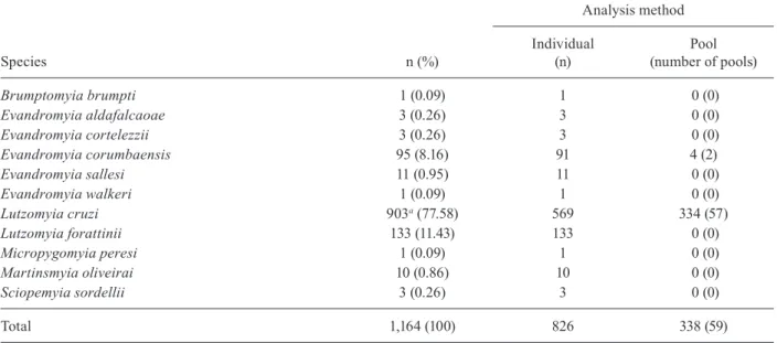
(8♂, 12♀), Psathyromyia bigeniculata (1♂, 2♀), Sci-opemyia sordellii (4♀), and Nyssomyia whitmani (1♀). Among the total number of females collected, 1,038 were investigated for natural infection by Leishmania using only PCR. Another 126 females collected with an aspirator were dissected for the study of flagellates and subsequently analysed by the same method. Thus, 1,164 females were analysed by PCR (Table II).
Only eight of the 1,164 females (0.69%) investigated exhibited a DNA band characteristic of Leishmania (300 bp). All naturally infected females were Lu. cruzi, caught
TABLE II
Distribution of the sandfly females investigated for natural infection by Leishmania according to species and type of analysis
Species n (%)
Analysis method
Individual (n)
Pool (number of pools)
Brumptomyia brumpti 1 (0.09) 1 0 (0)
Evandromyia aldafalcaoae 3 (0.26) 3 0 (0)
Evandromyia cortelezzii 3 (0.26) 3 0 (0)
Evandromyia corumbaensis 95 (8.16) 91 4 (2)
Evandromyia sallesi 11 (0.95) 11 0 (0)
Evandromyia walkeri 1 (0.09) 1 0 (0)
Lutzomyia cruzi 903a (77.58) 569 334 (57)
Lutzomyia forattinii 133 (11.43) 133 0 (0)
Micropygomyia peresi 1 (0.09) 1 0 (0)
Martinsmyia oliveirai 10 (0.86) 10 0 (0)
Sciopemyia sordellii 3 (0.26) 3 0 (0)
Total 1,164 (100) 826 338 (59)
on the same night in a single trap installed in the Maria Leite neighbourhood in June 2013. On this occasion, 272 sandflies were captured, 248 of them being males and 22 females, all of Lu. cruzi, and one male each of Lu. forattinii and of Mt. oliveirai. Thus, the natural infection rate of Lu. cruzi was 0.89% (8/903) based on the presence of Leishma-nia DNA. The parasite identified in all the amplified prod-ucts was L. (L.) amazonensis (200 bp and 140 bp) (Figure). Regarding the 126 females dissected for the study of flagel-lates, all were negative with both methods employed for the investigation of natural infection by Leishmania.
DISCUSSION
This study identified the natural infection of a sand-fly species by a Leishmania species not yet reported dur-ing a regular survey of collections. Periodic surveys for the detection of natural infection by Leishmania are im-portant for the understanding of the components of the parasite transmission chain. The detection of naturally infected sandfly species that have not previously been suspected as vectors of any Leishmania species demon-strates the need for studies to investigate their vectorial competence and the identification of permissive vectors (Kamhawi 2006, Paiva et al. 2007).
In the present study, the overall infection rate was 0.69% and the rate for Lu. cruzi alone was 0.89%. No flagellate forms were found in the dissected specimens and PCR was negative for all the pools analysed in this group. The females dissected were caught in a chicken coop close to the trap to which the positive females were attracted. In the chicken coop the females were collected with an aspirator while resting on the walls and birds. On the other hand, the light trap having light as its at-traction may attract females which have had blood meals on various animals, including mammals which serve as reservoirs of Leishmania.
Although Lu. cruzi and Lu. longipalpis females are morphologically indistinguishable constituting a complex of species (Young & Duncan 1994, Galati 2014), and de-spite the fact that Santos et al. (2003) reported the collec-tion of three Lu. longipalpis males in Corumbá, the find-ing of only Lu. cruzi males in the present investigation, as well as in previous studies (Galati et al. 1985, 1997, Santos et al. 1998, Pita-Pereira et al. 2008, Almeida et al. 2010, Casaril et al. 2014, Oliveira 2015), led us to identify all the females of this complex captured as Lu. cruzi.
Different methods with different degrees of sensitiv-ity and specificsensitiv-ity have been employed for the detection of natural infection in blood-feeding insects, such as dis-section for the direct study of flagellates, inoculation in experimental animal models and isolation of the parasite in a culture medium with dissected insects (Deane 1956, Lainson et al. 1985, Sherlock 1996). Although more ex-pensive in comparison with other methods, PCR is a prac-tical tool with high degrees of sensitivity and specificity and allows the grouping of individuals in pools (Schönian et al. 2003, Paiva et al. 2006, Savani et al. 2009). Howev-er, pooling may lead to the underestimation of the natural infection rate, as it is not possible to identify how many individuals were actually infected. In such cases, the cal-culation of the minimum infection rate (Paiva et al. 2006, 2010) and the estimation of the prevalence of infection using an algorithm (Katholi et al. 1995, Martín-Sánchez et al. 2006) have been employed. In order to estimate more accurately the possible natural infection rate, it was decided to analyse the specimens caught in light traps during regular collections from April 2013 individually.
This is the first report of the natural infection of Lu. cruzi by L. (L.) amazonensis and of the presence of this parasite in Corumbá. Although Lu. cruzi has been stud-ied little, the others reports of the finding of Leishmania DNA in wild females relate to L. (L.) infantum. Santos et al. (1998) found a 0.39% infection rate based on the dis-section of 3,575 specimens of Lu. cruzi. Another 1,013 sandflies of seven different species were dissected and no flagellate forms were found. Based on these findings, the authors implicated Lu. cruzi as a vector of L. (L.) in-fantum in Corumbá (Santos et al. 1998). In the same mu-nicipality, Pita-Pereira et al. (2008) found a 1.5% mini-mum infection rate in Lu. cruzi by L. (L.) infantum using the minicircle region of kDNA as the target of multiplex PCR with hybridisation. The minicircle region of kDNA has a high degree of sensitivity and is capable of detect-ing minimal quantities of Leishmania DNA (Smyth et al. 1992, Freitas-Lidani 2014). However, the minicircle region of kDNA only permits the identification of the genus of the parasite (Schönian et al. 2003).
For Lu. longipalpis, which is a confirmed vector of L. (L.) infantum, the first record of wild females naturally infected by L. (L.) amazonensis occurred in the city of Antônio João, located in the southeastern portion of MS, on the border with Paraguay (Paiva et al. 2006). Subse-quently, other authors also found Lu. longipalpis naturally infected by the same parasite in the city of Bonito (Savani et al. 2009). These two locations, Antônio João and Bo-nito, are respectively endemic for canine VL and CL.
Digestion of amplified products from internal transcribed spacer 1 region of Leishmania with HaeIII restriction enzyme. Lane 1: ladder marker with 100 bp; 2: negative control (reaction without DNA con-taining water); 3-10: sample of wild sandflies naturally infected by
Leishmania (Leishmania) amazonensis; 11: positive control by Leish-mania (LeishLeish-mania)infantum (MHOM/BR/1972/BH46); 12: positive control L. (L.)amazonensis (IFLA/BR/1967/PH8); 13: sample not di-gested by HaeIII; 14: negative control (DNA from nonfed F1 females);
The fact that the eight Lu. cruzi females were caught on a single night with a single CDC trap installed in a peridomicile area demonstrates that the parasite seems not to be dispersed throughout the urban area. Addition-ally, in this case, a common source is strongly suggested for natural infection because only nonengorged females were analysed. The observation of a rodent of the genus Dasyprocta, considered a secondary host of L. (L.) ama-zonensis (Lainson et al. 1994, Ashford 2000, Lainson & Shaw 2005), in the peridomicile area where the infected specimens were collected may explain these results. Fac-tors that can contribute to the presence of this rodent in the area include the location of the dwelling on the outskirts of the town, a yard fenced with barbed wire, the presence of fruit trees, and a chicken coop, the base of which was suspended approximately 20 cm above the ground, leav-ing a clearance in which the animal was observed.
L. (L.) amazonensis has been recorded in Bolivia, Brazil, Colombia, French Guyana and Paraguay. In Brazil, this parasite has been found in all regions, es-pecially the Amazon Region. However, it is likely that the geographical distribution of L. (L.) amazonensis is broader than is currently known and that it also extends into other countries of South America where its sandfly vector is found (Lainson & Shaw 1987, 2005, Grimaldi et al. 1989, MS/SVE 2010). This parasite is implicated as an etiological agent of CL (Lainson & Shaw 1987, 2005). However, there are also human cases of VL, diffuse or anergic leishmaniasis and post-kala-azar dermal leish-maniasis attributed to this parasite (Barral et al. 1991, Aleixo et al. 2006) as well as canine VL (Tolezano et al. 2007, Hoffmann et al. 2012).
In the first year of study (2012), two autochthonous cases of CL were reported in the municipality of Corumbá. In the two following years (2013-2014), there were no re-ported cases of CL. Between January-July 2015, only one case of mucosal leishmaniasis was reported (MS 2015). However, due to lack of studies on aetiology and/or ge-notyping of Leishmania species in Corumbá, it is not pos-sible infer that the aetiology of human cases of the disease. As observed for Lu. longipalpis, it is possible that Lu. cruzi also has a permissive character and permits infection by other species of the genus Leishmania, which suggests that the adhesion mechanism of the para-site through lipophosphoglycans is not species specific or may occur by means of other mechanisms (Volf & Myskova 2007). This has important implications for the transmission and evolution of the parasite, as it may con-tribute to the dispersal of Leishmania due to its ability to adapt to new vectors (Myskova et al. 2007), since the main vector of L. (L.) amazonensis, Bichromomyia fla-viscutellata (Mangabeira, 1942), has not yet been found in the region (Galati et al. 1985, 1997, Santos et al. 1998, Almeida et al. 2010, Casaril et al. 2014, Oliveira 2015).
The incrimination and subsequent confirmation of a species as a vector of Leishmania should be based on several criteria, the first of which is the discovery of nat-urally infected wild females through the detection of the flagellate forms of the parasite on more than one occa-sion (Killick-Kendrick & Ward 1981, Killick-Kendrick 1990). Another criterion is the isolation and typing of promastigotes from females that have not fed for more than 36 h (Ready 2013).
Due to its epidemiological complexity, cutaneous and VL, the aetiology of which is attributed to L. (L.) ama-zonensis is characterised as a disease that is difficult to control and requires specific measures depending on the area of occurrence. Therefore, besides the establishment of early diagnosis and treatment, the proper identifica-tion of the species of Leishmania and the determinaidentifica-tion of its area of distribution are essential to the planning and adoption of prevention measures and the reduction of the exposure of the human population to the vector (Dorval et al. 2006, MS/SVE 2010).
Considering that L. (L.) infantum and L. (L.) amazo-nensis were identified in Corumbá and the lack of studies on aetiology of human and canine cases of leishmaniases, both visceral and cutaneous forms, in addition to clinical and epidemiological aspects, attention special should be given to the identification by genotyping of the parasites.
Experimental infection studies, undertaken by this re-search group, with both species, are currently underway and are necessary to gain a better understanding of the parasite-vector interaction. Studies for the identification of reservoir hosts should also be conducted, since some wild and domesticated mammals are considered to be hosts of different species of Leishmania (Ashford 2000).
In short, L. (L.) amazonensis DNA was detected for the first time in Lu. cruzi collected in the urban area of Co-rumbá, an endemic area for VL and CL. The authors would like to emphasize the importance of this finding, since Lu. cruzi, a suspected vector of L. (L.) infantum and adapted to the urban environment, could contribute to the dispersion and urbanisation of L. (L.) amazonensis. It is further rel-evant the fact that this species has been found associated with human and canine VL cases. Therefore, these findings point to the need for further investigation into the possible role of this sandfly as vector of this parasite.
ACKNOWLEDGEMENTS
To the Zoonosis Control Centre of Corumbá, for technical assistance and help during capture of sandflies.
REFERENCES
Aleixo JA, Nascimento ET, Monteiro GR, Fernandes MZ, Ramos AMO, Wilson ME, Pearson RD, Jeronimo SMB 2006. Atypical American visceral leishmaniasis caused by disseminated Leish-mania amazonensis infection presenting with hepatitis and ade-nopathy. Trans R Soc Trop Med Hyg100: 79-82.
Almeida PS, Nascimento JC, Ferreira AD, Minzão LD, Portes F, Miran-da AM, FaccenMiran-da O, Andrade Filho JD 2010. Espécies de flebotomí-neos (Diptera, Psychodidae) coletadas em ambiente urbano em municípios com transmissão de leishmaniose visceral do estado de Mato Grosso do Sul, Brasil. Rev Bras Entomol54: 304-310. Aransay AM, Scoulica E, Tselentis Y 2000. Detection and
identifica-tion of Leishmania DNA within naturally infected sand flies by seminested PCR on minicircle kinetoplastic DNA. Appl Environ Microbiol66: 1933-1938.
Ashford RW 2000. The leishmaniases as emerging and reemerging zoonoses. Int J Parasitol30: 1269-1281.
Casaril AE, Monaco NZN, Oliveira EF, Eguchi GU, Paranhos Filho AC, Pereira LE, Oshiro ET, Galati EAB, Mateus NLF, Oliveira AG 2014. Spatiotemporal analysis of sandfly fauna (Diptera: Psy-chodidae) in an endemic area of visceral leishmaniasis at Panta-nal, central South America. Parasit Vectors7: 364.
da Silva OS, Grunewald J 1999. Contribution to the sand fly fauna (Diptera: Phlebotominae) of Rio Grande do Sul, Brazil, and Leish-mania (Viannia) infections. Mem Inst Oswaldo Cruz94: 579-582. Deane LM 1956. Leishmaniose visceral no Brasil. Estudos sobre
reservatórios e transmissores realizados no estado do Ceará, Serviço Nacional de Educação Sanitária, Rio de Janeiro, 162 pp. Dorval MEC, Oshiro ET, Cupollilo E, Camargo AC, Alves TP 2006.
Ocorrência de leishmaniose tegumentar americana no estado do Mato Grosso do Sul associada à infecção por Leishmania (Leish-mania)amazonensis. Rev Soc Bras Med Trop39: 43-46. El Tai NO, Osmar OF, El Fari M, Presber WH, Schönian G 2000.
Genetic heterogeneity of ribosomal internal transcribed spacer in clinical samples of Leishmania donovani spotted on filter paper as revealed by single-strand conformation polymorphisms and sequencing. Trans R Soc Trop Med Hyg94: 575-579.
Feliciangeli MD, Rodriguez N, Bravo A, Arias F, Guzman B 1994. Vectors of cutaneous leishmaniasis in north-central Venezuela.
Med Vet Entomol8: 317-324.
Freitas-Lidani KC, de Messias-Reason IJ, Ishikawa EAY 2014. A comparison of molecular markers to detect Lutzomyia longipal-pis naturally infected with Leishmania(Leishmania)infantum.
Mem Inst Oswaldo Cruz109: 442-447.
Galati EAB 2014. Phlebotominae (Diptera, Psychodidae): classificação, morfologia, terminologia e identificação de adultos. Available from: fsp.usp.br/egalati/ApostilaPhlebotominae_2014_vol_I.pdf. Galati EAB, Nunes VLB, Rego Jr FA, Oshiro ET, Rodrigues M 1997.
Estudo de flebotomíneos (Diptera, Psychodidae) em foco de leishmaniose visceral no estado de Mato Grosso do Sul, Brasil.
Rev Saude Publica31: 378-390.
Galati EAB, Rego Jr FA, Nunes VLB, Oshiro ET 1985. Fauna flebot-omínica do município de Corumbá, Mato Grosso do Sul, Brasil, e descrição de Lutzomyia forattinii, sp. n. (Diptera, Psychodidae, Phlebotominae). Rev Bras Entomol29: 261-266.
Gontijo CMF, Melo MN 2004. Leishmaniose visceral no Brasil: quadro clínico, desafios e perspectivas. Rev Bras Epidemiol7: 338-349. Grimaldi GJ, Tesh RB, McMahon-Pratt DA 1989. Review of the
ge-ographic distribution and epidemiology of leishmaniasis in the New World. Am J Trop Med Hyg41: 687-725.
Hoffmann AR, Navarro IT, Camargo Jr VE, Caldart ET, Breganó RM, Pereira PM 2012. Leishmania amazonensis in dog with clin-ical diagnosis of visceral leishmaniasis in Paraná state, Brazil - a case report. Semina33 (Suppl. 2): 3265-3270.
Johnson PT, McConnell E, Hertig M 1963. Natural infections of lep-tomonad flagellates in Panamanian Phlebotomus sandflies. Exp Parasitol14: 107-122.
Kamhawi S 2006. Phlebotomine sand flies and Leishmania parasites: friends or foes? Trends Parasitol22: 439-445.
Katholi CR, Toe L, Merriweather A, Unnasch TR 1995. Determining the prevalence of Onchocerca volvulus infection in vector popu-lations by polymerase chain reaction screening of pools of black flies. J Infect Dis172: 1414-1417.
Killick-Kendrick R 1990. Phlebotomine vectors of the leishmaniasis: a review. Med Vet Entomol4: 1-24.
Killick-Kendrick R, Ward RD 1981. Ecology of Leishmania. Parasi-tology82: 143-152.
Lainson R, Rangel EF 2005. Lutzomyia longipalpis and the eco-epide-miology of American visceral leishmaniasis, with particular refer-ence to Brazil - A Review. Mem Inst Oswaldo Cruz100: 811-827. Lainson R, Shaw JJ 1987. Evolution, classification and geographical
dis-tribution. In W Peters, R Killick-Kendrick (eds.), The leishmaniases in biology and medicine, Academic Press, London, p. 12-120. Lainson R, Shaw JJ 2005. New world leishmaniasis. In FEG Cox, D
Wakelin, SH Gillespie, DD Despommier (eds.), Topley & Wil-son’s microbiology and microbial infections: parasitology, 10th ed., Hodder Arnold ASM Press, London, p. 313-349.
Lainson R, Shaw JJ, Ryan L, Ribeiro RSM, Silveira FT 1985. Leishma-niasis in Brazil. XXI. Visceral leishmaLeishma-niasis in the Amazon Region and further observations on the role of Lutzomyia longipalpis (Lutz & Neiva, 1912) as the vector. Trans R Soc Trop Med Hyg79: 223-226. Lainson R, Shaw JJ, Silveira FT, de Souza AAA, Braga RR, Ishikawa EAY 1994. The dermal leishmaniases of Brazil, with special ref-erence to the ecoepidemiology of the disease in Amazonia. Mem Inst Oswaldo Cruz89: 435-443.
Martín-Sánchez J, Gállego M, Barón S, Castillejo S, Morillas-Marquez F 2006. Pool screen PCR for estimating the prevalence of Leishma-nia infantum infection in sandflies (Diptera: Nematocera, Phlebot-omidae). Trans R Soc Trop Med Hyg100: 527-532.
Michalsky EM, Fortes-Dias CL, Pimenta PF, Secundino NF, Dias ES 2002. Assessment of PCR in the detection of Leishmania spp in experimentally infected individual phlebotomine sandflies (Dip-tera: Psychodidae: Phlebotominae). Rev Inst Med Trop Sao Paulo 44: 255-259.
MS - Ministério da Saúde Brasil 2015. Sistema de Informação de Agra-vos de Notificação. Available from: dtr2004.saude.gov.br/sinanweb. MS/SVE - Ministério da Saúde/Superintendência de Vigilância Epi-demiológica Brasil 2010. Manual de vigilância da leishmaniose tegumentar americana, MS, Brasília, 180 pp.
Myskova J, Svobodova M, Beverley SM, Volf P 2007. A lipophospho-glycan-independent development of Leishmania in permissive sand flies. Microbes Infect9: 317-324.
Oliveira EF 2015. Vectorial capacity of Lutzomyia (Lutzomyia) cruzi
(Diptera: Psychodidae) for Leishmania (Leishmania) infantum, PhD Thesis, Universidade de São Paulo/Faculdade de Saúde Pública, São Paulo, 203 pp.
Paiva BR, Oliveira AG, Dorval MEMC, Galati EAB, Malafronte RS 2010. Species-specific identification of Leishmania in naturally infected sand flies captured in Mato Grosso do Sul state, Brazil.
Acta Trop115: 126-130.
Paiva BR, Secundino NFC, Nascimento JC, Pimenta PFP, Galati EAB, Andrade-Júnior HF, Malafronte RS 2006. Detection and identifi-cation of Leishmania species in field-captured phlebotomine sand-flies based on mini-exon gene PCR. Acta Trop99: 252-259. Paiva BR, Secundino NFC, Pimenta PFP, Galati EAB, Andrade-Júnior
HF, Malafronte RS 2007. Padronização de condições para de-tecção de DNA de Leishmania spp em flebotomíneos (Diptera, Psychodidae) pela reação em cadeia da polimerase. Cad Saude Publica23: 87-94.
Perez JE, Ogusuku E, Inga R, Lopez M, Monje J, Paz L, Nieto E, Arevalo J, Guerra H 1994. Natural Leishmania infection of Lut-zomyia spp in Peru. Trans R Soc Trop Med Hyg88: 161-164. Perruolo G, Rodríguez NN, Feliciangeli MD 2006. Isolation of
Leish-mania(Viannia)braziliensis from Lutzomyia spinicrassa (spe-cies group Verrucarum) Morales Osorno Mesa, Osorno and Hoy-os 1969, in the Venezuelan Andean Region. Parasite13: 17-22. Pita-Pereira D, Alves CR, Souza MB, Brazil RP, Bertho AL, de
in-fected Lutzomyia intermedia and Lutzomyia migonei with Leish-mania(Viannia)braziliensis in Rio de Janeiro (Brazil) revealed by a PCR multiplex non-isotopic hybridisation assay. Trans R Soc Trop Med Hyg99: 905-913.
Pita-Pereira D, Cardoso MAB, Alves CR, Brazil RP, Britto C 2008. Detection of natural infection in Lutzomyia cruzi and Lutzomyia forattinii (Diptera: Psychodidae: Phlebotominae) by Leishmania infantum chagasi in an endemic area of visceral leishmaniasis in Brazil using a PCR multiplex assay. Acta Trop107: 66-69. Ready PD 2013. Biology of phlebotomine sand flies as vectors of
dis-ease agents. Annu Rev Entomol58: 227-250.
Santos SO, Arias J, Hoffmann MP, Furlan MBG, Ferreira WF, Pereira C, Ferreira L 2003. The presence of Lutzomyia longipalpis in a focus of American visceral leishmaniasis where the only proven vector is Lutzomyia cruzi, Corumbá, Mato Grosso do Sul state.
Rev Soc Bras Med Trop36: 633-634.
Santos SO, Arias J, Ribeiro AA, Hoffmann MP, Freitas RA, Malac-co MAF 1998. Incrimination of Lutzomyia cruzi as a vector of American visceral leishmaniasis. Med Vet Entomol12: 315-317. Savani ES, Nunes VL, Galati EAB, Castilho TM, Zampieri RA,
Floeter-Winter LM 2009. The finding of Lutzomyia almerioi and
Lutzomyia longipalpis naturally infected by Leishmania spp in a cutaneous and canine leishmaniasis focus in Serra da Bodoque-na, Brazil. Vet Parasitology160: 18-24.
Schönian G, Nascreddin A, Dinse N, Schweynoch C, Schallig HD, Presber W, Jaffe CL 2003. PCR diagnosis and characterization of
Leishmania in local and imported clinical samples. Diagn Micro-biol Infect Dis47: 349-358.
Sherlock IA 1996. Ecological interactions of visceral leishmaniasis in the state of Bahia, Brazil. Mem Inst Oswaldo Cruz91: 671-683. Smyth AJ, Ghosh A, Hassan MQ, Basu D, De Bruijn MH, Adhya S,
Mallik KK, Barker DC 1992. Rapid and sensitive detection of
Leishmania kinetoplast DNA from spleen and blood samples of kala-azar patients. Parasitology105: 183-192.
Tauil PL 2006. Perspectivas de controle de doenças transmitidas por vetores no Brasil. Rev Soc Bras Med Trop39: 275-277.
Tolezano JE, Uliana SRB, Taniguchi HH, Araújo MFL, Barbosa JAR, Barbosa JER, Floeter-Winter LM, Shaw JJ 2007. The first records of Leishmania(Leishmania)amazonensis in dogs (Canis fami- liaris) diagnosed clinically as having canine visceral leishmania-sis from Araçatuba county, São Paulo state, Brazil. Vet Parasitol 149: 280-284.
Volf P, Myskova J 2007. Sand flies and Leishmania: specific versus
permissive vectors. Trends Parasitol23: 91-92.