Multicentre study highlighting clinical relevance of new high-throughput
methodologies in molecular epidemiology of
Pneumocystis jirovecii
pneumonia
F. Esteves1, B. de Sousa2, E. J. Calderón3, L. Huang4, R. Badura5, F. Maltez6, Q. Bassat7,8, Y. de Armas9, F. Antunes10and O. Matos11 1) NOVA Medical School/Faculdade de Ciências Médicas, Centro de Toxicogenómica e Saúde Humana (ToxOmics), Departamento de Genética, Universidade Nova de Lisboa, Lisbon, 2) Faculdade de Psicologia e de Ciências da Educação, Coimbra, Portugal, 3) CIBER de Epidemiologia y Salud Publica and Instituto de Biomedicina de Sevilla, Hospital Universitario Virgen del Rocio/CSIC/Universidad de Sevilla, Seville, Spain, 4) Division of Pulmonary and Critical Care Medicine & HIV/AIDS Division, San Francisco General Hospital, Department of Medicine, University of California San Francisco, San Francisco, CA, USA, 5) Centro Hospitalar de Lisboa Norte, Hospital de Santa Maria, Faculdade de Medicina, Universidade de Lisboa, 6) Centro Hospitalar de Lisboa Central, Hospital de Curry Cabral, Serviço de Doenças Infecciosas, Lisbon, Portugal, 7) ISGlobal, Barcelona Ctr. Int. Health Res. (CRESIB), Hospital Clínic Universitat de Barcelona, Barcelona, Spain, 8) Centro de Investigação em Saúde de Manhiça (CISM), Maputo, Mozambique, 9) Laboratory of Molecular Diagnostic, Pedro Kourí Hospital, Havana, Cuba, 10) Instituto de Saúde Ambiental, Faculdade de Medicina da Universidade de Lisboa and 11) Unidade de Parasitologia Médica, Grupo de Protozoários Oportunistas/VIH e Outros Protozoários, Global Health and Tropical Medicine, Instituto de Higiene e Medicina Tropical, Universidade Nova de Lisboa, Lisbon, Portugal
Abstract
Pneumocystis jirovecii causes severe interstitial pneumonia (PcP) in immunosuppressed patients. This multicentre study assessed the distribution frequencies of epidemiologically relevant genetic markers of P. jirovecii in different geographic populations from Portugal, the USA, Spain, Cuba and Mozambique, and the relationship between the molecular data and the geographical and clinical information, based on a multifactorial approach. The high-throughput typing strategy for P. jirovecii characterization consisted of DNA pooling using quantitative real-time PCR followed by multiplex-PCR/single base extension. The frequencies of relevant P. jirovecii single nucleotide polymorphisms (mt85, SOD110, SOD215, DHFR312, DHPS165 and DHPS171) encoded at four loci were estimated in ten DNA pooled samples representing a total of 182 individual samples. Putative multilocus genotypes of P. jirovecii were shown to be clustered due to geographic differences but were also dependent on clinical characteristics of the populations studied. The haplotype DHFR312T/SOD110C/ SOD215T was associated with severe AIDS-related PcP and high P. jirovecii burdens. The frequencies of this genetic variant of P. jirovecii were significantly higher in patients with AIDS-related PcP from Portugal and the USA than in the colonized patients from Portugal, and Spain, and children infected with P. jirovecii from Cuba or Mozambique, highlighting the importance of this haplotype, apparently associated with the severity of the disease and specific clinical groups. Patients from the USA and Mozambique showed higher rates of DHPS mutants, which may suggest the circulation of P. jirovecii organisms potentially related with trimethoprim-sulfamethoxazole resistance in those geographical regions. This report assessed the worldwide distribution of P. jirovecii haplotypes and their epidemiological impact in distinct geographic and clinical populations.
© 2016 European Society of Clinical Microbiology and Infectious Diseases. Published by Elsevier Ltd. All rights reserved.
Keywords: DNA pools, high-throughput molecular epidemiology, immunosuppressed patients, multilocus genotyping survey, Pneumocystis jirovecii, pneumonia
Original Submission: 29 December 2015; Revised Submission: 19 February 2016; Accepted: 13 March 2016 Editor: E. Roilides
Article published online: 26 March 2016
Corresponding author: O. Matos, Unidade de Parasitologia Médica, Grupo de Protozoários Oportunistas/VIH e Outros Protozoários, Global Health and Tropical Medicine, Instituto de Higiene e Medicina Tropical, Universidade Nova de Lisboa, Rua da Junqueira N° 100, 1349-008 Lisbon, Portugal
E-mail:[email protected]
Clin Microbiol Infect 2016; 22: 566.e9–566.e19
Introduction
Pneumocystis jirovecii pneumonia (PcP) is a major concern among human immunodeficiency virus (HIV) -infected persons and non-HIV-infected persons who are undergoing immunosup-pressive treatments related to malignancies, connective tissue diseases or organ transplantation [1–3]. Pulmonary coloniza-tion with P. jirovecii in patients presenting with diverse levels of immunodeficiency, primary respiratory disorders, or even in the immunocompetent general population, is also an important epidemiological issue, especially in terms of transmission[4–9]. Although a culture system to propagate P. jirovecii in vitro was developed in 2014, it still needs to be validated, disseminated and shown to be cost-effective for diagnostic purposes[10]. In the absence of a well-established culture system to isolate and maintain live organisms, previous efforts were made to un-derstand the patterns of transmission so as to develop methods of detection, intervention, characterization and management for P. jirovecii[11–19]. Recently, the de novo assembly of the P. jirovecii genome was published, opening the way to solve some critical issues, such as the identification of nutritional supplements for the development of reliable and cost-effective culture in in vitro systems, and detection of new targets for development of anti-PcP drugs and vaccines[20]. The multiplex amplification of genomic P. jirovecii DNA associated with single base extension (SBE) and DNA pooling was reported to be a reliable alternative high-throughput DNA sequencing tech-nique, allowing the calculation of the single nucleotide poly-morphism (SNP) allele frequencies in a large number of samples
[21,22]. DNA pooling is a reliable and time-saving method for genotyping screenings, in which equal amounts of DNA from a large number of individual samples are pooled and the SNP allele frequencies are estimated[21].
In the last two decades, several P. jirovecii DNA regions were studied and specific SNPs associated with parameters of P. jirovecii infection were identified [2,3,14–18,23–25]. The mitochondrial large subunit ribosomal RNA (mtLSUrRNA) is a conserved multicopy gene with a central role in basic meta-bolic mechanisms during translation, providing peptidyl
transferase activity to the mitochondrial ribosome
[11,14,18,19,22]. Genetic variations at base 85 of the mtLSUrRNA gene were recognized to be potentially associated with high P. jirovecii burden levels and unfavourable follow up of infection[21,22]. The dihydropteroate synthase (DHPS) and dihydrofolate reductase (DHFR) genes encode for two P. jirovecii central enzymes in the folate synthesis. Significant associations between the SNP at bases 165 and 171 of DHPS and the use of sulfa drugs for PcP prophylaxis[2,15,17,25]or failure of both trimethoprim-sulfamethoxazole treatment[26]
and trimethoprim-sulfamethoxazole or dapsone prophylaxis
[3], were reported. Additionally, P. jirovecii may evolve under pressure from DHFR inhibitors, such as trimethoprim or py-rimethamine, and mutations in this gene may contribute to
drug resistance [23,24,27]. The SNP at position 312 was
associated previously with PcP infection burden[18]. Super-oxide dismutase (SOD) is involved in the protective mecha-nisms of P. jirovecii against reactive oxygen radicals produced by alveolar macrophages or neutrophils [22,28]. The major genetic variations of the SOD locus (bases 110 and 215) are reported to be at linkage disequilibrium and associated with severity of PcP episodes[18,21,22,28].
This report is thefirst multicentre P. jirovecii molecular study in different geographic populations from four different conti-nents. The aims were: to evaluate the distribution frequencies of specific genetic markers in four P. jirovecii loci, in populations fromfive different geographic origins (Portugal, the USA, Spain, Cuba and Mozambique), including important genomic regions
involved in basic metabolic mechanisms, such as the
mtLSUrRNA, SOD, DHFR and DHPS and to epidemiologically assess the relationship between the molecular data and the geographical and clinical information.
Materials and Methods
Subjects and data
A cohort of 182 respiratory specimens tested previously and found to be positive for P. jirovecii by real-time quantitative PCR (qPCR) were included in the study. Specimens included bron-choalveolar lavagefluids and induced sputa (adult patients) or nasopharyngeal swabs (children) collected during routine diag-nostic procedures/clinical care in five different geographical locations (multicentre study). In each healthcare/diagnostic centre, data were collected using standardized data collection forms. The present study had the approval of the Institutional Review Boards/Ethical Committees from the involved in-stitutions. The clinical and demographic data are summarized in
Table 1.
Pneumocystis jirovecii burden was quantified in the 70 AIDS-related PcP episodes from Portugal by scoring the number of cysts observed by applying the semi-quantitative method of indirect immunofluorescence staining with monoclonal anti-bodies (MonoFluoTM kit P. jirovecii; Bio-Rad, Marnes-la-Coquette, France) and designated as low/moderate (one to three cysts in one field at × 1000) in 38 cases, and as heavy (four or more cysts in onefield at × 1000) in 32 cases. Follow up was possible in 53 of the 90 Portuguese patients with PcP. A follow up was considered positive when the patient showed a
favourable response to anti-P. jirovecii therapy and survived for at least 4 weeks after the diagnosis of PcP. Negative follow up was established either when there was a negative response to anti-P. jirovecii therapy (failure to improve clinically after administration of the drug for more than 10 days) or when the patient died during a PcP episode[18,21].
DNA pooling
After collection, the samples from thefive cohorts were pro-cessed and immediately stored at−20°C for further analysis. All specimens were subjected to DNA extraction using the Qiamp kit (Qiagen, Hilden, Germany). Molecular detection of P. jirovecii was performed by nested-PCR (nPCR) directed to the P. jirovecii large subunit mitochondrial rRNA (mtLSUrRNA) gene[7,12,29]. All respiratory specimens were confirmed to be positive for P. jirovecii by qPCR targeting the kexin-like serine protease (KEX1) gene of P. jirovecii[13,21].
Pneumocystis jirovecii DNA pools were planned based on geographical origin and clinical data. DNA quantification of each individual respiratory specimen was achieved using the qPCR targeting the KEX1 gene of P. jirovecii. The assay was performed in the 7300 Real-Time PCR System (Applied Biosystems, Foster City, CA, USA), using the TaqMan®Gene Expression with minor
groove binder probes FAM™ dye-labelled (Applied Biosystems): 2 min at 50°C, 10 min at 95°C and 50 amplification cycles of 15 s at 95°C and 1 min at 60°C using a 9-μL DNA sample, 1×
Taq-Man® Gene Expression Master Mix (Applied Biosystems), 1×
TaqMan® Gene Expression Assay (forward primer 50
-CAACCCTGTTCCAATGCCTAA-30, reverse primer 50
-CAA-CACCGATTCCACAAACAGT-30 and minor groove binder
probe 50-TGCTGGTGAAGTAGCTGCCGTTCGA-3’; Applied
Biosystems), in a 20-μL reaction volume. The baseline was taken from cycles three to 15 and the threshold was set at 0.02. The amount of P. jirovecii DNA present in each individual sample was calculated applying the standard P. jirovecii DNA pattern curve CT = −3.4323 log10 [KEX1] + 20.3610, in which CT is the
quantification cycles and [KEX1] is the concentration of the KEX1 fragments (ng/mL). This standard curve represents the relationship between P. jirovecii KEX1 gene fragment concentra-tion and qPCR CT values, previously estimated using serial
di-lutions of KEX1 PCR product suspensions quantified by the PicoGreen dsDNA quantification reagent method[13,21].
The respiratory specimens were diluted (1: 20) and the respective CTvalues were estimated and converted into P. jirovecii
DNA concentration (ng/mL) using the standard curve. The con-centration of KEX1 gene copies (copies/μL) was derived from the TABLE 1.Clinical information and demographic data of the groups of patients involved in the study
Country (area)
Patients
n Mean age(range) years
Male to female sex ratio Sampling collection, month year Type of respiratory sample Immune status
n Pneumocystisjirovecii detection Clinical data
Portugal (Lisbon) 108 40 (18–79) 1.78:1 March 2004 February 2012 88 BAL 20 IS 88 HIV (+)
20 HIV (−)a Microscopy (IF/mAb)and nPCR 90 microscopicallyconfirmed PcP
(70 HIV (+), 20 HIV (−)); 18 microscopically-negative for PcP, positive by nPCR (other pulmonary diseases) USA (San Francisco, CA)
30 Unvailableb Unvailableb February 2004
December 2012
13 BAL 17 IS
30 HIV (+) Microscopy (modified
Giemsa stain, Diff-Quik) and nPCR Microscopically confirmed PcP receiving mechanical ventilation and accompanying sedation Mozambique (Maputo) 22 3 months
(1–3 months) 1.75:1 November 2006October 2007
22 NP swabs 9 HIV (+) 6 HIV (−) severely malnourished 7 Unknownc Microscopy (Giemsa stain) and nPCR Microscopically negative for PcP, positive by nPCR presenting cough,
fever and distressd
Spain (Seville) 12 51 (18–87) 3:1 January 2006
July 2013
7 BAL 5 IS
7 HIV (+)
5 HIV (−) COPD Microscopy (Giemsastain) and nPCR
Microscopically negative for PcP, positive by nPCR colonized by P. jirovecii and diagnosed with other pulmonary diseases than PcP
Cuba (Havana) 10 5 months
(1–10 months) 0.25:1 July 2013August 2013
10 NP swabs 10 HIV (−) Microscopy (Giemsa
stain) and nPCR
Microscopically-negative for PcP, positive by nPCR presenting cough, fever and respiratory
secretionsd
Abbreviations: HIV (+), HIV-positive patients; HIV (−), HIV-negative patients; BAL, bronchoalveolar lavage; IS, induced sputum; IF/mAb, indirect immunofluorescence staining with
monoclonal antibodies; nPCR, nested PCR directed to P. jirovecii mtLSU rRNA gene; NP, nasopharyngeal; COPD, chronic obstructive pulmonary disease.
aSix patients with neoplasia,five organ transplantation recipients and nine patients with no available data to establish the immune status.
bAdult patients hospitalized with microscopically confirmed PcP without demographic information.
cPatients with no available data to establish immune status.
d
PicoGreen dsDNA quantification reagent method and KEX1 DNA fragment molecular weight (229 091.67 g/mol). As the KEX1 is a nuclear single-copy gene, the number of copies per microlitre corresponds to the P. jirovecii genome concentration. The average genome concentrations (P. jirovecii genomes/μL) of the pooled P. jirovecii samples were PT1 2.89 × 10⁶, PT2 8.74 × 10⁶, PT3 2.00 × 10⁶, PT4 1.69 × 10⁶, PT5 7.59 × 10⁶, PT6 2.51 × 10⁶, USA 1.42 × 10⁷, MOZ 1.74 × 10⁷, SPA 1.85 × 10⁷and CUB 1.13 × 10⁹. Equivalent amounts of DNA (1 × 10⁻⁵ng) from each of the individual samples were proportionally combined in the cor-responding pool, according to geographical origin and/or clinical data, as follows.
Portugal Pool 1 (PT1). Thirty-eight respiratory specimens from HIV-positive adult patients with AIDS-related PcP (positive microscopy, positive nPCR), presenting low/moderate parasite burden.
Portugal Pool 2 (PT2). Thirty-two respiratory specimens from HIV-positive adult patients with AIDS-related PcP (positive microscopy, positive nPCR), presenting high parasite burden.
Portugal Pool 3 (PT3). Eighteen respiratory specimens from HIV-positive adult patients PcP-negative (negative microscopy, positive nPCR) with other pulmonary diseases, colonized by P. jirovecii (subclinical infection).
Portugal Pool 4 (PT4). Twenty respiratory specimens from HIV-negative adult patients with PcP (positive microscopy, positive nPCR).
Portugal Pool 5 (PT5). Thirty-five respiratory specimens from HIV-positive adult patients with PcP, presenting clinical improvement (positive follow up).
Portugal Pool 6 (PT6). Eighteen respiratory specimens from HIV-positive adult patients with PcP who failed to improve clinically or died during the PcP episode (negative follow up).
USA Pool. Thirty respiratory specimens from HIV-positive patients with severe AIDS-related PcP (positive microscopy, positive nPCR).
Mozambique Pool. Twenty-two respiratory specimens from infants infected with P. jirovecii, presenting respiratory symp-toms (negative microscopy, positive nPCR).
Spain Pool. Twelve respiratory specimens from HIV-positive and HIV-negative adult patients colonized by P. jirovecii, pre-senting with other respiratory diseases than PcP (negative mi-croscopy, positive nPCR).
Cuba Pool. Ten respiratory specimens from HIV-negative young children infected with P. jirovecii, presenting respiratory symptoms (negative microscopy, positive nPCR).
Genotyping
The P. jirovecii DNA pools were studied using the multiplex-PCR (Mmultiplex-PCR)/SBE technique (in triplicate), as described previ-ously[21,22]. Four P. jirovecii hot spots (mtLSUrRNA, SOD, DHFR
and DHPS) were simultaneously amplified using an MPCR (T1
Thermocycler; Biometra, Göttingen, Germany), as follows: 10 min at 95°C, followed by 45 amplification cycles of 1 min at 95°C, 1 min 60°C and 1 min at 72°C, and afinal extension of 10 min at 72°C, using 4μL DNA sample, 2.5 U AmpliTaq Gold DNA polymerase (Applied Biosystems), 1.5 × reaction buffer (75 mMKCl, 15 mMTris–HCl (pH 8.3); Applied Biosystems), 0.5 mM deoxynucleoside triphosphates (dNTPs) (Applied Bio-systems), 4 mM MgCl2(Applied Biosystems), 0.01μg/μL bovine
serum albumin (Sigma–Aldrich, Cleveland, OH, USA), 0.75 μL dimethylsulphoxide (DMSO) (Sigma–Aldrich), 0.5 μM of
mtLSUrRNA primers (pAZ102-X and pAZ102-E), 1.4 μM of
SOD primers (MnSODFw and MnSODRw2), 0.7μM of DHPS
primers (DHPSFw1 and DHPSRv1), and 1.4 μM of DHFR
primers (FR 208 and FR 1018), in a 50-μL reaction volume.
Except for DHPSFw1 (50
-CGATGGGGGTGTTCATTCA-TATG-30) and DHPSRv1 (50
-GCCTTAATTGCTTGTTCTG-CAACC-30), all primers were described previously
[11,16,19,21–23,29].
The MPCR products (10 μL) were incubated with shrimp
alkaline phosphatase (2 U) (USB Corporation, Cleveland, OH, USA) and exonuclease I (4 U) (USB Corporation) for 1 h at 37° C (20μL reaction volume). After inactivation of the enzymes (15 min at 96°C), 5μL of treated MPCR products were used in the SBE reaction (15μL reaction volume): 1 min at 90°C, fol-lowed by 45 SBE cycles of 10 sec at 90°C and 20 sec 45°C,
using 4μL SNPStart Master Mix (GenomeLab SNPStart primer
extension kit; Beckman Coulter, Brea, CA, USA) and SBE-TAG
probes (2.7μM DHFR312, 0.3 μM mt85, 0.6 μM DHPS165, 1.2
μM SOD215, 2.1 μM SOD110, 0.9 μM DHPS171) (Eurofins
Genomics, Ebersberg, Germany). Except for DHPS165 (50
-GGATAAATATCTAACACCGTGCGTGTTGACTATTATTG
ATATTGGTGGGCAGTCT-30) and DHPS171 (50-CCAAAGT
TCTCAATGCTGCTTGCTGTTCTTGAATGGGGGGTCGT
TGACGACGACATCTATAGAAACAACATSTGAACCAG-30,
in which S corresponds to Deoxyinosine), all SBE-TAG probes were described previously[22]. The SBE products were treated with shrimp alkaline phosphatase (0.5 U) for 1 h at 37°C, fol-lowed by enzyme inactivation (15 min at 96°C). The MPCR/SBE products were analysed in a CEQ 8000-XL (Beckman Coulter)
[21].
The MPCR/SBE-DNA pooled products were characterized through length discrimination (nucleotides, nt) provided by the SBE-TAG probes (35 nt DHFR312, 47 nt mt85, 55 nt DHPS165, 64 nt SOD215, 75 nt SOD110, 82 nt DHPS171) and identified by the fluorescence-labelled ddNTPs (D1-red adenine, D2-black cytosine, D3-green guanine and D4-blue thymine). A reference positive control (GenomeLab SNPStart primer extension kit; Beckman Coulter) with four control peaks (29 nt D2-black cytosine, 35 nt D1-red adenine, 36 nt
D3-green guanine and 50 nt D4-blue thymine) was run in each SBE assay. The average normalized relative frequencies of the SNP alleles in each DNA pool sample were calculated by
dividing the maximum height values of fluorescence peaks
observed in the SBE products by the reference fluorescence
values of the positive control. Data analysis
To overcome the failure of normality necessary for the appli-cation of Student’s t-test, the Kruskal–Wallis non-parametric test was used to analyse the differences in the SNP frequency distribution variation across the pools. The significance level considered in all the statistical tests was 0.05[21,22].
The most representative multilocus genotypes (MLG) (SNP frequencies >32% for mt85 and 50% for the remaining SNP) of each pool were analysed and a dendrogram was computed using the software CLUSTAL W2 multiple sequence alignment (version 2.0.12).
Results
DNA pooling
According to the Kruskal–Wallis test, the median Cq values and genomes concentration were statistically different between several pools: PT1 versus PT2 (p 0.011); PT2 versus PT3 (p <0.001); PT2 versus PT4 (p <0.001); PT3 versus PT5 (p 0.002); PT3 versus MOZ (p 0.004); PT4 versus PT5 (p <0.001); PT4 versus USA (p 0.003); PT4 versus MOZ (p <0.001); PT4 versus SPA (p 0.035); PT4 versus CUB (p 0.011).
Genotyping
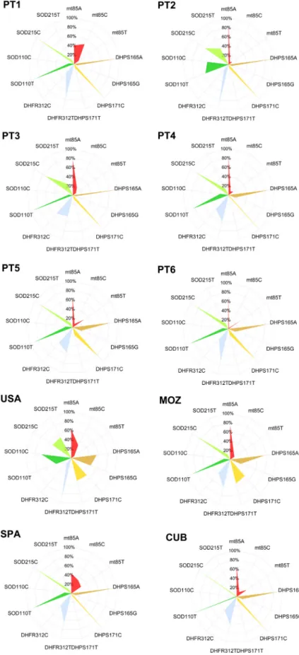
In all, 182 pulmonary specimens divided into ten DNA pools were analysed for the epidemiological distribution of P. jirovecii genotypes in distinct geographic regions and among patients presenting with different clinical conditions. The six SNP, located in the four genetic loci studied, were successfully characterized by the Multiplex-SBE/DNA pooling method in all DNA pools. Fluorescence peaks with 35 nt (DHFR312), 47 nt (mt85), 55 nt (DHPS165), 64 nt (SOD215), 75 nt (SOD110) and 82 nt (DHPS171) were detected in the SBE reactions and the average normalized relative frequencies of each SNP allele were estimated (Table 2, Fig. 1). Data analysis demonstrated several significant statistical differences in the frequency distri-bution of SNP among pools from different geographic origins and with distinct parameters of infection (Table 3).
The relationships between the most relevant putative MLG identified in the pools studied were analysed applying the neighbour joining method. The most representative putative
MLG (SNP frequencies >32% for mt85 and 50% for the TABLE
2. Pneumocy stis jirovec ii single nu cleotide p o lymo rphism all eles relative fre quencies distribution amon g th e differ ent po ols studied Pools Single nucleotide pol ymorphism relative frequencies (%) mtLSUrRNA DHP S DHFR SOD mt85A mt85C mt8 5T DH PS165A DHPS165G DHPS171C DHPS 171T DHFR312T DH FR312C SOD110T SOD11 0C SOD21 5C SOD215T PT1 40.1 48.1 11.8 92.1 7.9 91.4 8.6 73.3 26.7 89.0 11.0 92.0 8.0 PT2 84.9 6.1 9.0 92.1 7.9 91.4 8.6 85.1 14.9 53.6 46.4 61.4 38.6 PT3 74.3 18.0 7.7 93.9 6.1 91.1 8.9 47.4 52.6 91.2 8.8 72.2 27.8 PT4 79.3 8.7 12.0 84.8 15.2 96.7 3.3 58.6 41.4 81.4 18.6 84.7 15.3 PT5 63.7 8.2 28.1 80.2 19.8 82.8 17.2 55.3 44.7 85.7 14.3 88.8 11.2 PT6 68.6 2.2 29.2 84.1 15.9 86.6 13.4 69.7 30.3 87.5 12.5 86.8 13.2 USA 61.1 31.5 7.4 55.4 44.6 50.0 50.0 85.2 14.8 35.1 64.9 49.4 50.6 MO Z 72.5 16.6 10.9 75.1 24.9 47.2 52.8 61.5 38.5 86.6 13.4 87.4 12.6 SPA 42.8 32.0 25.2 89.8 10.2 95.2 4.8 65.4 34.6 85.4 14.6 91.4 8.6 CUB 63.0 11.4 25.6 86.7 13.3 91.9 8.1 60.9 39.1 97.2 2.8 97.6 2.4 Abbre viations: PcP, Pneu mocystis jirovecii pneumonia ; PT1, AIDS-re lated PcP Portugue se patie nts with low/mode rate parasite burden; PT2, AIDS-re lated PcP Port uguese patie nts with high para site burden; PT3, HIV-positive PcP-negative Portuguese patie nts with other pulmonar y disea ses, coloniz ed by P. jirovecii ; PT4, HIV-ne gative Portugues e patients with PcP; PT5, Por tuguese patien ts w ith PcP presenting positive follo w-up; PT6 , Port uguese patient s with P cP presenting negative -up; USA, AIDS-re lated PcP patie nts from USA; MOZ, Mozambic an infants infected with P. jirovecii , presenting respirato ry symptoms ; SPA, immunocompromised Spanish patie nts colonized by P. jirovec ii, prese nting other respirat ory disea ses than Pc P; CUB, HIV-ne gative youn g ch ildren in fected with P. jirovecii , presenti ng respira tory symptoms . The relat ive frequency of each polymor phis m was calcula ted by dividing the maximum height values of fluoresce nce peaks observe d in the single -base extension products by the reference fluorescence observe d in the positive control.
FIG. 1. Graphic representation of Pneumocystis jir-ovecii single nucleotide polymorphism alleles relative frequencies among the different pools studied. Radar charts were drawn with axes for each single nucle-otide polymorphism (represented from the centre to the periphery) and a scale in the range of 0-100% (10% intervals).
remaining SNP) were considered in the construction of a specific dendrogram (Fig. 2) in which three major clusters were
identified: cluster A with MLG from Spain (SPA-A, SPA-B),
Cuba and Portugal (PT1-A, PT3); cluster B with MLG from Portugal (PT1-B, PT2, PT4, PT5, PT6); cluster C with MLG from Mozambique and USA (USA-A,B,C,D).
Discussion
To our knowledge, this is the first report on P. jirovecii
mtLSUrRNA and DHPS gene variability in Mozambique, DHFR gene variability in Mozambique, Cuba and Spain, and SOD gene variability in the USA, Mozambique and Spain. The results strengthen the hypothesis that both geographic distances and clinical parameters have direct impact in the distribution of the genetic subtypes of P. jirovecii as demonstrated inFigs. 1 and 2. In the Portuguese pools, data on genome concentrations are consistent with P. jirovecii burden, with the pool PT2 (AIDS-related PcP patients with high parasite burden) pre-senting the highest P. jirovecii genome concentration. The high concentration of P. jirovecii genomes detected in the pools from Cuba and Mozambique suggests that both populations were infected with high burdens of P. jirovecii, which was not initially detected, probably due to the low sensitivity and difficulty in reading of the microscopic diagnostic method using Giemsa staining[30].
mtLSUrRNA
In Portugal, mt85C has been reported as the most frequent allele of the SNP mt85 among HIV-positive patients with PcP
(mt85C 58.0%–63.0%, mt85A 26.0%–43.0% and mt85T
18.0%–22.0%, time period 1997–2007) [12,18,19,22]. In the present study, except for the pool PT1, the Portuguese pools demonstrated mt85A as the most frequent allele. The allele mt85C was statistically associated with the pool PT1 (Tables 2 and 3). This finding is similar to previous reports, in which low/moderate P. jirovecii burden was more frequently observed among respiratory specimens with mt85C, whereas high bur-dens were more frequently detected in respiratory specimens
with mt85A or mt85T [18,19,21]. The differences observed
between the present and previous data may be the result of the
different time periods of the studies (2004–2013 and
1997–2007, respectively).
In the USA, the distribution pattern of mt85 (mt85C 31.5%, mt85A 61.1% and mt85T 7.4%) was similar to previous studies
(mt85C 42.9%–43.9%, mt85A 36.7%–50.0% and mt85T
7.1%–9.3%, in a population of PcP patients, time period
1986–1999)[14,31], particularly in San Francisco, with mt85C 25.0%, mt85A 50.0% and mt85T 7.0% (mixed genotypes 18.0%) (in HIV-positive patients)[14]. In the AIDS-related PcP patients from San Francisco, the distribution pattern of mt85 is relatively stable in different time periods (2004–2012 and 1995–1998).
Pools from Mozambique and Cuba showed similar distribu-tion patterns of mt85 (mt85C 16.5%, mt85A 72.5%, mt85T 10.9% and mt85C 11.4%, mt85A 63.0%, mt85T 25.7%, respec-tively). Two recent Cuban studies, in a population of children colonized by P. jirovecii (both time periods 2010–2013) demonstrated analogous distribution patterns of mt85, with
mt85C 18.0%–31.0%, mt85A 56.0%–68.0% and mt85T
25%–37% [5,6]. The similarity between the results from
Mozambique and Cuba may be due to the fact that both pools were constituted by populations of young children infected with P. jirovecii and presenting analogous clinical parameters.
Since 2003, several studies in Spain demonstrated mtLSUrRNA variation patterns (mt85C 40.0%–55.1%, mt85A 10.1%–18.2%
and mt85T 25.0%–36.7% in the general population, in
HIV-positive patients with PcP, and in HIV-negative patients with other concomitant pulmonary disorders, between 1995 and 2008)[4,8,9,12]. Except for the general population study (mt85C and mt85A both most prevalent), mt85C was the most prevalent allele in all the other populations, followed by mt85T and mt85A. The present data showed a change in the allelic distribution of mt85 in Seville, Spain, particularly an increase of the mt85A. These differences appear to be more related to the periods in which the populations were studied (2006–2013 in the present study, and 1995–2008 in the earlier studies) than with the characteristics of the population, since the studies of Montes-TABLE 3.Multiple comparisons of the Pneumocystis jirovecii
single nucleotide polymorphisms frequency distribution variation across the pools studied
SNPs Multiple comparison analysis (p values)a
mt85A PT1 vs. PT2 (0.0042); PT2 vs. SPA (0.0011);
SPA vs. MOZ (0.0053)
mt85C PT1 vs. PT2 (0.0442); PT1 vs. PT6 (0.0061); PT3 vs. PT6 (0.032);
PT2 vs. SPA (0.0451); PT6 vs. SPA (0.0131)
mt85T PT3 vs. PT5 (0.0462); USA vs. CUB (0.0353)
DHPS165A/G PT1 vs. USA (0.0191); PT2 vs. USA (0.0131);
PT3 vs. USA (0.0021); USA vs. SPA (0.0083)
DHPS171C/T NSb
DHFR312T/C PT2 vs. PT3 (0.012); PT3 vs. USA (0.0151)
SOD110T/C USA vs. CUB (0.0093)
SOD215C/T PT2 vs. CUB (0.0321); USA vs. CUB (0.0093)
Abbreviations: PcP, Pneumocystis jirovecii pneumonia; PT1, AIDS-related PcP Portuguese patients with low/moderate parasite burden; PT2, AIDS-related PcP Portuguese patients with high parasite burden; PT3, HIV-positive PcP-negative Portuguese patients with other pulmonary diseases, colonized by P. jirovecii; PT4, HIV-negative Portuguese patients with PcP; PT5, Portuguese patients with PcP presenting positive follow-up; PT6, Portuguese patients with PcP presenting negative-up; USA, AIDS-related PcP patients from USA; MOZ, Mozambican infants infected with P. jirovecii, presenting respiratory symptoms; SPA,
immunocompromised Spanish patients colonized by P. jirovecii, presenting other respiratory diseases than PcP; CUB, HIV-negative young children infected with P. jirovecii, presenting respiratory symptoms.
aFor the multiple comparisons of the pools, the p values presented are the adjusted
p values (Bonferroni correction) that took into consideration the number of
comparisons, i.e. the adjusted p value = p value k(k– 1)/2, with k = 10 the number
of pools to be compared is 45. The multiple comparisons were performed considering three groups of pools: (1) All ten pools; (2) only the six Portuguese pools; (3) only the four foreign pools.
Cano et al.[4]and Esteves et al.[12]focused on Spanish pop-ulations similar to those in the present study.
DHPS
In Portugal, a decline of DHPS gene mutation frequencies has been documented since the beginning of the 2000s. Frequencies of 24.7% DHPS165G and 22.5% DHPS171T were detected in a population of HIV-positive and HIV-negative patients with pul-monary disorders, between 1994 and 2001, in which DHPS mutations were more frequent in the time period 1994–1997 (33%) than in 1998–2001 (9%) (p 0.022)[32]. More recently, the proportion of mutant DHPS alleles in a population of HIV-positive patients was 7.0% DHPS165G and 9.0% DHPS171T, between 2001 and 2007[19]. The decline of DHPS mutant al-leles among the HIV-positive patients (pools PT1, PT2 and PT3, time period 2004–2012) may be attributed to the lack of exposure to trimethoprim-sulfamethoxazole in this population, after the decreased use of sulfa prophylaxis due to widespread use of potent combination antiretroviral therapy in Europe, in the late 1990s[2,12,32]. However, in the HIV-negative patients with PcP (pool PT4), the frequencies of DHPS mutants,
especially DHPS165G, appeared to be slightly higher than the ones reported in the HIV-positive patients. The reason for this observation is unclear. One possible explanation is that these groups are clinically distinct, reflecting different trimethoprim-sulfamethoxazole exposure, which may have an impact on the genetic variability of the DHPS locus in P. jirovecii[33,34].
In the USA, since 1998, several studies have reported DHPS mutations per PcP cases ranging from 26% to 81%, describing high overall frequencies of DHPS165G and DHPS171T in different time periods (7.4%–77.3% and 22.2%–59.4%, respec-tively, from 1976 to 2001[3,14,15,17,31,35]), especially in San Francisco (65.0% DHPS165G, 64.0% DHPS171T)[18]. In this city, several reports consistently described overall high frequencies of mutations in the DHPS gene (87.0%, 1995–1998 [14]; 81.5%, 1996–1999[17]; 81.4%, 1997–2002[15]). The high frequencies of DHPS mutants in the US pool (Fig. 1) is of concern and may reflect trimethoprim-sulfamethoxazole exposure of the AIDS-related PcP population and increased of sulfa-induced mutants, especially after the mid-1990s[3,14,23].
In the early 2000s, several studies described low frequencies
of DHPS mutations in African countries neighbouring
FIG. 2.Dendrogram showing the relationships between the Pneumocystis jirovecii putative multilocus genotypes (MLG) from different geographic origins, constructed on the base of the four polymorphic markers (mtLSUrRNA, DHPS, DHFR and SOD) and demonstrating that genetic differences between clusters associated mainly with geographic differences, but also with the clinical set up in the different pools. Dendrogram was computed using the software CLUSTAL W2 multiple sequence alignment (version 2.0.12). The most representative putative MLG of P. jirovecii in the pools (single nucleotide polymorphism frequencies higher than 32% for mt85 and 50% for the remaining single nucleotide polymorphism) studied were as follows:
PT1-A: mt85C/DHPS165A/DHPS171C/DHFR312T/SOD110T/SOD215C PT1-B, PT2, PT4, PT5, PT6: mt85A/DHPS165A/DHPS171C/DHFR312T/SOD110T/SOD215C PT3: mt85A/DHPS165A/DHPS171C/DHFR312C/SOD110T/SOD215C USA-A: mt85A/DHPS165A/DHPS171C/DHFR312T/SOD110C/SOD215T USA-B: mt85A/DHPS165A/DHPS171T/DHFR312T/SOD110C/SOD215T USA-C: mt85A/DHPS165A/DHPS171T/DHFR312T/SOD110C/SOD215C USA-D: mt85A/DHPS165A/DHPS171C/DHFR312T/SOD110C/SOD215C MOZ: mt85A/DHPS165A/DHPS171T/DHFR312T/SOD110T/SOD215C SPA-A: mt85A/DHPS165A/DHPS171C/DHFR312T/SOD110T/SOD215C SPA-B: mt85C/DHPS165A/DHPS171C/DHFR312T/SOD110T/SOD215C CUB: mt85A/DHPS165A/DHPS171C/DHFR312T/SOD110T/SOD215C
Mozambique (DHPS165G 7.1% and DHPS171T 0% in Zimbab-wean AIDS-related PcP patients, 1992–1993[36]; DHPS165G 1.9% and 10.0%, DHPS171T 1.9% and 6.7% in South African adult patients with PcP and in HIV-infected children, respec-tively, 2000–2003[37,38]). The low frequencies were attrib-uted to the lack of exposure to trimethoprim-sulfamethoxazole in the populations studied [37,39]. However, in South Africa rates of DHPS165G 44.0% and DHPS171T 41.1% were reported in HIV-infected adult patients suspected of having PcP (time
period 2006–2007) [38]. The results of the present study
showed similar high prevalence of DHPS mutants in Mozambi-can infants with P. jirovecii infection in an overlapping time period (2006–2007). In this region of southeastern Africa, the differences in the percentage of P. jirovecii DHPS mutations may
be due to different time periods of study, reflecting the
increased widespread empirical use of
trimethoprim-sulfamethoxazole, the mainstay of PcP treatment and prophy-laxis regimens in sub-Saharan Africa, in the late 2000s[38]. But it can also be attributed to an increased awareness by clinicians leading to higher rates of diagnosis and subsequently higher detection rates of DHPS mutations.
In contrast to the USA and Mozambique, pools from Spain and Cuba showed low frequencies of DHPS mutants (Fig. 1). In Spain, the reported frequencies of DHPS mutants vary from 0% in the general population[8]to 15% for both DHPS165G and DHPS171T in HIV-positive and HIV-negative patients with pul-monary disorders [12]. One study involving HIV-negative pa-tients with chronic pulmonary disease colonized by P. jirovecii showed frequencies of DHPS165G 21.4% and DHPS171T 14.3% (time period 2001–2002)[7]. Another study in AIDS-related PcP patients, detected frequencies of DHPS165G 16.1% and DHPS171T 12.8% (time period 2001–2003)[4]. These differ-ences may be due to the lack of exposure to trimethoprim-sulfamethoxazole in the general population, when compared with AIDS-related PcP patients, who are more likely to be treated with that drug combination in prophylactic or thera-peutic doses.
In Cuba, a recent study showed a frequency of 12.0% for both DHPS165G and DHPS171T in young children with whooping cough, colonized by P. jirovecii (between 2010 and 2013)[6]. This distribution pattern is consistent with the pre-sent results in Cuban HIV-negative young children infected with P. jirovecii, in a coincident time period (2013). The detection of low frequencies of DHPS mutations in Cuban children is most probably due to the lack of exposure of this specific population to trimethoprim-sulfamethoxazole.
DHFR
In Portugal, DHFR312T was the most prevalent DHFR allele, as has been found in previous studies[18,19,21]. The frequency of
DHFR312C in AIDS-related PcP patients (PT1 and PT2) was similar to those previously found in Portuguese HIV-positive
patients (21%–25%, 1997–2007 [19]; and 10%, 2001–2008
[21]). DHFR312C frequency was considerably higher in positive patients colonized by P. jirovecii (PT3) and HIV-negative PcP patients (PT4) that normally present lower P. jirovecii burdens than AIDS-related PcP patients (Fig. 1,
Tables 2 and 3). The difference observed may be due to the recently reported association between DHFR312T and higher P. jirovecii burdens, also found in the present study (p 0.01)[18]. A very low frequency of DHFR polymorphic sequences was reported in the PcP patients from the USA, between 1985 and
1998 (2.7%) [23]. In the present study, the frequency of
DHFR312C in the US patients was 14.8%. The reason for this increase is not obvious. It may eventually reflect the selective pressure of trimethoprim-induced polymorphisms caused by
the increased widespread use of
trimethoprim-sulfamethoxazole in the USA after the mid-1990s, or it may be linked to the association between DHFR312T and the higher P. jirovecii burdens normally found in AIDS-related PcP patients. Similar distribution patterns of DHFR312 were observed between the pools from Mozambique, Spain and Cuba. In a retrospective study from South Africa, DHFR312C was not observed in a heterogeneous population of patients with pul-monary disorders (2000–2003)[39]. In Mozambique, the high levels of DHFR polymorphic sequences detected in the present study may reflect the increased widespread empirical use of trimethoprim-sulfamethoxazole in the late 2000s and the se-lective pressure of trimethoprim-induced polymorphisms. SOD
In the present study, the distribution pattern of the SOD
polymorphic sequences in the Portuguese population
(SOD110C 8.8%–46.4% and SOD215T 8.0%–38.6%,
time-period 2004–2012) was identical to a previous report on
P. jirovecii multilocus genotyping in pooled DNA samples[21]. The highest frequencies of SOD110C (46.4%) and SOD215T (38.6%) were observed in the PT2 pool, supporting the hy-pothesis that the genotype SOD110C/SOD215T is linked to higher P. jirovecii burdens[18,21].
The US pool (AIDS-related PcP, most receiving mechanical ventilation) showed the most distinct distribution pattern of the SOD alleles (SOD110C 64.9%, SOD215T 50.6%). Again, the alleles SOD110C and SOD215T were statistically associated with this pool (Table 3), supporting also the relationship between the genotype SOD110C/SOD215T and more virulent PcP episodes.
The distribution patterns of the SOD alleles were similar among the pools from Mozambique (SOD110C 13.4%, SOD215T 12.6%), Spain (SOD110C 14.6%, SOD215T 8.6%) and Cuba (SOD110C 2.9%, SOD215T 2.4%). The pool from
Mozambique showed frequencies lower than those found in a previous study in Zimbabwean AIDS-related PcP patients, in
2004 (SOD110C 33.3%, SOD215T 33.3%) [28]. A possible
explanation of this difference is related to the distinct clinical populations studied, reflecting the possible association between more virulent PcP episodes, reported in Zimbabwe, and the genotype SOD110C/SOD215T.
Low frequencies of SOD110C and SOD215T were observed in the pools from Spain and Cuba. However, in a recent study involving young Cuban children with whooping cough, colo-nized by P. jirovecii, the frequencies of SOD110C and SOD215T
(time period 2010–2013) were much higher, 42.0% and 71%,
respectively[6]. The reason for this discrepancy is not obvious. We hypothesize that this difference is due to distinct sample sizes (190 respiratory specimens in the previous report and ten in the present study) biasing the results, and/or to different underlying concomitant pulmonary diseases, in both Cuban and Spanish colonized populations. The low frequencies of SOD110C and SOD215T alleles are usually associated with more virulent PcP episodes.
The relationships between the most frequent P. jirovecii pu-tative MLG detected in the different pools studied pointed out that clusters are mainly due to the geographic differences but also dependent on clinical characteristics of the populations studied (Fig. 2). MLG from pools corresponding to colonized patients from Portugal and Spain were grouped in the same cluster (cluster A). Also, the MLG from the pool from Cuba (infants infected with P. jirovecii) was included in this cluster but with a higher phylogenetic distance. The majority of the MLG detected in the Portuguese pools were clustered, corresponding to PcP cases (cluster B). Pneumocystis jirovecii from the USA and Mozambique were related in the same cluster because of the DHPS mutations present in both MLG (cluster C). However, within this cluster the MLG from Mozambique (infants infected with P. jirovecii) was considerably distant from the four major MLG detected in the pool from the USA (severe AIDS-related PcP cases).
The observation of different dihydropteroate synthase (DHPS) alleles in and between P. jirovecii populations carries epidemiological implications related to transmission patterns, sulfa drug exposure and geographical distribution of specific genotypes[14,15,17]. In general overview, patients living in the USA and Mozambique presented higher rates of DHPS mutants, which is clearly depicted inFig. 1. This fact suggests that the circulation of P. jirovecii haplotypes may be potentially related to trimethoprim-sulfamethoxazole resistance in those geograph-ical regions. The high frequencies of DHFR312T and SOD110C/ SOD215T detected in patients with AIDS-related PcP from Portugal and the USA endorse the importance of these genetic
variants of P. jirovecii, which were already associated with the virulence or severity of the disease (Table 3)[18,21]. Consid-ering the present data, P. jirovecii with the haplotype DHFR312T/ SOD110C/SOD215T is likely to be associated with more severe AIDS-related PcP cases and high P. jirovecii burdens as demon-strated in the pools from patients with severe PcP from Portugal (PT2) and the USA.
Conclusion
Geographic location and clinical parameters of the groups of patients studied as well as the time-period in which the samples are obtained, were confirmed as determinant epidemiological factors of P. jirovecii infection. The present study demonstrates that the multifactorial approach to PcP studies is a powerful high-throughput methodology for large-scale screening of P. jirovecii SNP of epidemiological relevance. These results convey a more complete picture of the worldwide distribution of P. jirovecii haplotypes and assess their epidemiological impact in different geographic populations of patients.
Transparency Declaration
The present study was supported by Fundação para a Ciência e a Tecnologia (FCT) project PTDC/SAU-MIC/116716/2010 and Associação para a Investigação e Desenvolvimento da Faculdade de Medicina de Lisboa. Dr Huang was supported by National In-stitutes of Health (NIH) grants HL087713 and HL090335. This work was developed in the framework of the‘Red Iberoamericana sobre Pneumocystosis’ (212RT0450) of The Ibero-American Programme for Science, Technology and Development (CYTED).
References
[1] Roux A, Canet E, Valade S, Gangneux-Robert F, Hamane S, Lafabrie A, et al. Pneumocystis jirovecii pneumonia in patients with or without AIDS, France. Emerg Infect Dis 2014;20:1490–7.
[2] Helweg-Larsen J, Benfield TL, Eugen-Olsen J, Lundgren JD, Lundgren B. Effects of mutations in Pneumocystis carinii dihydropteroate synthase gene on outcome of AIDS-associated P. carinii pneumonia. Lancet 1999;354(9187):1347–51.
[3] Kazanjian P, Locke AB, Hossler PA, Lane BR, Bartlett MS, Smith JW, et al. Pneumocystis carinii mutations associated with sulfa and sulfone prophylaxis failures in AIDS patients. AIDS 1998;12:873–8. [4] Montes-Cano MA, de la Horra C, Martin-Juan J, Varela JM,
Torronteras R, Respaldiza N, et al. Pneumocystis jiroveci genotypes in the Spanish population. Clin Infect Dis 2004;39:123–8.
[5] Monroy-Vaca EX, de Armas Y, Illnait-Zaragozí MT, Toraño G, Diaz R, Vega D, et al. Prevalence and genotype distribution of Pneumocystis
jirovecii in Cuban infants and toddlers with whooping cough. J Clin Microbiol 2014;52:45–51.
[6] Monroy-Vaca EX, de Armas Y, Illnait-Zaragozí MT, Diaz R, Toraño G, Vega D, et al. Genetic diversity of Pneumocystis jirovecii in colonized Cuban infants and toddlers. Infect Genet Evol 2014;22:60–6. [7] Calderón E, de la Horra C, Medrano FJ, López-Suárez A,
Montes-Cano MA, Respaldiza N, et al. Pneumocystis jiroveci isolates with dihy-dropteroate synthase mutations in patients with chronic bronchitis. Eur J Clin Microbiol Infect Dis 2004;23:545–9.
[8] Medrano FJ, Montes-Cano M, Conde M, de la Horra C, Respaldiza N, Gasch A, et al. Pneumocystis jirovecii in general population. Emerg Infect Dis 2005;11:245–50.
[9] Respaldiza N, Montes-Cano MA, Dapena FJ, de la Horra C, Mateos I, Medrano FJ, et al. Prevalence of colonisation and genotypic charac-terisation of Pneumocystis jirovecii among cysticfibrosis patients in Spain. Clin Microbiol Infect 2005;11:1012–5.
[10] Schildgen V, Mai S, Khalfaoui S, Lüsebrink J, Pieper M, Tillmann RL, et al. Pneumocystis jirovecii can be productively cultured in differentiated CuFi-8 airway cells. MBio 2014;5:e01186–14.
[11] Wakefield AE, Pixley FJ, Banerji S, Sinclair K, Miller RF, Moxon ER, et al. Detection of Pneumocystis carinii with DNA amplification. Lancet 1990;336(8713):451–3.
[12] Esteves F, Montes-Cano MA, de la Horra C, Costa MC, Calderón EJ, Antunes F, et al. Pneumocystis jirovecii multilocus genotyping profiles in patients from Portugal and Spain. Clin Microbiol Infect 2008;14:356–62. [13] Rohner P, Jacomo V, Studer R, Schrenzel J, Graf JD. Detection of Pneumocystis jirovecii by two staining methods and two quantitative PCR assays. Infection 2009;37:261–5.
[14] Beard CB, Carter JL, Keely SP, Huang L, Pieniazek NJ, Moura IN, et al. Genetic variation in Pneumocystis carinii isolates from different geographic regions: implications for transmission. Emerg Infect Dis 2000;6:265–72.
[15] Crothers K, Beard CB, Turner J, Groner G, Fox M, Morris A, et al. Severity and outcome of HIV-associated Pneumocystis pneumonia containing Pneumocystis jirovecii dihydropteroate synthase gene muta-tions. AIDS 2005;19:801–5.
[16] Tsolaki AG, Beckers P, Wakefield AE. Pre-AIDS era isolates of Pneu-mocystis carinii f. sp. hominis: high genotype similarity with contempo-rary isolates. J Clin Microbiol 1998;36:90–3.
[17] Huang L, Beard CB, Creasman J, Levy D, Duchin JS, Lee S, et al. Sulfa or sulfone prophylaxis and geographic region predict mutations in the Pneumocystis carinii dihydropteroate synthase gene. J Infect Dis 2000;182:1192–8.
[18] Esteves F, Gaspar J, Marques T, Leite R, Antunes F, Mansinho K, et al. Identification of relevant single-nucleotide polymorphisms in Pneumo-cystis jirovecii: relationship with clinical data. Clin Microbiol Infect 2010;16:878–84.
[19] Esteves F, Gaspar J, Tavares A, Moser I, Antunes F, Mansinho K, et al. Population structure of Pneumocystis jirovecii isolated from immuno-deficiency virus-positive patients. Infect Genet Evol 2010;10:192–9. [20] Cissé OH, Pagni M, Hauser PM. De novo assembly of the Pneumocystis
jirovecii genome from a single bronchoalveolar lavagefluid specimen from a patient. MBio 2012;4:e00428–12.
[21] Esteves F, Gaspar J, de Sousa B, Antunes F, Mansinho K, Matos O. Pneumocystis jirovecii multilocus genotyping in pooled DNA samples: a new approach for clinical and epidemiological studies. Clin Microbiol Infect 2012;18:E177–84.
[22] Esteves F, Gaspar J, De Sousa B, Antunes F, Mansinho K, Matos O. Clinical relevance of multiple single-nucleotide polymorphisms in Pneumocystis jirovecii Pneumonia: development of a multiplex PCR-single-base-extension methodology. J Clin Microbiol 2011;49: 1810–5.
[23] Ma L, Borio L, Masur H, Kovacs JA. Pneumocystis carinii dihydropter-oate synthase but not dihydrofolate reductase gene mutations corre-late with prior trimethoprim-sulfamethoxazole or dapsone use. J Infect Dis 1999;180:1969–78.
[24] Nahimana A, Rabodonirina M, Bille J, Francioli P, Hauser PM. Mutations of Pneumocystis jirovecii dihydrofolate reductase associated with failure of prophylaxis. Antimicrob Agents Chemother 2004;48:4301–5. [25] Kazanjian P, Armstrong W, Hossler PA, Burman W, Richardson J,
Lee CH, et al. Pneumocystis carinii mutations are associated with duration of sulfa or sulfone prophylaxis exposure in AIDS patients. J Infect Dis 2000;182:551–7.
[26] Mei Q, Gurunathan S, Masur H, Kovacs JA. Failure of co-trimoxazole in Pneumocystis carinii infection and mutations in dihydropteroate synthase gene. Lancet 1998;351(9116):1631–2.
[27] Costa MC, Esteves F, Antunes F, Matos O. Genetic characterization of the dihydrofolate reductase gene of Pneumocystis jirovecii isolates from Portugal. J Antimicrob Chemother 2006;58:1246–9.
[28] Miller RF, Lindley AR, Malin AS, Ambrose HE, Wakefield AE. Isolates of Pneumocystis jirovecii from Harare show high genotypic similarity to isolates from London at the superoxide dismutase locus. Trans R Soc Trop Med Hyg 2005;99:202–6.
[29] Wakefield AE. DNA sequences identical to Pneumocystis carinii f. sp. carinii and Pneumocystis carinii f. sp. hominis in samples of air spora. J Clin Microbiol 1996;34:1754–9.
[30] Esteves F, Calé SS, Badura R, de Boer MG, Maltez F, Calderón EJ, et al. Diagnosis of Pneumocystis pneumonia: evaluation of four serologic biomarkers. Clin Microbiol Infect 2015;21:379.e1–379.e10. [31] Ma L, Kovacs JA. Genetic analysis of multiple loci suggests that
mu-tations in the Pneumocystis carinii f. sp. hominis dihydropteroate syn-thase gene arose independently in multiple strains. Antimicrob Agents Chemother 2001;45:3213–5.
[32] Costa MC, Helweg-Larsen J, Lundgren B, Antunes F, Matos O. Mu-tations in the dihydropteroate synthase gene of Pneumocystis jiroveci isolates from Portuguese patients with Pneumocystis pneumonia. Int J Antimicrob Agents 2003;22:516–20.
[33] Totet A, Latouche S, Lacube P, Pautard JC, Jounieaux V, Raccurt C, et al. Pneumocystis jirovecii dihydropteroate synthase genotypes in immunocompetent infants and immunosuppressed adults, Amiens, France. Emerg Infect Dis 2004;10:667–73.
[34] Nahimana A, Rabodonirina M, Helweg-Larsen J, Meneau I, Francioli P, Bille J, et al. Sulfa resistance and dihydropteroate synthase mutants in recurrent Pneumocystis carinii pneumonia. Emerg Infect Dis 2003;9: 864–7.
[35] Beard CB, Roux P, Nevez G, Hauser PM, Kovacs JA, Unnasch TR, et al. Strain typing methods and molecular epidemiology of Pneumocystis pneumonia. Emerg Infect Dis 2004;10:1729–35.
[36] Miller RF, Lindley AR, Ambrose HE, Malin AS, Wakefield AE. Geno-types of Pneumocystis jiroveci isolates obtained in Harare, Zimbabwe, and London, United Kingdom. Antimicrob Agents Chemother 2003;47:3979–81.
[37] Zar HJ, Alvarez-Martinez MJ, Harrison A, Meshnick SR. Prevalence of dihydropteroate synthase mutants in HIV-infected South African chil-dren with Pneumocystis jiroveci pneumonia. Clin Infect Dis 2004;39: 1047–51.
[38] Dini L, du Plessis M, Frean J, Fernandez V. High prevalence of dihy-dropteroate synthase mutations in Pneumocystis jirovecii isolated from patients with Pneumocystis pneumonia in South Africa. J Clin Microbiol 2010;48:2016–21.
[39] Robberts FJ, Chalkley LJ, Weyer K, Goussard P, Liebowitz LD. Dihy-dropteroate synthase and novel dihydrofolate reductase gene muta-tions in strains of Pneumocystis jirovecii from South Africa. J Clin Microbiol 2005;43:1443–4.