UNIVERSIDADE DE LISBOA
FACULDADE DE CIÊNCIAS
DEPARTAMENTO DE BIOLOGIA ANIMAL
The role of GRIM-19 and STAT3 interaction in renal tumorigenesis:
clear cell renal cell carcinoma
Ana Isabel Mendes Dias
DISSERTAÇÃOMestrado em Biologia Humana e Ambiente 2012
UNIVERSIDADE DE LISBOA
FACULDADE DE CIÊNCIAS
DEPARTAMENTO DE BIOLOGIA ANIMAL
The role of GRIM-19 and STAT3 interaction in renal tumorigenesis:
clear cell renal cell carcinoma
Dissertação orientada pelo Professor Doutor Valdemar Máximo (Faculdade de Medicina da Universidade do Porto e IPATIMUP) e pela Professora Doutora Ana Maria Crespo (Faculdade de
Ciências da Universidade de Lisboa)
Ana Isabel Mendes Dias
DISSERTAÇÃOMestrado em Biologia Humana e Ambiente 2012
Agradecimentos
Ao longo do nosso percurso de vida há experiências que nos marcam e, de facto, poder fazer parte da família “IPATIMUPiana” é uma honra. Assim, os meus primeiros agradecimentos são dirigidos ao meu orientador Prof. Doutor Valdemar Máximo, a quem muito agradeço ter-me dado a oportunidade de realizar a minha tese de mestrado integrada num dos seus projectos, por toda a confiança que sempre depositou nas minhas competências e pela oportunidade de explorar sempre mais um pouco as questões que foram surgindo durante a realização deste trabalho.
Não menos grata estou à Doutora Paula Soares por me ter aceitado no seu grupo, “Cancer Biology”, e por nos proporcionar a todos realizar trabalho com qualidade, o que nos incentiva a querer ser mais e melhores.
Ainda, um agradecimento especial ao Professor Doutor Sobrinho Simões por ser o principal mentor do magnífico ambiente e nível de excelência que bem caracteriza o IPATIMUP. Obrigada, acima de tudo, pelo exemplo que é para todos nós.
Contudo, esta experiência não teria corrido tão bem se eu não estivesse envolvida pelo ambiente fantástico que se vive, diariamente, no grupo “Cancer Biology”. A todos tenho que agradecer a disponibilidade, entreajuda, alegria e também amizade. Em especial, tenho a agradecer ao João Vinagre e à Joana Nunes por me terem ensinado parte das técnicas que foram a base deste trabalho e por estarem sempre disponíveis a esclarecer dúvidas de última hora. Ao Doutor Jorge Lima agradeço-lhe por todos os conhecimentos que me transmitiu e a sua incansável ajuda. À Joana Silva e ao Ricardo Celestino agradeço toda a disponibilidade e atenção. À Ana Almeida, obrigado pelo companheirismo e apoio incondicional. Ao Rui Batista, que foi uma ajuda preciosa nesta última fase do trabalho, obrigada pelo apoio e pelos valiosos comentários. A todos os outros elementos obrigado pelas gargalhadas e boa disposição.
Mas porque a família “IPATIMUPiana” é muito grande, os meus agradecimentos também se estendem à Vânia Camilo, à Diana Sousa, ao Carlos Resende, ao Hugo Seca, à Patrícia Oliveira, ao Daniel Ferreira, à Bárbara Sousa e à Diana Martins, pelo apoio inesquecível que me deram e pela partilha de conhecimentos.
Sendo que, os últimos são os primeiros, não há palavras que possam descrever o apoio, a paciência e as palavras de incentivo que a minha família me deu e dá a cada momento. Aos meus pais e à minha madrinha agradeço acreditarem sempre em mim e ajudarem-me a ir sempre mais além e, também, por serem os meus exemplos de força e perseverança, os quais me movem e me ajudam a acreditar que o empenho, o esforço e a dedicação, a seu tempo, darão frutos. Aos meus avozinhos agradeço o seu carinho e a sua preocupação e desejo que possam acompanhar muitas mais etapas.
Por último, um agradecimento especial aos amigos de agora e de sempre, que estando longe ou perto, dão brilho à minha vida nos bons momentos mas sobretudo nos menos fáceis.
ABSTRACT
GRIM-19 is a gene that encodes a 16-kDa protein originally identified as a critical regulatory protein for interferon (INF)-ß and retinoic acid (RA)-induced cell death. It was also demonstrated that GRIM- 19 is involved in mitochondrial metabolism, as an integrant component of complex I of the mitochondrial respiratory chain (MRC). GRIM-19 appears, therefore, as a dual function protein involved in cell death and mitochondrial metabolism. Moreover, GRIM-19 downregulation was observed in all subtypes of renal cell carcinomas (RCC), particularly in clear cell renal cell carcinoma (ccRCC), in contrast with thyroid tumors in which GRIM-19 downregulation is specifically associated with oncocytic tumors (Hürthle cell tumors, mitochondrion-rich), suggesting a role for GRIM-19 in tumorigenesis. As GRIM-19 binds and inhibits the signal transducer and activator of transcription-3 (STAT3), which has been shown to be activated and playing a major role in several human tumors, it is tempting to advance that GRIM-19 may function as a tumor suppressor in those tumors. Recently it was reported that STAT3 is located in mitochondria and it appears to be involved in the regulation of mitochondrial metabolism. Thus, it is necessary to clarify if and how the interaction between GRIM-19 and STAT3 proteins interferes with main function of mitochondria: energy production and cell death regulation.
The aim of this work was to investigate the potential role of GRIM-19 as a tumor suppressor gene by studying its interaction with STAT3 protein and by clarifying the mechanism (s) responsible for its downregulation and lack of GRIM-19 expression in RCC, particularly in ccRCC.
Blocking GRIM-19 expression using a specific short hairpin RNAs (shRNA) in ccRCC derived cell lines (Caki-2 and 786-O) and in a human embryonic kidney cell line (HEK 293), we observed that GRIM-19 downregulation leads to an increase of phosphorylated STAT3, thereby increasing STAT3 activity in tumoral cells but not in non tumoral cells. Our data suggests that GRIM-19 expression, in RCC, is regulated through a repressive mechanism by transcription factors. We hypothesize a model of how GRIM-19 and STAT3 interaction could be linked with their dual functions and their cellular localization. GRIM-19 and p-STAT3Ser727 interaction in mitochondria may have a determinant role in ccRCC tumorigenesis but it needs further investigations.
Overall, we conclude that GRIM-19 and STAT3 interaction has a relevant role in renal tumorigenesis. In contrast with thyroid, GRIM-19 may have a broader role in kidney tumorigenesis playing a major role in cell morphology and cell metabolic remodeling.
RESUMO
O GRIM-19 é um gene que codifica uma proteína de 16kDa originalmente identificada como sendo importante na regulação da morte celular induzida por interferão e ácido retinóico. Posteriormente foi também demonstrado que a proteína GRIM-19 é parte integrante do complexo I da cadeia respiratória mitocondrial (CRM) participando no processo da fosforilação oxidativa. Deste modo, a proteína GRIM-19 parece exercer uma dupla função na célula, estando envolvida na regulação da morte celular e no metabolismo mitocondrial. Foi observada uma subexpressão em todos os sub-tipos de carcinomas de células renais (CCR), nomeadamente no carcinoma de células claras renais (CCCR), contrariamente aos tumores de tireóide, onde a subexpressão da proteína GRIM-19 está especialmente associada a tumores de tireóide com características oncocíticas (com células Hürthle -caracterizadas pela presença de um elevado número de mitocôndrias), sugerindo assim um papel do gene/da proteína GRIM-19 na tumorigénese.
Uma vez que a proteína GRIM-19 se liga e inibe o transdutor de sinal e activador de transcrição-3 (STAT3), o qual está ativado em muitos tumores humanos, é possível que o GRIM-19 funcione como gene supressor tumoral em tumores nos quais a proteína STAT3 desempenhe um papel importante. Por outro lado, foi recentemente descrito que a proteína STAT3 se pode também localizar na mitocôndria regulando a atividade dos complexos I e II da CRM e por conseguinte, o metabolismo celular. Assim, é necessário esclarecer se e como a interação destas duas proteínas interfere nas principais funções da mitocôndria: produção de energia e regulação da morte celular.
O principal objectivo deste trabalho foi investigar o papel do GRIM-19 como potencial gene supressor tumoral, nomeadamente através da sua interação com a proteína STAT3 na tumorigénese renal e ao mesmo tempo, identificar e perceber quais os mecanismos responsáveis pela sua subexpressão e/ou ausência de expressão da proteína GRIM-19 em CCR, particularmente em CCCR.
Bloqueando a expressão de GRIM-19 usando um “short hairpin” RNAs (shRNA) específico em linhas celulares derivadas de CCCR (Caki-2 e 786-O) e numa linha renal embrionária humana (HEK-293), observámos que a subexpressão da proteína GRIM-19 tem efeitos distintos em células tumorais e não-tumorais, quer em termos de remodelação metabólica quer quanto à forma como a proteína GRIM-19 interage com a proteína STAT3. Nos clones de 786-O shGRIM-19 a subexpressão da proteína GRIM-19 levou a uma alteração metabólica, tornando as células mais glicolíticas (maior consumo de glucose e maior produção de lactato e aumento de expressão da enzima glicolítica HK II e do
v
transportador de glucose GLUT1). Contudo, nas linhas celulares HEK não observámos esse efeito, mas sim menor expressão das proteínas glicolíticas.
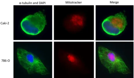
Através de ensaios de imunofluorescência e avaliação do potencial de membrana (JC-1), observámos que a subexpressão da proteína GRIM-19 implica uma reorganização ao nível do citoesqueleto da célula, assim como da sua rede mitocondrial; também se confirmou que a subexpressão da proteína GRIM-19 provoca um decréscimo da actividade mitocondrial a nível do complexo I da CRM. Assim, a proteína GRIM-19 parece ter um papel mais abrangente na tumorigénese renal.
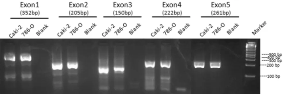
Os resultados deste trabalho permitiu-nos também propor um possível mecanismo pelo qual a expressão da proteína GRIM-19 é regulada em CCR. Inicialmente, verificámos que as linhas renais tumorais 786-O e Caki-2 tinham uma expressão distinta da proteína GRIM-19, a qual não se devia a mutações genéticas. Para além disso, realizámos Real-time PCR para fazer uma quantificação relativa de GRIM-19 mRNA, que mostrou também não haver qualquer tipo de alteração a nível transcripcional. Contrariamente, ao “alternative splicing” descrito por outros autores, obtivémos um transcripto normal do exão 1ao 5 com cerca de 402bp. Tendo em conta muitos estudos que identificaram que, em tumores de células renais, a maioria dos genes supressores tumorais estão subexpressos devido à hipermetilação do promotor, iniciámos uma análise do estado de metilação da provável região do promotor do gene GRIM-19 nas linhas Caki-2 e 786-O. Verificámos que a linha celular 786-O, a que tinha menor expressão da proteína GRIM-19, era a tinha uma fracção mais hipermetilada. Procedemos então a um tratamento de desmetilação com 5Aza-dC para ver se ocorria um aumento de expressão da proteína GRIM-19. Surpreendentemente, por Real-Time PCR, confirmou-se a análise prévia por Western blot, que mostrou que a linha celular 786-O não sofreu alterações mas que a linha celular Caki-2 sofreu um decréscimo de cerca de 50% de expressão do GRIM-19.Ao mesmo tempo, também por Real-Time PCR, observou-se um aumento muito significativo do STAT3 mRNA, particularmente na linha celular 786-O. Adicionalmente, procedemos a uma análise de um fragmento de ilhas CpG para tentar identificar quantas e quais as ilhas CpG que estavam metiladas. Verificamos que as linhas celulares tinham padrões de metilação distintos. Apesar de não se ter usado um método quantitativo do grau de metilação, poderemos afirmar que a linha celular Caki-2 mostrou evidências de estar mais metilada e que com o tratamento de desmetilação algumas ilhas identificadas mostram uma perda significativa desse padrão de metilação.
Todos estes resultados demonstram que a subexpressão da proteína/gene GRIM-19 em CCCR estará associada à hipermetilação do seu promotor (verificado por dois métodos diferentes). Sugerimos ainda que a transcrição do gene GRIM-19 poderá estar sob a
regulação de um mecanismo de repressão por acção de factores de transcrição, dado que, na região do promotor estudada, existem locais de ligação de vários factores de transcrição, incluindo o STAT3.
Este trabalho permitiu-nos também idealizar um modelo de interacção entre as proteínas GRIM-19 e STAT3 e como esta pode estar relacionada com a dupla função das mesmas e a sua localização celular. Serão necessários mais ensaios para confirmar a co-localização destas proteínas na mitocôndria e a sua interacção, bem como para saber se a presumível localização do p-STAT3Ser727 na mitocôndria, observada aquando de um ensaio de fraccionamento celular, poderá ser devida a um mecanismo de compensação em consequência da indução da subexpressão de uma subunidade do complexo I da CRM, GRIM-19.
Em conclusão, como já fora publicado, confirma-se que a subexpressão de GRIM-19 em CCCR não é justificada por mutações ou alterações a nível transcripcional. Por outro lado, o estado de metilação da hipotética região do promotor do gene GRIM-19 por si só também não explica porque é que a linha celular Caki-2 tem uma expressão maior do que a linha celular 786-O. Contudo, os resultados deste trabalho levantam pela primeira vez a hipótese de que a região do promotor do gene GRIM-19 estará hipermetilada e que poderá existir um mecanismo de repressão por factores de transcrição a regular a transcrição do GRIM-19 em CCCR. Por último, a interacção do GRIM-19 em particular com o p-STAT3Ser727 pode ter um papel determinante na tumorigénese de CCCR mas ainda são necessárias mais investigações para confirmar esta hipótese.
Os resultados deste trabalho sugerem que proteína /gene GRIM-19 terá um papel mais abrangente na tumorigénese de CCCR e também revelaram várias evidências de que este assunto merece um desenvolvimento mais aprofundado devido a uma potencial contribuição para terapias antineoplásicas.
Palavras-chave: Proteína mitocondrial; GRIM-19; STAT3; Tumorigénese; Carcinoma de células
TABLE OF CONTENTS
ABSTRACT………..iii
RESUMO………..…iv
ABBREVIATIONS………1
1.LITERATURE REVIEW...…………..………..………..………..7
1.1 Cancer - classical and additional hallmarks………..………..…..………7
1.1.1 Cancer cell energy metabolism……...……….8
1.2 Targets for cancer therapy…………...……….…..…...……...10
1.3 Regulation of mitochondrial metabolism and role of a nuclear gene…...…………...11
1.4 GRIM-19: a subunity of complex I of mitochondria………….……...….………..…...14
1.5 GRIM-19 and other proteins interaction………...………..…….……15
1.5.1 STAT3 and its interaction with GRIM-19………….……….…..……….15
1.6 GRIM-19 and tumorigenesis………….………...…………...19
1.7 GRIM-19 and kidney cancer……….…………..……….….20
2. RESEARCH AIMS and OBJECTIVES………...27
3. MATERIAL and METHODS………...………..…….31
3.1 Cell lines and cell culture conditions………...……….31
3.2 Nucleic acids extraction from cell lines………31
3.2.1 DNA extraction……….31
3.2.2 RNA extraction……….32
3.3 Molecular Analysis of GRIM-19 Gene………..32
3.3.1 Polymerase Chain Reaction (PCR)………..32
3.3.2 Electrophoresis in agarose gel………...…..………..……..33
3.3.3 DNA extraction from agarose gel...33
3.4 Automated Sequencing Analysis………...……….34
viii
3.5.1Short hairpin RNA expression plasmids………...………35
3.5.2 Reverse Transfection………...……….35
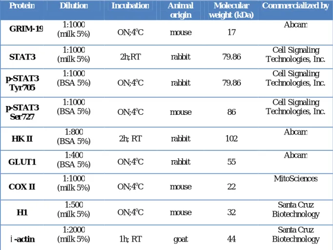
3.6 Western blot analysis………36
3.7 Reverse Transcriptase – Polymerase Chain Reaction (RT-PCR)……...………..37
3.8 Real-Time PCR / qPCR………...………..38
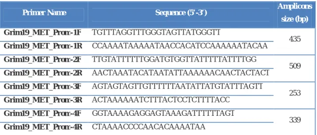
3.9 GRIM-19 gene promoter analysis……….39
3.9.1 Evaluation of methylation status...39
3.9.2 Treatment with a DNA demethylating agent, 5-aza-2’-deoxycytidine...40
3.9.2.1 Determination of the ideal concentration of 5-aza-2'-deoxycytidine…..….40
3.9.2.2 Demethylation treatment of DNA ………...…………...………….40
3.9.3 Bisulfite conversion and cleanup of converted DNA………..……….41
3.10 Membrane potential (JC-1)……….43
3.11Cell fractionation assay - cytosolic, mitochondrial and nuclear proteins…….………..43
3.12 Immunofluorescence- cytoskeleton, mitochondrial and nuclear staining………...44
3.13 In vitro cellular growth assay using BrdU incorporation assay……….44
3.14 In vitro cell death assay using TUNEL assay………..………45
3.15 Glucose and lactate quantification………..46
3.16 Statistical analysis………...46
4. RESULTS……….………49
4.1 The role of GRIM-19 in the renal tumorigenesis……….49
4.1.1 GRIM-19 and clear cell renal cell carcinoma cell lines - a model of study………...….49
4.1.2 Molecular analysis of GRIM-19 gene……….……50
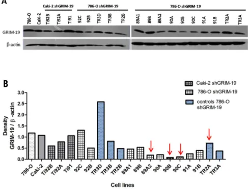
4.1.3 Analysis of tumor cell lines with stable knockdown of GRIM-19……….….52
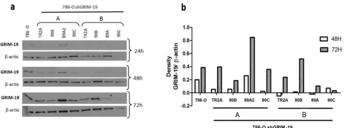
4.1.4.1 786-O cell line and shGRIM-19 clones……….………..…..56
4.1.4.2 HEK293 cell line and shGRIM-19 clone……….61
4.2 GRIM-19 protein expression and STAT3 protein expression, activation and localization………..……….63
4.2.1 Analysis of GRIM-19 and STAT3 interaction……….63
4.2.2 Proteins expression and localization – results of cell fractionation assay……65
4.3 Study of GRIM-19 promoter ……….……….68
4.3.1 Evaluation of methylation status………..………..68
4.3.2 Quantitative analysis by Real-Time PCR……….………….69
4.3.3 Analysis of bisulfite conversion sequencing………..71
5. DISCUSSION………..75
6. CONCLUSIONS and FUTURE PERSPECTIVES………..89
7. REFERENCES……….93
ABBREVIATIONS
aa –Amino acids
ANOVA- Analysis of variance ATP-Adenosine triphosphate 5aza-dC – 5-aza-2'-deoxycytidine BcL-2- B-cell lymphoma protein 2 BcL-XL- B-cell lymphoma-extra large BrdU- 5-bromo-2-deoxyuridine ccRCC- clear cell renal cell carcinoma cDNA- Complementary DNA
CpG- C and G are found on the same strand of DNA and are connected by a phosphodiester bond
COXII- Cytochrome C oxidase subunit II C- Cytosine
cyt c- cytochrome c
DAPI- 4'-6-Diamidino-2-phenylindole DHA-Dehydroascorbic acid
DMEM- Dulbecco's modified eagle medium DMSO- Dimethyl sulfoxide
DNA– Deoxyribonucleic acid
dNTPs- deoxyribonucleotide triphosphate dUTP-deoxynucleotide triphosphates ECL- Enhanced chemiluninescence EDTA-Ethylenediaminetetraacetic acid ETC- Electron transport chain
FACS-Fluorescence-activated cell sorting FAK-Focal-adhesion kinase
FBS- Fetal bovine serum
FCCP- Carbonyl cyanide 4-(trifluoromethoxy) phenylhydrazone FHM- Hypermethylated DNA fraction
2 FITC-Fluorescein isothiocyanate
FUM- Unmethylated DNA fraction
GW112 –Glycoprotein related to olfactomedin(OLFM4) G- Guanine
GFP- Green fluorescence protein GLUT1-Glucose transporter 1
GRIM-19 – Gene associated with Retinoid-Interferon-Induced Mortality 19 HIF-1- Hypoxia-inducible transcription factor 1
HIF-1α- Hypoxia-inducible transcription factor 1α H1- Histone 1
HK II- Hexokinase II
HPRT-Hypoxanthine phosphoribosyltransferase 1 HtrA2- HtrA serine peptidase 2
IFN-β- Interferon- β
JAK- Janus tyrosine kinases
JC-1- Tetraethylbenzimidazolylcarbocyanine iodide kb- kilobases
LDH-A- Lactate dehydrogenase A ΔΨm- Mitochondrial membrane potential Mcl-1- Myeloid cell leukemia sequence 1 MMP-2- Matrix metalloproteases 2 MMP-9- Matrix metalloproteases 9 MRC- Mitochondrial respiratory chain mRNA- Messenger RNA
mtAA- mitochondrial ascorbic acid MTC1- Monocarboxylate transporter 1 mtDNA- mitochondrial DNA
NADPH-Nicotinamide adenine dinucleotide phosphate NADH- Nicotinamide adenine dinucleotide
nDNA- nuclear DNA
NOA- Non-oncogene addiction
NOD2- Nucleotide oligomerization domain 2 OXPHOS- Oxidative phosphorylation PBS- Phosphate buffered saline PCR- Polymerase Chain reaction
PDK1- Pyruvate dehydrogenase kinase 1 p53- protein 53
PFA-Paraformaldehyde
p-STAT3 – Phosphorylated STAT3
p-STAT3 Ser727 – Phosphprylation in residue serine at position 727 of STAT3 p-STAT3 Tyr705 – Phosphprylation in residue tyrosine at position 705 of STAT3 qPCR- quantitative real time Polymerase Chain reaction
RA- Retinoic acid
RB- Retinoblastoma tumor suppressor gene RCC- Renal cell carcinoma
RCT- Renal cell tumors RNA- Ribonucleic acid RNAi- RNA interference ROS- Reactive oxygen species
RPMI- Roswell Park Memorial Institute rRNA- ribosomal RNA
RT-PCR- Reverse Transcriptase – Polymerase Chain Reaction RT- Room temperature
SEM- standard error of the mean Ser727 – Serine residue at position 727
SDS-PAGE – Sodium dodecyl sulfate polyacrylamide gel electrophoresis SH2- Src Homology 2
shCT- short hairpin control
shGRIM-19- short hairpin RNA specific for GRIM-19 shRNA – short hairpin RNA
4
STAT- Signal Transducer and Activator of Transcription STAT3 - Signal Transducer and Activator of Transcription 3 TFs- Transcription factors
TMRE-Tetramethylrhodamine ethyl ester tRNA -Transfer RNA
TUNEL- Terminal deoxynucleotidyl transferase dUTP nick end labeling Tyr705- Tyrosine residue at position 705
WCE- Whole cell extraction
VEGF- Vascular endothelial growth factor VHL- Von Hippel-Lindau
1.1 Cancer - classical and additional hallmarks
Luo et al. [1] states that “Cancer is a complex collection of distinct genetic diseases
united by common hallmarks”.
Currently, there is no doubt that cancer arises through a multistep process. It is stated that cancer development is consistent with Darwinian principles meaning that, cancer evolves through random mutations and epigenetic changes that alter these pathways followed by the clonal selection of cells that can survive and proliferate under circumstances that would normally be deleterious [1].
In 2000, Hanahan and Weinberg published a review that outlined six classical cancer hallmarks: unlimited proliferation potential, self-sufficiency in growth signals, insensitivity to antigrowth signals, evading apoptosis, sustained angiogenesis (attract new blood vessels to supply with nutrients and oxygen) and tissue invasion and metastasis (to distal organs) [2]. However, due to the progress of the last decade, it has been now proposed six additional cancer hallmarks: DNA damage, replication stress, proteotoxic stress, mitotic stress, metabolic stress and oxidative stress. Despite, these are not responsible to initiating tumorigenesis, they are common features of different types of tumors and they are collectively referred as the stress phenotypes of cancers [1].
Indeed, these both classic and additional hallmarks interplay in a functional point of view, promoting tumorigenesis and suppressing oncogenic stress, as elucidated in figure 1. For instance, tumor cells use preferentially glycolysis which provides them advantages such as adaptation to hypoxia (low oxygen environment) and the acidification of microenvironment allowing tumor invasion as well as suppressing immune surveillance [3]. Tumors are also characterized to have chromosome instability phenotypes that can result from defects in different pathways involved in mitosis thus, inducing mitotic stress and consequently promoting aneuploidy (altered chromosome number) [1]. On the other hand, the mentioned stress phenotype can also influence the proteotoxic stress phenotype that is counteracted by the heat shock protein response pathway, the responsible for the proper folding and/or proteolytic degradation of proteins [1, 4]. Moreover, elevated levels of reactive oxygen species (ROS) promote DNA damage that normally cause senescence or apoptosis but that is overcomed by tumor cells [1].
8
Figure 1. The Hallmarks of Cancer. Classical cancer hallmarks originally proposed by Hanahan and
Weinberg [2] (top half, white symbols) and evasion of immune surveillance proposed by Kroemer and Pouyssegur [3]. Additional hallmarks that depict the stress phenotypes of cancer cells (lower half, colored symbols). Indications (black arrows) of the functional interplays among these hallmarks that promote the tumorigenic state and suppress oncogenic stress (adapted from [1])
1.1.1 Cancer cell energy metabolism
The complex connection between metabolism and proliferation remains an exciting area of research. It is possible that additional metabolic pathways have not been described yet. Understanding this important aspect of biology is likely to have a major impact on our understanding of cell proliferation control and cancer. Furthermore, exploiting the metabolic dependencies of cancer cells will be an essential step forward for cancer treatment.
It is well known that under aerobic conditions, non-proliferating (differentiated) cells process glucose, first to pyruvate via glycolysis in the cytosol and afterwards to carbon dioxide through oxidative phosphorylation in the mitochondria; under anaerobic conditions, glycolysis is the favored pathway to produce energy [5]. On the contrary, tumors cells have a characteristic glycolytic phenotype. As described by Otto Warburg, more than 50 years ago, tumor cells tend to convert most glucose to lactate, even in the presence of sufficient oxygen levels to support mitochondrial oxidative phosphorylation [6]. This phenomenon is designed aerobic glycolysis or Warburg effect (an equivalent of fermentation used by many unicellular organisms to proliferate). However, the efficiency of ATP production is higher by oxidative phosphorylation than by metabolism of glucose to lactate, generation of 36 ATPs against 2 ATPs per molecule of glucose. This raises the question of why a less
efficient metabolism, at least in terms of ATP production, would be selected by proliferating cells. Two possible explanations are: inefficient ATP production is perhaps a problem only when resources are scarce or most likely, proliferating cells to replicate all of their cellular contents need glucose for other additional requirements rather than just for ATP production (as source of energy). Besides to support cell growth and division, cells also needs glucose for synthesis of macromolecular precursors such as acetyl CoA which is required for fatty acids and NADPH for synthesis of amino acids and nucleotides [5].
The excessive generation of lactate observed in the Warburg effect would appear to be an inefficient use of cellular resources. In fact, for each lactate excreted, a cell wastes three carbons that may be used for either ATP production or macromolecular precursor biosynthesis. However, there are evidences that tumors have a heterogeneous cellular metabolism since some cells seem to use the excess lactate as a fuel for mitochondrial oxidative phosphorylation. Indeed, Sonveaux et al. [7] reported a “metabolic symbiosis” between hypoxic and aerobic cancer cells. Basically, it is assumed that lactate produced by hypoxic cells during glycolysis is taken up by aerobic cells (which are closer to the blood vessels) which then use it as their main substrate for oxidative phosphorylation. To explain this process, the authors evaluated the monocarboxylate transporter 1 (MTC1), located at the cell plasma membrane, as the principal responsible to sustain the symbiosis process. As illustrated in figure 2, with MTC1 inhibition, oxidative tumor cells switch from lactate oxidation to glycolysis avoiding adequate glucose delivery to hypoxic cells, which die from glucose starvation and become more susceptible for radiotherapy treatment [7].
Undoubtedly, this metabolic reprogramming is orchestrated by hypoxia-inducible transcription factor1 (HIF-1) through the transcriptional activation of key genes encoding metabolic enzymes, such as: GLUT1, which encodes a glucose transporter that increases glucose uptake during glycolysis [8]; hexokinase II, which catalyze the irreversible first step of glycolysis (glucose is phosphorylated to glucose-6-phosphate) [9]; LDHA, encoding lactate dehydrogenase A, which converts pyruvate to lactate [10] and PDK1, encoding pyruvate dehydrogenase kinase 1, which inactivates the enzyme responsible for conversion of pyruvate to acetyl-CoA, thereby shunting pyruvate away from the mitochondria [11,12].
10
Figure 2. Lactate uptake and schematic representation of the suggested metabolic symbiosis in tumors (adapted from [7]).
Moreover, HIF-1 is also responsible to induce a transcriptional program by stimulating angiogenesis through the induction of VEGF-A and angiopoietin-2 [13]. Hence, the role played by the increased activity in HIF-1 in cancer development will continue to be a target of investigation specially due to its connection with mitochondrial alterations.
1.2 Targets for cancer therapy
Nowadays, Science continues to have a big challenge which is: “The Path Ahead to New Cancer Therapeutics” [1].
Several efforts have been done to achieve progresses in this field. Of note, there are many approaches, like the Cancer Genome Atlas [14], that aim to characterize, in a large number of cancer types, genomic alterations including copy-number variation, transcriptional profiles, epigenetic modifications and DNA sequence alterations. Even if it is expensive, these are a kind of approaches very important to yield new information on human cancer, aid biomarker discovery and help to stratify patients. It can be done, for example, by identification of common alterations in oncogenes and tumor suppressors for further functional analysis and uncover oncogene addiction pathways that can be targeted [14, 15,16]. However, this kind of studies have limitations as they do not have in account non coding regions analysis as well as analysis of the non-oncogenes function. The latter
comes from a recent concept that cancer cells are addicted to both oncogenes and non-oncogenes. This phenomenon was termed by Solimini et al. [17] as “non-oncogene addiction” (NOA). Indeed, NOA genes, like oncogenes, support tumorigenesis but they do not undergo oncogenic mutations or have significant functional genomic alterations in tumors. Thus, NOA genes and pathways provide important targets for antitumor therapies [1].
Ideally, researchers expect to discover a “druggable genome” which represents proteins with enzymatic functions that can be easily assayed or targeted. Important progresses have been done and now the RNA interference (RNAi) itself seems to be the cancer therapy of the near future [18]. But, regardless to the target proteins, there are evidences that targetting metabolic pathways ( part of tumor- intrinsic NOA) will give extra advances in cancer therapy as there are some potential candidates that are overexpressed in certain cancer types such as the proteins: GLUT1, HKII, phosphoglycerate dehydrogenase (PHGDH) and LDH-A [19].
For instance, GLUT1 is overexpressed in many cancer types including renal cell carcinomas (RCCs) that exhibit loss of the von Hippel-Lindau (VHL) tumor suppressor gene [20,21]. Interestingly, a recent study identified a series of small molecules that inhibit GLUT1 and selectively kill VHL deficient RCCs [21]. Same was observed with the other enzymes in other tumors [19].
However, this apparent inhibition efficiency leads with an important issue/future challenge. The question remains whether inhibition the abovementioned proteins, also expressed in normal tissues, will be effective in diminishing tumors without imparting significant toxicity to normal tissues [19].
Overall, promising advances in nanomedicine seem to overcome these limitations regarding drug delivery directly to targeted cells and tissues. For example, carbon nanotubes may be a very efficient tool thus it is a good candidate as part of a new era of cancer therapy [22].
1.3 Regulation of mitochondrial metabolism and role of a nuclear gene
Mitochondria are organelles responsible for energy production required for cellular metabolism (producing most of the cellular ATP (adenosine-5′-triphosphate) via the oxidative phosphorylation (OXPHOS)). However, they have also an essential role in cell death regulation [23, 24].
12
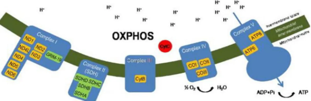
In fact, mitochondrion is a special organelle and semi-autonomous as it possess its own genome which encodes some machinery necessary for their replication, transcription, translation and protein assembly [24, 25]. Each mitochondrion has about two to ten molecules of its own DNA, the mitochondrial DNA (mtDNA). Its 16.5kb circular double-stranded molecule encodes 22 transfer RNAs (tRNAs) and 12S and 16S ribosomal RNA (rRNA), as well as 13 proteins (highlighting in orange boxes, see Figure 4) which are part of the mitochondrial respiratory chain (MRC) and OXPHOS system (Figure 3) [23, 26]. Despite having their own DNA, mitochondria are not independent from the nuclear DNA (nDNA) because most mitochondrial proteins are encoded by nDNA which are imported into the mitochondria [24]. GRIM-19 is an example of a nuclear gene that encodes a mitochondrial protein.
The nDNA encodes approximately 80 OXPHOS proteins, required for the mitochondrial metabolic pathways as well as for all of the enzymes required for mitochondrial biogenesis including mtDNA polymerase γ (POLG), RNA polymerase, mtDNA transcription factors and ribosomal proteins, among others [27].
As shown in Figure 3, the mitochondria respiratory chain (MRC) is composed by four complexes (complex I- IV) involved in the electron transport chain and complex V (ATP Synthase) which is responsible for ATP synthesis [28]. There, it can be possible to identify which genes/proteins are related with each complex.
Figure 3. Schematic representation of oxidative phosphorilation system (OXPHOS) in the mitochondria. In the scheme are represented the genes/ proteins present in each complex. Orange boxes
highlighting mtDNA encoded OXPHOS proteins and green boxes highlinting nDNA –encoded mitochondrial proteins such as GRIM-19 subunit (adapted from [23]).
Several studies published to date show that mutations in mitochondrial proteins (regardless of codified by nuclear or mitochondrial genes) are commonly associated with hereditary and sporadic tumors. It is also known that tumor cells acquire distinctive
metabolic features which confers them some growth advantage (Warburg effect or aerobic glycolysis) [29, 30]. The Warburg effect (decrease of MRC activity and increase of glycolysis which means that cancer cells produce lactate even in the presence of oxygen) has been demonstrated in different type of tumors and in fact, it has been explored clinically for detection of tumors by Fluorodeoxyglucose positron emission tomography [23,29]. Although this metabolic shift is now being considered a hallmark of cancer cells, its origin remains to be established. Nonetheless, it is tempting to speculate that mutations in genes coding mitochondrial proteins force cells towards glycolysis thus leading to their particular metabolic profile.
Interestingly, a nuclear gene coding a mitochondrial protein that has emerged to be a key player in growth and apoptosis was a gene associated with retinoid-IFN-induced mortality (GRIMs), particularly GRIM-19. This gene was identified by a research group interested in unveiling the molecular basis of cell death associated with exposure to interferon (IFN)-β and retinoic acid (RA) (a derivative of vitamin A) [31].IFNs and RA have both been shown to suppress tumor growth thus they are used in the treatment of several cancers [32-34].
In 2000, by employing an anti-sense knock-out approach, Angell et al. [34] were able to isolate genes associated with retinoid-IFN-mortality (GRIM),among which was 19. The authors verified that the antisense 19 (reduce intracellular levels of GRIM-19) was able to confer a strong growth advantage to cells when exposed to the combination of IFN-β and RA treatment [34].
Interestingly, overexpression of GRIM-19 was able to reduce cell viability (only resistant cells form colonies), while cells expressing moderate levels of GRIM-19 were significantly more susceptible to cytotoxic effect of IFN/RA. GRIM-19 seemed to induce apoptosis under IFN/RA combination [34].
At that time, GRIM-19 gene was characterized to have a 552 base pairs (bp) cDNA and to codify a protein with 16kDa [26]. On the other hand, and also in 2000, Chidambaram
et al. [35] identified the chromosomal location of human GRIM-19- 19p13.1 – 13.2. Once
that human chromosome 19p13.2 locus was associated with genes that can suppress prostate and thyroid tumor cell growth [36, 37], then it was suggested that GRIM-19 could be one candidate as tumor suppressor gene [35].
In fact, like other tumor suppressor proteins such as p53, mutations in GRIM-19 gene and absence of its protein expression have been observed in different types of tumors [38-45]. Notably, mutations in the GRIM-19 gene were identified in a particular histotype of thyroid tumors and this was the first nuclear gene associated with Hürthle
(mitochondrion-14
rich) cell tumors to be reported [38]. More recently, it has been observed a very weak or complete lack of GRIM-19 protein expression in renal tumor cells but curiously no mutations were observed [39]. Furthermore, several studies shown that GRIM-19 interaction with STAT3 protein are relevant in tumor progression [40-45] and recent findings describe that STAT3 also seems to regulate mitochondrial metabolic function. Therefore, further investigations must be done to disclose how those proteins may be involved in the mitochondria metabolic shift [46, 47].
1.4 GRIM-19: a subunit of complex I of mitochondria
One year after GRIM-19 identification, Fearnley et al., [48] reported an additional protein of the 42 MRC complex I subunits described in bovine heart mitochondria. In their study it was observed a new subunit (B16.6) of MRC complex I that corresponded to the bovine homolog of the GRIM-19 human protein (83% of identity).
Later, Huang et al. [49] using different tumoral cell lines (cervical, breast, kidney and neuronal pheochromocytoma cells) to re-examine whether the localization of GRIM-19 is exclusively on mitochondria, showed that GRIM-19 protein is exclusively located on the cytoplasm with no immunofluorescence staining of GRIM-19 in the nucleus. Furthermore, the authors investigated the localization of GRIM-19 at a sub mitochondrial level using antibodies for complexes subunits of MRC [49]. As shown in Figure 4, absence of GRIM-19 (GRIM -/-) induced a drastic decrease of complex I expression while complex II expression was not affected. Interestingly, at complex III level occurred also a certain decrease of expression. These results demonstrate that in fact, GRIM-19 is an essential component of MRC as it not only ensures complex I assembly but it could also influence assembly of other complexes thus interfering with the electron transport chain, leading with mitochondrial failure [49].
Figure 4. Effect of GRIM-19 in the expression of other complexes subunits. Western blot analysis
Some studies of knockout studies, in blastocytes of mice [49], and knockdown, in
Xenopus embryos [50], of GRIM-19 gene, demonstrate that this gene is essential for
assembly and activity of complex I. However, while these results did not elucidate how it occurs, Lu & Cao [51] addressed this question and decided to study the functional domains of GRIM-19 protein. From this study, it was concluded that N-terminal domain ( aa 1-60) is essential for mitochondrial localization of GRIM-19 and also for its incorporation to complex I; the middle region (aa 70-100) is required for electron transfer activity of complex I and the last C-terminal (10 aa) promotes GRIM-19 assembly to complex I of MRC [51].
In addition, it was also demonstrated that GRIM-19 is essential for early embryonic development in mice, as genetic ablation of this gene by homologous deletion leads to embryonic lethality by day 9.5, particularly due to oxidative phosphorylation (OXPHOS) failure [49].
All these evidences show that GRIM-19 gene has a double role as it is involved in cellular death induced by IFN-β /RA as well as in the mitochondria metabolism. Nonetheless, it was recently published that GRIM-19 is present in the nucleus, mitochondria or cytoplasm [52, 53]. Indeed, it not completely clarified this question related with GRIM-19 localization. However, as referred by Máximo et al. [54], this variable localization could reflect the different functions of GRIM-19 in cellular biology, as part of MRC, of apoptosis pathway induced by IFN-β /RA and also its interaction with cytosolic proteins such as STAT3 protein.
1.5 GRIM-19 and other proteins interaction
There are several studies confirming some partners of GRIM-19 involved in growth suppression such as GW112 [55], HtrA2 [56], NOD2 [57], p16Ink4a [58] and in particular STAT3 protein [40,41,59].In fact, the interaction of GRIM-19 with the latter protein has been investigated in several studies in order to understand how these proteins interact. It was observed, in different types of human tumors, an overexpression of STAT3 protein and by inducing overexpression of GRIM-19 it seems to inhibit STAT3 protein activity in a translational level and also increase apoptosis of tumor cells.
1.5.1 STAT3 and its interaction with GRIM-19
STATs (Signal Transducers and Activators of Transcription) are key mediators of cytokine signaling and whose activation occurs through Janus Kinase (JAK) pathway.
16
Subsequently, STAT proteins are translocated to the nucleus where they exert their DNA-binding activity and thus, inducing transcription of genes involved cell proliferation, differentiation, cell survival and development [60].
STAT3 is one STAT family member that is involved in embryonic development [61], cell growth and anti-apoptosis [62]. Usually, STAT3 is tightly regulated by feedback inhibitors. However its constitutive activation has been documented and directly contributes to oncogenesis in cells transformed by viruses, oncogenes and autocrine growth factors [41, 60] and it was observed in tumors [60, 63, 64]. STAT3 is responsible for the expression of some genes involved in cell proliferation like cyclins B1 and D1, cdc2, c-myc, and antiapoptotic proteins like Bcl-XL, Bcl-2 and Mcl-1, p21WAF1/CIP1 [60].
In general, STATs (and in particular STAT3) are predominantly in a latent state in the cytoplasm. When a ligand binds to the extracellular domain of the cytokine receptors and activates them, the intracellular receptor associated JAK is activated by autophosphorylation. This leads to phosphorylation of tyrosine residue (Tyr705) in monomeric and unphosphorylated STAT3, which becomes active and ready to dimerize by the SH2 (Src-homology 2) domains from each one. The dimerization is essential for STAT3 because allows its translocation to the nucleus where it exerts its biological function through DNA binding [60, 65, 66]. However, there are evidences that another phosphorylation, on the serine residue (Ser727) is important to STAT3 activation, but its relevance is not completely clear.
Some studies pointed that phosphorylation at the serine residue (Ser727) is fundamental to the optimal transcriptional activation of STAT3 after the tyrosine residue (Tyr705) phosphorylation [67, 68]. Other studies showed that Ser727 phosphorylation can be essential for STAT3 signalling, once its blockage can eliminate this signalling [60,69,70]. Moreover, there are studies indicating that Ser727 phosphorylation may result in STAT3 signalling activation independently of Tyr705 phosphorylation under certain conditions [71-73]. More recently, Wegrzyn et al. described that STAT is present in mitochondria and the Ser727 phosphorylation was reported to be important to this localization, independently of the function in the nucleus [47].
In summary, STAT3 activation and function lead to a lot of questions and it is necessary further investigation to clearly understand this issue.
In 2003, the first evidences that STAT3 and GRIM-19 are interacting proteins were shown. First, Lufei et al. [40], using a yeast two hybrid assays (Y2H), identified GRIM-19 as specific STAT3 interacting protein (do not interact with STAT1 or STAT5) and this interaction was demonstrated both in vivo and in vitro [40]. Afterwards, Zhang et al. [41]
using the same technique to identify other 19 protein partners, confirmed GRIM-19/STAT3 interaction. Indeed, both studies confirmed that GRIM-19 interacts specifically with STAT3, but not with other STATs (1, 2 and 5a STAT) family members [40, 41].
Zhang et al. [41] also observed that after IFN/RA treatment (inducers of GRIM-19 expression), more STAT3 was co-immunoprecipitated with GRIM-19 in comparison with unstimulated cells, which was supposed to be caused by an increase of GRIM-19 expression [41].
These two studies presented some contradictory aspects related to the primary domains of STAT3/GRIM-19 binding and to the mechanism by which GRIM-19 blocks STAT3 activity. Nevertheless, both studies identified STAT3 as an intervenient of the GRIM-19 death-inducing pathway and GRIM-19 as a novel inhibitor of STAT3 [40, 41].
Due to this ambiguous issue related with GRIM-19 role in oncogenic cell proliferation and its effects in a constitutively active STAT3, Kalakonda et al. [59] developed a study using a human prostatic cell line (PC3 cells). The authors observed that by inducing STAT3 activation (enhance oncogenic features) and when GRIM-19 vector was introduced and cells expressed it, colony formation by these cells decreased. This capacity of GRIM-19 protein to inhibit STAT3 function, particularly, the expression of endogenous genes involved in cell growth control was also observed in vivo as GRIM-19 was able to inhibit tumor formation [59].
Nonetheless, in 2010, Nallar et al. [53] identified one of the structural elements responsible for GRIM-19 antitumoral function. The authors identified a motif of four amino acids: glutamic acid, aspartate, methionine and proline – QDMP- in N-terminus of GRIM-19, considering it as the main responsible for STAT3 repression. The authors observed that by promoting a deletion in this motif the capacity of this mutated GRIM-19 to suppress growth was lower than the wild-type GRIM-19. Therefore, so far, N-terminus is the major responsible for GRIM-19 function in cell growth (inhibition of genes involved in cellular cycle regulation) [53] and motility/invasion (inhibition of proto oncogenes such as src) [74]. Interestingly, a tumor-derived mutation described by our group [38], located at N-terminus (lysine converted to an asparagine in amino acid 5 – K5N) was also unable of inhibit colony formation in soft-agar and limit cell growth [53].
Still back in 2009, it was shown that STAT3 is present in mitochondria and has a regulatory function in OXPHOS, particularly in MRC complexes I and II [47]. Based in previous studies reporting GRIM-19 interaction with STAT3 [40, 41] and GRIM-19 effects on MRC [48, 49], the authors supposed that STAT3 might also co-localize with GRIM-19 in mitochondria. Thus, using an SDS-PAGE, the hypothesis was confirmed, although the
18
co-localization observed was relatively smaller in mitochondria comparing to the cytosol fraction. Using STAT3 knockout cells (STAT3-/-) it was shown that STAT3 is essential for complexes I and II activity, since their activity was significantly decreased in these cells and STAT3a (a STAT3 isoform) restoration were able to restore the activity of these complexes [47].
Recently, a study demonstrated more evidences that mitochondrial STAT3 (p727 STAT3) is necessary for the maximal activity of complex I and II activity in the MRC [75]. Moreover, it was suggested that this regulation is direct and independent of STAT3 transcriptional activity (Figure 5). Basically, overexpression of mitochondrial –targeted STAT3 blocks partially the electron flow within complexes I and II that does not impair mitochondrial membrane potential nor enhance the production of reactive oxygen species (ROS). Consequently, less ROS and lack of cytochrome c translocation from mitochondria inner membrane into the cytosol attenuates apoptosis. Thus, it is possible to enhance cell viability even under stress conditions (such as, absence of oxygen) [75].
Figure 5. A novel protective mechanism mediated by mitochondrial STAT3 that is independent of its canonical activity as a nuclear transcription factor During stress conditions, such as cardiac
ischemia, STAT3 works both as a signaling molecule involved in regulation of cardioprotective gene expression and as a direct modulator of complex I of the mitochondrial electron transport chain. Y705, tyrosine 705; C-I, -II, -III and -IV, respiratory complex I, II, III, and IV (adapted from [76]).
Indeed, it is necessary to investigate which is the mechanism by which STAT3 regulates complex I and II of the MRC. This will be a crucial aspect to clarify what is the role of STAT3 in the cellular metabolism once that it seems to be a function apart from the role as transcription factor in the regulation of nuclear genes expression.
1.6 GRIM-19 and tumorigenesis
Several studies reported absence or reduction of GRIM-19 protein expression in different types of tumors [38,39 ,42 ,43,52].
In thyroid tumors, there is a consensual relation between downregulation of GRIM-19 protein and oncocytic tumors, the Hürthle cell tumors as described by Máximo et al. [38]. Furthermore, Gong et al. [42] analyzed in human colorectal tissues the expression of GRIM-19 and STAT3 and they observed that GRIM-19 expression was lower in tumor samples compared to normal colorectal tissues. At same time, it was concluded that STAT3 expression is negatively correlated with GRIM-19 expression [42], which is an inhibitor of STAT3 protein [40,41]. Same results were observed by Zhang et al. [43] in primary human prostate carcinomas by performing an immunohistochemical assay. Moreover, the same authors investigated the opposite effects of STAT3 and GRIM-19 in cell growth in vitro and
in vivo. The authors constructed a STAT3-specific short hairpin RNA (shRNA – a RNA
interference molecule) in order to reduced STAT3 expression in prostate cancer PC-3M cells. In parallel, they enhanced GRIM-19 expression and observed an inhibition of the STAT3-dependent genes and a suppression of cell growth in a synergistic way. The same conjugation, shSTAT3 and GRIM-19 overexpression, were tested in vivo from which was possible to observe tumors with smaller size than the controls and by TUNEL assay, these smaller tumors presented a higher number of apoptotic cells [43].
Another study in human cervical cancer, the most common gynecologic neoplasm in women, confirmed once more GRIM-19 involvement in cancer [44]. Zhou (Y) et al. [44] demonstrated the existence of a correlation between GRIM-19 downregulation and high basal levels of STAT3 and STAT3 target genes (cyclin B1, Bcl-2-L1). On the other hand, it was demonstrated that the restoration of GRIM-19 levels in HeLa cells induces an efficient tumoral suppression due to inhibition of invasive factors (MMP-2 and MMP-9) and angiogenic factors (VEGF) [44]. More recently, similar results were observed in a study involving human glioma cells [45]. GRIM-19 expression negatively regulated the phosphorylation of STAT3 and prevented translocation of STAT3 from the cytoplasm to the nucleus in malignant glioma cells. This study shown not only that GRIM-19 expression regulates the activation of STAT3 and the expression of many STAT3-dependent genes but it also suggests that GRIM-19 induces glioma cell apoptosis through a STAT3-independent mechanism[44]. Interestingly, in contrast to the mentioned cervical cancer study [44], GRIM-19 seems to suppress glioma cell migration probably through inhibition of MMP-9, but not MMP-2[45].
20
In lung tumors was also reported by Zhou (A) et al. [52] that in non-small cell lung cancer (NSCLC), GRIM-19 was significantly lower (24.3%) than in normal lung tissues [52]. These authors observed that GRIM-19 expression correlates with clinicopathological factors of lung cancer (positive rate of GRIM-19 in clinical stages I and II was higher than in stages III and IV or NSCLC). It was also observed that in normal lung tissues GRIM-19 is primarily located in the cytoplasm, but in lung cancer tissues it is predominantly located in the nucleus [52]. It corroborates with the idea that GRIM-19 translocates from the cytoplasm to the nucleus in order to inhibit STAT3 protein function related with tumorigenesis [40, 41].
In summary, all the mentioned facts reinforce the idea that GRIM-19 is an essential protein in the progress of tumorigenesis. Thus, GRIM-19 is a double-edged sword [39] - first as a mitochondrial protein, responsible to ensure energy production at the cellular level. On the other hand, it is also a protein with suppressor activity as it inhibits expression of the transcription factor STAT3. Therefore, absence of GRIM-19 promotes an excessive cell growth due to deregulation of the two aforesaid pathways, giving an advantage to tumors cells.
1.7 GRIM-19 and kidney cancer
Over the last three decades, kidney cancer incidence has been rising in Europe and the United States each year. In fact, kidney cancer is diagnosed in approximately 271000 people worldwide and 116000 persons have died from the disease [77].Thereby, it accounts for 2% of the adult malignancies being more frequent in men.
Of note, renal cell carcinoma (RCC) comprise 80-85% of the kidney cancer [77] and its higher incidence (Western and Eastern Europe countries, Scandinavia, Italy, North America, Australia and New Zealand) might be associated with the most associated risk factors of kidney cancer such as obesity, smoking and hypertension [78].
From the five major histological subtypes of RCC, including type 1 papillary RCC, type 2 papillary RCC, chromophobe RCC, oncocytomas, clear cell renal cell carcinoma (ccRCC) is the most common histological subtype[79]. Typically, this subtype of RCC is characterized by the presence of cells with a clear cytoplasm that are arranged in sheets, acini or alveoli with a prominent thin-walled vasculature (Figure 6A) [79, 80].Since its isolation in 1993 by Latif, clear cell RCC became the most well studied type of inherited renal cell carcinoma whose main cause is a von Hippel Lindau (VHL) mutation [81]. VHL gene regulate activation of hypoxia pathway so, inactivation of VHL gene promotes
expression of transcription factor which induces expression of Hypoxia associated-genes (HIF), such as VEGF (vascular endothelial growth factor) which is responsible to enhance angiogenesis process. This explains the characteristic high vascularity of the histological subtype ccRCC [82]. However, HIF-1α has many other targets which are associated with human tumorigenesis, such as genes involved in survival, motility, extra-cellular matrix modification and glucose metabolism [83].
Under hypoxia conditions, VHL loss of function leads, in aerobic conditions, to HIF-1α-dependent metabolic shift from oxidative to glycolytic metabolism, through increased glucose uptake and lactate production associated with a reciprocal decrease in mitochondrial respiration (Warburg effect) [84]. Additionally, in RCC it has been reported a reduced levels of mitochondrial DNA and respiratory chain proteins as well as increased levels of glycolytic enzymes [85-87]. GLUT1 protein expression has been demonstrated at the earliest stages of tumor formation [88] and high lactate dehydrogenase A (LDHA) and low mitochondrial respiratory chain content are both associated with poor prognosis in advanced RCC [89,90]. Those aspects show evidences of a link between VHL /HIF- α, mitochondrial dysfunction and tumorigenesis but it still needs further investigation to be consider a robust interrelation.
Figure 6. Two of the five histological subtypes of renal cell carcinoma. A) Clear cell renal cell
carcinoma (ccRCC) showing cells with a clear cytoplasm which is histologically very distinct from, for example B) oncocytoma, benign tumor, in which cells have an eosinophilic cytoplasm due to a high number of mitochondria (adapted from [79]).
Recently, a review of Arai et al., on renal tumorigenesis, reported that it is necessary to develop more studies to identify other key molecules to use in kidney cancer prevention, diagnosis and therapy [91]. Interestingly, in 2006, Alchanati et al. [39] demonstrate that GRIM-19 may act as a central molecule in RCC, regardless of the histotype of the tumors. By proteomic analysis, the authors observed differences between the proteins present in
22
samples from clear cell RCC and normal renal tissues. Then, they focused their study on one particular spot which was observed constantly in the normal tissues but absent in tumor. They conclude that the spot analyzed corresponded to GRIM-19 protein. By Western blot, using a specific antibody for the GRIM-19 protein, they confirmed a complete lack or reduction of GRIM-19 expression in eleven of the clear cell RCC samples in contrast to its abundant expression in the paired normal samples [39]. However, in opposite to thyroid tumors [38], no mutations were identified in renal cell carcinomas except in one out of six cases of clear cell RCC analyzed [39]. These results indicate that GRIM-19 downregulation probably occurs through inhibition of transcription rather than by genetic mutations.
Nevertheless, there are studies showing that STAT3 is involved in the activation of the HIF-1. Like VHL gene inactivation, the loss of GRIM-19 expression may also contribute to deregulation of HIF-1 expression, in this case, through interaction with STAT3 thus, promoting tumoral angiogenesis [92].
The study by Alchanati et al. [39] also gives rise to the hypothesis that, at least, in RCC, alterations related with GRIM-19 protein are not related with excessive number of mitochondria in tumor cells. However, this protein seems to be crucial in the renal tumorigenesis as recently observed by immunohistochemistry assay performed by our group (unpublished data). Moreover, Alchanati et al. also stated that clear cell RCC is the histological sub-type that presents the highest downregulation of GRIM-19 [39]. Furthermore, there are already some evidences that STAT3 plays also an important role in the tumorigenesis and proliferation of renal tumors however STAT3 activation in the different histotypes of renal tumors still needs to be clarified [93].
Notably, the list of tumor-related genes silenced by DNA hypermethylation during renal carcinogenesis has been increasing [91]. As an example, almost twenty years ago, Herman et al. have demonstrated DNA hypermethylation of the VHL gene in 19% of examined RCC tumors [94]. Treating a renal cancer cell line with 5-aza-2’-deoxycytidine resulted in re-expression of the VHL gene. Thus, the VHL gene became, after RB gene, the second known tumor-suppressor gene silenced by DNA methylation [90]. Indeed, more studies have been focusing this issue and last year, Morris et al. published a study when they identified epigenetically inactivated candidate tumor suppressor genes in renal cell carcinoma [95]. The authors used a genome-wide strategy combining two techniques (methylated DNA immunoprecipitation (MeDIP) and whole-genome array analysis in combination with high-density expression array analysis) to identify genes that are frequently methylated and silenced in RCC [95]. A recent review by Henrique et al. summarizes the current knowledge about epigenetic mechanisms in renal cell tumors
(RCTs). The authors referred a list of commonly methylated gene promoters in RCTs, whose genes are involved in diverse pathways such as hormonal response, signal transduction, tumor invasion, angiogenesis and apoptosis [96].
Indeed, the detection of promoter region hypermethylation and transcriptional silencing has facilitated the identification of candidate renal cell carcinoma (RCC) tumor suppressor genes [95]. As GRIM-19 is stated to be a potential tumor suppressor gene [35] and the mechanism responsible for GRIM-19 protein downregulation in RCC needs further investigation, it is tempting to speculate whether GRIM-19 expression and function is not also affected by some epigenetic inactivation.
It is crucial to understand the etiopathogenesis of the tumors, since it will help to achieve better diagnosis, prognosis and possibly contribute for new therapeutic development. Hence this work has one main interest which is to improve the knowledge about kidney cancer etiopathogenesis, where the role of GRIM-19 as tumor supressor gene may be essential. The principal question is: why is GRIM-19 downregulation observed in all subtypes of renal cell carcinoma (RCC), particularly in clear cell renal cell carcinoma (ccRCC) whereas in thyroid tumors, downregulation of GRIM-19 is mainly observed in oncocytic tumors (Hürthle cell tumors, mitochondrion-rich).
Therefore, with this study we propose to answer the following questions:
a) what is the role of GRIM-19 in renal tumorigenesis;
b) how does GRIM-19 protein expression interfere with the expression, activation and localization of STAT3 protein, particularly in the context of renal tumorigenesis;
3.1 Cell lines and cell culture conditions
Caki-2 (ATCC number: HTB-47 TM) is a tumorigenic cell line isolated from a primary renal carcinoma by J. Fogh. 786-O (ATTC number: CRL-1932TM) is a tumorigenic cell line which was derived from a primary clear cell adenocarcinoma by RD Williams.
HEK 293 (ATCC number: CRL-1573 TM) is an immortalized cell line derived from embryonic kidney tissue by FL Graham.
All mentioned cells lines are epithelial and adherent cell lines (see more details in www.ATCC.com).
Caki-2 cell line was maintained in McCoy’s 5a medium; 786-O cell line as well the cell lines derived from it shControl (shCT) and shGRIM-19 were maintained in RPMI 1640; HEK 293 cells and their derived shCT and shGRIM-19 were maintained in DMEM. All media were supplemented with 10% (v/v) inactivated and filtered fetal bovine serum (FBS), 1% (v/v) penicillin/streptomycin (PenStrep) and 0.5% (v/v) fungizone. All cells were routinely maintained at 37°C, 5% CO2 in a humidified incubator and cultured as a monolayer.
Regarding transfections, stable clones (shCT and shGRIM-19) were generated by selection with puromicin (2μg/mL) which was added to the respective medium of each parental cell line.
All media and FBS were purchased from PAA as part GE Healthcare (UK); Trypsin-EDTA, PenStrep and fungizone were purchased from GIBCO, as part of Invitrogen Life Technologies (Carlsbad, California, USA).
3.2 Nucleic acids extraction from cell lines
3.2.1 DNA extraction
This procedure was performed using Invisorb® Spin Tissue Mini Kit. The process was done accordingly to the manufacturer’s instructions (Invisorb® Spin Tissue Mini Kit, Invitek, Berlin, Germany) for DNA isolation from 10-106 eukaryotic cells/cell pellets.
Extracted DNA was quantified using NanoDrop® ND-1000 Spectrophotometer (NanoDrop Technologies, Inc., Delaware, USA) and DNase/RNase-free distilled water (Gibco,Invitrogen, Carlsbad, CA, USA) as blank.
![Figure 1. The Hallmarks of Cancer. Classical cancer hallmarks originally proposed by Hanahan and Weinberg [2] (top half, white symbols) and evasion of immune surveillance proposed by Kroemer and Pouyssegur [3]](https://thumb-eu.123doks.com/thumbv2/123dok_br/15599894.1051894/24.892.236.657.105.420/hallmarks-classical-hallmarks-originally-proposed-weinberg-surveillance-pouyssegur.webp)
![Figure 2. Lactate uptake and schematic representation of the suggested metabolic symbiosis in tumors (adapted from [7])](https://thumb-eu.123doks.com/thumbv2/123dok_br/15599894.1051894/26.892.232.679.106.477/figure-lactate-schematic-representation-suggested-metabolic-symbiosis-adapted.webp)