FACULDADE DE CIÊNCIAS DE LISBOA
DEPARTAMENTO DE BIOLOGIA ANIMAL
ROLE OF THE NODAL SIGNALING PATHWAY IN
LEFTRIGHT MORPHOGENESIS OF THE ZEBRAFISH
(Danio rerio) HEART
Maria Inês Medeiros de Campos Baptista
DOUTORAMENTO EM BIOLOGIA
(Biologia do Desenvolvimento)
2009
FACULDADE DE CIÊNCIAS DE LISBOA
DEPARTAMENTO DE BIOLOGIA ANIMAL
ROLE OF THE NODAL SIGNALING PATHWAY IN
LEFTRIGHT MORPHOGENESIS OF THE ZEBRAFISH
(Danio rerio) HEART
Maria Inês Medeiros de Campos Baptista
Tese Orientada por:
Professora Doutora Sólveig Thorsteinsdóttir e
Professor Doutor Alexander Schier
DOUTORAMENTO EM BIOLOGIA
(Biologia do Desenvolvimento)
2009
Os trabalhos apresentados nesta tese foram realizados com o
apoio financeiro da Fundação para a Ciência e a Tecnologia (bolsa de
referência SFRH/BD/11801/2003) e NIH grant R01 GM56211.
1) Para a elaboração da presente tese de doutoramento foi usado integralmente, como capítulo, um artigo científico publicado numa revista científica internacional indexada. Uma vez que este trabalho foi realizado em colaboração com outros investigadores, e de acordo com o disposto no nº 1 do Artigo 41º do Regulamento de Estudos Pós‐Graduados da Universidade de Lisboa, publicado in Diário da República 2ª série – N.º 209 ‐ 30 de Outubro de 2006, esclareço que participei integralmente na concepção e execução do trabalho experimental, na interpretação dos resultados e na redacção do manuscrito.
2) Porque nesta tese está integrado um artigo científico publicado na revista Developmental Dynamics (Campos‐Baptista MI, Holtzman NG, Yelo D and Schier AF ‐ Nodal signaling promotes the speed and directional movement of cardiomyocytes in zebrafish. Developmental Dynamics, 237:3624 – 3633; 2008 ‐ Capítulo II), a redacção dos capítulos subsequentes foi efectuada de acordo com as normas dessa mesma revista. Os capítulos III e IV correspondem a uma compilação de resultados considerados relevantes para esta tese, ainda não publicados.
AGRADECIMENTOS ... xiii RESUMO ...xvi ABSTRACT ... xxii LIST OF ABREVIATIONS ... xxiv CHAPTER I: INTRODUCTION ... 1 1. LEFT‐RIGHT ASYMMETRY IN VERTEBRATES ... 3 1.1. Leftright axis organization ... 3 1.1.1. Situs solitus, situs inversus and heterotaxia ……...………...…..………… 4 1.2. Early steps of leftright asymmetry ………...………. 5 1.2.1. Brown and Wolpert: the Fmolecule model …………...………... 7 1.3. Structures involved in leftright asymmetry determination ……….……… 7 1.3.1. Monocilia and the node ………. 8 1.4. Nodal flow ………. 9 1.4.1. Generation of the nodal flow ………. 10 1.4.2. Theoretical model for nodal flow generation ………..………. 11 1.4.3. Nodal flow in the zebrafish kupffer’s vesicle ………. 14 1.5. Interpreting the nodal flow ……….. 16 1.5.1. Chemical gradient model ……….. 16 1.5.2. Twocilia model ……… 18 1.5.3. NVP’s and Ca2+ concentration ……… 19 1.6. Asymmetry before the nodal flow ………. 20 1.7. Asymmetric cascade of gene expression ………... 22 1.7.1. Nodal signaling pathway ………... 23 1.7.2. Components of the Nodal signaling pathway ……… 25 1.7.2.1. Nodals ……….………..……… 25 1.7.2.2. Convertases ………... 26 1.7.2.3. Receptors ……… 27 1.7.2.4. Co‐receptors ………. 27 1.7.2.5. Antagonists ……… 28 1.7.2.6. Cytoplasmic and nuclear factors ……….. 29
1.7.3. Upstream and downstream of Nodal signaling ………... 30 1.7.4. Nodal signaling in development ……….. 31 1.7.4.1. Mesendoderm induction ………... 31 1.7.4.2. Neural patterning ……….. 32 1.7.4.3. Left‐right axis determination ………. 33 1.7.4.4. Body axis rotation and body wall closure ……….. 35 1.7.4.5. Neurological asymmetries ………... 36 1.7.4.6. Novel roles for the Nodal signaling pathway ……… 41 1.7.5. Midline barrier model ………. 42 1.7.5.1. Involvement of lefty1 in the midline barrier ………. 43 1.7.5.2. Role of lefty1in the midline barrier ………...…..44 1.7.5.3. Production of lefty1 in the midline ………. 45 1.7.5.4. Cell death and the midline barrier ……….. 46 1.7.6. Asymmetric organ development ………. 46 1.7.6.1. Pitx2 ………...… 47 1.7.6.2. SnR and Nkx3.2 ……… 49 1.7.6.3. Other asymmetrically expressed genes ………... 49 1.7.7. Heart asymmetric morphogenesis ……… 51 2. AIMS AND STRUCTURE OF THE THESIS ………. 54 3. REFERENCES ……… 56 CHAPTER II: NODAL SIGNALING PROMOTES THE SPEED AND DIRECTIONAL MOVEMENT OF CARDIOMYOCYTES IN ZEBRAFISH ………...………... 82 CHAPTER III: BILATERAL AND RIGHT‐SIDED EXPRESSION OF NODAL CONFIRM ROLE IN PROMOTING SPEED AND DIRECTIONAL MOVEMENT OF CARDIOMYOCYTES AND NEED OF CARDIAC ROTATION FOR PROPER HEART CONE FORMATION ………... 95 CHAPTER IV: LEFTWARD DISPLACEMENT OF THE PROSPECTIVE ENDOCARDIUM IS THE FIRST VISIBLE SIGN OF ASYMMETRY DURING LEFT‐RIGHT MORPHOGENESIS OF THE ZEBRAFISH HEART ………. 122 CHAPTER V: DISCUSSION ………... 137
ACKNOWLEDGEMENTS _________________________________________________________________________________________________
I would like to thank Alex Schier for outstanding supervision. For teaching me how to do real, honest, high‐quality science and being the pier I look up to and the kind of scientist I want to be. For being not only my boss but also my friend. For allowing me the privilege of learning from him and enjoying such intellectually stimulating scientific atmosphere. Thanks Alex for being my true Father in science.
I thank Deborah Yelon for great collaboration and support and all the Schier and Yelon lab members for the great work atmosphere and thoughtful discussions.
I thank Sólveig Thorsteinsdottir for internal supervision, prompt help with all the details of writing and submitting a thesis and for all the input, critic and comments. More importantly, I thank Sólveig for being that special teacher that inspired me during classes and showed me how beautiful and amazing Developmental Biology is.
I thank all my friends, without whom going through my PhD would have been unbearable. So many friends, I cannot take the risk of trying to enumerate them all. They know who they are! I could not not mention though my dearest Sarita, for all the late nights in the lab, for crying and laughing with me (tu não é os Shawn!?; Maria Albertina!!), for always being there. Lu, Vitor, Sara, Daniel, Janicos, Vera, Zezito, Flávia, Tiago, Bernardo, Maurício (gajo!), Horácio, Sónia, Laura, Edgar, Claúdia, Patrick, Rita, Miguel, Joana, Kika, Cátia, Rogério, Paula, Bruno, Steve, Si… so many and so many more. As I said, I shouldn’t risk trying to name them all. Thank you, thank you all!
I would like to thank my piers, my foundation, my “porto‐de‐abrigo”, my origins, the reason of my existence and the genesis of who I am: my Family.
I thank David, my best friend, my companion, metade‐da‐minha‐laranja, my husband. For always being there for me, with extreme love, tenderness, understanding and patience.
that is having Clara, my sweet little niece, in my life. Obrigada mana!
I thank Tai‐Tai, Duarte, Mané, Sara, Tono, Nary, Pi, Joana, Mimi, Ivone, Susana, Zezinha, João, Nanda, Kiki for the constant support and always making distance seem smaller with all their love and bem‐querer.
Last but not least, I thank my parents. My parents! I should reformulate this phrase: I TRY to thank my parents, today and always, for words fail me… there will never be a tangible way to thank them. Love you!
Embora externamente os vertebrados se apresentem como organismos simétricos, esta aparência é enganadora. Os vertebrados desenvolveram a capacidade de distinguir o lado direito do lado esquerdo do corpo e existem assimetrias óbvias na maneira como os seus órgãos internos se dispõem no interior da cavidade corporal e são formados. No adulto estas assimetrias são facilmente observáveis aquando da análise de órgãos como o coração, o estômago e os intestinos e estes apresentam características morfológica e estruturalmente assimétricas. De igual forma, os órgãos internos distribuem‐se assimetricamente dentro da cavidade corporal e assumem posições unilaterais, como sejam o coração e o estômago do lado esquerdo e o fígado do lado direito, por exemplo. A forma como os órgãos internos se dispõem dentro da cavidade corporal é conservada em todos os vertebrados e é necessária para o funcionamento correcto e adequado desses órgãos.
Para que o eixo esquerdo‐direito (ED) se forme é necessário que os eixos anterior‐posterior (AP) e dorsal‐ventral (DV) já tenham sido estabelecidos. Só então é que os órgãos internos se podem distribuir correctamente no interior do organismo. Quando os órgãos internos se dispõem de forma correcta dentro da cavidade corporal, o organismo apresenta situs solitus. Se ocorrerem anomalias na distribuição dos órgãos internos o situs do organismo é afectado e pode levar a diferentes tipos de distribuição dos órgãos internos. Nos casos de situs inversus o eixo ED está invertido relativamente aos outros dois eixos e os órgãos internos ocupam uma posição diametralmente oposta ‘aquela que normalmente exibiriam. Pode suceder de os órgão internos se distribuírem de forma discordante, sendo que alguns se encontram na posição correcta, enquanto que outros estão mal posicionados. Estes casos denominam‐se de situs ambiguous ou heterotaxia e dois exemplos de heterotaxia são o isomerismo esquerdo e o isomerismo direito. Nestes casos particulares os órgãos internos exibem dois lados esquerdos ou dois lados direitos, respectivamente. 1 em 8000 adultos nascem com deficiências causadas
pelo estabelecimento incorrecto do eixo ED, deficiências essas que comportam graves implicações para a saúde do indivíduo. Das possíveis anomalias de assimetria, situs inversus é a única classe cujos efeitos não afectam a saúde do indivíduo, uma vez que os órgãos internos continuam a funcionar normalmente, pese embora a sua disposição invertida. Por outro lado, isomerismos, casos de inversão individual (por exemplo, dextrocardia, em que apenas o coração está incorrectamente posicionado, no lado direito) ou outros tipos de heterotaxia, são frequentemente letais ou fonte de graves problemas. Por exemplo, dextrocardia está associada com malformações cardíacas dramáticas tais como defeitos no septo ventricular, formação de apenas um ventrículo, etc. E’, portanto, um problema fundamental perceber como é que nos vertebrados o plano corporal, inicialmente simétrico, adquire uma assimetria ED estereotípica.
Embora os biólogos já se interessem pelo estabelecimento da assimetria esquerda‐direita há bastantes anos, só recentemente é que se começou a perceber em mais detalhe como é que este processo decorre, nomeadamente ao nível genético e molecular. Com base nesse conhecimento, os estabelecimento da assimetria ED divide‐se em 4 passos: quebra de simetria ao nível do nódulo, transferência dos sinais de assimetria do nódulo para a placa mesodérmica lateral (PML), expressão assimétrica de moléculas‐sinal (tais como moléculas da família dos TGF‐ß e morfogénese assimétrica dos órgãos internos, induzida por essas moléculas‐sinal. Para o propósito desta tese, o focus da nossa pesquisa incide no último passo de estabelecimento da assimetria ED, ou seja, na indução de morfogénese assimétrica por parte de moléculas‐sinal expressas unilateralmente na PML. Por forma a abordar este problema, recorremos ao estudo do estabelecimento de assimetria no coração do peixe‐zebra e ‘a análise de como esta é induzida pela cascada genética Nodal.
Nodals são membros da família de factores de crescimento de transformação (TGF‐ß), secretados extracelularmente e necessários para a formação de mesoderme e endoderme, bem como para os estabelecimento de assimetria ED em vertebrados. A cascada genética activada por Nodal sinaliza para o interior da célula através de receptores e co‐receptores do tipo EGF‐CFC (tal como oneeyed pinhead,
Estudos em pacientes com defeitos congénitos de assimetria demonstraram que os genes Nodal são necessários para o estabelecimento correcto da assimetria em humanos. No peixe‐zebra, o gene Nodal southpaw (spaw) é expresso no lado esquerdo da PLM, sendo que este é também o tecido no qual o coração de forma.
O coração é o primeiro órgão a formar‐se e a funcionar durante a embriogénese dos vertebrados e, no peixe‐zebra, este órgão inicia a sua formação através da diferenciação de precursores cardíacos na placa mesodérmica lateral anterior (PMLA), do lado esquerdo e do lado direito do embrião. Os precursores cardíacos correspondem a células do miocárdio e do endocárdio que, após diferenciados, migram em direcção ‘a linha mediana do embrião e se fundem, para formar uma estrutura simétrica, o cone cardíaco. O cone cardíaco corresponde a um disco, formado por células do miocárdio, centrado num lúmen, formado por células do endocárdio, inicialmente orientado ao longo do eixo DV. Rapidamente, o cone cardíaco reorienta‐se e inicia um processo de extensão que leva ‘a formação do tubo cardíaco e posiciona a base do cone (que vai formar a região venosa) do lado esquerdo da linha mediana. O apex deste cone vai originar a região arterial do coração. Posteriormente, o tubo cardíaco inicia uma série de dobras e movimentos rotacionais que transformam a estrutura inicialmente linear em duas câmaras cardíacas: o átrio e o ventrículo.
Por forma a que o cone cardíaco, uma estrutura simétrica, dê origem ao tubo cardíaco, assimetricamente posicionado do lado esquerdo da linha mediana, têm que decorrer uma série de eventos morfogenéticos que quebrem a simetria inicial. Existem vários fenómenos, visualmente perceptíveis, que ocorrem nesta fase inicial de quebra de simetria: rotação, no sentido horário, do cone cardíaco; deslocamento do lúmen para o lado esquerdo da linha mediana; extensão das células anteriores posicionadas no lado esquerdo para a esquerda; involução das células posicionadas no lado direito em direcção ao lado esquerdo.
O gene Nodal spaw é expresso do lado esquerdo da PMLA, num domínio adjacente ao cone cardíaco e, na altura em que a simetria é quebrada, o gene lefty2 já foi
activado na porção esquerda do cone cardíaco. Desta forma, na altura em que a estrutura cardíaca inicia a aquisição de assimetria, a cascada genética Nodal está presente, tanto na placa mesodérmica lateral como no próprio cone cardíaco. Como é que a expressão assimétrica de Nodal induz a morfogénese assimétrica do coração ao nível celular?
Os nossos estudos confirmaram, através da análise de embriões mutantes para o co‐receptor de Nodal oep, as observações anteriores de que, na ausência de Nodal, o cone cardíaco não exibe os processos morfogenéticos indicativos de quebra de simetria. Recorrendo a microscopia confocal de alta resolução obtivemos filmes do desenvolvimento inicial do coração do peixe‐zebra que, quando reconstruídos em 3‐dimensões e analisados, nos permitiram caracterizar pormenorizadamente as características cinéticas destas células. Quantificámos a velocidade, deslocamento, ângulo de movimento e outros componentes relevantes na deslocação dos cardiomiócitos. Os nossos estudos revelaram que, na ausência da cascada genética Nodal, as células cardíacas perdem a capacidade de se mover. A velocidade exibida pelas células é altamente reduzida quando comparada com aquela obtida para o tipo‐selvagem. Da mesma maneira, enquanto que no tipo‐selvagem as células exibem um movimento determinado, linear e direccionado para o lado esquerdo, nos mutantes LZoep o pouco movimento efectuado pelos cardiomiócitos é aleatório e não direccionado.
Em linhas gerais, o nosso trabalho demonstra que Nodal é responsável pela promoção do movimento e da velocidade dos cardiomiócitos, ao mesmo tempo que introduz informação direccional para que esta deslocação seja feita para o lado esquerdo do embrião. Estas observações foram subsequentemente confirmadas pelo estudo e análise de embriões com diferentes interferências ao nível da cascada genética Nodal, nomeadamente por a exibirem bilateralmente ou apenas no lado direito da placa mesodérmica.
Preliminarmente, também tentámos perceber qual é o primeiro processo, dos vários visualmente perceptíveis, a ocorrer durante a quebra de simetria no coração do peixe. Os nosso resultados sugerem que a deslocação do lúmen para o lado esquerdo do embrião seja o primeiro sinal visível de assimetria no coração e que a rotação do
estes três processos já terem decorrido. Palavraschave: coração, esquerdo‐direito, assimetria, nodal, peixe‐zebra.
Although externally bilaterally symmetric, vertebrates display asymmetry in the way their internal organs are shaped and distributed within the body cavity. The Nodal signaling pathway has long been implicated in the establishment of left‐right asymmetry in vertebrates. Nodals are members of the TGF‐β super‐family that signal to the interior of the cell to activate downstream targets, such as Nodal itself and lefty2, a pathway inhibitor. Many of the genes in the Nodal‐signaling pathway have a conserved asymmetric expression pattern in the left lateral plate mesoderm (LPM). Little is known on how this asymmetric gene expression translates into asymmetric morphogenesis. To address this question I studied the process of asymmetry acquisition in the zebrafish heart. The heart is the first organ to form and function in vertebrates. Initially, the bilateral fields of cardiomyocytes on the left and right LPM migrate toward the midline to fuse and form the heart cone, a symmetric ring‐shaped structure centered in a lumen. The cone tilts and extends to form the heart tube. During heart tube formation symmetry is broken and the cardiac structure develops toward the left of the embryo. The Nodal gene southpaw is expressed adjacently to the cardiomyocytes, whereas lefty2 is expressed in the left side of the heart.
In this thesis I analyzed how Nodal expression instructs cells to break organ symmetry. Studies on embryos lacking Nodal signaling (LZoep mutants) revealed that Nodal promotes the speed and directional movement of cardiomyocytes (Chapter II). Analyses of embryos with bilateral Nodal expression (ntl morphants) or Nodal expressed solely in the right LPM (polaris morphants) confirmed and extended these results (Chapter III). Preliminary results suggest that asymmetry initiates with a leftward displacement of the lumen (Chapter IV). Taken together, my results indicate that asymmetric Nodal signaling regulates the speed and guides the movement of heart cells and thus establishes left‐right asymmetry during organogenesis.
ALPM AVE Bmp Cmlc2 DFC ES FR Hpf IFT KV Lft2 LPM LR LZoep Ntl NVP Oep PFP PCD Pol RNA Shh Smo Spaw Sqt YSL Anterior Lateral Plate Mesoderm Anterior Visceral Endoderm Bone morphogenetic protein Cardiac myosin light chain 2 Dorsal Forerunner Cells Embryonic Stem Fasciculus Retroflexus Hours‐post‐fertilization Intra‐Flagellar Transport Kupffer’s Vesicle Lefty2 Lateral Plate Mesoderm Left‐Right Late Zygotic one‐eyed‐pinhead No tail Nodal Vesicular Parcel One‐eyed‐pinhead Prospective Floor‐Plate Primary Ciliary Dyskinesia Polaris Ribonucleic Acid Sonic hedgehog Smoothened Southpaw Squint Yolk Syncytial Layer
Chapter I
1. LeftRight Asymmetry in Vertebrates
Although vertebrates originate from an initially bilaterally symmetrical embryo, they developed the ability to differentiate left and right‐handedness. Left‐ right (LR) asymmetries are evident, especially in the development and disposition of internal organs. Asymmetries along the LR axis are readily apparent in the adult, where internal organs such as the heart, stomach, and intestines all have a characteristic asymmetric structure and are asymmetrically positioned within the body cavity. These and other organs form during development by following complex patterns of loops and turns that result in stereotyped positioning (reviewed by Fujinaga, 1997). Interestingly, the direction of these loops and turns and the relative positioning of organs within the body cavity appear to be conserved in all vertebrates, suggesting that this particular asymmetric structure and arrangement is necessary for the normal function of the internal organs. For example, the asymmetric development of the digestive system (which is particularly complex in vertebrates) allows it to be packed more efficiently within the body cavity. Likewise, the heart has to be asymmetric for proper and efficient blood pumping. 1.1. LeftRight Axis Organization
The vertebrate body plan is organized according to the establishment of three body axes: antero‐posterior (AP), dorso‐ventral (DV) and left‐right (LR), the latter being the last to be determined. Although all three axes are associated with morphological asymmetry, LR asymmetry provides a unique opportunity to address cellular and molecular mechanisms underlying asymmetry generation, for several reasons. First, it is possible to study this process from the initial symmetry‐breaking step to the final stages, including asymmetric organogenesis. Second, being the last axis to be determined, the LR axis is more accessible and the more amenable to experimental manipulation. Finally, defects arising from abnormal LR patterning are less severe than those originating from abnormal AP or DV patterning, which makes experimental approaches easier.
1.1.1. Situs solitus, Situs inversus and Heterotaxia
Correct placement of the internal organs inside the body cavity is known as
situs solitus (Figure 1.1A). Deviations in LR axis determination during
embryogenesis result in a wide spectrum of abnormal laterality phenotypes, generally classified as either situs inversus or situs ambiguous. Situs inversus is a condition in which the LR axis is reversed in alignment with the other two body axes, resulting in a mirror image of normal body and organ situs (Figure 1.1D). Situs
ambiguous, also known as heterotaxia, is a much broader category that refers to any
combination of discordant normal and abnormal LR asymmetries. An example of heterotaxia is left and right isomerism, where internal organs are placed as if there were two right or two left sides (Figure 1.1B and 1.1C).
_________________________________________________________________________________________________
Figure 1.1. Asymmetric disposition of visceral organs in humans. The normal disposition of
organs is called situs solitus (A). The right lung has three lobes, whereas the left lung (indicated in a different color for clarity) has two. In addition, the apex of the heart points to the left side, the liver is on the right side, and the stomach and the spleen are on the left side. Although not shown in the figure, the gut coils counterclockwise in the abdominal cavity. In right isomerism (B), also called asplenia syndrome, the heart and lungs are double‐right (as indicated by the structure of the heart chamber and by both lungs having three lobes), as is the liver, which is generally found in a midline position. The stomach may be located on either side or in the midline, and the spleen is absent. In left isomerism (C), also called polysplenia syndrome, the heart and lungs are double‐left, the liver may be double‐left, located in a midline position, or normal, and the stomach is usually found in a midline position. There is always more than one spleen (termed splenules), although multilobulated single spleen may also occur. Situs inversus refers to the complete mirror‐image reversal of organ asymmetry (D). Since laterality defects are highly variable, the figure depicts simplified cases, and is not intended to portray accurately the whole range of possible defects. The term situs inversus and right and left isomerism can also be used to describe laterality defects in individual organs, even if
they are not included in specific syndromes. Adapted from Brown and Anderson, 1999. From Capdevila et al., 2000.
_________________________________________________________________________________________________
Altered LR asymmetry is at the base of several human birth defects (1 in 8000 live births) with significant implications for the health of the individual (reviewed in Casey and Hackett, 2000). Of these, situs inversus is the only class with no major effects on the health of the individual. In contrast, isomerism (symmetrical organ
situs), single organ inversions (such as dextrocardia) or heterotaxia often have
serious consequences. For instance, isolated dextrocardia with the abdominal viscera normally positioned is associated with various combinations of severe cardiac malformations (ventricular septal defects, single ventricle, etc). Understanding how the body‐plan switches from initial symmetry to a stereotyped LR asymmetry in vertebrates is a fundamental problem.
1.2. Early Steps of LeftRight Asymmetry
Although LR asymmetry has fascinated biologists for many years, only recently progress has been made toward understanding this process at the genetic and molecular levels. In general terms, establishment of left‐right asymmetry can be divided into four steps: initial breaking of LR symmetry in or near the node, transfer of LR‐biased signals from the node to the lateral plate mesoderm (LPM), LR asymmetric expression of signaling molecules, such as transforming growth factor‐β (TGF‐β) related molecules, and LR morphogenesis of the visceral organs induced by these signaling molecules (Figure 1.2). Studies on different vertebrates have contributed for this view of the establishment of LR asymmetry. Xenopus laevis and chick misexpression studies provided important insights while studies in zebrafish and mouse have helped develop important models.
_________________________________________________________________________________________________ _________________________________________________________________________________________________ Figure 1.2. Four steps of leftright asymmetric morphogenesis.
In the first step, initial symmetry is broken in a process that might involve nodal flow (the node is represented in grey; direction of the flow is indicated with red arrows). In the second step, LR asymmetric signals are transferred (arrows) from the node to the LPM. Asymmetric expression of nodal and
lefty in the LPM is established in the third
step. The last step involves asymmetric morphogenesis. Three distinct patterns can result from asymmetric morphology: directional looping of a tube (pattern I), differential lobation (pattern II) and one‐ sided regression of a structure (pattern III). Red and green represent LR asymmetric signals; grey represents parts of the embryo that are LR asymmetric. Adapted from Hamada et al, 2002.
1.2.1. Brown and Wolpert: the F-Molecule Model
In the early 1990s, Wolpert and colleagues proposed a theoretical solution for the breaking of symmetry and generation of LR asymmetry, known as the F molecule model (Brown and Wolpert, 1990). They postulated that a chiral protein, the F-molecule, is tethered with respect to the AP and DV axes and thus automatically aligns the LR axis along with the other two main axes. According to this model, in the absence of such molecule, symmetry should be maintained.
_________________________________________________________________________________________________ Figure 1.3 – The Brown and Wolpert (1990) model.
A chiral molecule (schematized by an F) tethered with respect to the other axes can orient the direction of the LR axis. Adapted from Mercola and Levin, 2001. _________________________________________________________________________________________________
However, observations that in the absence of symmetry break the result was random asymmetry and not symmetry, lead to other hypothesis, depicted below. Interestingly, the F‐molecule model is supported by recent observations (see section 1.4.1.1). 1.3. Structures Involved in LeftRight Asymmetry Determination Recent observations suggest that the rotational movement of cilia in the node might be involved in breaking symmetry in vertebrates.
1.3.1. Monocilia and the Node
Cilia are projections from the apical cell surface, derived from a basal body, the centriole, and membrane bound. They possess a microtubule cytoskeleton, named the ciliary axoneme, wrapped by a ciliary membrane. The ciliary axoneme consists of a ring of nine doublet microtubules surrounding a central pair or a missing pair of microtubules and acts as a rail for the transport of membrane organelles and protein complexes over long distances.
In the animal kingdom, depending on the presence or absence of the central pair of microtubules, axonemes fall into two major groups: 9+2, in which the nine doublet microtubules surround a central pair of singlet microtubules, and 9+0, in which the central pair is missing (Porter, 1957; Satir, 2005). The molecular motors responsible for ciliary movement are the axonemal dyneins and these are missing in the 9+0 cilia (Figure 1.4).
_________________________________________________________________________________________________
Figure 1.4 – Two types of cilium. 9+2 cilia contain a ring
of nine peripheral doublets of microtubules plus a pair of central microtubules, whereas 9+0 cilia, also known as primary cilia or monocilia, lack the central pair of microtubules. 9+2 cilia are typically found in the respiratory epithelium, reproductive system (for example, the oviduct) and central nervous system (for example, ependyma). 9+0 cilia are found in various organs, including skin (keranocytes), kidney (renal tubular epithelial cells) and blood vessels (in endothelial cells). 9+0 cilia are also found in early mouse embryos, in particular on the cells of the midline structures, such as prechordal plate, node and notochordal plate (Sulik et al., 1994). Adapted from Hamada et al., 2002.
_________________________________________________________________________________________________
Contrary to 9+2 cilia, 9+0 cilia are immotile. Whereas epithelial cells may possess several hundred 9+2 motile cilia, 9+0 are usually solitary, being also known as primary cilia or monocilia. They have a widespread distribution among the cells of
the body and can be found on epithelial cells, such as the kidney tubule, the bile duct, the endocrine pancreas and the thyroid, and in non‐epithelial cells such as the chondrocytes, fibroblasts, smooth muscle cells, neurons and Schwann cells.
In Kartagener’s syndrome, a relatively rare human genetic disorder in ciliary structure and function, also known as primary ciliary dyskinesia (PCD), the dynein arms are missing from the microtubules of the molecular motors. Half of the patients have their organs with reverse orientation, immotile sperm and defective cilia in their airway. This phenotype indicated a possible link between cilia and LR asymmetry determination (Afzelius, 1976), although it was not known which cilia were relevant for this process.
Because 9+0 cilia are in general immotile, it was surprising to find that unique motile 9+0 cilia are present in the transient, liquid‐filled midline structure, formed during gastrulation, the embryonic node (Sulik et al., 1994).
The mouse embryonic node is the equivalent to the Spemann organizer in amphibians, the Hensen’s node in chick and the Kupffer’s vesicle in zebrafish.
The mouse node arises after the DV and AP axes have been defined and, as in primary cilia, only one cilia exists per node cell, with 9+0 axonemes. However, node monocilia possess dynein arms with LR dynein (Supp et al., 1997; Supp et al., 1999) and are motile. The ventral monocilia project into the extra‐embryonic space and rotate rapidly, in a clockwise direction (when viewed from the ventral side) (Nonaka et al., 1998). The rotational movement of nodal monocilia is unique, since most motile cilia and flagella only move back and forth. The particular kinetics of the nodal monocilia generates a leftward flow of the extra‐ embryonic fluid around the node – a phenomenon called “nodal flow” ‐ and might transport an unknown molecule that could act as a left‐side determinant. 1.4. Nodal Flow
Several studies suggested the “nodal flow” to be important for LR determination (Harvey, 1998).
The nodal flow is impaired in several mouse mutants, such as those deficient in the kinesin super‐family proteins KIF3a or KIF3b. KIFs move various cargo and the KIF3 complex is composed of a heterodimer of the motorproteins KIF3a and KIF3b, and an associated protein KAP3. These motorproteins are essential for ciliogenesis in the node cells and Kif3a and kif3b knockout mice either completely lack cilia or have very short cilia, only found sporadically in the ventral node. These mice showed randomized situs (Nonaka et al., 1998; Marszalek et al., 1999; Takeda et al., 1999).
Leftright dynein (Lrd), a mouse axonemal dynein gene, has been identified as the
gene responsible for the inversus viscerum (iv) phenotype, consisting of randomization of LR determination. In iv homozygous mice embryos, the nodal cilia seldom move and appear very rigid (Okada et al., 1999). Nodal cilia cannot rotate because of the mutation in Lrd and thus cannot generate the leftward nodal flow necessary for LR determination.
Polaris, a gene directly involved in the microtubule‐dependent transport process
called intra‐flagellar transport (IFT), is required for the maintenance of cilia integrity (Murcia et al., 2000). Polaris mutant mice lack the primary cilia in the node and exhibit LR randomization (Murcia et al., 2000).
Other experiments have supported the involvement of the nodal flow in LR determination. Local injection of methylcellulose to increase the viscosity of the extracellular node fluid leads to LR defects, in Xenopus (Schweickert et al., 2007). Culturing mouse embryos under conditions of artificial nodal flow, to the right, reversed LR asymmetry indicating that the direction of the flow of the extra‐ embryonic fluid determines the subsequent LR asymmetry of the mouse embryos (Nonaka et al., 2002).
1.4.1. Generation of the Nodal Flow
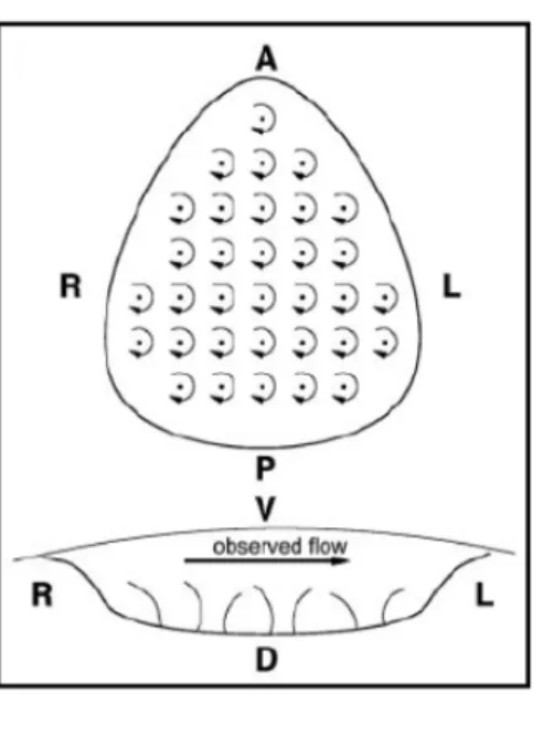
How the clockwise motion of the node monocilia drives a leftward flow in the node is an intriguing question. In the mouse, the node is a roughly pear‐shaped depression on the surface of the embryo, when viewed from the ventral side, covered by the Reichert’s membrane, which makes it a closed structure. It is filled
with extra‐embryonic liquid and arrayed over its base are a few tens of monocilia. When viewed from above (ventral view) these monocilia rotate clockwise (Figure 1.5). Since the node is a closed structure, how does the fluid re‐circulate inside the node, as it must, in order to generate the observed leftward flow? _________________________________________________________________________________________________
Figure 1.5 – Mouse Node. Ventral and posterior sketch
views of the node of the mouse embryo, and its rotating cilia, showing also the experimentally observed leftward nodal flow. Adapted from Cartwright et al., 2004. _________________________________________________________________________________________________ 1.4.2. Theoretical Model for Nodal Flow Generation Cartwright and colleagues approached this problem, and established a model for the fluid‐dynamical basis of the embryonic development of LR asymmetry in the mouse node. If the monocilia were to rotate around its vertical axes (Figure 1.6A), a set of vortices would be generated, one per cilium, and not a directional flow in the fluid above. Instead, the resulting flow would consist of a cellular network of vortices, in which general circulation would only occur at the edges of the network of cilia (Figure 1.6A). Elsewhere other than the edges the movement would be vortical. This scenario does not correspond to the general leftward flow above the cilia that has been experimentally observed. It has been suggested that the elongated pear‐shape of the node is the key factor, defining the leftward nodal flow (Nonaka et al., 1998; Nonaka et al., 2002). A triangle is the geometric shape that most resembles the node shape. However, switching from a rectangular array of
cilia to a triangular array of cilia does not qualitatively change the flow field (Figure 1.6B). The flow field is still vortical within the triangular array, with a general circulation only at the edges, as described for the rectangular array.
One other possible way to generate a leftward nodal flow could relay on the shape of the cilia. A directional flow would be produced if cilia were shaped like oars and feathered during part of the rotation. This does not seem to be the case since cilia are cylindrical in cross section. To hypothesize how a directed and circulating flow within the node can be produced by cylindrical cilia, one can use the analogy of a kitchen blender. When the blender is held vertically in the fluid it is mixing, with the blades rotating in a horizontal plane, this utensil generates a surface flow that corresponds to a vortex around the stem. If the blender is tilted, then a general flow is generated, in the direction in which the blades are turning when they are closest to the surface. If one considers this scenario in the node, all cilia would be tilted in the same direction, inclined and forming an angle to the horizontal. When sweeping out circles, the tilted cilia would generate a directional flow across the chamber above them, due to the fluid overhead being entrained in their direction of rotation (Figure 1.6D). The greater the tilt of the cilia, the stronger the directional flow above the vortices. In order to obtain the observed leftward flow, and considering the cilia rotate clockwise, the monocilia have to be tilted toward the posterior end of the embryo.
_________________________________________________________________________________________________
Figure 1.6 – Flow generated by different models of cilia positioning. (A) Vortical flow structure produced by
a single rotlet. (B) Rectangular array of rotlets with vertical axes, showing cellular structure of vortices with a general circulation only occurring at the edges. (C) Triangular array of rotlets with vertical axes, to correspond more closely to the shape of the node. As in
(B), general circulation is observed only at the edges. (D) Results of tilting the rotlet axes: array of tilted rotlets with a tilt angle of approximately 24°, showing directional flow above and below the array. Adapted from Cartwright et al., 2004. _________________________________________________________________________________________________
Cartwright and colleagues proposed that the monocilia tilt at an angle ranging from 5° to 25° from the vertical. Recent studies revealed that the monocilia in the node have an axis of rotation tilted of 10° to 40° to the posterior from the vertical angle (Buceta et al., 2005). It is worth noticing that the posterior tilting of the node cilia allows the cilia to orient along the anterior‐posterior and dorsal‐ventral axes, fulfilling one central condition of the F molecule model, proposed by Brown and Wolpert. The clockwise rotation of the cilia is an intrinsic chiral activity and thus the cilia have been proposed as an F structure in vertebrates (Nonaka et al., 2005; Okada et al., 2005). The cilia make a leftward swing away from the surface and a rightward sweep near the surface. According to hydrodynamics, a stationary surface retards the movement of fluids by shear resistance. Thus, the rightward sweep is lees effective than the leftward swing in generating fluid movement (Figure 1.8). It has been shown that the cilia in the mouse node rotate in a clockwise direction, when viewed from the ventral side, and that these cilia are tilted toward the posterior end of the embryos (Nonaka et al., 2005; Okada et al., 2005). The net flow at the node is from the right to left side and is driven by the effective stroke of the tilted cilia.
_________________________________________________________________________________________________
Figure 1.8 – Leftward flow is generated by the posterior tilt of nodal cilia.
(A) The trajectory of the tips of nodal cilia (red circles on the white ellipse) is shifted toward the
posterior when compared to the root of the cilia (yellow circles). (B) A scanning electron micrograph of ciliated cells of the ventral node of the rabbit embryo is shown. The root of a cilium is most frequently found toward the posterior of the cell. Bar, 5µm. (C) A hydrodynamic mechanism generates the leftward flow. Due to a gradient of shear resistance, a cilium cannot efficiently drive the extra‐embryonic fluid when it makes a rightward movement in the proximity of the surface. These figures are modified from Okada et al, 2005. Adapted from Hirokawa et al., 2006.
_________________________________________________________________________________________________
This nodal flow has been demonstrated in Xenopus (Schweickert et al., 2007), medaka, Oryzias latipes, (Tanaka et al., 2005; Blum et al., 2007) zebrafish and rabbit (Okada et al., 2005) and it is thus proposed to be the initial break of LR asymmetry for all vertebrates (Nonaka et al., 1998; Nonaka et al., 2002; Okada et al., 2005). 1.4.3. Nodal Flow in the Zebrafish Kupffer’s Vesicle
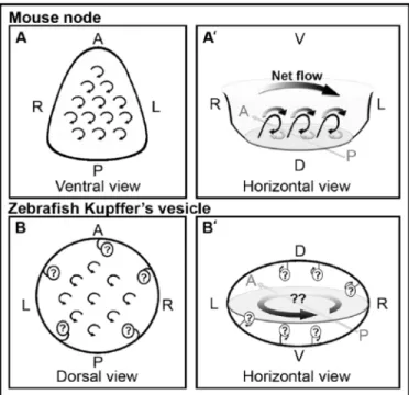
In zebrafish, the Kupffer’s vesicle (KV) is assumed to be the analogous structure to the mouse node in terms of LR patterning (Essner et al., 2002). KV is a transient ciliated organ derived from the dorsal forerunner cells (DFCs) (Cooper and D'Amico, 1996; D'Amico and Cooper, 1997), a group of cells that remains at the margin while the dorsal blastoderm involutes, during gastrulation. While the ciliated surface at the ventral floor of the mouse is relatively flat, KV is a hollow sphere containing cilia that project both from the dorsal roof and the ventral floor (Amack et al., 2007; Kreiling et al., 2007). Several studies reported that cilia rotate counterclockwise when viewed from the apical side (Figure 1.7B), which is the opposite of what is observed in the mouse node (Figure 1.7A and A’) (Kramer‐ Zucker et al., 2005; Shu et al., 2007).
_________________________________________________________________________________________________
Figure 1.7 – Models for the net flow inside zebrafish Kupffer’s vesicle compared with the planar mouse node. A, A’, B, B’: Schematic diagrams
of predicted nodal flow at the mouse node, and in zebrafish Kupffer’s vesicle.
(A) The ventral view of the mouse node
showing clockwise rotation of the cilia (as viewed from the apical side of the cells). (A’) Horizontal view of the mouse node (as viewed from the posterior side of the embryo) showing the posterior tilted cilia (thin, black arrows), which cause the dominant leftward flow (thick black arrows) and diminish the rightward flow (thick, gray arrows) due to the surface interactions. The net flow results in unidirectional flow from right to left (gradient arrow).
(B) Dorsal view of zebrafish Kupffer’s
rotation of the cilia (as viewed from the dorsal side of the vesicle). (B’) Horizontal view of the zebrafish Kupffer’s vesicle assuming counterclockwise rotation of the cilia (as viewed from the posterior side of the embryo). Flow inside Kupffer’s vesicle would be predicted to be counterclockwise (gradient arrows). However, this illustrates how conflicting flow paths would be produced if cilia on all surfaces rotate similarly. A, anterior; D, dorsal; L, left; R, right; P, posterior; V, ventral. Adapted from Baker et al., 2008.
_________________________________________________________________________________________________
This was puzzling, since it suggested that the mechanism for LR determination was not conserved in zebrafish. Likewise, the fluid dynamics inside the zebrafish Kupffer’s vesicle was not completely understood.
Having the two ciliated surfaces of the dorsal roof and the ventral floor facing each other, with cilia rotating in the same direction would not generate the strong leftward flow (Kramer‐Zucker et al., 2005), circular and counterclockwise at the center of the vesicle, as observed by bead movement and computational analysis (Figure 1.7B’) (Essner et al., 2005; Kawakami, 2005; Ellertsdottir et al., 2006; Kreiling et al., 2007; Shu et al., 2007).
Recent studies have shown that cilia on all cells in Kupffer’s vesicle rotate clockwise, when viewed from the apical side of the cells (Okabe et al., 2008). This seems to differ from the previous report (Kramer‐Zucker et al., 2005) but cilia on the dorsal roof were observed from the basal side of the cells, making the rotation appear different from those on the ventral floor. Thus, all cilia in Kupffer’s vesicle rotate clockwise similar to what is observed in the mouse node (Nonaka et al., 2002; Okada et al., 2005). Although cilia rotate in opposite directions at the dorsal roof and the ventral floor, the dominant flow inside the zebrafish Kupffer’s vesicle is counterclockwise (when viewed from the dorsal side of the KV). This correlates with cilia movement on the dorsal roof structure, since cilia are more numerous at the dorsal roof (Kreiling et al., 2007; Okabe et al., 2008).
The current model for fluid flow in the zebrafish Kupffer’s vesicle is reasonably comparable to the descriptions of flow within the mouse node, making the nodal flow as the initial break of LR asymmetry a conserved mechanism to all vertebrates.
1.5. Interpreting the Nodal Flow
As mentioned earlier, after left‐right symmetry is disrupted in or near the node and left‐right asymmetry is initiated, it is necessary to transfer the left‐right biased signals from the node to the lateral plate mesoderm. Although the nodal flow has been widely accepted to be the mechanism breaking symmetry in all vertebrates, it is not yet well understood how the nodal flow directs left‐right asymmetry. Two non‐mutually exclusive models have been proposed to explain how the information from the leftward nodal flow is interpreted at the level of the lateral plate mesoderm: the chemical gradient model and the “two cilia”/physical stimulation model.
1.5.1. Chemical Gradient Model
The chemical gradient model appeared first and, as the name suggests, is based on the formation of chemical gradients. It proposes that the directional flow transports particles/molecules (morphogens) and produces a concentration gradient along the ventral node (Nonaka et al., 1998; Okada et al., 2005). The chemical morphogen(s) accumulate only on the left side. In this model, simply by sensing the morphogen the cells can tell whether they are on the left side of the embryo. There are phenotypic differences between mouse mutant embryos lacking cilia and those with immotile cilia. Mutant mice with immotile cilia exhibit a complex pattern of expression of genes that are normally specific to the left side while mice without cilia have a higher tendency to exhibit bilateral patterns of gene expression in the LPM (Nonaka et al., 1998; Marszalek et al., 1999; Takeda et al., 1999; Murcia et al., 2000). In the chemical gradient model these differences can be explained if some (or all) node monocilia serve as the sensor for the chemical morphogen. Recent studies reported the presence of membrane‐sheated vesicles in the node that could correspond to the chemical morphogen. These particles, coined Nodal Vesicular Parcels (NVPs), bud from the node cells and have a diameter of 0.3‐ 5µm. Due to the mechanism by which NVPs are released and then caught, such big particles avoid circling within the ventral node (Tanaka et al., 2005). NVPs appear as
if they are leaving the tip of a bending rod (perhaps a microvilium) much like a whip. Ultra‐structurally, NVPs seem to comprise multiple lipophilic granules bounded by a membrane. NVPs are actively released from all regions of the ventral node into the middle of the laminar fluid flow, several micrometers above the ciliated surface, where the flow rate is fastest. This allows NVP to avoid being hit by rotating cilia in the initial phase. They are transported to the left side by the nodal flow and then hit the ciliated surface, burst and are fragmented. NVPs are absorbed by the surface of nodal crown cells on the left side and release their content, namely Shh and retinoic acid (RA), two molecules involved in LR asymmetry determination (Schilling et al., 1999; Tsukui et al., 1999; Wang et al., 2004), and shown to be present in NVPs (Tanaka et al., 2005). Cilia contain shh receptor, Smoothened (Smo). Because the turnover of NVPs seems to be delayed in Kif3a mutants (that lack cilia) compared with the iv/iv mutant (that has immotile cilia) it is plausible that a physical interaction between an NVP and a cilium is essential for the burst of the vesicles to occur. Studies on a hypomorphic allele of fibroblastgrowthfactor 8 (Fgf8) in mouse implicated FGFs in left sidedness promotion (Meyers and Martin, 1999). Blocking of FGFs with pharmacological and biochemical methods blocked the production of NVPs (Tanaka et al., 2005), suggesting that FGF signaling in the node is required for the production of NVPs (Figure 1.9). _________________________________________________________________________________________________ Figure 1.9 – The flow of NVPs Protruding microvillum picks up an NVP and releases it into the nodal flow. This event is stimulated by FGFR signaling. The NVP is transported to the left by the fluid flow and is hit by the ciliated surface on the left periphery of the node, releasing its contents onto the surface of nodal crown cells. Figure was modified from Tanaka et al, 2005 and adapted from Hirokawa et al, 2006.
As with most novel findings, this work provides possible explanations and raises several questions. This model respects the original idea of a morphogen being made in the node and carried to the left. It also presents a plausible mechanism to explain FGF function in mouse LR determination. NVPs, as presented in this work, provide a mechanism that could deliver asymmetric hedgehog signaling at the mouse node. However, no asymmetric hedgehog expression has ever been reported, as one would expect from the proposed model. Patched‐1 expression (up‐regulated by hedgehog) is symmetrical in the mouse node (Zhang et al., 2001). Mice lacking shh exhibit bilateral Nodal expression due to defects on the midline (Tsukui et al., 1999) rather than at the node, suggesting that shh is not the only LR morphogen in NVPs. These are a few questions showing that the precise relationship between hedgehog signaling and NVPs is still far from being clear. 1.5.2. TwoCilia Model The physical stimulation model considers a second type of non‐motile nodal cilia, placed in the peripheral region of the central node, expressing polycystin‐2 and having mechanosensory properties. It has been suggested that these immotile cilia could act as a sensor for the direction of the flow upon physical stimulation. This model is now commonly known as the “two‐cilia” model, since one type of cilia generates the flow and the other senses it (Tabin and Vogan, 2003). Upon monocilia physical stimulation, the information of flow direction is relayed by the induction of an asymmetric, left‐sided elevation of the intracellular of Ca2+ on the
left periphery of the ventral node (McGrath et al., 2003). Propagation of the increased Ca2+ concentration through the LPM may affect the balance of Lefty/Nodal
TGF‐β signaling, establishing the “leftness” of the LPM and inducing left‐side specific gene cascades. However, the detailed molecular mechanism of these signal transduction events is still under investigation.
mutant embryos with immotile cilia and those lacking cilia. These exhibit a complex expression pattern of genes that are normally specific to the left side of the LPM or tend to have bilateral gene expression patterns, respectively (Nonaka et al., 1998; Marszalek et al., 1999; Takeda et al., 1999; Murcia et al., 2000). This model is based on the idea that some nodal cilia would have sensory function, but this does not necessarily imply that the nodal cilia sense mechanical stimulus by flow.
The monocilia may serve as the sensor for chemical molecules. Both scenarios are supported: the cilia of the nasal epithelium sense chemical molecules. However, early LR signaling is disrupted by mutations in human polycystic disease (PKD) genes that are involved in mechanosensation by the monocilia of the renal epithelial cells (Murcia et al., 2000; Pennekamp et al., 2002). Some conceptual issues remain unanswered for the two‐cilia hypothesis. In this model, each ciliated cell should detect the directionality of flow by using the monocilium as a sensor. However, because of the symmetrical shape of the ventral node, the node cells on both sides can similarly sense the flow coming from the right side. It is thus not possible to convey global information on laterality. Further information, such as communication with surrounding cells, would be required to know whether they are on the left or on the right side of the node.
1.5.3. NVPs and Ca2+ Concentration
A connection between the release of NVPs and elevation of Ca2+
concentration on the left periphery of the ventral node was established by pharmacological studies.
Treatment of mice embryos with an FGFR inhibitor not only completely suppresses NVPs release as it also abolishes Ca2+ elevation. It did not perturb the leftward nodal
flow, indicating that the flow by itself is insufficient for left specific Ca2+ elevation.
Treatment with either Indian hedgehog (Ihh), retinoic acid (RA) or Shh (all factors symmetrically present within the embryonic node), was able to rescue calcium signaling at the node. Intriguingly, Shh and RA specifically rescued left‐sided Ca2+
signaling, while Ihh stimulated bilateral Ca2+ signaling. Analysis of the production of NVPs revealed that Shh and RA rescued the production of NVPs that were carried by the nodal flow toward the left side but, strangely, Ihh did not rescue the production of NVPs. These results suggest that Ihh specifically stimulates Ca2+ while Shh and RA activated left‐sided Ca2+ signaling through their rescue of NVP production. Antibodies against Shh and RA detect both Shh and RA in NVPs being released from floor of the node. The precise role of Shh and RA in NVP production is unclear. The ability of Ihh, but not Shh, to bilaterally activate Ca2+ signaling is also difficult to
explain. Both molecules are capable of activating the LR pathway in chick (Pathi et al., 2001), albeit ihh less efficiently than shh. Both are expressed at the mouse node and loss of both genes, or of their common receptor smoothened (Smo), results in loss of the left‐sided nodal cascade (Zhang et al., 2001). Still many questions remain to be answered and further investigation in the field is necessary. 1.6. Asymmetry Before the Nodal Flow Although the leftward flow of NVPs presents itself as a good candidate for the initial mechanism breaking left‐right asymmetry in mouse embryos, this does not seem to be the case in other organisms (reviewed by Levin, 2005). Monocilia in the ventral node or its counterpart structures are evolutionarily conserved (Essner et al., 2002) but the leftward nodal flow might not be the event breaking symmetry in some vertebrates, such as frog, birds and fish.
In frog, the establishment of the left‐right axis is linked to the formation of the dorsal‐ventral axis, thus happening very early during development. The DV axis is initiated by sperm entry during fertilization, followed by a cytoplasmic rotation during the first cells cycle (Gerhart et al., 1989). Thus, laterality has already been generated 2 hours after fertilization in frog eggs. During the initial cleavages of fertilized eggs the maternal RNA of H+/K+‐ ATPase is already localized
asymmetrically and this localization is essential for left‐right determination (Levin et al., 2002).