Novel and revisited approaches in nanoparticle systems for buccal drug delivery
Ana S. Macedo, Pedro M. Castro, Luís Roque, Natália G. Thomé, Catarina P. Reis, Manuela E. Pintado, Pedro Fonte
PII: S0168-3659(20)30014-6
DOI: https://doi.org/10.1016/j.jconrel.2020.01.006
Reference: COREL 10102
To appear in: Journal of Controlled Release
Received date: 23 October 2019
Revised date: 2 January 2020
Accepted date: 4 January 2020
Please cite this article as: A.S. Macedo, P.M. Castro, L. Roque, et al., Novel and revisited approaches in nanoparticle systems for buccal drug delivery, Journal of Controlled
Release (2020),https://doi.org/10.1016/j.jconrel.2020.01.006
This is a PDF file of an article that has undergone enhancements after acceptance, such as the addition of a cover page and metadata, and formatting for readability, but it is not yet the definitive version of record. This version will undergo additional copyediting, typesetting and review before it is published in its final form, but we are providing this version to give early visibility of the article. Please note that, during the production process, errors may be discovered which could affect the content, and all legal disclaimers that apply to the journal pertain.
Novel and revisited approaches in nanoparticle systems for buccal
drug delivery
Ana S. Macedo1,2, Pedro M. Castro3, Luís Roque2, Natália G. Thomé4,5,Catarina P. Reis6,7, Manuela E. Pintado3, Pedro Fonte4,5,8*
1
LAQV, REQUIMTE, Department of Chemical Sciences - Applied Chemistry Lab, Faculty of Pharmacy, University of Porto, Rua de Jorge Viterbo Ferreira 228, 4050-313 Porto, Portugal
2
CBIOS, Universidade Lusófona Research Center for Biosciences & Health Technologies, Campo Grande 376, 1749-024 Lisboa, Portugal
3
CBQF — Centro de Biotecnologia e Química Fina — Laboratório Associado, Escola Superior de Biotecnologia, Universidade Católica Portuguesa/Porto, Rua Arquiteto Lobão Vital, 172, 4200-374 Porto, Portugal
4
Center for Marine Sciences (CCMar), University of Algarve, Gambelas Campus, 8005-139 Faro, Portugal
5
Department of Chemistry and Pharmacy, Faculty of Sciences and Technology, University of Algarve, Gambelas Campus, 8005-139 Faro, Portugal
6
iMed.ULisboa, Faculdade de Farmácia, Universidade de Lisboa, Av. Prof. Gama Pinto, 1649–003 Lisboa, Portugal
7
Instituto de Biofísica e Engenharia Biomédica, Faculdade de Ciências, Universidade de Lisboa, Campo Grande, 1749–016 Lisboa, Portugal
8
iBB - Institute for Bioengineering and Biosciences, Department of Bioengineering, Instituto Superior Técnico, Universidade de Lisboa, 1049-001 Lisboa, Portugal
*Corresponding author: prfonte@ualg.pt
1 Abstract
The buccal route is considered patient friendly due to its non-invasive nature and ease of administration. Such delivery route has been used as an alternative for the delivery of drugs that undergo first-pass metabolism or are susceptible to pH and enzymatic degradation, such as occurs in the gastrointestinal tract. However, the drug concentration absorbed in the buccal mucosa is often low to obtain an acceptable therapeutic effect, mainly due to the saliva turnover, tongue and masticatory movements, phonation, enzymatic degradation and lack of epithelium permeation. Therefore, the encapsulation of drugs into nanoparticles is an important strategy to avoid such problems and improve their buccal delivery. Different materials from lipids to natural or synthetic polymers and others have been used to protect and deliver drugs in a sustained, controlled or targeted manner, and enhance their uptake through the buccal mucosa improving their bioavailability and therapeutic outcome. Overall, the main aim of this review is to perform an overview about the nanotechnological approaches developed so far to improve the buccal delivery of drugs. Herein, several types of nanoparticles and delivery strategies are addressed, and a special focus on pipeline products is also given.
Keywords: Buccal delivery; Nanoparticle; Polymer; Lipid; Drug delivery; Permeation
enhancer.
List of abbreviations and acronyms:
BSA - Bovine serum albumin; C – Concentration in the donor phase; Ch-PLA – Choline polylactid acid; CLSM – Confocal laser scanning microscopy; CMC –
Carboxymethyl cellulose; D – Diffusion coefficient; Da – Dalton; EFF-Cg – C-glycosyl flavonoid enriched fraction of Cecropia glaziovii; ER – Enhancement ratio; ɛ -
Fractional area; GRAS - Generally recognized as safe; h – Length of the membrane; Log P – Logarithm of the partition coefficient o/w; HPMC - Hydroxypropyl
methylcellulose; J – Permeation flux; K – Partition coefficient; LMWH - Low
molecular weight heparin; MTT - 3-(4,5-Dimethylthiazol-2-yl)-2,5-diphenyltetrazolium bromide; MCG – Membrane coating granules; NLC - Nanostructured lipid carriers; o/w – Oil-in-water; o/w/o – Oil-in-water-in-oil; PdI – Polydispersity index; PEG -
2 Polyethylene glycol; PEG-b-PLA - Poly(ethylene glycol)methyl
ether-block-polylactide; PLA - Polylactic acid; PLGA - Poly(lactic-co-glycolic acid); PVA – Polyvinyl alcohol; PVP – Polyvinylpyrrolidone; SC – Sodium cholate; SDGC - Sodium deoxyglycocholate; SDTC - Sodium deoxytaurocholate; SGC - Sodium glycocholate; SLN - Solid lipid nanoparticles; STC - Sodium taurocholate; w/o – Water-in-oil; w/o/w – Water-in-oil-in-water.
Table of Contents:
1. Introduction... 3
2. Buccal drug delivery ... 4
2.1. Anatomophysiology of the oral cavity... 4
2.2. Advantages of buccal drug delivery... 8
2.3. Disadvantages of buccal drug delivery ... 9
3. Buccal drug delivery systems ... 11
4. Nanoparticles as tools for buccal drug delivery ... 12
5. Permeation enhancers and protease inhibitors in nanoparticle formulations ... 16
6. Nanoparticle systems for buccal drug delivery ... 18
6.1. Lipid-based nanoparticles ... 19
6.1.1. Liposomes... 21
6.1.2. Solid lipid nanoparticles ... 23
6.1.3. Nanostructured lipid carriers ... 25
6.2. Polymer nanoparticles... 27
6.2.1. Natural polymers ... 27
6.2.2. Synthetic polymers ... 29
6.3. Hybrid nanoparticles ... 33
7. Walkthrough on pipeline products... 35
8. Conclusion and future perspectives ... 37
Acknowledgments ... 38
9. References... 38
Journal Pre-proof
3
1. Introduction
Up until the 1940s, conventional dosage forms including topical, parenteral, and oral formulations, such as suspensions, solutions, tablets and capsules were the most used to deliver drugs. However, these dosage forms are not devoid of disadvantages, for instance, the invasive nature and risk of infection of injections, and the short therapeutic effect of some conventional formulations requires high dosages to maintain the therapeutic effect. For example, the oral administration of drugs usually require high dosages due to the first-pass metabolism, but that often result in hepatotoxicity and undesirable side-effects [1]. Thus, it is necessary the development of formulations that allow to improve the bioavailability and attain an optimal therapeutic effect.
Efforts have been made towards the use of different delivery routes, and in the development of new drug delivery systems to overcome the drawbacks of conventional administration routes [2]. Therefore, the administration of drugs through the oral mucosa, particularly the buccal and sublingual mucosa, has been attracting a great interest [3, 4]. The main advantage of using the buccal route is the direct access of drugs (such as propranolol, nifedipine, etc.) to the systemic circulation by the internal jugular vein, eliminating the hepatic first-pass metabolism and mitigating possible side-effects. Nevertheless, the buccal drug delivery still faces challenges such as low permeability and a smaller absorptive surface area, in contrast to the high absorptive surface area of the small intestine [5, 6].
The drug delivery technology is becoming progressively more sophisticated and the current approaches focus on the influence of drug pharmacokinetic profiles on the therapeutic efficacy, as well as on the importance of drug targeting to the specific action site. Among those emerging technologies, nanotechnology is in the front line for the delivery of drugs by the buccal route [7]. The nanocarriers are able to increase the bioavailability of the loaded drugs due to their ability to remain in systemic circulation for a longer period of time, by presenting a controlled drug release profile, resulting in steady-state plasma concentration and reduced side-effects [8, 9]. Also, the use of the nanoparticles allow the delivery of therapeutic proteins, since they can protect them from enzymatic degradation, and display a controlled or sustained release increasing its bioavailability [10, 11].
The nanoparticle systems may be formed by several materials from lipid to natural and synthetic polymers or hybrid materials, which provides to the delivery systems different
4 properties and behaviors. Also, permeation enhancer and protease inhibitors are excipients that may be added to formulations to increase the uptake of drugs through the buccal mucosa, and inhibit the degradation of therapeutic proteins, respectively. Thus, both the nanocarriers and formulation excipients are often loaded into polymer matrices or hydrogels for buccal delivery to increase the bioavailability of drugs.
The main aim of this review is to give an overview of the current state-of-the-art of the buccal delivery of nanoparticle systems and address the most promising strategies for drug delivery through the buccal mucosa. The nanoparticle buccal formulations in clinical trials or in the market are also addressed.
2. Buccal drug delivery
2.1. Anatomophysiology of the oral cavity
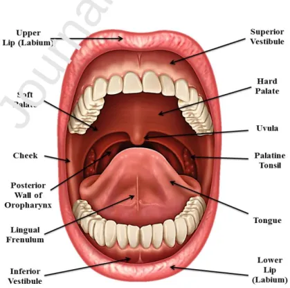
The oral cavity corresponds to the area of the mouth delineated by the lips, cheeks, floor of the mouth, soft palate and hard palate (Figure 1). It is one of the most used sites of drug administration since it is the first part of the digestive system, and it is also a chemosensory organ used as a secondary respiratory channel [12].
Figure 1. Anatomy of the oral cavity.
5 The oral mucosa has a total surface area of 170 cm2, and in some areas of the mouth it is involved in the mastication of food, such as the mucosa in the gum and the hard palate that represent 25% of the oral mucosa. The oral mucosa is formed by two layers, the deeper lamina propria and the superficial stratified squamous epithelium (Figure 2). The mucosa is protected by a keratinized epithelium with different levels of cell maturation, depending on the region of the oral cavity. The keratinized epithelium is found in the hard palate, gums and in some regions of the tongue dorsal surface [7]. In the keratinized part of the oral mucosa, the epithelium is constituted by four layers: basal, prickle, granular and keratinized layers. The non-keratinized epithelium covers the internal lips and cheeks, the soft palate, the ventral exterior of the tongue, and the sublingual mucosa. It is more flexible than the keratinized epithelium to enable the chewing and speech [13]. Furthermore, the oral epithelium is formed by a layer of 40 to 50 cells and its thickness is variable. The thickest epithelium is the buccal mucosa ranging from 500-600 µm thick, followed by the lining of the mouth and gums with a thickness ranging from 500-250 µm, and the thinnest layer is the floor of the mucosa with 100-200 µm [14, 15]. The superficial epithelial cells have intracellular vesicles called membrane coating granules (MCGs) that produce distinct types of lipids depending on the epithelium location, and therefore, play a key role in the permeability of substances. Non-polar lipids are derived from the lamellate MCGs and are found in the keratinized epithelium, whereas polar lipids are derived from MCGs present in the nonkeratinized epithelium [16, 17].
6
Figure 2. Layers of the oral mucosa.
The delivery of drugs through the oral mucosa may be subdivided into sublingual delivery through the sublingual mucosa, and the delivery through the buccal mucosa, since they are highly vascularized areas in the oral cavity and account for 60% of the oral mucosa. They are also delivery sites for the treatment of several affections of the oral cavity, such as fungal infections, ulcers and periodontal diseases [18, 19].
The saliva is a biologic fluid present in the oral cavity produced by the submandibular, the parotid and the sublingual glands, along with other minor submucosa glands. This fluid has several properties and is continuously secreted, dispersed and removed from the oral cavity. Such properties are high shear during eating and swallowing, the presence of electrolytes and organic molecules that maintain the local pH, and the presence of proteins with different types of antibacterial properties [20]. The renovation cycle of saliva influences the amount of drug present in the absorption site. The high turnover of the saliva contributes to a short residence time of the drug within the oral cavity, leading to poor drug absorption. Besides its composition, the saliva pH influences the dissolution and concentration of drugs. The physiologic pH of the oral fluids is between 6.0-7.5, but it can drop to 5.5 mainly in presence of some infections [21]. Also, the pH and salivary constituents are dependent on the saliva flow rate which varies with the period of the day and food intake, which increases the saliva production leading to a dilution of the drug and, therefore, influencing its absorption and therapeutic effect.
The epithelial cells are also immersed in a substance called mucus, that is secreted by major and minor salivary glands as part of the saliva. The mucus layer has a thickness of 50-450 µm thick and is mainly constituted by water, glycoproteins, enzymes, electrolytes, and macromolecular components known as mucins [22]. These large molecules are rich in carbohydrates and have a molecular weight ranging from 0.5 to 20 MDa, and may interact with each other to form a three-dimensional network which acts as a lubricant of the oral mucosa, contributing to the cell movement between neighbor cells and to the protection against the disruption of cell junctions [23]. The sulfate residues and the sialic acid in the mucins are charged negatively and bind to form an organized gel network at physiological pH. The mucins form a gel-like structure at the surface of the oral epithelium and can both enhance or hinder the absorption of drugs depending on the used carrier [23, 24].
7 Despite having a smaller number of enzymes when compared with the gastrointestinal tract, the enzyme degradation in the buccal and the sublingual regions is still the main concern for an effective drug delivery. Different enzymes such as dehydrogenases, carboxypeptidases and aminopeptidases are present in the buccal mucosa. The latter is the major metabolic obstacle for the buccal delivery of peptides, since their proteolytic activity is associated with the degradation of several therapeutic peptides [23].
Another important organ in the oral cavity is the tongue formed by skeletal muscle layered by a mucous membrane and represents about 15% of the surface of the oral mucosa. The extrinsic tongue muscles are responsible for the movement of the tongue, and the anterior two thirds of the tongue are in the oral cavity, whereas the remaining one third lies in the pharynx. The tongue moves the food in the mouth during the mastication to assist in the swallowing, and is also an obstacle to the absorption of drugs [25].
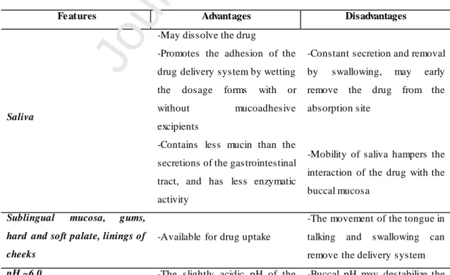
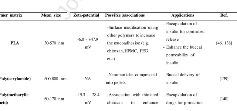
The development of delivery systems for buccal delivery rely on the thorough understanding of the anatomophysiology of the oral cavity, and knowledge of formulation design to overcome limitations and use the advantages of this delivery route [26]. The Table 1 summarizes the most relevant features of the oral cavity that influence the drug delivery by the buccal route.
Table 1. Features of the oral cavity involved in the buccal delivery of drugs.
Fe atures Advantages Disadvantages
Saliva
-May dissolve the drug
-Constant secretion and removal by swallowing, may early remove the drug from the absorption site
-Promotes the adhesion of the drug delivery system by wetting the dosage forms with or without mucoadhesive excipients
-Contains less mucin than the secretions of the gastrointestinal tract, and has less enzymatic activity
-Mobility of saliva hampers the interaction of the drug with the buccal mucosa
Sublingual mucosa, gums, hard and soft palate, linings of cheeks
-Available for drug uptake
-The movement of the tongue in talking and swallowing can remove the delivery system
pH ~6.0 -The slightly acidic pH of the -Buccal pH may destabilize the
8
saliva increases the dissolution of drugs that are weak acids, enhancing their absorption
drug or lead to its early release from the delivery system
-Easy modification of the pH value in the buccal cavity
Keratinized mucosa
-Low permeability ensures topical effect of highly potent drugs
-It is a barrier for drug absorption
Non-keratinized mucosa
-More permeable than the keratinized mucosa (like the buccal membrane and the sublingual area)
-Allow to obtain high plasma concentrations of drugs, increasing potential side effects
Oral cavity -Easily accessible route of
administration
-It is relatively thick, and absorption may be low to be useful for drug delivery
Surface area
-Large enough to allow drug absorption of drugs with appropriate physicochemical properties
-Lower vascularization than the other delivery routes
2.2. Advantages of buccal drug delivery
The administration of drugs through the buccal route shows better patient acceptance when compared with vaginal, rectal and ocular routes, improving the patient compliance to treatment due to the ease and comfort of the administration [26, 27]. The advantages of the buccal delivery include the direct absorption of drugs into the systemic circulation due to the good blood irrigation of the oral cavity, the avoidance of significant degradation of the drug as occurs by the high enzyme content and acid environment present in the gastrointestinal tract when drugs are absorbed in the intestine, and also the avoidance of the hepatic first-pass metabolism [28]. In addition, the rate of drug absorption when administered by the buccal route is not influenced by the gastric emptying rate as observed in the oral administration. Additionally, the oral mucosa is generally more permeable to drugs than other epithelia, and the easy removal of the delivery system is important to avoid irritative or toxicity effects in the buccal membrane.
9 For the delivery of poorly absorbed drugs, permeation enhancers may be also used in the formulations to increase the systemic availability of the drug, without causing permanent damage to the mucosa [29].
2.3. Disadvantages of buccal drug delivery
Despite the advantages, the buccal delivery has disadvantages and restrictions that hamper the drug delivery. Not all drugs are suitable for buccal delivery, for instance, drugs that are unstable at the oral pH, that have a bitter taste or odor, and drugs that can cause allergic reactions should be avoided.
The absorption rate of the drug and its elimination by involuntarily swallowing of the delivery system and food or liquids ingestion, may decrease the amount of absorbed drug, decreasing the blood concentration which may not be enough to attain a therapeutic effect [6]. The absorption rate of the drug depends on the surface area, the permeability coefficient and the drug concentration available in the oral mucosa surface. The accessible surface area for drug absorption in the oral cavity is small, about 50 cm2 for the buccal mucosa and 27 cm2 for the sublingual mucosa [30].
Regarding the concentration of the drug available, it is important to understand the complex environment of the oral cavity, since there are several factors which reduce the drug absorption (Figure 3).
10
Figure 3. Factors hampering the buccal uptake of drugs.
The saliva is the most relevant hampering factor, since its renovation cycles may dilute the drug concentration at the absorption site leading to a low drug amount at the surface of the buccal mucosa [31]. Also, the swallowing of the saliva or the ingestion of food may cause the removal of the drug from the absorption site (Figure 3). This requires the patients to do frequent administrations of the drug to achieve the desirable therapeutic effect [26]. Another important limitation is the irregular distribution of the delivery system within the mouth and the saliva. Also, talking, eating and chewing can lead to poor drug distribution within the oral cavity, affecting the release rates from the delivery system or retention times [32].
As aforementioned, the buccal delivery may lead to drug degradation, decreasing its bioavailability. For instance, the buccal mucosa expresses less P-glycoprotein than the intestine, but the cytochrome P450 3A4 is similarly expressed in the oral mucosa and in the small intestine, so it is an additional barrier to overcome by the new drug delivery systems administered through the buccal route [28, 33]. Another drawback is that the prolonged interaction of some drug delivery systems with the buccal mucosa may locally produce irritation and toxicity.
Despite the oral mucosa being easily available, attaining a systemic effect by administration of drugs topically is still unsuccessful, since the mucosa is also an
11 efficient barrier to the uptake of drugs due to its cellular and lipid composition, and to the physiologic parameters that hamper the drug absorption [13]. Overall, the new buccal drug delivery systems must address and solve these disadvantages.
3. Buccal drug delivery systems
To circumvent the disadvantages associated with buccal drug delivery, the drug delivery systems for buccal administration should have high mucoadhesive properties, mechanical strength, high resistance to the flushing action of the saliva, and release the drug towards the mucosa in a sustained or controlled manner. Furthermore, the formulation should protect the drug from the oral pH and enzymatic degradation. The loading of drugs into polymer matrices such as hydrogels, films, and nanoparticles may overcome these disadvantages [34]. The mucoadhesivity and mechanical strength can be achieved by using anionic and cationic polymers. Anionic polymers bind to the
mucin proteins by hydrogen bonds through the hydroxyl groups.
Carboxymethylcellulose (CMC) [35, 36] and alginate [37, 38] are the most commonly used anionic polymers for buccal drug delivery.
The cationic polymers increase mucoadhesivity by interacting with the negatively charged portions of the mucus. Chitosan forms thiol/sulfide bonds with the cysteine groups in the mucin. Mortazavian et al. developed a thiolated N-dimethyl ethyl chitosan by multivariate design [39, 40]. The tensile strength and bioadhesion force were analyzed as dependent variables. The study showed an increase in tensile strength and bioadhesion force with the increase on chitosan concentration in the formulations. The optimized formulation had a tensile strength of 5.24 kg/mm2 and a bioadhesion force of 2.35 N. Ex vivo permeation studies through rabbit mucosa showed that a higher amount of permeated insulin was also found for the optimized formulation, compared to the N-dimethyl ethyl chitosan and chitosan nanoparticles. Rencber et al. developed nanoparticles coated with chitosan and the cationic copolymer Eudragit for the delivery of fluconazole to treat oral candidiasis [41]. Nanoparticles had a size of approximately 200 nm and a zeta potential of +30 mV. The selected formulation delivered topically fluconazole to the oral mucosa of rabbits infected with candida albicans and achieved a completed healing after 3-5 days of formulation application. Further thiolation of chitosan has been described to improve the mucoadhesivity of formulations, and
12 consequently, their bioavailablity [42]. Enzyme inhibitors can also be added to nanoparticles prevent enzymatic degradation. Nevertheless, polyacrylic acid [43] and chitosan derivatives [44] have also shown to decrease enzymatic activity within the oral cavity.
4. Nanoparticles as tools for buccal drug delivery
Therapeutic effect is attained when drugs permeate membranes and reach the target site in an enough concentration to cause a pharmacodynamic effect. In the buccal administration, drugs must diffuse the mucus layer and reach the buccal epithelium to be absorbed [45, 46]. Small and lipophilic drugs (log P 1.6-3.3) are usually well absorbed through the oral mucosa, and drugs with higher log P values are less absorbed due to their poor water solubility. Lipophilic small drugs permeate the oral mucosa through the transcellular route. The hydrophilic large molecules are less successfully delivered through the oral mucosa (non-keratinised buccal mucosa and sublingual mucosa), so their preferred permeation route is the paracellular pathway due to the amphiphilic nature of the intercellular lipids. Furthermore, the salivary pH affects the molecule charge and its hydrophilic/hydrophobic nature, which possible hinders its absorption [47].
The use of nanoparticles as drug carriers is a good strategy to overcome the drawbacks associated with buccal drug delivery. In fact, the nanocarriers may present several advantages such as the increase of the diffusion rate of the drug across the mucus layer, protection of the drug from degradation, and from the drug dilution in the saliva since the nanoparticles adhere to the buccal mucosa prolonging the buccal residence and contact time with the mucosa [48]. In addition, nanocarriers may avoid drug elimination and oral clearance, and have a controlled and/or prolonged drug release profile, resulting in a decreased number of administrations, which improves patient compliance [49].
The film formed by the saliva on the surface of the mucosa hinders the permeation of lipophilic substances through the epithelium, whereas it enables the permeation of hydrophilic compounds. Due to the aqueous nature of the saliva, nanoparticles designed with hydrophilic polymers have a favorable permeation [50]. Nanoparticles with neutral charge or positively charged display better mucoadhesion due to the negative charges provided by the sialic acid in the mucus. The intimate contact with the mucus results in
13 longer retention times and higher drug dosage at the administration site. However, the turnover of the mucosal cells contributes to lower absorption of carriers, especially the lipophilic. Furthermore, the nanoparticle size and arrangement of the mucus modify the diffusion kinetics [51]. The salivary pH is also important for the controlled release of the drug from the nanocarrier. Permeability decreases for drugs ionised at low pH values, and the permeation of ionised drugs may be improved using strategies that increase the nonionized fraction of the drugs [50].
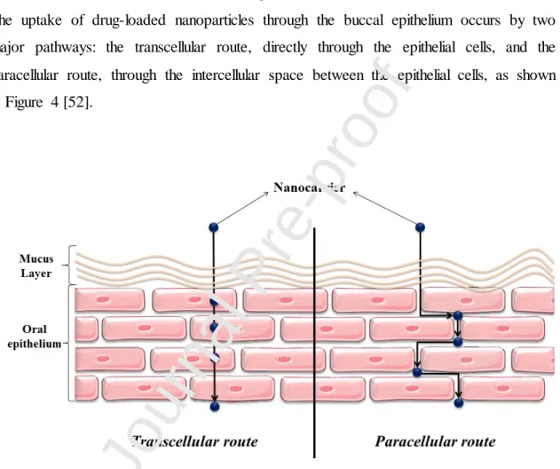
The uptake of drug-loaded nanoparticles through the buccal epithelium occurs by two major pathways: the transcellular route, directly through the epithelial cells, and the paracellular route, through the intercellular space between the epithelial cells, as shown in Figure 4 [52].
Figure 4. Routes of drug-loaded nanoparticle uptake through the buccal mucosa.
A previous study with the fluorescent probe fluorescein isothiocyanate showed that the paracellular transport is the most commonly used by large molecules, and the mucus within the intercellular spaces acts as an additional barrier [53]. The rate of permeation depends not only on the physicochemical properties of the drug, but also on the type of vehicle and whether permeation enhancers are present (see Section 5). Also, it has been shown that materials often use more than one permeation route to cross the epithelial
14 barrier during absorption [3]. The permeation flux of drugs through the buccal mucosa can be written as Eq. 1.
Eq. (1)
Where J is the permeation flux through the paracellular route, D is the diffusion coefficient of the drug, h is the length of the membrane, ɛ is the fractional area of the paracellular route, and C is the drug concentration in the donor compartment.
The Eq. 2 describes the permeation of drugs through the transcellular route.
Eq. (2)
Where the flux, J, depends on the diffusion coefficient (D) and the partition coefficient (K) of the drug, through the transcellular path (1-ɛ), across the length of the hydrophobic membrane (h). It has also been suggested that some nanoparticles cannot permeate the buccal mucosa through the paracellular route, since this route is restricted to lipophilic substances with a molecular weight below 1000 Da, and its fractional area is reduced compared to the transcellular route [54]. The ex vivo permeability of nanoparticles loading drugs may be carried out in continuous perfusion chambers, such as Ussing chambers, Franz cells and Grass-Sweetana [55]. A study by Goswami et al. showed that the paracellular transport is carried out through aqueous pores with a size of 18-22 Å for the buccal mucosa, and 30-53 Å for the sublingual mucosa [56]. In this study the authors used polyethylene glycol (PEG) as model hydrophilic permeant to study the relationship between increasing molecular weights and permeability through oral porcine mucosa.
In the literature, just a few studies have described the preferred permeation route of nanocarriers. Overall, the studies that show evidence of effective buccal drug delivery using nanocarriers showed a particle size of approximately 100 nm and narrow polydispersity index (PdI), and have either a lipid-based or polymer nature, and one or more permeation enhancers are usually included to promote drug delivery. Al-Dhublab, B. developed a zolpidem loaded nanosphere impregnated film [57]. The nanospheres had a polymer matrix of poly(lactic-co-glycolic acid) (PLGA) disperse in a film of hydroxypropyl methylcellulose (HPMC), Eudragit® LR 100, and Carbopol 974 P. Ex
15
vivo studies were carried out in Franz cells with rabbit buccal mucosa using simulated
saliva as the receptor medium at 37 ± 0.2 ºC. The highest flux was observed for the film containing 7.5 % of Eudragit® LR 100 (93.87±17.43 µg/cm2/h), compared to the film containing 10% Eudragit® LR 100 (75.39±12.53 µg/cm2/h). Although similar zolpidem concentration was used in both formulations, pharmacokinetics studies showed higher plasma peak for the zolpidem-nanosphere film (52.54 ng/mL) compared to the oral solution (32.34 ng/mL).
The delivery of therapeutic proteins and peptides by the buccal mucosa has gained popularity over the years as a non-invasive alternative. Mainly because these drugs have high molecular weight that hinders their permeation through the intestinal epithelium, and suffer enzymatic degradation in the gastrointestinal tract. Nanoparticles have took the lead on the development of proteins delivery systems due to their ability to facilitate the buccal uptake and protect their bioactivity. Morales et al. developed a film containing insulin-coated nanoparticles by the co-precipitation of valine, and a sorbitan monostearate in propanol solution was used as the anti-solvent [58]. The nanoparticles were incorporated in two films, one containing Eudragit® RPOL, and another one containing Eudragit® RPOL combined with HPMC. The permeation mechanism was tested ex vivo using EpiOral, a buccal mucosa model used to determine the permeation routes of molecules. It contains 8-11 cell layers of primary buccal keratinocytes on fibroblast and collagen matrix [59]. The film containing Eudragit® RPOL and the insulin-coated nanoparticles showed a permeation flux of 0.34 g/h/cm2 and a lag-time of 7.81 min, compared to a permeation flux of 0.07 g/h/cm2 and lag-time of 11.72 min observed for the formulation containing the insulin-coated nanoparticles and the Eudragit® RPOL and HPMC combined. The study suggested that the positive charges in the polymer chain of the Eudragit® RPOL enhanced the permeability of insulin by i) disturbing the lipid layers in between the epithelial cells and ii) increased the retention time with the buccal mucosa, creating a reservoir of drug in the proximity of the epithelial cells [58], suggesting the paracellular route to be the preferred permeation mechanism. Similarly, chitosan has been proposed to have the same permeation enhancement mechanism, due to the positive charges of the polymer chain [60, 61]. Chitosan based thermosensitive gels for the delivery of erythropoietin have also been developed, and showed a good formulation and protein stability over time [62, 63]. Recent studies carried out by Mahdizadeh Barzoki et al. proposed coated nanoparticles with thiolated chitosan for the delivery of insulin through the buccal mucosa [40]. A
16 mean particle size of 148 nm, zeta potential of 15.5 mV, PdI of 0.26 and an association efficiency of 97.56% were observed. Insulin was also conjugated with phosphate and encapsulated in flexible nanovesicles for buccal drug delivery by Xu et al [64]. The formulation had a mean particle size of 85.84 ± 2.38 nm and a zeta potential -26.2 ± 0.5 mV, and the deformability was also assessed. The formulation increased the permeation of insulin through porcine buccal mucosa through the deposition of the insulin-phosphate conjugates that was further enhanced by the deformability of the nanovesicles. The nanovesicles displayed both transcellular and paracellular transport as evidenced by confocal laser scanning microscopy (CLSM). Further analysis of the receptor medium with transmission electron microscopy showed intact nanovesicles after the permeation study.
5. Permeation enhancers and protease inhibitors in nanoparticle
formulations
As aforementioned, the buccal mucosa is a semi-permeable membrane that acts as a barrier for most drugs. Thus, in addition to nanoparticles, strategies to improve drug bioavailability include the use of permeation enhancers and protease inhibitors as excipients in nanoparticle delivery systems. The permeation enhancers are chemicals that change the barrier physicochemical properties and open a pathway for drug uptake, whereas protease inhibitors circumvent the enzymatic barrier present in the mucus layer allowing the successful delivery of drugs [65, 66].
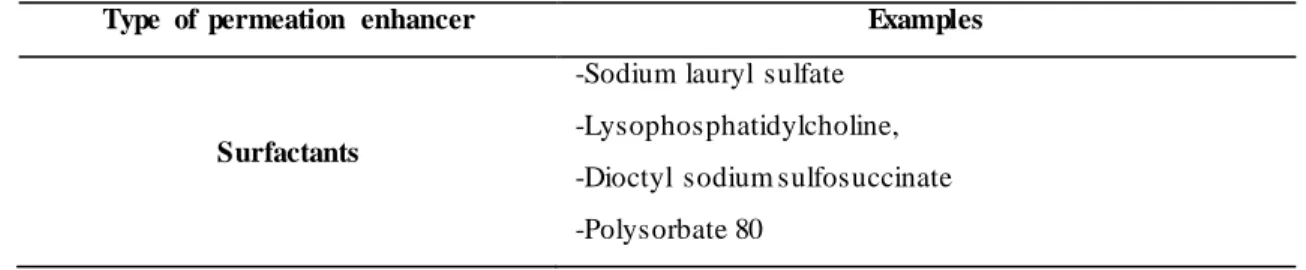
The permeation enhancers need to be compatible with other formulation excipients, display immediate permeation, increase the drug uptake, be nontoxic, and have no pharmacological effect [29]. There are different types of permeation enhancers used in buccal delivery to increase the uptake of drugs as shown in Table 2.
Table 2. Types of permeation enhancers used in buccal drug delivery.
Type of permeation enhancer Examples
Surfactants
-Sodium lauryl sulfate -Lysophosphatidylcholine, -Dioctyl sodium sulfosuccinate -Polysorbate 80
17 -Glyceryl monolaurate -Poloxamer 407 Fatty acids -Sorbitan laurate -Sodium caprylate -Sucrose palmitate -Lauryl choline -Oleic acid -Caprylic acid -Lauric acid Mucoadhesi ve Polymers -Chitosan -Polycarbophil
-Sodium carboxymethylcellulose and derivatives
The permeation enhancers may increase the drug uptake by 4 major mechanisms: i) increase the drug partitioning, ii) by interaction with the cell protein domains within the epithelium, iii) extraction of the intercellular lipids, and iv) increase the solubility of the drug in the vehicle or in the delivery system. It was described that the increase in drug uptake by the paracellular route using permeation enhancers is caused by the extraction of the intercellular lipid lamellae between adjacent cells that form the buccal epithelium, creating a space for macromolecules go through [66]. The mucus rheology is also affected, so usually permeation enhancers decrease its viscosity and elasticity parameters and enable the diffusion of molecules with high molecular weight. The permeation of poorly soluble drugs might also be improved in the presence of permeation enhancers [67]. Patil et al. produced insulin-loaded alginic acid nanoparticles, and nicotinamide was added to the formulation as permeation enhancer for sublingual delivery [67]. The insulin-loaded nanoparticles had an average size of 200 nm, low PdI (<0.25), and a high association efficiency of about 95%. The Fourier transform Infra-red spectroscopy spectra, differential scanning calorimetry, and Circular dichroism results showed a good interaction between the alginic acid and insulin, confirming its stability.
Protease inhibitors are also commonly used in formulations to improve the delivery of therapeutic proteins and peptides, by avoiding the degradation by the enzymes present in the saliva. Also, protease inhibitors may change the pH value within the oral cavity resulting in lower enzymatic activity, or even change the conformation of the peptide or protein, or by bonding to the protein, thus reducing the accessible sites to enzymatic degradation. Protease inhibitors such as aprotinin, amastatin, bestatin, boroleucine and
18 puromycin have been widely used [68]. In the case of aprotinin, it has been used for buccal peptide delivery [29, 66]. Furthermore, the association between mucoadhesive nanoparticles and protease inhibitors has shown to be advantageous to protect the drug and improve its therapeutic effect [69].
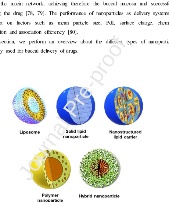
6. Nanoparticle systems for buccal drug delivery
The incorporation of drugs into nanoparticles allows to overcome limitations of buccal delivery, such as the barrier properties of the buccal epithelium, the undesired swallowing due to saliva turnover and the masticatory movements [23, 70, 71]. Nanocarriers are versatile delivery systems that might be used to load and delivery different drugs, using several matrices and production techniques. The qualitative composition of the encapsulating agent is of paramount importance regarding permeability, release profile, adhesion to buccal epithelium or even for targeting specificity. The Figure 5 shows the most common types of nanoparticles used for buccal drug delivery. The optimization of the nanoparticle formulation can lead to the production of a buccal delivery system that presents good stability, safety and effectiveness. A proper nanoparticle formulation must be able to maintain intimate contact between the carried drug and the buccal mucosa, assuring the permeation of the drug. Moreover, the optimization of nanoparticle formulation must guarantee the permeation enhancement along with the stability and protection from premature degradation of the carried drug.
Nanocarriers are typically delivered as aqueous suspensions or incorporated into a gel matrix or film. Gels and films are three-dimensional polymeric network cross-linked that can be tailored to display specific features. Several types of adhesive gels may be used to deliver drug-loaded nanoparticles [72]. Intelligent hydrogels with thermosensitive [73-75] and pH responsive properties [76] have been developed for buccal delivery of drugs. Cross-linked polyacrylic acid has been used for buccal drug delivery due to its high mucoadhesivity and controlled drug release [51]. For instance, nanoparticles may be dispersed in formulations containing mucoadhesive polymers that assure a higher residence time of the carriers within the buccal mucosa, therefore increasing the probability of drug permeation across the epithelium [34]. The cationic polymers are preferable among others due to the establishment of electrostatic bonds with the negatively charged molecules composing mucin present in the saliva [77]. The
19 improvement of the buccal drug delivery has been achieved by the development of formulations presenting mucoadhesive properties by using different types of polymers, such as sodium alginate, guar gum, hydroxy ethyl cellulose, methyl cellulose and polyethylene glycol. Such polymers are able to interact with the mucus layer originating strong intermolecular hydrogen bonding, increasing the penetration of the polymer through the mucin network, achieving therefore the buccal mucosa and successfully delivering the drug [78, 79]. The performance of nanoparticles as delivery systems is dependent on factors such as mean particle size, PdI, surface charge, chemical composition and association efficiency [80].
In this section, we perform an overview about the different types of nanoparticles commonly used for buccal delivery of drugs.
Figure 5. Types of nanoparticles used for buccal drug delivery.
6.1. Lipid-based nanoparticles
The drug dissolution in biologic fluids is one of the key factors for a high bioavailability. The absorption of hydrophobic drugs after oral administration is limited
20 by the dissolution rate, which is the limiting step to attain high blood levels of the drug. Hence, using a lipid matrix with a high surface area to carry drugs for buccal delivery may be a good strategy to improve absorption and overall bioavailability of the carried drugs [81]. The lipids used to prepare the nanoparticles are generally recognized as safe (GRAS), and thus with good biocompatibility and tolerability properties. The lipid nanoparticles have been used to deliver drugs with a controlled release, mostly lipophilic drugs, since they are relatively easy to produce with robust scale-up ability and, if properly tailored, can be targeted to specific tissues or organs [82]. The high-speed homogenization, sonication and high-pressure homogenization are the most common methods to prepare lipid nanoparticles [83]. The high pressure homogenization is a robust process that can easily be scaled-up, but the lipid nanoparticles may be widely polydisperse regarding their size [83]. On the other hand, the high-speed homogenization and sonication offers a narrower PdI but are more laborious and harder to scale-up.
The lipid-based nanoparticles may be classified into liposomes, solid lipid nanoparticles (SLN) and nanostructured lipid carriers (NLC), according to the type and/or blend of lipids used. Liposomes are generally composed by one (unilamelar vesicles) or more (multilamelar vesicles) bilayers with amphiphilic behavior, enclosing a hydrophilic core, and the phospholipids are the most common amphiphilic entrapping agents [84]. The SLN core matrix is composed of a lipid that is solid at room temperature, which increases both their stability and the association efficiency of drugs, when compared with liposomes [85]. Finally, the NLC were designed to improve the characteristics of SLN, namely the drug association efficiency and also the size dispersion and a more sustained release profile [81]. The NLC are prepared using a blend of solid and liquid lipids, which may also increase the solubilization of loaded drugs. Moreover, due to the presence of lipids in different physic states, the drug diffusion from NLC is usually biphasic, comprising an initial burst release and a posterior slower release of the drug. The improved association efficiency and tailored drug delivery kinetics can be obtained by changing the relative amounts of liquid and solid lipids of NLC [86].
In the following subsections, it is addressed the different lipid-based nanoparticles for buccal delivery of drugs.
21 6.1.1. Liposomes
The liposomes were introduced in 1970 as a breakthrough delivery system that allowed the targeting of therapeutic molecules [87]. They were developed to improve the pharmacokinetic and toxicity profile of drugs, by increasing their permeability, stability and allowing drug controlled release [88]. The nature of liposomes as lipid nanoparticles is considerably different from SLN or NLC, with intrinsic advantages, and the most important one is their ability to load both lipophilic and hydrophilic molecules, due to their amphiphilic character [89, 90].
Liposomes loading vitamin B6 were developed to improve the absorption and bioavailability of the drug [91]. The produced liposomes were dispersed in a mucoadhesive buccal film of HPMC and sodium carboxymethyl cellulose (CMC) to improve the residence time and, therefore, the duration of contact with the buccal epithelium and improving permeation. The release assay indicated that the buccal film and liposomes contributed to a prolonged release of vitamin B6 (72.6 % at 6h), when compared with the film without liposomes (96.37 % at 30 min), which indicated that liposome structure was not significantly affected by the solvent casting procedure used to produce the films. The ex vivo permeability assay performed in chicken pouch showed that the vitamin B6 loaded into liposomes-film conjugation presented a lower permeability flux (36.89 %) across the membrane when compared with vitamin B6 dispersed in the film or with a vitamin B6 solution.
In another study, it was attempted to increase the buccal permeability of silymarin, using a blend of lecithin, stearyl amine and cholesterol as encapsulating agents [92]. The permeability across chicken cheek pouch showed that the liposomes increased significantly the silymarin permeability across the buccal mucosa when compared with a silymarin solution. It was suggested that the permeability observed would have been superior if the epithelial cells of the cheek pouch were still metabolically active.
The buccal mucosa is also a suitable delivery route for immunization purposes, however the continuously renewed mucus layer and the activity of lysozyme, proteins and glycoprotein mucins hinders the success of the formulations [93]. Both physical and chemical barriers prevent antigens from crossing the epithelial layer, and from being presented to antigen-presenting cells. Aiming to develop a conceptually new, non-invasive, vaccination method, Zhen et al. associated mannose-PEG-cholesterol with lipid A liposomes, and used bovine serum albumin (BSA) as model immunogenic
22 protein [94]. Previously, they demonstrated the efficiency of the system as adjuvant for protection and presentation of antigens, specifically to immunocytes, located in the oral mucosa [95]. Nevertheless, due to a low vaccination success, the system was used to produce microneedles to promote a more effective presentation of antigens in the buccal mucosa. The microneedles have already proved to be effective to deliver topical vaccines, by piercing the skin and reaching the epidermal or dermal layer, in a painless manner [96, 97]. Since the buccal mucosa is considerably more absorptive than skin, the use of microneedles as buccal delivery systems may be a promising approach. The liposomes were prepared by emulsification followed by freeze-drying, using a blend of soy phosphatidylcholine / mannose-PEG-cholesterol / stearylamine / monophosphoril lipid A (100/5/10/1, molar ratio). After freeze-drying and re-hydration, the liposomes were poured into the microneedle array inverse molds in a reduced pressure environment. The microneedles were then dried in a desiccator containing anhydrous CaCl2. After rehydration, it was observed that the liposomes presented an average size
of 200 nm, which is suitable for buccal permeation. The surface charge was almost neutral, and the association efficiency remained close to 40% even after 180 days of storage. The in vitro release assay performed in phosphate buffer saline pH 7.4, 37 ºC, showed that only about 40% of BSA was released from the microneedles after 48 h, indicating they are good buccal depot delivery systems. In addition, the incorporation of calcein, a fluorescent agent that does not permeate the cellular membrane within the microneedles demonstrated that the developed delivery systems facilitated the uptake of antigens by immune cells via mannose receptor-mediated phagocytosis. Moreover, after
in vitro administration in mice, the BSA-loaded microneedles induced an effective
immune response either by Th1 or Th2 lymphocytes, establishing both systemic and mucosal immunity. A similar strategy showed to be effective in the buccal immunization against hepatitis B virus, inducing a stronger immune response when compared with subcutaneous or intradermal administration [98].
In another study, Chen et al. prepared a novel delivery system of self-assembled liposome in a multi-layered fibrous mucoadhesive membrane for the delivery of carvedilol [99]. The system consisted of an electro spun layer that formed polyvinylpyrrolidone (PVP) phospholipids liposomes upon contact with water, an adhesive layer formed by HPMC, CMC, and PEG 400. The liposomes sizes were below 100 nm, and a narrow PdI was obtained (approximately 0.15). The association efficiency of the drug varied between 30% and 68% for the multi-layered film and it
23 was 91% for the liposomes. The zeta potential varied between 12 mV and 20 mV, showing that the system was positively charged. The in vitro permeation test using porcine buccal mucosa showed the liposomes prepared by the conventional method achieved a cumulative amount permeated of carvedilol of 42.1µg/cm2 at 5h, while the liposome prepared by electro spun showed a cumulative amount permeated of carvedilol of 18.0 µg/cm2, whereas the formulation showed 21.8 µg/cm2. The pharmacokinetic study showed a 154% increase in the relative bioavailability of the buccal formulation compared to an intragastric administration of a carvedilol suspension.
Recently, a new study showed the potential of soy lecithin liposomes incorporating bile salts (sodium cholate [SC], sodium taurocholate [STC], sodium glycocholate [SGC], sodium deoxyglycocholate [SDGC], or sodium deoxytaurocholate [SDTC]) as edge activators for the delivery of insulin across TR146 buccal cells [100]. The prepared formulations had a mean particle size of 140-150 nm and an association efficiency of 66%-78%. All the formulations showed an enhancement ratio (ER) superior to the insulin solution, being the highest ER 5.24 observed for the formulation SDGC-incorporated liposome (p < 0.001). A similar permeation profile was observed by CLSM when the liposomes were loaded with the fluorescent probe fluorescein isothiocyanate combined with insulin. Higher intensity was observed for the formulation SDGC liposomes, followed by SC liposomes > SDTC liposomes > SGC liposomes > STC liposome. A similar trend was observed in the permeated fluxes: SDGC liposomes showed a permeated flux of 27.6 ng/cm2/h, compared to SC liposomes, 16.5 ng/cm2/h > SDTC liposomes, 15.9ng/cm2/h > SGC liposomes 10.34, ng/cm2/h > STC liposome, 8.4 ng/cm2/h, compared to a flux of 5.2 ng/cm2/h of the insulin solution.
6.1.2. Solid lipid nanoparticles
The SLN have been extensively studied as delivery systems for a wide array of drugs and aiming different delivery routes such as parenteral, oral, pulmonary, nasal or ocular [101-104]. When buccal mucosa delivery is aimed, it is important to assure enough contact time between SLN and the epithelium, to overcome premature swallowing (i.e. saliva turnover, chewing, tongue movements and phonation) [105]. Curcumin is a curcuminoid present in turmeric and has diverse therapeutic activities (e.g. antioxidant,
24 antimicrobial, chemotherapeutic against several types of cancer and anti-inflammatory), however the rapid hepatic metabolism and poor chemical stability, demand a delivery route different from the oral one. To enhance the mucoadhesion of curcumin-loaded SLN to the buccal mucosa, and aiming the treatment and management of lesions related with oral cancer, curcumin-loaded SLN were incorporated into a polycarbophil/poloxamer 407 mucoadhesive gel [106]. The SLN were prepared by hot melt followed by high-shear dispersion (12,000 rpm) and high-speed homogenization. The in vitro release studies revealed that curcumin released from the delivery system (~10% after 5h) was significantly slower (p < 0.05) when compared with curcumin-loaded SLN (~28% after 5h) or curcumin-curcumin-loaded gel (~48% after 5h). It was also reported that the ex vivo permeation and retention test, performed using chicken buccal mucosal tissue, revealed that the curcumin carried by the delivery system did not significantly permeate the mucosa, being therefore indicated for local delivery on the oral cavity. The lack of permeability was most likely due to the low hydrophilicity of curcumin. Even though the curcumin loaded into SLN did not reach the systemic circulation in significant concentrations, 21% of curcumin was recovered from the dissected chicken buccal mucosa after 3 h of contact, indicating that the mucoadhesive formulation associated with SLN not only assured the protection of curcumin against the oral enzymatic and mechanical activity, but also promoted the permeation through the buccal basal cells. The results were significantly higher when compared with the basal penetration of a curcumin solution (2% after 3h) and slightly higher than curcumin-loaded SLN (18% after 3 h). It was concluded that SLN contributed to a higher adhesion and superior permeation of curcumin across the buccal basal cells. Moreover, the higher permeation of curcumin administered with SLN was also associated with the well-known permeation enhancer ability of Poloxamer F-127 [107, 108]. The latter can change the morphology of the barrier created by intercellular lipids by disrupting tight junctions. Since there are no tight junctions in the buccal epithelium, the permeability enhancement was most likely associated with the removal of lipids due to surfactant activity of the Poloxamer. The clinical evaluation of the formulation was also performed after administration in 10 patients with erythroplasia. When asked to describe the reduction of pain level associated to buccal lesions, patients treated with curcumin-loaded SLN reported significantly (p < 0.05) higher values (20, 60 and 90% after 3, 7 and 10 days of treatment, respectively) when compared to curcumin incorporated in a conventional gel (16.6, 36.7 and 63.3% after 3, 7 and 10 days,
25 respectively). The reduction of the lesion size was also superior (p < 0.05) for patients treated with curcumin-loaded SLN (67.5 and 94.3 after 2 and 4 weeks, respectively) when compared with patients treated with the curcumin gel (25% and 66.67% after 2 and 4 weeks, respectively).
In another study, to increase the residence time through mucoadhesion to buccal mucosa, it was created a sponge-like dosage form, based on polycarbophil, loaded with SLN for buccal delivery of curcumin, as an attempt to improve the previously described mucoadhesive gel [109-111]. The in vitro mucoadhesion assay performed on a mucin-enriched agar plate (pH 6.8) demonstrated that the polycarbophil sponge-like matrix formulation showed no displacement from the initial spot on the agar/mucin plate, even after 24h with a 30º inclination [112]. Such absence of displacement indicates good characteristics of mucoadhesion. The in vivo mucoadhesion residence time assessment, performed in six healthy volunteers, showed that curcumin-loaded SLN incorporated within the matrix presented a residence time of 15 ± 2.5 h. The high residence time was associated with the close interaction between the system and mucin caused by interpenetration of the polymer and the mucus [113]. The cohesive forces between mucin and the formulation occur through hydrogen bonds and van der Waals forces, and the mucoadhesion was enhanced due to the system being a solid dosage form compared to liquid or semi-solid mucoadhesive formulations [114, 115]. The curcumin release was tested both in vitro (using a 12-14 kDa dialysis membrane) and in vivo (in five healthy adult volunteers), and as expected, the curcumin-loaded SLN incorporated within the matrix presented a significantly slower release of drug content (~15% after 6 h) when compared with curcumin-loaded SLN (50% after 6 h). In addition, it was showed that SLN-polycarbophil sponge-like matrix offered a sustained release of curcumin in vivo, and curcumin was still detectable in the saliva of volunteers 15 h after administration of the delivery system.
6.1.3. Nanostructured lipid carriers
The NLC were developed to overcome inherent disadvantages of SLN. The entrapping matrix is a blend of solid and liquid lipids, that provides distinct characteristics of NLC when compared with other lipid-based nanoparticles [116]. The NLC matrix is more disorganized compared to the matrix composing SLN (Figure 5), which results in a
26 higher entrapment of drugs, and thus higher association efficiency due to more free spaces where the drug can be entrapped, and a slower drug release [117].
In a previous study, domperidone was encapsulated into NLC for drug delivery in the oral cavity aiming to increase the permeability of domperidone across the buccal epithelium [118]. The domperidone-loaded NLC were prepared by high pressure homogenization, using a blend of palmitic acid (solid lipid) and oleic acid (liquid lipid). Both in vitro (TR146 human buccal carcinoma cells) and ex vivo (excised porcine buccal mucosa) domperidone uptake studies indicated that nearly 10% of domperidone was able to permeate the buccal epithelium. Moreover, 11.48 ± 7.19% of domperidone carried by NLC reached the cytoplasm and 17.99 ± 2.24% crossed the TR146 cell monolayer and reached the basolateral side. Even though the free domperidone in vitro and ex vivo permeability performance were not evaluated in this study, the obtained results are promising since domperidone is extensively metabolized in the liver (first-pass effect), leading to a reported low oral bioavailability, 12.7 to 17.6% of drug administered per os in capsules or tablets [119].
In another study, the NLC prepared using Precirol® ATO 5 (solid lipid) and Miglyol®812 (liquid lipid) were used to deliver ibuprofen across the buccal mucosa [120]. The ibuprofen-loaded NLC were also incorporated within a mucoadhesive hydrogel matrix to increase the residence time and contact of nanoparticles with the buccal mucosa. The in vitro release assay revealed that the NLC promoted a slower release of ibuprofen, when compared with an ibuprofen solution and that the mucoadhesive gel had a role on the hindrance of ibuprofen release.
Kraisit and Sarisuta developed NLC for the delivery of triamcinolone acetonide using the Box-Behnken design by hot homogenization [121]. Spermaceti, soybean oil and polysorbate 80 were used to produce the NLC, and a particle size below 200 nm, a zeta potential of -5.91 to -20.83 mV, and an association efficiency of 80% were observed. Increasing the lipid and surfactant showed a decrease in particle size. The incorporation of triamcinolone acetonide in the NLC matrix was confirmed by energy-dispersive X-ray spectroscopy. The penetration of the loaded NLC was assessed by CLSM in the porcine buccal mucosa, and Nile red-loaded NLC were found at a 180 µm depth at 8h after permeation.
Regardless the advantages of NLC as carriers, more studies are needed to demonstrate their potential to deliver drugs into the buccal epithelium.
27
6.2. Polymer nanoparticles
The polymers used to produce nanoparticles can be obtained either from natural or synthetic sources [122]. The ideal polymers for nanoparticle production must be biodegradable, biocompatible and with good drug entrapment and release properties. Moreover, when nanoparticles are produced as buccal delivery systems, the polymers must be mucoadhesive to increase the residence time of the delivery system, enhancing the drug uptake and the amount that reaches the systemic circulation [123]. The mucoadhesion is mostly obtained either by the formation of electrostatic interactions, and by the formation of hydrogen bonds with the mucus layer. Since mucin presents a negative charge, the cationic polymers are preferable for production of mucoadhesive nanoparticles. Moreover, due to the hydrophilic nature of mucin, the polymers that present a higher number of functional groups capable of establishing hydrogen bonds are also preferable [124].
6.2.1. Natural polymers
The polymers with natural origin, either directly extracted or chemically modified, are commonly used in the pharmaceutical industry to produce drug delivery systems due to their inherent advantages such as low toxicity, biodegradability, and availability at low price, especially when compared with synthetic polymers [125, 126]. Also, some natural polymers present innate targeting characteristics, delivering drugs to specific cells, tissues or organs. Nonetheless, natural polymers present also some disadvantages, being prone to microbial contamination along with heterogeneity regarding its physicochemical composition, since most polymers with natural origin are extracts. The most common sources are plants (e.g. guar-gum, starch, pectins, locust bean gum, gum acacia, psyllium and arabic gum), algae (alginate and carrageenan), bacteria (e.g. xanthan gum, gellan and curdlan), fungus (e.g. scleroglucan, pullulan, chitin) and animal (e.g. gelatine type A and B, chitin and shellac).
Charged polymers can be used as backbone to prepare nanoparticles, simply by adding oppositely charged cross-linkers upon vigorous stirring. Previously, to develop and optimize polymer nanoparticles as buccal delivery systems, it was used chitosan, pectin and alginate as matrix polymers and tripolyphosphate and zinc as negative and positive
28 counterions, respectively [127]. The polymer:counterion proportion was thoroughly studied to achieve the most stable and suitable nanoparticles for buccal delivery. The stability was determined by assessing the nanoparticle aggregation and disintegration in contact with simulated salivary fluid, along with the variation of PdI, hydrodynamic diameter and zeta potential. It was observed that the alginate nanoparticles presented good stability during 120 min in contact with artificial saliva, since some parameters did not significantly change throughout the course of the assay. Even though the chelation of zinc ions would be expected due to the high content of phosphates in artificial saliva, it was suggested that the presence of calcium in the dispersion media prevented the zinc chelation and maintained the structure of alginate nanoparticles intact, as expected to occur in vivo [128]. The size of pectin nanoparticles significantly decreased after contact with artificial saliva, indicating either erosion and/or shrinking due to the formation of additional cross-linking with the ions present in the artificial saliva. The size variation is an indicator of the relatively poor stability in contact with artificial saliva, and can potentially lead to a premature release of the drug due to erosion [128]. Zinc has been widely reported as cytotoxic to other types of cells [129, 130]. Nevertheless, the zinc counterion can be easily replaced by other non-toxic bivalent cation such as calcium. Moreover, alginate and pectin nanoparticles are not expected to induce relevant cytotoxicity when tested in vivo due to the presence of mucus and high saliva turnover that hinder the rapid high concentration of toxic substances within the buccal cells [131, 132].
In another study, it was evaluated the effectiveness of nystatin loaded into alginate particles with different sizes loaded into a toothpaste [79]. The beads were produced by extrusion/external gelation and the micro- and nanoparticles by emulsification/internal gelation, obtaining anionic and monodispersed particles. The encapsulation of nystatin in polymeric particles showed the prolonged release and the high inhibitory effect of Candida albicans over one year when compared to nystatin alone. This study was the base to another study in which PLGA, polylactic acid (PLA) and alginate nanoparticles were able to encapsulate nystatin [133]. All the polymers were bioadhesive and stable over 6 months. The produced alginate, PLA and PLGA nanoparticles also showed to be efficient encapsulation systems for nystatin, with an association efficiency of 70%. No toxic effects of the nanoparticles were observed in a
S. cerevisiae model, and a high adhesivity to oral mucosal was achieved. The adhesive
capacity of the alginate, PLGA and PLA nanoparticles was assessed in an in vitro