Biodiversity and characterization of Staphylococcus species isolated from a small
manufacturing dairy plant in Portugal
José C. Soares, M. Rosário Marques, Freni K. Tavaria, Joana O. Pereira, F. Xavier Malcata, Manuela M. Pintado
⁎
CBQF/Escola Superior de Biotecnologia, Universidade Católica Portuguesa, Rua Dr. António Bernardino de Almeida, P-4200-072, Porto, Portugal
a b s t r a c t
a r t i c l e i n f o
Article history:
Received 28 October 2010
Received in revised form 19 January 2011 Accepted 8 February 2011 Keywords: Staphylococcus Multiplex-PCR ARDRA Enterotoxin Antibiotic profile Virulence factors
The level and the diversity of the staphylococcal community occurring in the environment and dairy products of a small manufacturing dairy plant were investigated. Species identification was performed using different molecular methods, viz. Multiplex-PCR, amplified ribosomal DNA restriction analysis (ARDRA), and sodA gene sequencing. The main species encountered corresponded to Staphylococcus equorum (41 isolates, 39.0%), S. saprophyticus (28 isolates, 26.7%) and S. epidermidis (15 isolates, 14.3%). Additionally, low incidence of enterotoxin genes was obtained, with only 9 strains (8.6%) being positive for one or more toxin genes. With regard to antimicrobial resistance, 57.1% of the isolates showed at least resistance against one antibiotic, and 28.6% were multi-resistant, which might accomplish resistance for up to 6 antibiotics simultaneously. These results provided evidence that the presence of Staphylococcus species in dairy environment are mostly represented by S. equorum and S. saprophyticus, and illustrate that carrying antimicrobial resistance genes has become reasonably widespread in cheese and dairy environment.
© 2011 Elsevier B.V. All rights reserved.
1. Introduction
The species of the Staphylococcus genus are ubiquitously dissemi-nated in the environment, with a number of species inhabiting specific ecological niches. They are found living naturally on the skin and mucous membranes of warm-blooded animals and humans, which generally imply a commensal or symbiotic relationship with their host. Staphylococci are also isolated from a wide range of foodstuff such as meat, cheese and milk, and from environmental sources such as soil, sand, air and water (Heikens et al., 2005; Kloos and Schleifer, 1986). It was demonstrated that some strains have important technological value, namely, Staphylococcus xylosus, S. carnosus, and S. equorum—all included in the coagulase-negative staphylococci (CNS) group. In particular situations, some species can represent a medical risk, especially if they enter into the host tissue through skin traumas barrier, like inoculation by needles, or implantation of medical devices. Thus, in the last decades, staphylococci have also emerged as important and potential pathogens, mainly in immunocompromised patients, premature newborns, and patients with implanted biomaterials (Heikens et al., 2005). S. aureus is a major human pathogen that causes a wide range of diseases. In addition to external lesions and systemic infections, S. aureus is also responsible for toxin-mediated diseases, such as toxic shock syndrome (TSS) and staphylococcal food poisoning (SFP) (Blaiotta et al., 2006; Resch et al., 2008). Therefore, they pose a health risk not only for humans, but also as etiological agents of mastitis in
veterinary medicine. The exact distinction between clinical importance, pathogenic and/or contaminating isolates is sometimes problematic and complex (Piette and Verschraegen, 2009). However, since staphylococci are widespread, it has become increasingly imperative to exactly identify them at the genus and species level for general public health. Moreover, the high prevalence of staphylococci, mostly from fermented foodstuffs, and their pathogenic potential has been reported, which are both of a major concern in food context (Coton et al., 2010; Even et al., 2010; Irlinger, 2008). Accurate measurement of the impact, sources, transmission mechanisms and control options for the staphylococcal species are also important in the food chain, since control strategies for staphylococci are largely unknown (Zadoks and Watts, 2008).
The aim of the present study was the characterization of the staphylococcal microflora isolated from a small cheese dairy plant. The characterization was based on the staphylococci enumeration from cheese and dairy environment, molecular differentiation of the wild isolates, antibiotic susceptibility profile, detection of 19 kinds of super-antigen (SAg), namely, 9 staphylococcal enterotoxins (SEs) (SEA-E, SEG-I and SER) and 9 staphylococcal enterotoxin-like (SEl) (SElJ-Q and SElU) and also the toxic shock syndrome toxin (TSST-1), and on the presence of other virulence factors.
2. Materials and methods
2.1. Sampling procedure and microorganism isolation
Samples were collected from a manufacturing cheese plant located at “Herdade dos Esquerdos”. This unit is located in the center-east
⁎ Corresponding author. Tel.: +351 22 558 00 97; fax: +351 22 509 03 51. E-mail address:mmpintado@esb.ucp.pt(M.M. Pintado).
0168-1605/$– see front matter © 2011 Elsevier B.V. All rights reserved.
doi:10.1016/j.ijfoodmicro.2011.02.008
(Monforte) area of Portugal. The samples were collected at different stages of cheese manufacture, considering sampling from different sources, viz. cheese maker hands, milk from the cooling (milking room) and storage (cheese plant) tanks, cheese making vat, rectangular stainless steel table (where the curd is pressed and moulded), moulds, ripening chambers (ground, wall, and shelves), support and brush of the washing machine, packaging machine, and soft cheese (Prado) throughout the ripening period. Additionally, samples were also taken from cheeses with visual defects, mainly, soft (Prado) and semi-soft categories (Nisa and Merendeira de Nisa), with 30 days of maturation. The batches were manufactured from raw ewe's milk, without the addition of starter culture and using traditional methods.
Surfaces and equipments were sampled during processing and after the cleaning and disinfection procedure following the International Organization for Standardization (ISO 18593:2004). After processing, the unit was cleaned and decontaminated with an alkaline chlorinated (4.5%, v/v) solution. The surfaces were sampled by swabbing a surface of 100 cm2. The microorganisms were enumerated by spread plating
in Baird Parker medium (BPM, Lab M, Lancashire, UK) at 37 °C for 48 h. For each sampling point, 5 to 10 colonies on plates of BPM were randomly selected by using a Harrison disc (Harrigan and McCance, 1976) and plated on Brain Heart Infusion agar (Difco, Detroit, USA). The strains were preliminary screened by Gram staining and catalase test and stored at−80 °C in Nutrient broth (Biokar Diagnostics, Beauvais, France) containing 30% of sterile glycerol until further analysis. A total of 119 isolates were obtained for further characterization.
2.2. DNA preparation
DNA template was isolated by the guanidine–isothiocyanate extraction method as described byCuny and Witte (1996).
2.3. Reference strains
The type strains used in this study were: S. lentus LMG 19109, S. aureus ATCC 25923, S. epidermidis ATCC 14990, S. capitis subsp. capitis ATCC 27840, S. hominis subsp. hominis ATCC 27844 S. lugdunensis ATCC 43809, S. haemolyticus ATCC 29970, S. chromogenes LMG 19102, S. caprae LMG 19123, S. xylosus ATCC 29971, S. warneri ATCC 27836, S. hyicus LMG 19101, S. saprophyticus subsp. saprohpyticus ATCC 15305, S. sciuri subsp. sciuri ATCC 29062, S. simulans ATCC 11631, S. equorum ATCC 43958, Micrococcus luteus ATCC 10240, and M. lylae ATCC 27569.
In addition, as positive controls for SAg genes, detection from the type strains of S. aureus were used as follows: R5371/00 (SEA, SEG, SEH, SEI, SElM, SElN, SElO, SElU and TSST-1), R5460/00 (SEB, SEG, SEH, SElM, SElN, SElO, SElU and TSST-1), FRI 472 (SED, SEG, SElJ, SElM, SElN, SElO and SER), 3169 (SEC, SED, SElJ, SElL, SElO, SER and TSST-1), and FRI 913 (SEA, SEC, SEE, SElL, SElK, SElQ and TSST-1) obtained by Prof. A. Løvseth (Section of Feed and Food Microbiology, National Veterinary Institute, Ullevålsveien, Oslo, Norway).
2.4. Staphylococcus identification 2.4.1. Multiplex-PCR
The total collection of the isolates obtained was submitted to Multiplex-PCR in two separate PCR runs. Amplifications were performed form each isolate with the primers Tstag422/Tstag765 (Martineau et al., 2001), Sap1/Sap2, STAA-AuI/STAA-AuII, STAE-EpF/STAE-EpR, STAS-SiI/ STAS-SiII, STAH-HyI/STAH-HyII and STAC-ChrI/STAC-ChrII (Forsman et al., 1997) allowing the identification of the Staphylococcus genus in the two mixes and of the S. saprophyticus, S. aureus and S. epidermidis species in mix 1, and S. simulans, S. hyicus and S. chromogenes species in mix 2, respectively.
Previously, a 1226-bp fragment was amplified with St210 (5′-CCCGGAGGAAGAGAAAGAA-3′) and St1436 (5′-GGAATATCAACCTGT-TATCCATCG-3′) universal primers from the 23S rRNA gene and used
as PCR internal control for assessing the DNA quality. Multiplex-PCR reactions were performed in afinal volume of 10 μL according to the following conditions: 50 ng of the template DNA, 5μL of 10× Multiplex Master Mix (QIAGEN), and 1μL of primer mixture 1 or 2 as appropriated (0.5μM of each primer). PCR amplifications were performed in a DNA thermal cycler Techne TC-512 (Cambridge, UK). Amplification cycles included an initial denaturation at 94 °C for 5 min, followed by 40 cycles of denaturation at 94 °C for 30 s, annealing at 57 °C for 90 s, extension at 72 °C for 90 s, and afinal extension step at 72 °C for 10 min. The amplified PCR products were resolved by electrophoresis in a 2% agarose (Invitrogen, Paisley, UK) gel at 150 V for 60 min and visualized by ethidium bromide staining.
2.4.2. ARDRA
The 56 Staphylococcus spp. isolates unidentified by the Multiplex-PCR were submitted to ARDRA analysis. Two universal primers, 27F (5 ′-AGAGTTTGATCCTGGCTCAG-3′) and 1436R (5′-GGAATATCAACCTGT-TATCCATCG-3′) were used to amplify a 3.5 kb fragment containing the 16S rRNA gene, 16S–23S intergenic spacer region, and about 1436 bp of the 23S rRNA gene. Amplification reactions were performed in a total volume of 15μL containing: 100 ng of DNA solution, 0.75 U TaKaRa (TaKaRa Bio, Shiga, Japan), 7.5μL 2× GC buffer I, 2.5 mM of each dNTP, 0.2μM of each primer and adjusted to the final volume with water. The PCR mixtures were denatured (3 min at 95 °C) and then subjected to 30 cycles of amplification (30 s at 94 °C, 6 min at 57 °C, and 5 min at 72 °C) plus one additional cycle at 72 °C for 10 min. Amplification products were analysed on 1% (w/v) agarose gel. For restriction digests, 6–12 μL of each amplicon was digested with 5 U of VspI, PvuII, SspI, XmnI, EcoRV and HindΙΙΙ (Fermentas, York, UK) endonucleases for 12 h at 37 °C followed by enzyme inactivation at 80 °C for 20 min. Restriction fragment patterns were separated by electrophoresis on 1% agarose gel at 150 V for 2 h and visualized by ethidium bromide staining.
2.4.3. sodA amplification and sequencing
Complementary identification of one representative isolate previously identified by Multiplex or ARDRA and the 11 unidentified isolates was performed by sodA gene sequencing as described byPoyart et al. (2001). PCR amplifications were performed in a final volume of 50 μL containing 300 ng of DNA template, 0.7μM each primer, and 25 μL NZYTaq 2× Green Master Mix (NZYTech). The PCR mixtures were denatured (3 min at 95 °C) and then subjected to 30 cycles of amplification (40 s at 95 °C, 80 s at 40 °C, and 60 s at 72 °C) plus afinal chain elongation cycle at 72 °C for 5 min. PCR products were resolved by electrophoresis on a 1% agarose gel stained by ethidium bromide, and purified by using the ExoSAP-IT (USB) and directly sequenced with the d1 and d2 primers. Sequence comparisons against international databanks were performed using BLAST (Altschul et al., 1990).
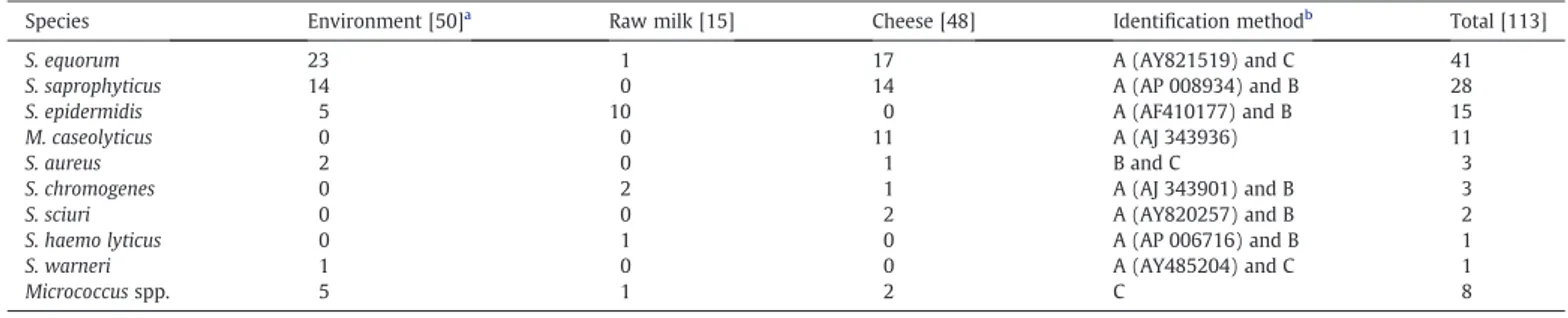
The combination of these methods allowed the identification of nine species (Table 1), reflecting the complex microflora of these cheese dairy plant.
2.5. Multiplex-PCR for detection of SAg genes
The primers used to detect the SAg genes and Multiplex-PCR reactions were prepared as described byHwang et al. (2007).
2.6. Antibiotic susceptibility test
Antimicrobial susceptibility testing were performed by the agar dilution method following the guidelines of the National Committee for Clinical Laboratory Standards (NCCLS, 2002) using a multipoint inoculator MastScan (SCAN 400, 2003). The minimum inhibitory concentrations (MICs) of the following antimicrobial agents and dilution range (inμg/mL) were determined: ampicillin (0.125–16), erythromycin (0.125–32), gentamycin (0.125–64), penicillin G (0.125–16), neomycin (0.25–16), tetracycline (0.125–16), cephalexin (0.25–32), and oxacillin
(0.125–32). Quality control was assessed with S. aureus ATCC 29213. Plates were incubated at 37 °C and read at 24 h. MICs were expressed as the lowest dilution that completely inhibited growth of the bacterial isolate. Resistance breakpoints were applied accordingly to NCCLS (2002).
2.7. Other virulence factors
Simultaneous detection of clumping factor and protein A was per-formed using the Staphytect Plus kit (Oxoid, Basingstoke, UK) according to the manufacturer's instruction. Strains to be tested for coagulase production were incubated by adding a loopfull of the overnight BHI culture to 0.3 mL of rabitt plasma (Biomérieux, Marcy-l'Etoile, France) in a test tube. For DNase activity, strains were inoculated on DNase Test Agar (Pronadisa, Madrid, Spain) with Toluidine Blue and incubated at 37 °C for 24 h. DNase activity was indicated by pink to red zones surrounding the colonies. S. aureus ATCC 25293 and S. epidermidis ATCC 14990 were used as positive and negative controls, respectively. Hemolytic activity was performed on Columbia agar (Biomérieux) supplemented with 5% of horse blood and examined after 24 and 48 h of incubation. S. aureus ATCC 25293, which displayβ-hemolysis and S. epidermidis ATCC 14990 with γ-hemolysis, were used as controls. Gelatinase activity was detected using a 12% (w/v) gelatine medium (yeast extract 10 g/L, tryptone 15 g/L, and gelatine at 120 g/L from bovine skin; Sigma). After overnight growth, cultures were transferred to tubes containing 4 mL of gelatine medium. The tubes were incubated at 30 °C for 7 d. If the bacteria did not produce gelatinase the medium remained solid, while the presence of sufficient gelatinase turned the medium liquid, even when placed at 4 °C for 24 h. For lipase activity the strains were inoculated on Tween 80 agar media (Tween 80 10 g/L, yeast extract 2.5 g/L, tryptone 5 g/L, calcium chloride
2 g/L, and agar 15 g/L) and incubated at 30 °C for 7 d. A positive result was indicated by the appearance of a white precipitate around the colonies.
2.8. Statistical analysis
The average and standard deviations were calculated for each experimental parameter, pertaining to the staphylococcal microbio-logical characterization. Statistical analysis was performed using descriptive statistics, T-test for paired and independent samples and Fischer's Exact Test. A value of p lower than 0.05 was considered significant. The statistical analyses were performed using Statistical Package for Social Sciences (SPSS, ver. 16.0).
3. Results
3.1. Cheese plant and staphylococcal viable counts
The BPM counts and distribution of the isolates are shown inFig. 1. The counts ranged from 2.6 to 4.2 log cfu/100 cm2. The highest counts
were obtained for samples collected from the ripening chambers, namely the shelves, as well as from the washing and packaging machine. The results revealed that staphylococci were isolated from 11 (58%) of the environmental samples analyzed, all of them sampled after cheese manufacture. However, in cleaned and sanitized equipment surfaces the BPM counts were below the detection limits, revealing that the cleaning procedure was very efficient in removing the staphylococcal microflora. Exception was the brush of the washing machine, which exhibited 2.6 log cfu/100 cm2 representing a potential source of future batch contamination—nevertheless, the cleaning procedure has a statistically
Table 1
Distribution and identification method of the staphylococci isolated from the small processing unit.
Species Environment [50]a
Raw milk [15] Cheese [48] Identification methodb
Total [113]
S. equorum 23 1 17 A (AY821519) and C 41
S. saprophyticus 14 0 14 A (AP 008934) and B 28
S. epidermidis 5 10 0 A (AF410177) and B 15
M. caseolyticus 0 0 11 A (AJ 343936) 11
S. aureus 2 0 1 B and C 3
S. chromogenes 0 2 1 A (AJ 343901) and B 3
S. sciuri 0 0 2 A (AY820257) and B 2
S. haemo lyticus 0 1 0 A (AP 006716) and B 1
S. warneri 1 0 0 A (AY485204) and C 1
Micrococcus spp. 5 1 2 C 8
a
Total isolates. b
A: sodA gene sequencing (GenBank accession numbers;http://www.ncbi.nlm.nih.gov); B: Multiplex-PCR; C: ARDRA.
0 1 2 3 4 5 RMT TT ST CV CMH SST M
RC1A RC1B RC1C RC2A RC2B RC2C RC3A RC3B RC3C WMBA WMB PC Sample site Log 10 cfu/100cm 2
Fig. 1. BPM counts from samples taken throughout cleaned and sanitized surfaces ( ) and from surfaces during cheese manufacture (■) in the cheese processing unit. RMT, room milk tank; TT, transport tank; ST, storage tank; CV, cheese vat; CMH, cheese maker hands; SST, stainless steel table; M, moulds; RC1A, ripening chamber 1 (shelves); RC1B, ripening chamber 1 (wall); RC1C, ripening chamber 1 (ground); RC2A, ripening chamber 2 (shelves); RC2B, ripening chamber 2 (wall); RC2C, ripening chamber 2 (ground); RC3A, ripening chamber 3 (shelves); RC3B, ripening chamber 3 (wall); RC3C, ripening chamber 3 (ground); WMBA, washing machine (support); WMB, washing machine (brush) and PC, packaging machine.
significant effect (pb0.05) in the reduction of staphylococci, as expected (Fig. 1).
Fig. 2illustrates the BPM counts obtained from the raw milk and throughout the ripening of soft (Prado) cheese. The BPM counts revealed a significant increase (pb0.05) in raw milk from cooling tank (milking room location) towards storage tank (dairy plant location), essentially due to refrigeration of the former. Maximum counts of 7.1 and 6.7 log cfu/g were obtained in thefirst week of ripening both in surface and core samples, respectively, and decreased until the end of ripening. The cheese surface had significantly (pb0.05) higher counts when compared to the cheese core for the same ripening period reflecting a better adaptation to this microenvironment. However, higher levels of contamination by staphylococci were found in cheeses with visual defects (results not shown) analyzed with counts ranging from 7.1 to 8.0 and 6.1 to 7.3 log cfu/g in the surface and core, respectively, at 30 d of ripening.
3.2. Molecular identification 3.2.1. Multiplex-PCR assays
After preliminary screening for Gram-positive and catalase-positive, the isolates were obtained and submitted to Multiplex-PCR to identify the bacteria belonging to the Staphylococcus genus, and in particular to the S. aureus, S. epidermidis, S. saprophyticus, S. chromogenes, S. hyicus and S. simulans species by yielding specific DNA fragments of 420 bp, 240 bp, 221 bp, 250 bp, 300 bp and 220 bp, respectively (Fig. 3). Six strains failed to amplify the 1226-bp internal control (data not shown) from the 23S rRNA gene and were rejected. A total of 105 strains amplified the 370 bp fragment, confirming that they belong to the Staphylococcus genus and eight did not originate any fragment, showing that they belong to another genus of Gram-positive cocci. Throughout 105 positive Staphylococcus isolates, 28 (26.7%) were identified as S. saprophyticus by producing a PCR product of 221 bp. Among these, 14 were isolated from visual defect cheeses, and 14 from the environment. Fifteen isolates (14.3%) gave amplification through S. epidemidis specific primers by producing an expected band of 240 bp, 10 from raw milk andfive from the environment. Three strains were identified as S. aureus by giving the specific fragment of 420 bp, one isolated from cheese with visual defects and two from the packaging machine surface. The specific PCR product of 250 bp, which permits identification of S. chromogenes was amplified in three strains, one from visual defect cheese surface, and two from the raw milk.
3.2.2. ARDRA
The 56 isolates unidentified by Multiplex-PCR were further charac-terized by ARDRA. To include more variable regions a longer sequence
was used which includes: 16S rDNA, 16S–23S rDNA intergenic spacer region, and about 1400 bp of the 5′-end of the 23S rDNA resulting in a PCR product of 3.5 kb. The ARDRA profiles obtained after endo>nuclease restriction enabled the differentiation between all the type strains used. In ARDRA analysis, more than one PCR product was detected in the staphylococcal type strains although in all cases one band predominated (3.5 kb fragment), since staphylococcal species can contain several rrn operons (Forsman et al., 1997). This is the reason for the sums of the restriction fragments were sometimes higher than 3.5 kb (as seen in
Table 2).
No restriction site was found for EcoRV (data not shown) endo-nuclease; however, the Micrococcus type strains used have a restriction site. Consequently, EcoRV can be used as marker for discrimination between Staphylococcus and Micrococcus genus. The eight isolates which did not amplify the 370 bp in Multiplex-PCR corresponding to the Staphylococcus genus were here identified as Micrococcus spp.
Using just a single restriction enzyme alone, differentiation of all type strains could not be achieved. Discrimination of the reference strains into distinct groups and band profiles obtained are demon-strated inTable 2. Partial differentiation of staphylococcal species into distinct groups was achieved with PvuII, VspI and SspI. Nevertheless, complete differentiation was only achieved by introducing the digestion profiles obtained with HindIII and XmnI endonucleases. The most discriminative in vitro digestion was the restriction with HindIII endonuclease (suitable to distinguish between most of the studied species), generating from 3 to 5 bands depending on the particular species. The ARDRA profiles allowed distinguishing 4 additional species within the staphylococcal isolates. Most of the isolates belong to the S. equorum (41 strains, 39.0%) species. This species was mostly isolated from environment (51.1%) but also from cheese samples (37.0%) and from raw milk (7.2%). Moreover, two isolates from cheese were identified as S. sciuri, and one isolate from the environment was classified as S. warneri. Finally, one isolate from the raw milk was recognized as S. haemolyticus revealing a low incidence of these last mentioned three species.
3.2.3. sodA sequencing
To verify and complement the data obtained by Multiplex-PCR and ARDRA the sodA gene was partially sequenced. In order to confirm at the species level the strains previously identified the sodA gene sequences of one representative strain were determined. The species groups identified by Multiplex-PCR and ARDRA were in good agreement with
3 4 5 6 7 8 M1 M2 7 15 30 Surface Core Log 10 cfu/g
Fig. 2. BPM counts obtained from milk storage and Prado cheese throughout the ripening period sampled at 7, 15 and 30 days. M1 represents morning milk collected from the cooling tank (4–8 °C); and M2 represents morning milk collected from the storage tank in the cheese dairy plant after 2–3 h.
Fig. 3. Multiplex-PCR amplifications obtained with Staphylococcus-, S. aureus-, S. epidermidis-, S. saprophyticus-, S. chromogenes-, S. hyicus-, and S. simulans-specific primers. Mix 1: lane 1, S. aureus ATCC 25923; lane 2, S. epidermidis ATCC 14990 and lane 3, S. saprophyticus ATCC 15305; and Mix 2: lane 4, S. chromogenes LMG 19102; lane 5, S. hyicus LMG 19101; lane 6, S. simulans ATCC 11631 and lane M, 100-bp molecular ladder (Invitrogen).
the sodA gene sequences obtained. In addition, the sodA gene sequences of 11 unidentified isolates were also determined. They were identified as Macrococcus caseolyticus and the sodA sequences of the representa-tive strains showed identity values of 100 to 98%. All of them represent cheese isolates.
3.3. Detection of SAg genes
The Multiplex-PCR results for SAg detection reveal that only three different toxin genes were detected: SEH, SElJ, and TSST-1 (data not shown); nine of the isolates examined were found to be positive for one or more toxin genes and demonstrated discernible genotypic variability, subdividing them into four groups according to the toxin gene profile. From the positive strains, eight possessed only one type of toxin gene viz. one SEH, three SElJ and four TSST-1; whereas the remaining strain harbored more than one toxin gene simultaneously viz. SElJ with TSST-1 (data not shown). The classical and most common staphylococcal toxins (SEA–SEE) were not detected in these isolates. With regard to species distribution, three S. equorum and two for each of the remaining strains of S. epidermidis, S. saprophyticus and S. aureus harbour toxin genes.
3.4. Antibiotic susceptibility test
The spectrum of activity of each isolate was tested against eight antibiotics. Among the isolates, the overall antimicrobial susceptibility profiles revealed that the highest percentage of resistance was detected for penicillin (30.5%), followed by ampicillin (24.8%), erythromicin (22.9%), and tetracycline (15.2%). The lowest resistance was exhibited for cephalexin (6.7%), gentamycin (6.7%) and oxacillin (4.8%). Interestingly, all strains were sensitive to the clinically important antibiotic neomycin (data not shown). S. aureus isolates demonstrated resistance only against theβ-lactamic antibiotics tested, viz. penicillin and ampicillin. According to Table 3 resistance to at least one of the antibiotics tested was encountered in 57.1% of the isolates. Resistance was more frequent among S. saprophyticus species with 78.6% of resistance, followed by S. aureus, S. equorum and S. epidermidis strains exhibiting more than 50% resistance to antibiotics. The S. chromogenes and S. haemolyticus species were sensitive to all the antibiotics tested. With regard to multiple resistances (resistance to at least two antibiotics), 30 (28.6%) isolates are multi-resistant with strains of S. equorum and S. saprophyticus reaching up to six antibiotic resistances per strain, whereas, S. epidermidis (26.7%) was resistant to a maximum of three antibiotics (Table3). Multiple resistant strains even included the strains of S. aureus, S. sciuri and S. warneri.
3.5. Virulence factors
All staphylococcal isolates were subject to numerical analysis of some essential virulence factors (results not shown). Three strains (2.9%) were positive for both latex agglutination and coagulase test tube. These strains, identified as S. aureus, exhibited a negative reaction to lipase and gelatinase activity, demonstratedβ-hemolysis and were positive for DNase test. Only few isolates showed lipase (2 isolates, 1.9%) and gelatinase (12 isolates, 11.4%) activities, revealing a low incidence of these virulence factors among the isolates. Fifteen isolates (14.3%) demonstratedβ-hemolytic activity, three (2.9%) were α-hemolytic and the great part (89 isolates, 84.8%) of the isolates presents non-hemolytic activity.
4. Discussion
In this study, the biodiversity of the staphylococcal community of a small unit manufacturing traditional cheese plant was evaluated by the identification and characterization of 105 Staphylococcus isolates recovered from cheese related products and the environment. Members of this group are widespread in nature and frequently isolated from a wide range of foodstuffs, especially ripened fermented foods such as cheeses and sausages (Coton et al., 2010).
The surfaces of equipment used for food handling or processing are recognized as possible support for microbial growth, and biofilms residing on such surfaces are recognized nowadays as potential sources of contamination (Somers et al., 2001). In the environment, BPM counts ranged from 2.6 log cfu/100 cm2to 4.2 log cfu/100 cm2(Fig. 1). The most Table 2
Restriction fragment patterns of Staphylococcus type strains with VspI, PvuII, SspI, XmnI and HindIII restriction endonucleases. Species Restriction endonucleases fragments (bp)
VspI PvuII SspI XmnI HindIII
S. aureus 1800, 1000, 500 2400, 700, 600 2800, 1800, 1600 1900, 1000, 400, 250 1300, 1200, 850, 200 S. capitis 1800, 1700 2600, 900 2800, 1700 1900, 1000, 400, 250 1300, 850, 600, 450, 400 S. caprae 1800, 1700 2600, 900 2800, 1700 1900, 1000, 400, 250 1300, 650, 350 S. chromogenes 1800, 1700 2600, 900 3500 2000, 1000, 450 1800, 1300, 430 S. epidermidis 1800, 1000, 500 2600, 900 3500 1900, 1000, 400, 250 1300, 650, 450 S. equoruom 1800, 1700 2600, 900 3500 1800, 1000, 300, 200, 150 1900, 1500, 200 S. haemolyticus 1800, 1000, 1200, 500 2600, 900 2800, 1800, 1600 2000, 1000, 450 1300, 750, 500, 400 S. hominis 1800, 1000, 500 2600, 900 2800, 1500 2000, 1000, 450 1300, 850, 700, 350 S. hyicus 1800, 1700 2400, 700, 600 2800, 1700 2000, 1000, 450 1300, 600, 500 S. lentus 1800, 1700 2400, 700, 600 2800, 1700 1900, 1000, 400, 250 1500, 1300, 650, 200 S. lugdunensis 1800, 1300, 900, 500 2400, 700, 600 3500 1700, 1600, 200 1300, 750,700, 650 S. saprophyticus 1800, 1700 2400, 700, 600 3500 1900, 1000, 400, 250 1500, 1300, 650, 200 S. sciuri 1800, 1700 2400, 700, 600 3500 2000, 1000, 450 1600, 1100, 850, 600 S. simulans 1800, 1700 2600, 900 1900, 1600 1900, 1000, 400, 250 1700, 1300, 600 S. xylosus 1800, 1700 2400, 700, 600 2800, 1700 2000, 1000, 450 1500, 1300, 650, 200 S. warneri 1800, 1700 2400, 700, 600 2800, 1700 1900, 1000, 400, 250 1300, 800, 350 Table 3
Incidence of (multiple) antibiotic resistance among staphylococci strains isolated from the small processing unit.
Species No. of isolates Resistant strains Number of strains resistant against a number of antibiotics Multiresistant strains 1 2 3 4 5 6 S. equorum 41 23 15 2 1 1 2 2 8 S. saprophyticus 28 22 9 7 4 1 – 1 13 S. epidermidis 15 8 4 3 1 – – – 4 S. aureus 3 2 – 2 – – – – 2 M. caseolyticus 11 2 2 – – – – – 0 S. sciuri 2 2 – 1 1 – – – 2 S. warneri 1 1 – – 1 – – – 1 S. chromogenes 3 0 – – – – – – 0 S. haemolyticus 1 0 – – – – – – 0 Total 105 60 30 15 8 2 2 3 30
colonized samples of environment were the ripening chambers, the washing and the packaging machine. Accordingly, staphylococci were not detected on sanitized and cleaned equipment surfaces; exception was the brush of the washing machine, revealing that the cleaning procedure was efficient in removing the staphylococcal microflora. The results are relevant, because when these microorganisms are present in the environment their population can increase rapidly during cheese manufacture and ripening and achieve high numbers in thefinal product —a concern to public health, since some of these strains are recognized as foodborne pathogens.
The BPM counts in cheese related products ranged from 4.0 log cfu/mL in raw milk to 6.0 (cheese surface) or 5.7 (cheese core) log cfu/g after 30 d of ripening (Fig. 2). Consistency in the BPM counts during the ripening period was probably due to its capacity to withstand a wide range of temperatures, pH, and low aw. However, the highest counts were
ob-tained in thefirst week of ripening and then decreased until the end of ripening. Such a decrease in bacterial counts may be ascribed to physico-chemical conditions prevailing in the cheese environment during maturation, i.e. decrease of moisture, increase in salt content, and de-crease in pH that are unfavorable for microbial growth. The results revealed viable counts on the same order of other Portuguese raw ewe's milk cheese varieties such as Serra da Estrela (Macedo et al., 1995), Terrincho (Pintado et al., 2008), and Picante da Beira Baixa (Freitas et al., 1999). However, the viable counts on cheeses with visual defects (data not shown), after 30 d of maturation, were higher, which could possibly reflect a positive correlation involving this group of microorganisms with cheese deterioration. The staphylococcal levels found are of technological relevance, since this cheese variety is made directly from raw ewe's milk without any type of thermal processing.
The isolates were identified using the discriminatory power of Multiplex-PCR, ARDRA and determination of the sodA gene sequences. The staphylococcal flora of the environment and cheese related products were dominated by S. equorum (39.0%) and S. saprophyticus (26.7%) species.Coton et al. (2010) and Even et al. (2010)also showed a high prevalence of S. equorum and S. saprophyticus species in food-related samples. These two species were also dominant on the surfaces of some smear Italian cheeses (Fontana et al., 2010). Some studies even suggested the inclusion of S. equorum as part of starter cultures for smear ripened cheeses and typical Swiss semi-hard cheeses (Bockelmann, 2002; Irlinger, 2008). These two species were found both in cheese related and environmental samples. The appearance of S. saprophyticus on cheese is typical, especially on acid curd and goat cheeses. This is a ubiquitous species, which is normally involved in acute urinary tract infections in young adult women (Bockelmann and Hoppe-Seyler, 2001; Irlinger, 2008). The prevalence of these two species indicates that they may be capable for adaptation to food plant environment. Furthermore, since cheese processing receive considerable manual handling, it is not surprising tofind S. epidermidis as it is commonly found living on the skin and the mucous membranes of humans and accounts for the majority of infections caused by CNS (Couto et al., 2001). This species was present in raw milk (10 isolates) and environmental samples (five isolates) and tend to disappear in thefinal cheese product. Eleven strains of M. caseolyticus (10.5%) were also isolated. This species was identified in several milk or cheese samples or even in traditionally fermented sausages generally as minor or sporadical species (Bonomo et al., 2009; Fontana et al., 2010; Giannino et al., 2009; Piessens et al., 2010). To our knowledge this species had never been identified from traditional Portuguese cheeses previously. The remaining isolates were assigned as S. aureus (3 strains), S. chromogenes (3 isolates), S. sciuri (2 isolates), S. haemolyticus (1 isolate) and S. warneri (1 isolate). The S. aureus species is generally recognized as producer of a great number of toxins, and is the best known staphylococcal pathogen. Accordingly, S. aureus is rarely found in fully ripened cheeses, because it usually tends to disappear during the maturation period due the conditions prevailing in cheese microenvironment, e.g. low awin combination with low pH and
produc-tion of bacteriocins (Jorgensen et al., 2005). The S. chromogenes, S. sciuri,
S. haemolyticus and S. warneri species have also sporadically been isolated in French cheese related samples (Coton et al., 2010) or also from fermented sausages and manufacturing environments (Leroy et al., 2010). Another important element is the detection of SAg genes, which is a major concern in food context as these can lead to SFP and has been generally linked to the S. aureus strains. However, some studies suggest that more attention should be addressed to the CNS group (Valle et al., 1990, Vernozy-Rozand et al., 1996; Zell et al., 2008).Udo et al. (1999)already detected enterotoxin genes in CNS isolated from the hands of restaurant workers, and suggest that such strains may contribute to food poisoning. Regardless of the great inconsistency concerning the prevalence of enterotoxigenic staphylococci, SEA–SEE are generally the most frequently observed enterotoxins implicated in SFP (Normanno et al., 2005). However, in this study these toxin genes were never detected. We detected only SEH, SElJ and TSST-1 toxin genes among the staphylococci collection. One or more enterotoxin genes were carried by 8.6% of the isolates, two coagulase positive staphylococci (CPS) and seven CNS (data not shown). The results are in agreement with other studies: 5.3% (Vernozy-Rozand et al., 1996) and 13.6% (Valle et al., 1990) of CNS isolated from goat milk and cheese, respectively. Among the CNS species isolated S. equorum was the prevalent enterotoxigenic strain. Likewise,Zell et al. (2008)revealed a high incidence of toxin detection in S. equorum species among CNS isolated from food or starter cultures.
The susceptibility to clinically relevant antibiotics should be tested since staphylococci could reach high numbers in cheese related samples, and include a remarkable ability to adapt antibiotic use by acquiring resistances and transfer those resistance determinants to humans via the food chain (Berger-Bächi and McCallum, 2006). In this study, the number of antibiotic resistances is notably higher for S. saprophyticus, S. equorum and S. epidermidis species (Table 3). This finding is in agreement with previous studies showing that these species isolated from food exhibit higher rates of antibiotic resistances (Even et al., 2010; Resch et al., 2008). From these, S. saprophyticus presents a great number of multi-resistant strains (46.4%) followed by S. epidermidis (26.7%). Therefore, it is interesting to consider similarly to Resch et al. (2008) an association concerning the incidence of antibiotic resistance in food-associated staphylococci and their relatedness to (opportunistic) pathogenic species. The incidence of antibiotic resistances respecting the source of isolation (results not shown) was not confirmed (pN0.05). Similarly,Resch et al. (2008)
also found that the origin might be of secondary importance in determining the antibiotic resistance in food-associated staphylococci. Regarding the clinical importance of antibiotics no resistance was demonstrated for neomycin and the great part are sensitive to oxacillin, gentamicin and cephalexin. This is in agreement to published data through marked low rate of resistance for neomycin and gentamicin in staphylococci isolated from various foods (Martín et al., 2006; Resch et al., 2008). The resistance to oxacillin encoded by mecA gene was detected only infive isolates. The mecA determinant is mainly associated to S. aureus and, very recently, to S. epidermidis, which may cause resistance to all β-lactamic antibiotics, which results in severe problems in the treatment of human infections (Simeoni et al., 2008). A remarkable level of antibiotic resistance was observed, 60 (57.1%) and 30 (28.6%) of the isolates demonstrate resistant and multi-resistant strains, respectively, as seen inTable 3. The high prevalence of resistance was registered for penicillin (30.5%) as expected (data not shown). Similarly, high levels of resistance to this antibiotic for staphylococci isolated from different sources (Acco et al., 2003; Benhassen et al., 2003; Even et al., 2010) has also been reported. Therefore, the majority of antimicrobial resistance in the staphylococcal community could be due to the production of β-lactamases. This is not surprising since penicillin became one of the most commonly used antibiotics to treat infections in humans and animals. The second-highest resistance was observed for ampicillin (24.8%), followed by erythromycin (22.9%) and tetracycline (15.2%). Likewise,
isolated from fermented foodstuffs. The broad use of ampicillin and tetracycline as antimicrobial agents of ovine mastitis may explain the selective pressure, which generated these resistances in staphylococci (Lollai et al., 2008).
The importance of evaluating the combination of virulence traits among staphylococci has been recently emphasized both in human and veterinary medicine (Zecconi et al., 2006). Therefore, a small percentage of the isolates (17.1%) showed hemolytic activity—from these, 15 strains wereβ-hemolytic and three were α-hemolytic. It is difficult to state how hazardous β-hemolytic strains are for the consumer, since it should be also noted that the hemolytic activity can be influenced by synergistic effects of various hemolysins (Zell et al., 2008). The lypolitic and gelatinase activities were very low, viz. only 1.9% and 11.4%, respectively; S. epidermidis accounted for almost 65% of the gelatinase positive strains.
5. Conclusion
Our study highlighted the high diversity of Staphylococcus species isolated from a small unit manufacturing dairy plant. They showed a high capacity to colonize the surfaces, equipments and dairy related products. The predominance of S. equorum and S. saprophyticus species possibly suggests high adaptation properties of these species on this type of environment. Hygienic and safety traits, such resistance to relevant antibiotics and enterotoxigenic potential must be taken into account, since any undesirable trait could affect the safety and hygienic quality of the product. Therefore, we conclude that a considerable fraction of staphylococci presents resistances. However, it should be noted that the toxigenic potential is very low. Thus, identification of staphylococcal species and the characterization of the potential risks constitute afirst step towards food safety assessment of food-related staphylococci.
References
Acco, M., Ferreira, F.S., Henriques, J.A.P., Tondo, E.C., 2003. Identification of multiple strains of Staphylococcus aureus colonizing nasal mucosa of food handlers. Food Microbiology 20, 489–493.
Altschul, S.F., Gish, W., Miller, W., Myers, E.W., Lipman, D.J., 1990. Basic local alignment search tool. Journal of Molecular Biology 215, 403–410.
Benhassen, S., Messadi, L., Benhassen, A., 2003. Identification et caractérisation des espèces de Staphylococcus isolées de lait de vaches atteintes ou non de mammite. Annales de Médecine Vétérinaire 147, 41–47.
Berger-Bächi, B., McCallum, N., 2006. State of the knowledge of bacterial resistance. Injury 37, 20–25.
Blaiotta, G., Fusco, V., von Eiff, C., Villani, F., Becker, K., 2006. Biotyping of enterotoxigenic Staphylococcus aureus by enterotoxin gene cluster (egc) polymorphism and spa typing analyses. Applied and Environmental Microbiology 72, 6117–6123. Bockelmann, W., 2002. Development of defined surface starter cultures for the ripening
of smear cheeses. International Dairy Journal 12, 123–131.
Bockelmann, W., Hoppe-Seyler, T., 2001. The surfaceflora of bacterial smear-ripened cheeses from cow's and goat's milk. International Dairy Journal 10, 1–8. Bonomo, M.G., Ricciardi, A., Zotta, T., Sico, M.A., Salzano, G., 2009. Technological and
safety characterization of coagulase-negative staphylococci from traditionally fermented sausages of Basilicata region (Southern Italy). Meat Science 83, 15–23. Coton, E., Desmonts, M.-H., Leroy, S., Coton, M., Jamet, E., Christieans, S., Donnio, P.-Y., Lebert, I., Talon, R., 2010. Biodiversity of coagulase-negative staphylococci in French cheeses, dry fermented sausages, processing environments and clinical samples. International Journal of Food Microbiology 137, 221–229.
Couto, I., Pereira, S., Miragaia, M., Sanches, I.S., de Lencastre, H., 2001. Identification of clinical staphilococci isolates from humans by internal transcribed spacer PCR. Journal of Clinical Microbiology 39, 3099–3103.
Cuny, I., Witte, W., 1996. Typing of Staphylococcus aureus by PCR for DNA sequences flanked by transposon Tn916 target region and ribosomal binding site. Journal of Clinical Microbiology 43, 1502–1505.
Even, S., Leroy, S., Charlier, C., Zakour, N.B., Charcornac, J.-P., Lebert, I., Jamet, E., Desmonts, M.-H., Coton, E., Pochet, S., Donnio, P.-Y., Gautier, M., Talon, R., Le Loir, Y., 2010. Low occurrence of safety hazards in coagulase negative staphylococci isolated from fermented foodstuffs. International Journal of Food Microbiology 139, 87–95. Fontana, C., Cappa, F., Rebecchi, A., Cocconcelli, P.S., 2010. Surface microbiota analysis of Talleggio, Gorgonzola, Casera, Scimudin and Formaggio di Fossa Italian cheeses. International Journal of Food Microbiology 138, 205–211.
Forsman, P., Tilsaia-Timisjärvi, A., Alatossava, T., 1997. Identification of staphylococcal and streptococcal causes of bovine mastitis using 16S–23S rRNA spacer regions. Microbiology 143, 3491–3500.
Freitas, A.C., Pais, C., Malcata, F.X., 1999. Technological optimisation of Picante cheese using microbiological, chemical and physical criteria. Journal of Food Engineering 41, 163–175.
Giannino, M.L., Marzotto, M., Dellaglio, F., Feligini, M., 2009. Study of microbial diversity in raw milk and fresh curd used for Fontina cheese production by culture-independent methods. International Journal of Food Microbiology 130, 188–195. Harrigan, W.F., McCance, M.E., 1976. Laboratory Methods in Food and Dairy
Microbiology. Academic Press Inc., London.
Heikens, E., Fleer, A., Paauw, A., Florijn, A., Fluit, A.C., 2005. Comparison of genotypic and phenotypic methods for species-level identification of clinical isolates of coagulase-negative staphylococci. Journal of Clinical Microbiology 43, 2286–2290. Hwang, S.Y., Kim, H.K., Jang, E.J., Kwon, N.H., Park, Y.K., Koo, H.C., Jung, W.K., Kim, J.M.,
Park, Y.H., 2007. Novel multiplex PCR for the detection of the Staphylococcus aureus superantigen and its application to raw meat isolates in Korea. International Journal of Food Microbiology 117, 99–105.
Irlinger, F., 2008. Safety assessment of dairy microorganisms: coagulase-negative staphylococci. International Journal of Food Microbiology 126, 302–310. ISO 18593, 2004. Microbiology of food and animal feeding stuffs—horizontal methods
for sampling techniques from surfaces using contact plates and swabs. Jorgensen, H.J., Mørk, T., Rørvik, L.M., 2005. The occurrence of Staphylococcus aureus on
a farm with small-scale production of raw milk cheese. Journal of Dairy Science 88, 3810–3817.
Kloos, W.E., Schleifer, K.H., 1986. Genus IV. In: Sneath, P.H.A., Mair, N.S., Sharpe, M.E., Holt, J.G. (Eds.), Staphylococcus. : Bergey's Manual of Systematic Bacteriology, 2. Williams and Wilkins, Baltimore, USA, pp. 1013–1035.
Leroy, S., Giammarinaro, P., Chacornac, J.-P., Lebert, I., Talon, R., 2010. Biodiversity of indigenous staphylococci of naturally fermented dry sausages and manufacturing environments of small-scale processing units. Food Microbiology 27, 294–301. Lollai, S.A., Ziccheddu, M., Di Mauro, C., Manunta, D., Nudda, A., Leori, G., 2008. Profile
and evolution of antimicrobial resistance of ovine mastitis pathogens (1995–2004). Small Ruminant Research 74, 249–254.
Macedo, A.C., Malcata, F.X., Hogg, T.A., 1995. Microbiological profile in Serra ewe's cheese during ripening. The Journal of Applied Bacteriology 79, 1–11.
Martín, B., Garriga, M., Hugas, M., Bover-Cid, S., Veciana-Nogués, M.T., Aymerich, T., 2006. Molecular, technological and safety characterization of Gram-positive catalase-positive cocci from slightly fermented sausages. International Journal of Food Microbiology 105, 148–158.
Martineau, F., Picard, F.J., Ke, D., Paradis, S., Roy, P.H., Quelette, M., Bergeron, M.G., 2001. Development of a PCR assay for identification of staphylococci at the genus and species levels. Journal of Clinical Microbiology 39, 2541–2547.
National Committee for Clinical Laboratory Standards, 2002. Performance standards for antimicrobial disk and dilution susceptibility tests for bacteria isolated from animals; approved standard, 2th, NCCLS document M31-A2; Pennsylvania, USA. Normanno, G., Firinu, A., Virgilio, S., Mula, G., Dambrosio, A., Poggiu, A., Decastelli, L.,
Mioni, R., Sucuota, S., Bolzoni, G., Di Giannatale, E., Salinetti, A.P., La Salandra, G., Bartoli, M., Zuccon, F., Pirino, T., Sias, S., Parisi, A., Quaglia, N.C., Celano, G.V., 2005. Coagulase-positive staphylococci and Staphylococcus aureus in foods products marketed in Italy. Food Microbiology 98, 73–79.
Piessens, V., Supré, K., Heyndrickx, M., Haesebrouck, F., DeVliegher, S., Coillie, Van, 2010. Validation of amplified fragment length polymorphism genotyping for species identification of bovine associated coagulase-negative staphylococci. Journal of Microbiological Methods 80, 287–294.
Piette, A., Verschraegen, G., 2009. Role of coagulase-negative staphylococci in human disease. Veterinary Microbiology 134, 45–54.
Pintado, A.I.E., Pinho, O., Ferreira, I.M.P.L.V.O., Pintado, M.M.E., Gomes, A.M.P., Malcata, F.X., 2008. Microbiological, biochemical and biogenic amine profiles of Terrincho cheese manufactured in several dairy farms. International Dairy Journal 18, 631–640. Poyart, C., Quesne, G., Boumaila, C., Trieu-Cout, P., 2001. Rapid and accurate
species-level identification of coagulase-negative staphylococci by using the sodA gene as a target. Journal of Clinical Microbiology 39, 4296–4301.
Resch, M., Nagel, V., Hertel, C., 2008. Antibiotic resistance of coagulase-negative staphylococci associated with food and used in starter cultures. International Journal of Food Microbiology 127, 99–104.
Simeoni, D., Rizzotti, L., Cocconcelli, P., Gazzola, S., Dellaglio, F., Torriani, S., 2008. Antibiotic resistance genes and identification of staphylococci collected from the production chain of swine meat commodities. Food Microbiology 25, 196–201. Somers, E.B., Johonson, M.E., Wong, A.C.L., 2001. Biofilm formation and contamination
of cheese by nonstarter lactic acid bacteria in the dairy environment. Journal of Dairy Science 84, 1926–1936.
Udo, E.E., Al-Bustan, M.A., Jacob, L.E., Chugh, T.D., 1999. Enterotoxin production by coagulase-negative staphylococci in restaurant workers from Kuwait City may be a potential cause of food poisoning. Journal of Medical Microbiology 48, 819–823. Valle, J., Gomez-Lucia, E., Piriz, S., Goyache, J., Orden, J.A., Vadillo, S., 1990. Enterotoxin
production by staphylococci isolated from healthy goats. Applied and Environ-mental Microbiology 56, 1323–1326.
Vernozy-Rozand, C., Mazuy, C., Prevóst, G., Lapeyre, C., Bes, M., Brun, Y., Fleurette, G., 1996. Enterotoxin production by coagulase-negative staphylococci isolated from goat's milk and cheese. International Journal of Food Microbiology 30, 271–280. Zadoks, R.N., Watts, J.L., 2008. Species identification of coagulase-negative staphylococci:
genotyping is superior to phenotyping. Veterinary Microbiology 68, 1100–1109. Zecconi, A., Cesaris, L., Liandris, E., Daprà, V., Piccinini, R., 2006. Role of several
Staphylococcus aureus virulence factors on the inflammatory response in bovine mammary gland. Microbial Pathogenesis 40, 177–183.
Zell, C., Resch, M., Rosenstein, R., Albrecht, T., Hertel, C., Hertel, C., Götz, F., 2008. Characterization of toxin production of coagulase-negative staphylococci isolated from food and starter cultures. International Journal of Food Microbiology 127, 246–251.