0
Universidade de Lisboa
Faculdade de Farmácia
MODULATION OF FEAR BY TRANSCRANIAL DIRECT
CURRENT STIMULATION (tDCS)
João Manuel de Oliveira Mota
Mestrado Integrado em Ciências Farmacêuticas
1
Universidade de Lisboa
Faculdade de Farmácia
MODULATION OF FEAR BY TRANSCRANIAL DIRECT CURRENT STIMULATION (tDCS)
João Manuel de Oliveira Mota
Monografia do Mestrado Integrado em Ciências Farmacêuticas apresentada à Faculdade de Farmácia da Universidade de Lisboa
Orientador: Doutor Dimitri de Bundel, Professor Auxiliar
Co-orientador: Doutora Cristina Luzia Dias de Mello Sampayo, Professora Auxiliar
2 Com a colaboração da instituição:
Vrije Universiteit Brussel
3
Resumo
Contexto: Embora a psicoterapia de exposição seja promissora no tratamento de doenças do foro
nervoso e transtornos de medo como a perturbação de stress pós-traumático (PTSD), a maioria dos doentes não adquire total remissão da doença. Vários estudos sugerem a estimulação por corrente direta transcraniana (tDCS) como uma terapia capaz de facilitar o processo de extinção do medo, tendo este procedimento interesse como adjuvante para o aumento de eficácia da psicoterapia.
Objetivo: O objetivo deste estudo foi explorar o efeito de tDCS repetitivo na aquisição e
consolidação da extinção do medo, usando o paradigma do condicionamento e extinção do medo em ratinhos. Numa segunda experiência, teve-se como objetivo o estudo do mecanismo de ação do tDCS no córtex pré-frontal (PFC), através do uso de um procedimento de microdiálise em ratinhos, de modo a analisar o efeito do tDCS na libertação de neurotransmissores no PFC.
Métodos: Uma semana pós-cirurgia, ratinhos C57BL/6J machos (N=31) foram sujeitos a um
procedimento de condicionamento do medo por via auditiva, seguido de tDCS anodal a 0.2 mA, 2 x 20 min/dia, durante cinco dias. Posteriormente, um protocolo de extinção foi executado, avaliando a memória de extinção 1 e 21 dias após a extinção. Num outro procedimento, ratinhos (N=6) foram submetidos a microdiálise através de perfusão com fluído cerebrospinal artificial por uma sonda microdialítica, colocada no PFC, sendo recolhidas amostras antes, durante, e após estimulação.
Resultados: Os resultados demonstraram diferença significativa entre os grupos experimentais na
aquisição da memória de extinção. Aquisição acelerada da extinção foi observada em ratinhos sujeitos a tDCS, no entanto, não foram observados resultados significativos na retenção da extinção. Foram também observados uma redução do medo contextual nos ratinhos sujeitos a tDCS, assim como uma correlação entre o efeito de extinção e o patamar de base de medo em ratinhos. Os resultados da microdiálise não foram conclusivos.
Conclusão: Estes resultados demonstram potencial para o uso de tDCS como adjuvante na
extinção do medo. No entanto, serão necessários mais estudos para clarificar o seu mecanismo de ação, os seus efeitos na retenção da extinção em modelos patológicos, e o seu efeito a longo prazo.
4
Abstract
Background: Although there is promise for exposure-based psychotherapy in the treatment of
anxiety and trauma-related disorders such as post-traumatic stress disorder (PTSD), most patients fail to achieve full remission. Several studies suggest that transcranial direct current stimulation (tDCS), a safe and non-invasive technique, can be used to facilitate the fear extinction process. Therefore, this procedure seems to be of interest to potentiate the efficacy of exposure-based therapy.
Aim: The aim of this master thesis was to explore the effect of repeated tDCS on the acquisition
and consolidation of fear extinction, using the fear conditioning and extinction paradigm in mice. In a second experiment, we aimed to help unravel the mechanism of action of tDCS in the prefrontal cortex (PFC) by exploring the effects of tDCS on the release of dopamine, serotonin and noradrenaline in this brain region, using a microdialysis procedure in mice.
Methods: Following one week of recovery from surgery, male C57BL/6J (N=31) were subjected
to auditory fear conditioning, followed by 2 x 20 min/day anodal tDCS at 0.2 mA for five consecutive days. Extinction training was then performed, and retention of fear extinction was evaluated 1 day and 21 days afterwards. Microdialysis experiment was performed in mice (N=6) by perfusing a microdialysis probe, placed during surgery in the PFC, with artificial cerebrospinal fluid. Samples were collected before, during, and after tDCS stimulation.
Results: The results demonstrated a significant difference between the experimental groups in the
acquisition of extinction. Accelerated acquisition of extinction was observed in mice subjected to tDCS, whereas, no significant effects on the retention of extinction were observed. Also, a reduction in contextual fear levels in tDCS mice could be observed, as well as a significant correlation between extinction effect and baseline level of fear in mice. Microdialysis results were not conclusive.
Conclusion: These results show potential for the use of tDCS as an adjuvant to extinction training.
However, further studies are necessary to clarify its mechanism of action and the effects on extinction retention in a model of PTSD, and long-term effect on retention of extinction memory.
5
Table of Contents
List of Abbreviations ... 8
1. Introduction ... 11
1.1. Studying Fear – Fear Conditioning and Fear Extinction 11 1.1.1. Neural Pathways of Fear Acquisition ... 14
1.1.2. Neural Pathways of Fear Extinction ... 15
1.1.3. Fear Extinction and Neurotransmitters ... 16
1.2. PTSD: Disease and Current Treatments 18 1.2.1. Physiopathology of PTSD... 19
1.2.2. Current Treatments for PTSD ... 20
1.3. Transcranial Direct Current Stimulation (tDCS) 21 1.3.1. Mechanism of Action of tDCS ... 22
1.3.2. Parameters of Stimulation and Safety ... 24
1.3.3. Advantages and Disadvantages ... 25
1.3.4. Current Applications of tDCS ... 26
1.3. tDCS in the Modulation of Fear and in PTSD 27 2. Aims ... 28
3. Materials and Methods ... 29
3.1. Animals and Housing 29 3.2. Fear Conditioning Experiment 29 3.2.1. tDCS Surgery ... 29
3.2.2. Fear Conditioning ... 30
3.2.3. Transcranial Direct Current Stimulation (tDCS) ... 32
3.3. Microdialysis Experiment 33 3.3.1. Microdialysis Surgery... 33
3.3.2. aCSF and AOIII preparation ... 33
6
3.3.4. Ultra-High-Performance Liquid Chromatography (UHPLC) ... 34
3.4. Perfusion 35 3.5. Statistical analysis 35 4. Results ... 36
4.1. Fear Conditioning Experiment 36 4.2. Microdialysis Experiment 41 5. Discussion... 43
6. Conclusions ... 45
7. Bibliographical References ... 46
7
Table of Contents - Figures
Figure 1 – Main phases of the fear conditioning and extinction procedure. ... 13
Figure 2 - Brain circuitry mediating fear acquisition and expression. ... 15
Figure 3 - Brain circuitry mediating fear extinction. ... 16
Figure 4 - Representation of anodal tDCS. ... 22
Figure 5 – Illustration of where the plastic screws and the electrode holder were positioned on the skull of mice and representation of a tubular plastic jacket similar to the one used in the experiments. ... 30
Figure 6 – Schematic representation of the time frame of the fear conditioning experiment... 32
Figure 7 - Graphs for the habituation, fear conditioning and fear retrieval procedures. ... 37
Figure 8 - Graphs for the fear retrieval, fear extinction and early recall of extinction procedures. ... 38
Figure 9 - Graph for the extinction effect analysis. ... 39
Figure 10 - Graphs for the late recall of extinction and fear renewal procedures. ... 40
Figure 11 – Linear regression of the results representing the correlation between baseline freezing and extinction effect. ... 41
Figure 12 - Graphs for the noradrenaline (NA), dopamine (DA) and serotonin (5-HT) release during the microdialysis procedure. ... 42
8
List of Abbreviations
5-HT - Serotonin
a-tDCS - Anodal tDCS
aCSF - Artificial cerebrospinal fluid
ANOVA - Analysis of variance
AOIII - Antioxidant III
BA - Basal amygdala
c-tDCS - Cathodal tDCS
Ca2+ - Calcium ion
CaCl2.6H2O - Calcium chloride hexahydrate
CeA - Central nucleus of the amygdala
CeL - Lateral subdivision of the central amygdala
CeM - Medial subdivision of the central amygdala
CBT - Cognitive behavioural therapy
CR - Conditioned response
CS - Conditioned stimulus
DA - Dopamine
dACC - Dorsal anterior cingulate cortex
DC - Direct current
DSM-V - Diagnostic and statistical manual of mental disorders, 5th edition
EMDR - Eye movement desensitization and reprocessing
9
GABA - gamma-Aminobutyric acid
HAB - Habituation
HAC - Hospital antiseptic concentrate
IL - Infralimbic cortex
IP3 - Inositol 1,4,5-triphosphate
ITC - Intercalated cells
KCl - Potassium chloride
LA - Lateral amygdala
LTD – Long-term depression
LTM - Long-term memory
LTP - Long-term potentiation
MgCl2.H2O - Magnesium chloride monohydrate
mGluR - Metabotropic glutamate receptor
mPFC - Medium prefrontal cortex
NA - Noradrenaline
Na2EDTA - Disodium ethylenediaminetetraacetate dihydrate
NaCl - Sodium chloride
NaH2PO4 - Monosodium phosphate
NaOAc.3H2O - Sodium acetate trihydrate
NMDA - N-methyl-D-aspartate
PBS - Phosphate-buffered saline
10
PL - Prelimbic cortex
PTSD - Post-traumatic stress disorder
RM - Repeated measures
s.e.m - Standard error of the mean
SSRI - Selective serotonin
STM - Short-term memory
tDCS - Transcranial direct current stimulation
TFCBT - Trauma focused cognitive behavioural therapy
UHPLC - Ultra-high-performance liquid chromatography
UPLC - Ultra performance liquid chromatography
UR - Unconditioned response
US - Unconditioned stimulus
11
1. Introduction
Fear is an emotion induced by a certain cue or context that the brain perceives as dangerous, triggering innate or learned defensive mechanisms (1). These responses to threatening situations have a high phylogenetic and ontogenetic conservation, also having strong roots in human evolution, as they were essential for survival and protection against predators (2,3).
Fear differs from anxiety, with fear a response to an imminent or perceived threat, and anxiety a vague anticipation of a future menacing event, in absence of an immediate threat (DSM-V). Both are, however, intimately related in anxiety and trauma-related disorders, which are behavioural disturbances characterized by excessive and persistent fear and anxiety (4). Nowadays, these disorders are one of the most prevalent psychiatric disorders in the world, contributing to 26.8 million disability adjusted life years in 2010 (5,6) and a lifetime prevalence of approximately 20-30% (5). It is also associated with impaired workplace performance and hefty economic and healthcare costs (7), higher drug and alcohol abuse (8), and correlated with various health risks such as cardiovascular events (9–11).
1.1. Studying Fear – Fear Conditioning and Fear Extinction
Fear is a physiological response to threatening situations. When a fearful stimulus is present, the body reacts accordingly, leading to autonomic responses (such as increases in the blood pressure, heart and respiration rate, and in the liberation of metabolic energy sources), behavioural reactions (such as freezing or spasm-like twitches and fight, flight or avoidance reactions), and hormonal responses (with the release of stress hormones such as glucocorticoids like cortisol, or catecholamines like adrenaline, noradrenaline or acetylcholine) (12–15). However, fear can become excessive, growing beyond control, to the point of interfering with daily life, in which case an anxiety disorder or trauma-related disorder may be present.
A lot of research has been done in the past decades regarding the neurocircuits underlying anxiety disorders and its modulation. One of the most famous experiments regarding behavioural modulation is Pavlov’s experiment with dogs, in which he induced a conditioned response
12 (salivation), by coupling an unconditioned stimulus (food smell) to a conditioned stimulus (metronome sound) (16). Other well-known experiments were “Little Albert experiment”, performed by Watson, conditioning a phobia in an emotionally stable child and showing the importance of fear generalization in fear expression (17), and the “Little Peter experiment” performed by Mary Cover Jones, conditioning the desensitization of a phobia in a child (18). These experiments on classical conditioning allowed the thrive of psychological therapy and animal behaviour studies among others with great significance to the scientific world, having also good correlation as a translational model to humans (19,20).
Pavlovian fear conditioning is still widely used to study emotional learning and fear in both humans and rodents. In classical fear conditioning a neutral conditioned stimulus (CS, a tone or a light), is paired with an aversive unconditioned stimulus (US, an electrical shock), eliciting an unconditioned response (UR) and creating an association between both (21). After one or several pairings, a conditioned response (CR) occurs even when the CS is presented in the absence of the aversive US (21–23). When performed in rodents, the most commonly analysed response is freezing behaviour, an immobile state as a reaction to an aversive event, which is easily analysed and relatable to their fear expression (23–26).
The general procedure of fear conditioning in mice consists of an acquisition and a consolidation phases. (27,28). The acquisition phase is a form of associative learning, where mice learn to associate a previously neutral stimulus with an aversive meaningful stimulus (CS-US association), acquiring fearful emotions (21). The new short-term memory (STM) formed during the acquisition of conditioning is labile and is converted into a long-term memory (LTM) during the consolidation phase (29). Consolidation is a molecular and cellular process with the aim to stabilize the fear memory, maintaining them over time through both structural and molecular changes (29–32). As a fearful memory can be acquired during conditioning, fear memories are extinguished during the fear extinction process. Fear extinction is by definition a decline in conditioned fear responses following repetitive nonreinforced exposure to the conditioned stimulus (33). In practical terms, the CS is repeatedly presented in the absence of the US (34–36). Extinction sessions do not erase the previously established fearful memory, rather than that, a new “safe” memory is created, countering the original fear memory and inhibiting the expression of fear (34–36). Being extinction
13 the creation of a new memory, it also consists of an acquisition and a consolidation phase, although the involved neural pathways are different.
The biggest issue hindering fear extinction performance is that fear memories are easily prone to reoccur after extinction learning (34,35,37). Extinguished fear responses can spontaneously recover with the passage of time (Spontaneous recovery), can suffer renewal when tested in a context that differs from the one of extinction (Fear renewal) or even be reinstated following unexpected exposure to the US (Fear reinstatement) (33,35–38). The extinction of a fearful memory is a fragile process and the strength of the memory depends on many factors, including timing relative to conditioning, the intensity of the extinction procedure and stress (7,36).
The previously stated phenomena can be seen and better understood with the help of Figure 11.
Figure 1 – Main phases of the fear conditioning and extinction procedure. The strength of fear expression is shown
14
1.1.1. Neural Pathways of Fear Acquisition
The most important neuronal structures for both fear acquisition and fear extinction are the amygdala, medial prefrontal cortex (mPFC) and hippocampus.
A large body of evidence from lesion, inactivation, pharmacological and neurophysiological studies point to the amygdala as a crucial neural structure implied in fear expression, in the CS-US acquisition, and in fear extinction (39–41). The amygdala is composed of various nuclei, such as the basolateral complex of the amygdala (BLA), which consists of the lateral amygdala (LA) and the basal amygdala (BA). During conditioning, there is a convergence in the LA of auditory (CS), nociceptive and somatosensory (US) inputs from the thalamus and the cortex (42), resulting in processing and integration of the information and leading to synaptic plasticity in the LA. The LA is directly connected with the central nucleus of the amygdala (CeA), and indirectly via the BA and the intercalated cells (ITCs). The CeA can be subdivided into a lateral subdivision (CeL) and a medial subdivision (CeM), being the CeM responsible for controlling the expression of the conditioned response, through its projections to downstream areas such as the periaqueductal gray, the lateral hypothalamus and the paraventricular nucleus of the hypothalamus (29,39,40,43–48), mediating defensive behaviour (freezing, fight/flight response), autonomic and endocrine responses respectively (12,36,39).
The mPFC is an important structure in mediating fear acquisition and expression. It can be split into its prelimbic (PL) and infralimbic (IL) subdivisions, which respectively regulate the expression and suppression of fear (36,49).The PL (homologue of the dorsal anterior cingulate cortex (dACC)), is a critical structure for fear expression (49,50). As such, inactivation of the PL has been shown to reduce fear expression, whereas stimulation of the PL has been found to increase fear expression (49,51). Furthermore, this structure is involved in the use of contextual information to modulate the response to the CS and context-specific association (51). During fear expression, the BA receives projections from associative cortices such as the PL which will go on to modulate the BA projections to the CeM, regulating fear expression (48).
The hippocampus is a region of the brain involved in explicit memory process and in the encoding of context during fear conditioning, interacting also with the BA during the encoding of emotional
15 memories and conveying information about the context of the fearful event during fear expression (52,53).
The previously stated circuitries can be seen and better understood with the help of Figure 12.
Figure 2 - Brain circuitry mediating fear acquisition and expression [Adapted from (48)].
1.1.2. Neural Pathways of Fear Extinction
The fear extinction process is modulated by intra-amygdalar networks and the IL (homologue of human ventromedial prefrontal cortex (vmPFC)) (49). During extinction, the LA receives sensorial thalamic or cortical inputs, connecting to the BA. The BA is important for the extinction process, since it was found that inactivation of the BA blocks the acquisition of extinction (50,54). The BA contains extinction encoding neurons (55,56) which can supress the activity of the CeM via gamma-Aminobutyric acid (GABA) inputs in the ITC (50,54). Activity of the CeM is also inhibited by the CeL (50). The IL is another region critical in extinction, triggering protein kinases and protein synthesis necessary for long-term extinction memory (49). Increased activity of IL neurons has been shown during extinction training, whereas electrical stimulation of the IL was found to facilitate extinction of fear responses (57,58). Conversely, inactivation of the IL prior to extinction training and lesions of the vmPFC have been associated with impaired retention of fear
16 extinction (49,59), indicating the importance of the IL in the retention rather than the acquisition of extinction (49,50). The IL mediates the retention and expression of extinction memories via projections to the inhibitory ITC, and may also synapse directly on BA neurons. Both routes inhibit CeM activity through GABAergic inputs, thus reducing fear expression to the LA input (32,41,48). The hippocampus is involved in the contextual aspects of extinction via its projections to both the IL and the BA. Therefore, extinction memories are modulated by the hippocampus and encoded in the amygdala and prefrontal cortex. It is thought that extinction training can produce long lasting changes in synaptic plasticity in this circuit, ultimately reducing fear responses (32,41,48). The previously stated circuitries can be seen and better understood with the help of Figure 13.
Figure 3 - Brain circuitry mediating fear extinction [Adapted from (48)].
1.1.3. Fear Extinction and Neurotransmitters
Even though the molecular process of fear expression and extinction are not yet completely understood, the importance of several neurotransmitters has been acknowledged. Aside from the previously explained glutamatergic and GABAergic systems, other neurotransmitters, such as serotonin (5-HT), dopamine (DA) and noradrenaline (NA) play an important role in the fear extinction process (48).
17 The serotoninergic system can modulate the extinction circuitry via ascending 5-HT projections from the raphe nuclei in the midbrain to the amygdala, hippocampus and mPFC (60,61). 5-HT2A
receptors are expressed in the HPC, mPFC and LA, regulating their excitability through activation of inhibitory GABAergic interneurons or pyramidal cells. 5-HT1A receptors are mainly
concentrated in the CeA where their activation produces anxiolytic effects, but it is also expressed in different regions, where it may yield different effects (61). It has been shown that activation of 5-HT1A receptors in the amygdala or systemic activation of 5-HT2A receptors may facilitate
extinction learning, suggesting a role of these receptors in the fear extinction paradigm (62,63). The roles of other 5-HT system components in fear extinction have not been extensively explored, but 5-HT3 receptors have also been associated with fear extinction. The pharmacological
antagonism of 5-HT3 receptors has been found to improve fear extinction in rodents (64).
Different studies have implicated a crucial role for dopamine (DA) in the consolidation of fear extinction. The dopaminergic system innervates the forebrain via mesocortical/mesolimbic dopaminergic projections to the mPFC and amygdala (48,65) and elevated DA levels in the mPFC are associated with extinction training (66,67). Stimulation of dopaminergic signal transduction with a DA precursor or a DA transporter blocker was found to facilitate the consolidation of fear extinction (68,69). Conversely, impairments in extinction retrieval and consolidation of extinction were observed after blockade of the D2 receptor in the mPFC (IL subregion) (70) and after
micro-infusion of the D1 receptor antagonist, before or after extinction training, in the IL part of the mPFC
(71).
Additionally, the noradrenergic system is crucial for the formation and maintenance of both fear and extinction memories (72). Noradrenaline acts in brain regions relevant in extinction such as the mPFC, contributing to the facilitation of fear extinction (66,73). There is evidence that boosting noradrenaline levels enhances fear extinction and that antagonizing noradrenaline impairs extinction (74).
18
1.2. PTSD: Disease and Current Treatments
Fear is a key component of anxiety and trauma-related disorders such as Post-traumatic stress disorder (PTSD). PTSD is classified by the DSM-5 as a “Trauma and stressor-related disorder”, being a highly prevalent and debilitating neuropsychiatric disorder. Exposure to a traumatic event is a prerequisite for the diagnosis of PTSD, being it defined as exposure to actual or threatened death, serious injury or sexual violence (4). For a diagnosis of PTSD to be made, symptoms have to persist for more than a month (75).
Not necessarily everyone who has been exposed to a traumatic event develops PTSD. Whereas it has been estimated that about 70% of the world population has experienced at least one traumatic event in their lifetime (76), most individuals respond with resilience and only a minority (0.5-14.5%) will develop PTSD. The prevalence of this disease presents high variation among countries, gender and social group, being women and military veterans more prone to develop PTSD (77–79).
This neuropsychiatric disorder is characterised by the development of a blend of distinctive symptoms divided into four clusters. The first cluster is characterised by a predominant fear-based reexperiencing and intrusive thoughts flowing a triggering cue, emotional, and behavioural symptoms such as dissociative states. The second cluster encompasses avoidance of feelings and emotions related to the trauma and anything that can cause a recollection of it. The third cluster is mostly related to negative cognitions and anhedonia or dysphoric mood states. Finally, the fourth cluster is described primarily by hyper-arousal and reactive-externalizing symptoms with a high rate of insomnia and constant hypervigilance. Some individuals may exhibit combinations of these states or alternate between them (4).
PTSD is often poorly recognised, since most individuals have symptoms that meet diagnostic criteria for at least one other mental disorder, such as depressive, bipolar or anxiety disorders. Comorbid substance abuse and conduct disorders are also very common among PTSD sufferers. As a result of that, PTSD and associated anxiety disorders are dramatically undertreated and are poorly recognized. (4).
19
1.2.1. Physiopathology of PTSD
The physiopathology and neurobiology of PTSD is not yet completely understood. In PTSD, the normal fear response system seems to have abnormalities in its regulation and presents a hyperactive function of identifying fearful stimuli. It is known that three of the most important structures involved in fear acquisition and extinction (hippocampus, mPFC and amygdala) have their functionalities altered in PTSD patients (14,52).
Acute intense trauma or chronic stress can disrupt the amygdala function, causing hyper responsiveness to non-specific cues associated with intrusive memories, with activation of endogenous fear responses (14). This amygdala hyperactivity is not necessarily related to a physical hypertrophy, but may be related to a diminished mPFC activation (52).
Neuroimaging studies have consistently reported diminished mPFC activation during recollection of stressful events in subjects with PTSD when compared to control participants (80). PTSD symptom severity has also been found to be inversely correlated with mPFC activation, with increased psychophysiological responses in PTSD subjects (81). Being the mPFC an important structure in mediating fear extinction, impaired mPFC functioning may explain the deficit in extinction learning, the inability to extinguish learned fear response and show adequate safety learning, as the diminished durability of fear extinction memories observed in PTSD patients (7,14). The impaired activity in the mPFC may also influence activity in the amygdala, which may be normal under inhibitory control by the mPFC, but a breakdown of this process could contribute to the psychopathology of PTSD (82).
Reduced hippocampal volumes have been observed in people who suffer from PTSD compared with either trauma or non-trauma exposed subjects (53). Smaller hippocampal volumes may also predict more chronic and severe manifestations of the disease (83), whereas greater hippocampal volumes are related with a more positive treatment response (84). Although hippocampal volume does not show considerable changes during PTSD onset, studies suggest hippocampal volume is a heritable vulnerability for PTSD (53,84). The reduced hippocampal activity may cause an impaired capacity to distinguish between similar inputs and grouping of multiple contexts or items together even if they are dissimilar, contributing to generalization of fear (85).
20
1.2.2. Current Treatments for PTSD
The current treatment strategies for PTSD consist of psychological and pharmacological interventions (86).
Psychological interventions include cognitive-behavioural therapy (CBT), eye movement desensitization and reprocessing (EMDR), stress management and psychoeducation. Psychological interventions are often recommended as a first-line treatment in the management of PTSD (86,87).
CBT is a highly recommended psychological treatment for PTSD, and this type of intervention can be trauma focused or non-trauma focused (87,88). TFCBT focuses on exposure therapy and cognitive restructuring, having the modification of negative appraisals, correction of the autobiographical memory, and removal of the problematic behavioural and cognitive strategies as its main goals (89). Exposure therapy is based on the reduction of anxiety after exposure to the fearful cues, causing important changes in negative thinking patterns associated with the disease. Cognitive restructuring seeks the identification and modification of dysfunctional thoughts associated to the trauma (90). Non-TFCBT usually focuses on techniques to reduce anxiety such as relaxation techniques, guided dialogue and critical thought stopping, asides from the usual psychoeducation (90).
EMDR is another empirical procedure for the treatment of PTSD, albeit controversial (79,87,88). This intervention is an integrative trauma-focused therapy, combining exposure therapy with a series of guided eye movements, allowing the reprocessing of the traumatic experiences (79,90). Even though there is no agreed mechanism by which EMDR is thought to operate, the eye movement associated to the exposure therapy may make the exposure more tolerable to the patient, achieving good results in the processing of traumatic memories and how people react to them (91). Pharmacological interventions are mostly used as a second-line treatment for when psychotherapy is insufficient, or when there is co-morbid moderate-severe depression (86). These interventions can be used isolated or as an adjunct to psychotherapy therapies (92). Recommended pharmacological interventions include venlafaxine or selective serotonin reuptake inhibitors (SSRI), such as paroxetine or fluoxetine. Tricyclic antidepressants (86–88,92,93) and
21 antipsychotics like risperidone can be considered for off-label PTSD treatment when they have severe and debilitating symptoms and their symptoms have not responded to other drug or psychological treatments (87).
Other drugs have received attention and been studied for its usefulness in this disease, being for example benzodiazepines and mood stabilizers used as off-label medicines for the treatment of PTSD in patients with certain kinds of symptoms (92), and prazosin used in symptoms of sleep disturbance (94,95). There is however not enough evidence for their efficacy in the treatment of PTSD.
Current therapies for PTSD remain insufficient to achieve full remission of the disease, with only about one third of the patients achieving full remission and a tendency towards uniformly low values of stable recoveries (96). This demonstrates the need for additional research for novel treatment strategies based on the underlying fear circuitry.
Other emergent non-pharmacologic techniques have been on the rise as new strategies in the treatment of PTSD and in facilitation of extinction. Device-based neurostimulation techniques such as transcranial direct current stimulation (tDCS) have been considered of interest and continue to be thoroughly studied for their potential effects in the treatment of neuropsychiatric disorders (97).
1.3. Transcranial Direct Current Stimulation (tDCS)
The field of electrical brain stimulation dates to even before the discovery of electrical phenomena, with the first reported case occurring in the roman empire, when Scribonius Largus described the use of electric torpedo fishes for treatment of headaches. In the 18th century, Luigi Galvani invented the direct current (DC) battery, which was used for the first time for clinical applications by his nephew, Giovanni Aldini. Aldini’s work using DC stimulations as treatment for depression marked the beginning of the era of DC stimulation for neurological and psychiatric conditions. These techniques were abandoned for some time due to controversial results and lack of understanding of its principals, nonetheless, lately they’ve revealed to be a promising tool in the research of neural circuits and treatment of several neuropsychiatric conditions (98). Almost 40
22 years ago, Merton and Morton performed the first non-invasive cerebral frontal cortex stimulation, demonstrating the possibility of modification of brain functions by electrical fields without the need for surgery (99). However, only in 1998 when Priori and his colleagues investigated the influence of DC in the brain via transcranial magnetic stimulation, the usage of DC was promoted and modern tDCS era began (98).
Inside the domain of technologies for low-intensity brain stimulation, tDCS can be classified as a non-invasive transcranial electric stimulation technique. The tDCS technique is performed by applying a weak constant current flow directly through the brain via electrodes, creating an electric field. This technique requires two electrodes: a stimulation electrode, placed above the cephalic region of interest, and a reference electrode, which can be placed either on the scalp or extracephalic. With this, two forms of stimulation can be distinguished: anodal stimulation (a-tDCS), where the stimulation electrode is the positively charged anode, and cathodal stimulation (c-tDCS), where the stimulation electrode is the negatively charged anode (97,100).
The electric field created in the brain by tDCS [Figure 4] has been stated to create neuroplastic changes in cortical excitability depending on the applied current polarity (100,101).
Figure 4 - Representation of anodal tDCS using an apparatus connected to a 9-volt current source (a), which creates
an electrical field in the brain (b) [Adapted from (102)].
1.3.1. Mechanism of Action of tDCS
When an electric field in the brain is created due to tDCS, a shift in the polarisation of the neurons resting membrane potential occurs, creating subthreshold perturbations. These perturbations do not suffice to induce an action potential by themselves, however, since neurons in the brain are
23 spontaneously active and their activity depends on a large number of inputs, a small shift in membrane voltage may induce changes in firing rates of individual neurons. Since this technique modulates spontaneous neuron activity instead of firing action potentials, it is considered as functionally selective, being even more effective and selective when paired with neuronal plasticity (97,103).
Causing an alteration of membrane polarization, the mechanism of action of tDCS is highly polarity dependent. Surface anodal tDCS (a-tDCS) typically produces an inward current flow at the cortex, producing apical dendritic hyperpolarization and somatic depolarization of pyramidal cortical neurons. Cathodal stimulation acts opposingly, causing an outward current flow at the cortex which results in somatic hyperpolarization of pyramidal cortical neurons and apical dendritic depolarization. These effects have been observed consistently, mostly in studies of the motor cortex, however, these results cannot be extrapolated to other regions of the brain (97,102,104).
Short duration tDCS is known to induce neuronal excitability changes, but these effects are brief, not outlasting the stimulation period. Longer stimulation durations can, however, induce long term responses comparable to neuroplasticity, with synaptic changes resembling long-term potentiation (LTP) and long-term depression (LTD) (97,103).
The molecular mechanism of tDCS and its long-term effects are still largely unknown. However, it has been shown that blockade of N-methyl-D-aspartate (NMDA) receptors by an antagonist diminishes tDCS effects, whereas an agonist was found to facilitate tDCS effects, independently of the applied polarity. This suggests tDCS neuroplastic mechanism may be mediated by calcium-dependent synaptic plasticity of glutamatergic neurons (97,104). The fact that tDCS can locally reduce GABA transmission, which reduces glutamatergic plasticity in-vitro, also points in the direction of this theory (97,103,105).
Beyond local effects, it has been described that neuronal networks react to DC more sensitively than single neurons and tDCS may intervene at connectional levels such as functional connectivity, synchronization and oscillatory behaviours. In addition, tDCS modulates resting membrane potential not only at the synaptic level but also along the whole axons which may result in non-synaptic effects. These non-non-synaptic processes may be based on changes in conformation and
24 function of several axonal molecules and could also contribute to the lasting effects of tDCS (97,103).
Aside from neurons, almost all tissues and cells are sensitive to electric fields and may be influenced by tDCS. In this concern, tDCS may elicit changes in non-neuronal tissues in the brain, such as endothelial cells, lymphocytes or glial cells (97,103). Astrocytic Ca2+/IP3 (inositol
1,4,5-triphosphate) signalling, for instance, was reported to be implicated in the synaptic plasticity induced by tDCS in the cerebral mouse cortex and hippocampus (97,106).
Moreover, neurotransmitters such as serotonin, dopamine and noradrenaline revealed an important impact in tDCS-induced plasticity. Alteration of the activity of these systems considerably impacts stimulation effects and LTP (97,104).
1.3.2. Parameters of Stimulation and Safety
Optimal tDCS parameters are critical for procedure safety and should result in stimulation protocols inducing adequate and effective neural modulation of the target region with no significant side effects (107). The essential stimulation parameters for tDCS procedure, independently of applied polarity, are stimulation intensity, electrode montage, and duration of the stimulation (97).
The intensity of an electric current is defined as the quantity of charge passing a conductor per unit of time and is measured in ampere (A). Differences in applied intensity alter the current density, which is defined as the amount of electric current per unit electrode size (A/m2), being a key factor in the strength of the created electrical field. They also alter charge density, which is the amount of electric charge per unit electrode size (C/m2) (108). While higher current densities and charge densities have shown to lead to stronger tDCS effects, they also raise the possibilities of occurrence of adverse effects (97,107–110).
The current density also depends on the electrode montage, since differences in electrode area of contact with the site of stimulation can alter the current density considering a constant intensity. Small changes in the anatomical placement of electrodes and neuroanatomy of the brain may alter
25 the orientation of the electric field, strongly influencing the diffusion of the current and thus the results of the stimulation (97,103).
Likewise, the duration of the stimulation also changes tDCS effects. It is shown that short duration stimulations do not lead to long lasting effects, while an increase in the duration of stimulation can lead to stronger and lasting effects. Nonetheless, long duration stimulations have been associated with non-linear, partially reversed effects, being the control of the stimulation time crucial for the safety and efficacy of the procedure (97,107). Furthermore, repeated stimulations have shown to induce more robust and long-lasting results, which may be caused by accumulation of physiological effects (97,107).
In humans, a weak current (1-2 mA) is applied for 1 – 40 min when performing tDCS (electrode size between 25 and 35 cm2) and is considered safe (108). The current is kept constant throughout the protocol, except at the beginning and the end, where the current is ramped up and down during 10-30 s (97,107,110).
1.3.3. Advantages and Disadvantages
The most notable advantage of tDCS is the fact that the technique is non-invasive, being surgery not necessary for modulating brain function. This makes the technique more pleasant and tolerable when comparing with other brain stimulation techniques, also making it suitable for administration outside hospital context (97). Additionally, tDCS is easy to use, safe and inexpensive. This all has sparked the interest in tDCS the past decades (97,110).
Another advantage of tDCS are the associated mild adverse effects when performed correctly. The most common adverse effects associated with the use of tDCS are dizziness and itchiness, discomfort or/and skin burns in the area under the electrode (97).
The same qualities that represent the promise of tDCS may give rise to its danger. Its efficacy and ease in access have the potential for long-lasting and potentially irreversible damage despite its safety, especially if wrongfully used. Its simplicity may also lead to the assumption that precision, careful control of its parameters, training, and use of the correct protocols is not essential, which is most certainly not true (110–112).
26 The creation of a current flow with tDCS also comes as a disadvantage. While the targets of stimulation are often assumed to be under the electrodes, current flow spans through all cortical and sub-cortical areas between and around the electrodes, causing a lack of focality and specificity. Nonetheless, this disadvantage can be partially dismissed with the use of high definition tDCS or by pairing stimulations with a specific task (97,112,113).
Another peril of this technique is the lack of standardization. Even though there are margins for standard parameterization for stimulation, the utilized parameters vary between different studies. Aside from the external factors such as stimulation intensity, electrode area and positioning, and sample size, there are also intrinsic factors of the subjects such as genetic polymorphism, stress level and physiological state that may create variation in the results. The abundance of variety in parameters among studies and lack of standardized protocols difficult the task of extrapolating studies results to clinical applications (97,114).
1.3.4. Current Applications of tDCS
Procedures for brain stimulation via electrical currents have been known for a long time and their modulatory effects have been broadly studied. In the last decades, however, tDCS has received more attention, being considered a therapy of interest in various pathologies. Several studies focus on tDCS therapy for treatment of neuropsychiatric disorders such as major depressive disorder (115), schizophrenia, obsessive-compulsive disorder, epilepsy (116,117), various genres of addiction, anxiety disorders, Alzheimer’s disease, Parkinson’s disease (118), and in stroke rehabilitation (119) (97,103).
To date, no clinical indication for tDCS use has achieved a “definitely effective” level of evidence. Nonetheless, probable efficacy is conferred for the clinical effect of anodal tDCS over the left dorsolateral prefrontal cortex with the cathode over the right orbitofrontal region in non-resistant major depressive disorders (103). Some tDCS devices have already been approved for treatment in the European Union (EU) and some other countries for that indication (97). Efficacy of tDCS in other pathologies has been reported in various studies, but most of them are either pilot studies,
27 studies that don’t meet certain quality requirements to achieve a level of evidence, or studies with no conclusive results, which shows the need for further research in this area (97,103).
1.3. tDCS in the Modulation of Fear and in PTSD
Various treatments for PTSD have been investigated, but the pathology is sometimes refractory to them, and only a minority can achieve full remission (97). Models suggest that the mPFC is highly involved in fear extinction, being its diminished activation an acquired characteristic of PTSD, which may cause inhibition of the extinction of installed trauma memories (7,82,120). With this in mind it is only logical that this region would be a target for stimulation therapies such as tDCS (97).
Various studies have explored the effects of tDCS in the modulation of neuronal pathways in neuropsychiatric disorders, including PTSD (97). Evidence shows that tDCS can affect the fear circuitry and expression when targeting the PFC. Anodal tDCS over the right dorsolateral PFC after fear retrieval was shown to enhance fear memory reconsolidation (121), while cathodal tDCS over the left dorsolateral PFC was found to disrupt fear memory consolidation (122).
Successful extinction has been already associated with stimulation of the vmPFC, with several studies on mice to analyse the effect of increased neuronal activity in the IL in fear expression. Van’t Wout and colleagues concluded that tDCS was associated with accelerated late extinction, with no effects on extinction recall, in a study they performed with anodal tDCS stimulation (2 mA) targeting the vmPFC (123). In a following pilot study, anodal tDCS (2 mA, 10 min) was applied, over the vmPFC to modulate extinction in veterans with PTSD and assess its effect in extinction recall, combining tDCS with virtual reality exposure therapy, with non-significant, but promising effects (124).
These data emphasize the potential of tDCS to alter fear memories and enable the efficacy of fear extinction learning. Nonetheless, further research on this asset is required in order to advance in the investigation of the potential of tDCS in the treatment of PTSD, to assess stimulation parameters and timing regarding behavioural training, and to enable translation into clinical practice (125,126).
28
2. Aims
Several studies suggest that tDCS can be used to facilitate the fear extinction process. In this study, the effect of repeated tDCS on the fear extinction is explored, as well as its effects on the late recall of extinction and fear renewal, using the fear conditioning and extinction paradigm in mice. The second aim of this study is to help unravel the mechanism of action of tDCS in the prefrontal cortex. Several pharmacological studies have demonstrated the involvement of neurotransmitters in the mechanism of fear extinction. Given the role of these transmitters in extinction, significant effects of tDCS on neurotransmitter release would contribute to explain significant effects on behaviour. Recently it was discovered that tDCS has an effect on the dopaminergic transmission in the striatum and the ventral tegmental area. The aim of this pilot study is to explore the effects of tDCS on the release of dopamine, serotonin and noradrenaline in the prefrontal cortex. These results can help to better understand the effects of tDCS and how it affects the prefrontal cortex.
29
3. Materials and Methods
3.1. Animals and Housing
Male C57BL6/J (seven weeks old; Janvier, Le Genest-Saint-Isle, France) mice were used in all experiments. Mice were housed in groups of four to five before surgery and were single housed after surgery. They had access to food and water ad libitum and were maintained in a 12h light/dark cycle under stable laboratory conditions of humidity (30-70% relative humidity) and temperature (19-25ºC). One week prior to surgery mice were habituated to being handled by the researcher. All animal experiments were in accordance with the guidelines for animal experiments (KB 2013, Directive 2010/63/EU) and were approved by the Ethical Committee for Animal Experiments of the Vrije Universiteit Brussel (ECD-16-213-2).
3.2. Fear Conditioning Experiment
3.2.1. tDCS Surgery
Mice (N=31) were firstly anesthetized with isoflurane 3.5% in an induction chamber for 2 min and positioned on a stereotaxic apparatus, on which anaesthesia was maintained with isoflurane (1.5-2%). Subsequently, 0.1 mL meloxicam 10% (Metacam®, Boehringer-Ingelheim Vetmedica GmbH) was administered subcutaneously and ophthalmic ointment (Duratears®, Novartis) was applied on the eyes.
After disinfection with ethanol 70%, an incision was made to expose the skull. Two small holes were drilled on the left and right posterior halves of the brain, up to the dura mater, for placement of two plastic screws (Plastics one®, Bilaney Consultants GmbH), which were fixated with tissue glue (Vetbond®, 3M Deutchland GmbH). Afterwards, a small tubular electrode holder (2.1 mm internal diameter, Dixi Medical) was placed 1 mm left and 1 mm anterior to bregma, aligning the sutures of the skull. The electrode holder was fixated to the skull and connected to the screws with a glass ionomer cement (GC Fuji I, GC Europe N.V.) for better fixation and stability. The skull was further occluded with dental cement (Dentalon® plus, Kulzer GmbH) to obtain better stability
30 of the head stage. The placement of both the screws and the plastic holder can be observed in Figure 5.
Following surgery, 0.5 mL of saline solution (0.9% NaCl, Baxter) was administered intraperitoneally and mice were placed in a heated chamber at 30 ºC for recovery until fully awake. Mice were kept single-housed post-surgery for the remainder of the experiment.
Figure 5 – (A) Illustration of where the plastic screws and the electrode holder were positioned on the skull of mice
(represented by the rings). (B) Representation of a tubular plastic jacket similar to the one used in the experiments [Adapted from (127)].
3.2.2. Fear Conditioning
All the fear conditioning experiments were carried out in a fear conditioning apparatus comprising a test box (17 cm width, 17 cm length, 24 cm height) placed within an isolation cubicle for sound attenuation (Ugo basile® Isolation cubicle 46000-590 and information panel). Two different contextual configurations were used (A: grey walls, metal grid, washed with acetic acid 1%, 125 lux, fan 100%; B: checkered walls, ground floor, washed with hospital antiseptic concentrate (HAC) (1 – 3%), 15 lux, fan 100%). All experiments were recorded and controlled with the use of a video tracking system (EthoVision® XT, Noldus).
Fear conditioning experiments started one week after surgery. On day 1, mice were subjected to a habituation session in context B. After 2 min of acclimation to the box, mice were exposed to five tone presentations (CS-, semi-randomly assigned as 2.5 or 7.5 kHz, 80 dB, 30 s), with randomized intervals between tone presentations ranging between 20-120 s. Following the last tone presentation, there was a 1 min no-stimulus period before the mice returned to their home cage.
31 On day 2, mice were subjected to an auditory fear conditioning procedure in context A. After 2 min of acclimation to the box, mice received 5 pairings of one tone (CS+, assigned as 2.5 or 7.5 kHz depending on the previously assigned CS-, counterbalanced across experimental groups) with an unconditioned stimulus (US: 1 mA scrambled foot shock for the last 2 s of tone presentation). The 5 CS- tones (2.5 or 7.5 kHz, 80 dB, 30 s) were presented intermittently, before each CS+/US association, but never coinciding with the US. The interval between tone presentation was randomized between 20-120 s. Following the last pairing, there was a 1 min no-stimulus period. On day 4, conditioned mice were subjected to a fear retrieval session in context B. After 2 min of acclimation to the box, mice received 4 blocked presentations of the CS- (2.5 or 7.5 kHz, 80 dB, 30 s) followed by 4 blocked presentations of the CS+ (2.5 or 7.5 kHz, 80 dB, 30 s) with a 20-120 s randomized interval between tone presentations. The last tone presentation was followed by a 1 min no-stimulus period before mice returned to their home cage. Mice were then separated into equivalent sham and tDCS groups based on their fear baselines.
On day 10, mice were subjected to a fear extinction procedure in context B. After 2 min of acclimation to the box, mice received 4 blocked presentations of the CS- (2.5 or 7.5 kHz, 80 dB, 30 s) followed by 8 blocked presentations of the CS+ (2.5 or 7.5 kHz, 80 dB, 30 s) with a 5 s interval between tone presentations. At the end of extinction training, there was a 1 min no-stimulus period.
The next day (day 11), mice were subjected to an early recall of extinction in context B. After 2 min of habituation to the box, mice were exposed to 4 blocked presentations of the CS- (2.5 or 7.5 kHz, 80 dB, 30 s) followed by 4 blocked presentations of the CS+ (2.5 or 7.5 kHz, 80 dB, 30 s) with a 20-120 s randomized interval between tones. The last tone presentation was followed by a 1 min no-stimulus period before mice returned to their home cage.
Late recall of extinction and a fear renewal test were performed 21 days post extinction training. Firstly, mice were subjected to a late recall of extinction in context B, to test whether the fear response would return over time. One hour and thirty minutes later, mice were subjected to a fear renewal session in context A, to test whether renewal of the fear response would occur when CS- and CS+ were presented in the original conditioning context. Both tests were analogue to the previously described early recall of extinction.
32 Freezing behaviour during the experimental procedures was analysed using an automated video monitoring system (EthoVision® XT, Noldus) and was defined as the immobility above a threshold (0.3% maximum difference of pixels between two consecutive frames for a duration of 1 second or more). The percentage of time spent freezing during the presentation of the tones was used as a measure for fear expression. All video files were reanalysed, and time incorrectly analysed as freezing by the software was subtracted manually from the total freezing time.
The time frame and applied protocol of the previously stated stages of the fear conditioning procedure can be seen and better understood with the help of
Figure 16.
Figure 6 – Schematic representation of the time frame of the fear conditioning experiment.
3.2.3. Transcranial Direct Current Stimulation (tDCS)
Mice were semi-randomly allocated into tDCS (N=16) and sham (N=15) groups according to their fear baselines, attained during the fear retrieval session, in order to obtain two groups with similar expression of fear for accurate comparison after the tDCS procedure.
Mice were briefly anesthetized with isoflurane for electrode attachment but were awake during tDCS/sham stimulation. The plastic electrode holder base was filled with saline solution (NaCl 0.9%, Baxter), before an anodal electrode (3.5 mm2 contact area, Dixi Medical) was screwed into
33 the holder base. A cathodal electrode (3.54 cm2 contact area, MedCat), serving as reference
electrode was attached to the ventral thorax and fixated using paper tape. Conductive paste (Ten20, Weaver) was used on the cathodal electrode, to allow good conductance. tDCS was applied to awake and freely moving mice at a current intensity of 200 µA for 20 minutes two times a day, for five consecutive days (from day 5 to day 9), using a 9V battery-driven direct current stimulator (Université France – Comté, Besancon, France). The current intensity was ramped for 10 s, creating a fade-in and a fade-out to avoid any damage due to a sudden voltage change. In the sham group, no current was delivered, but mice underwent the same procedures as tDCS mice. The animals were monitored during and after tDCS stimulations to detect any possible abnormal behaviours related to the procedure.
3.3. Microdialysis Experiment
3.3.1. Microdialysis Surgery
Mice had a pre-surgery treatment exactly like described on 3.2.1. tDCS Surgery.
The skulls of the mice were disinfected with ethanol 70% and an incision was made to the skull. For probe implantation, a hole was drilled -0.9 mm lateral and +1.75 mm anterior to bregma, up to the dura mater, and afterwards, the dura mater was perforated with the use of a needle. After placement of the screws like specified in 3.2.1. tDCS surgery protocol, a microdialysis probe (Microbiotech, MAB 4.6.2. PES) was inserted into the previously drilled hole under a 15º angle, 3.3 mm ventral from the dura mater, being ideally placed into the mPFC. Afterwards, the procedure was continued like specified on 3.2. tDCS surgery protocol. Mice had at least two days to recover from surgery before the start of the microdialysis experiments.
3.3.2. aCSF and AOIII preparation
Artificial cerebrospinal fluid (aCSF, containing 147 mM NaCl, 3 mM KCl, 1 mM MgCl2.H2O, 1.2
34 containing 0.27 mM Na2EDTA and 0.1 M glacial acetic acid) were freshly prepared before each
experiment.
3.3.3. Microdialysis
During microdialysis experiments, mice (N=6; NtDCS=4; NSham=2) were placed in their home cage
with access to food and water. The inlet tube of the microdialysis probe was connected to a perfusion pump (CMA 400 Syringe Pump, CMA/Microdialysis, Solna, Sweden) and the outlet tube was placed inside a vial for collection of the samples. After a 2h habituation period, the probe was perfused with aCSF at a flow rate of 0.5 µL/min and samples were collected each 20 minutes. After collection of 6 baseline samples, mice were subjected to anodal tDCS or sham stimulation like described in 3.4. Transcranial Direct Current Stimulation (tDCS) for 20 minutes. One sample was collected during the stimulation. Samples were collected for three hours after the stimulation, making a total of 17 samples used for statistical analysis. Six baseline samples were collected and the average of the last three baseline samples was considered the baseline value for each neurotransmitter. All the collected samples were completed with 2.5 µL of AOIII and analysed with an ultra-high-performance liquid chromatography (UHPLC) system.
3.3.4. Ultra-High-Performance Liquid Chromatography (UHPLC)
An UHPLC system (ALEXYS analyser, Antec Scientific) was used to measure dopamine, noradrenaline and serotonin concentrations. The system consists of an Acquity ultra performance liquid chromatography (UPLC) column in line filter (2.1 mm, 0.2 µm) and an Acquity UPLC ethylene bridged hybrid C18 column (100 x 1.0 mm; 1.7 µm; Waters, Milford), connected to a DECADE Elite electrochemical detector. Clarity Chromatography Software (Data Apex, Prague) was used for data acquisition.
The mobile phase, composed of 92.5% buffer solution (150 mM NaOAc.3H2O; 20 mM citric acid;
12 mM sodium decanesulfonate; 0.5 mM Na2EDTA; set to pH 5.5 in MilliQ) and 7.5% acetonitrile,
35 A standard range was prepared to set up a calibration curve to determine the concentration of each neurotransmitter in the samples, according to the table in Annex 1.
3.4. Perfusion
Mice were sacrificed right after the fear renewal session or right after the microdialysis experiment with a pentobarbital overdose by intraperitoneal injection (250 mg/kg in 0.9% saline). Mice were transcardially perfused, first with phosphate-buffered saline (PBS) (0.01%) for blood removal and then with 4% (w/v) paraformaldehyde (PFA) in PBS for 5 minutes (1.5 min PBS and 3.5 min PFA). Brains of the mice were then collected, post-fixated overnight in the same PFA solution, and then stored in tris buffer for further analysis. The brains of mice used in the microdialysis experiment were sliced using a vibratome to verify the probe location.
3.5. Statistical analysis
Statistical analysis was performed using GraphPad Prism Software (α = 0.05) and values are expressed as means ± standard error of the mean (s.e.m.).
Statistical analysis of the fear conditioning experiments was performed by repeated measures (RM) two-way ANOVA. A D’Agostino-Pearson omnibus K2 normality test was performed to assess whether the data are well-modelled by a normal distribution and to assume normality in following statistical tests. Tukey’s multiple comparisons post-hoc test was used when comparing within-group effects, and Bonferroni’s multiple comparisons post-hoc test was used when comparing effects between-group effects. The existence of a correlation between baseline freezing (%) and the extinction effect (%) was analysed using a linear regression of the scattered data with a Pearson product-moment correlation coefficient.
Statistical analysis of the microdialysis experiments was performed by a RM two-way ANOVA with pair-wise comparisons for individual time points in independent experimental groups. To compare the tDCS group with the sham group, multiple t-tests were performed. Statistical significance was determined using a Bonferroni correction.
36
4. Results
4.1. Fear Conditioning Experiment
As stated before, this experiment had the aim to explore the effect of repeated anodal tDCS stimulations on short and long-term fear extinction memory.
One week after surgery, mice were subjected to a habituation session to the CS- stimulus [Interaction (F(6, 174) = 0.5497; p = 0.7698); Time (F(6, 174) = 1.415; p = 0.2113); Column Factor (F(1,29) = 0.4722; p = 0.4974)]. During the habituation procedure, mice showed a very low level of fear, consistent along all CS- presentations [Figure 7 – A].
The next day, mice were exposed to a discriminative fear conditioning procedure during which a foot shock was associated with every CS+ presentation and where the CS- was presented intermittently [Interaction (F(11, 319) = 0.9476; p = 0.4947); Time (F(11, 319) = 131; p < 0.0001); Column Factor (F(1, 29) = 0.6745; p = 0.4182)]. During conditioning, a significant increase in the freezing response was observed with every consecutive tone presentation. [Figure 7 – B].
On day four, a fear retrieval test was performed [Interaction (F(2, 58) = 0.1549; p = 0.8569); Time (F(2, 58) = 81.44; p < 0.0001); Column Factor (F(1, 29) = 0.7069; p = 0.4074)]. A significant time effect can be observed, and the discrimination between stimuli can be assumed for both sham mice (p = 0.0003) and tDCS mice (p < 0.0001) [Annex 2]. With the fear retrieval test results, a baseline fear response was established for mice, which was used to create two experimental groups with similar fear baselines [Figure 7 – C].
37
Figure 7 - Graphs for the habituation (A), fear conditioning (B) and fear retrieval (C) procedures. The level of fear is
represented by the percentage of freezing in each stage of the procedure. Fear retrieval CS- and CS+ values are presented as the average of four tone presentations. The statistical analysis of the results was performed with a RM two-way ANOVA. A Tukey’s multiple comparisons post-hoc test was performed to analyse the discrimination between stimuli in the fear retrieval test. Error bars are expressed as means ± s.e.m.
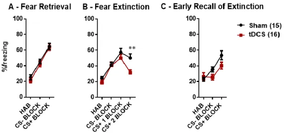
The day following the last stimulation (day 10), extinction training was performed [Interaction (F(3, 87) = 3.348; p = 0.0227); Time (F(3, 87) = 39.81; p < 0.0001); Column Factor (F(1, 29) = 3.248; p = 0.0819)]. No significant treatment effect in the overall extinction procedure was observed, however, in the Bonferroni’s multiple comparisons post-hoc test a significant difference between the experimental groups was found in the 2nd CS+ BLOCK (p = 0.0064) [Annex 3]. The
significant interaction factor shows that both groups behave differently in time. Furthermore, a significant within-group effect between CS+ 1 BLOCK and CS+ 2 BLOCK was solely identified in tDCS group (p = 0.0001), but not in the sham group (p = 0.3365) [Annex 4]. These results suggest a facilitation in the acquisition of extinction by mice subjected to tDCS in comparison to sham mice [Figure 8 – B].
The next day, extinction memory was assessed in an early recall of extinction test [Interaction (F(2, 58) = 3.836; p = 0.0272); Time (F(2, 58) = 26.82; p < 0.0001); Column Factor (F(1, 29) = 1.897; p = 0.1790)]. No significant difference between the experimental groups could be observed [Figure 8 – C], although the Bonferroni’s multiple comparisons test revealed a barely significant effect of treatment during CS+ presentation. Overall, these results show no significant differences
38 in retention of extinction memory between the experimental groups [Annex 5]. Moreover, a significant interaction factor shows that both groups behave differently in time. [Annex 6].
Figure 8 - Graphs for the fear retrieval (A), fear extinction (B) and early recall of extinction (C) procedures. The level
of fear is represented by the percentage of freezing in each stage of the procedure. All CS- and CS+ values are presented as the average of four tone presentations. The statistical analysis of the results was performed with a RM two-way ANOVA. Tukey’s multiple comparisons post-hoc test was performed to analyse within-group effects. Bonferroni’s multiple comparisons post-hoc test was performed to analyse between-group effects: * p < 0.05; ** p < 0.01. Error bars are expressed as means ± s.e.m.
In order to assess the effects of extinction on mice [Figure 9], a comparison between the fear responses to CS+ presentation before and after treatment was performed [Interaction (F(1, 29) = 2.938; p = 0.0972); Time (F(1, 29) = 24.19; p < 0.0001); Column Factor (F(1, 29) = 1.554; p = 0.2225)]. A Bonferroni’s multiple comparisons post-hoc test revealed a significant within-group effect between levels of fear in fear retrieval and extinction recall in tDCS mice (p < 0.0001), while no significant difference could be observed in sham mice (p = 0.0673) [Annex 7].
39
Figure 9 - Graph for the extinction effect analysis. The level of fear is represented by the percentage of freezing in
each stage of the procedure. All CS+ values are presented as the average of four tone presentations. The statistical analysis of the results was performed with a RM two-way ANOVA. Bonferroni’s multiple comparisons post-hoc test was performed to assess whether the extinction effect was significant or not: : * p < 0.05; ** p < 0.01; *** p < 0.001; **** p < 0.0001. Error bars are expressed as means ± s.e.m.
A late recall of extinction [Figure 10 – A] and a fear renewal test [Figure 10 – B] were conducted 21 days after extinction training.
Firstly, mice were subjected to 4 CS- tone presentations followed by 4 CS+ tone presentations in the extinction context [Interaction (F(2, 58) = 0.344; p = 0.7104); Time (F(2, 58) = 37.47; p < 0.0001); Column Factor (F(1, 29) = 2.627; p = 0.1159)]. No significant differences were observed between the experimental groups during the late recall of extinction.
One hour and a half later, mice were subjected to the latter procedure in the conditioning context, to test renewal of the fear response [Interaction (F(2, 58) = 2.942; p = 0.0607); Time (F(2, 58) = 3.906; p = 0.0256); Column Factor (F(1, 29) = 7.701; p = 0.0096)]. A significant effect of treatment was observed. And this difference can be barely observed during the acclimation period (HAB, p = 0.0734), being that difference even greater, and reaching significance, during CS- presentation
40 (p = 0.0043) [Annex 8]. These results suggest beneficial effects of tDCS on contextual renewal and generalization of the fear response.
Figure 10 - Graphs for the late recall of extinction (A) and fear renewal (B) procedures. The level of fear is represented
by the percentage of freezing in each stage of the procedure. All CS- and CS+ values are presented as the average of four tone presentations. The statistical analysis of the results was performed with a RM two-way ANOVA. Tukey’s multiple comparisons post-hoc test was performed to analyse within-group effects. Bonferroni’s multiple comparisons post-hoc test was performed to analyse between-group effects: ** p < 0.01. Error bars are expressed as means ± s.e.m.
In order to assess a possible correlation between the baseline freezing levels and the extinction effect, a linear regression analysis between the percentage of each one was performed [Figure 11]. The baseline freezing percentage was calculated based on the time mice spent freezing during the CS+ presentation of the fear retrieval test. The extinction effect was considered the difference between the freezing behaviour of mice after and before the fear extinction procedure, (% of freezing extinction retrieval - % of freezing fear retrieval).
In sham mice, the linear regression showed no correlation between baseline freezing and extinction effect [F(1, 13) = 0.02851; p = 0.8685; Pearson r = -0.04678, 95% CI = -0.546 to 0.4769]. On the opposite, in tDCS mice, the linear regression of the results demonstrated a significant negative linear correlation between baseline freezing and extinction effect [F(1, 14) = 8.752; p = 0.0104; Pearson r = -0.6202, 95% CI = -0.8535 to -0.1798]. These data show that in the tDCS group, mice with a higher baseline freezing response demonstrated a lower level of fear expression after the

![Figure 2 - Brain circuitry mediating fear acquisition and expression [Adapted from (48)]](https://thumb-eu.123doks.com/thumbv2/123dok_br/15134466.1011205/16.918.219.728.273.541/figure-brain-circuitry-mediating-fear-acquisition-expression-adapted.webp)
![Figure 3 - Brain circuitry mediating fear extinction [Adapted from (48)].](https://thumb-eu.123doks.com/thumbv2/123dok_br/15134466.1011205/17.918.205.728.467.739/figure-brain-circuitry-mediating-fear-extinction-adapted.webp)
![Figure 4 - Representation of anodal tDCS using an apparatus connected to a 9-volt current source (a), which creates an electrical field in the brain (b) [Adapted from (102)].](https://thumb-eu.123doks.com/thumbv2/123dok_br/15134466.1011205/23.918.256.666.611.815/figure-representation-apparatus-connected-current-creates-electrical-adapted.webp)