A combined petrographic and geochemical
metrological approach to assess the provenance
of the building limestone used in the Batalha
Monastery (Portugal)
Yufan Ding
1, Jose Mirao
1, Pedro Redol
2, Luis Dias
1, Patricia Moita
1, Emma Angelini
3, Sabrina
Grassini
3, Nicola Schiavon
11
HERCULES Laboratory, University of Évora, Largo Marquês de Marialva, 8, 7000 Évora,
Portugal
2
Direcao-Geral do Patrimonio Cultural, Mosteiro da Batalha, Batalha, Portugal
3Politecnico di Torino, Corso Duca degli Abruzzi, 24, 10129 Torino, TO, Italy
Abstract – To verify the provenances of limestones that
were used for construction and restoration of the Batalha Monastery, limestone quarries in central Portugal were investigated. Samples were collected from quarries and monastery in field investigation. The elements-alignment of calcium and strontium based on XRF result has suggested the source of fragments from different parts the monastery. Observation of the thin sections has supplemented petrographic evidence for this identification. XRD, TGA were also used for acquiring mineral information and chemical composition of the stones. This origin tracing result supplies foundation for further research of stone decay and reservation of the Batalha Monastery.
I. INTRODUCTION
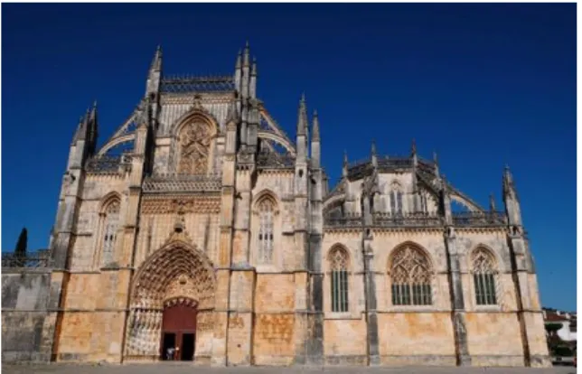
These guidelines include complete descriptions of the fonts, spacing, and related information for producing your proceeding manuscripts. The Monastery of Batalha, known as “Mosteiro Santa Maria da Vitoria” in Portugal, was constructed for commemorating the victory over Castile (the main the Kingdom of present Spain). It is considered an architectural masterpiece showing the perfect mixture of Gothic and Manueline style. The initial construction began in 1386 and took two centuries to finish. In 1840, after the monastery has been nearly abandoned due to the earthquake in 1755 and war damage by Napoleonic troops in 1810 a first restoration program started [1]. The Batalha Monastery was added to the list of World Heritage by UNESCO. Over a century has passed since the last large-scale restoration was completed. The Batalha Monastery shows high degrees of stone decay mainly due to bio-deterioration processes which are known to play an ever increasing role in stone decay both in urban and rural environments [2]. In order to plan a correct conservation strategy for the protection of monastery, it is necessary to obtain knowledge of the stone
that were used to construct it [3].
According to Aires Barros, the two original limestone quarries used for the construction of Batalha Monastery in the 15th century were the “Pidiogo” and “Valinho do Rei” located respectively 8km and 7.5km east from the monastery [4]. Soares et al indicated four quarries served as the main building stone source for the restoration during the 19th century: a) the “Reguengo do Fetal” quarry provided the works from 1840 to the mid-eighties; b) From 1854 until the end of the restoration activities, the “Carvalhos” quarry was mainly used; c) in the last decade of the 18th century, the “Cabeço do Roxo” and the “Outeiro de Sebastião” quarries were also suppling the materials [6].
II. METHODS AND MATERIALS
A. Field Investigation and Sample Collection
The location of Pidogo and Valinho do Rei quarries were marked In a document released by Batalha Municipal [5]. Reguengo do Fetal, Cabeço do Roxo and Outeiro de Sebastião were recorded in the book “O restauro do
Mosteiro da Batalha” [6], whereas their exact location was lost nowadays and was discovered by the authors. Field trips were made to these quarries (Fig. 2). 25 samples were collected, among which 13 samples were from Valinho do Rei and Pidiogo which were believed to be the original quarries for construction of monastery in 15th, 12 from the restoration quarries Reguengo do Fetal, Cabeço do Roxo and Outeiro de Sebastião. By special permission of the Direção-Geral do Patrimonio Cultural and the Mosteiro da Batalha authorities, 12 pieces of detached stone fragments were collected for destructive / non-destructive characterization respectively according to the protocol. The collected stone fragments came from various parts of the monastery, as labelled in Fig. 3.
a b
Fig. 2. Photo records, latitude and longitude of a. Valinho do Rei (39°39'32.5"N 8°44'58.1"W) and b.
Pidiogo (39°39'15.7"N 8°44'27.9"W) quarries
Fig.3 Locations of samples collected from Batalha Monastery a. Royal cloister west gallery 3rd window outside (ground floor level) b. Royal cloister roof top (roof level) c. Church north aisle eaves arch (roof level) d. Church roof railing (roof level) e. Church south carved baluster (roof level)
B. Thin section Petrography
Thin sections of the stone samples were obtained using the following procedure: stones were cut into cuboid with a cross-sectional area of 2cm*3cm, the cross-sectional surface was polished by 220# sand paper, 400# and 1000# SiC powder with water sequentially. The polished stone surface was glued to the glass slide with epoxy resin and epoxy hardener mixed at the ratio of 2:0.9. After the epoxy glue consolidated, the stone were to 0.1~0.2 mm thickness, and polished using 400# and 1000# SiC powder with water till the thickness of the stone section reached 0.025mm.
Thin sections were observed by Optical Microscopy using a LEICA DM2500P.
C. X-ray Diffraction (XRD)
Stone samples were hand milled into powder in an agate mortar. The characterization was carried out using a X-ray diffractometer BRUKER D8 Discover, with a Cu Kα source and operating at 40 kV and 40 mA. Scans were run from 3 to 75 ° 2θ, with 0.05 2θ step and 1 s/step measuring time by point. Diffract-EVA software package (BRUKER/AXS GmbH, Germany) and the PDF-2 database files (ICDD, Denver, USA). software with PDF-2 mineralogical database was utilized to interpret XRD spectra. The semi-quantification was done using the Reference Intensity Ratio by Hubbard et al [7].
D. Thermal Gravimetric Analysis (TGA)
The TGA analyses were conducted on a TG-DTA NETZSCH STA 449F3 Jupiter. The temperature of the sample was programmed at a heating rate of 10oC/min, increasing from 40oC to 1000oC, while the mass of the
sample was monitored against time or temperature. For stone samples which contains absorbed or crystallized H2O,
carbonate minerals and hydroxide minerals, thermal decomposition with gaseous reaction can be tested.
E. X-ray fluorescence (XRF)
XRF analyses were performed operating a Benchtop EDXRF Bruker S2 PUMA using a methodology similar to that adopted by Georgiou et al [8]. Quantifications were obtained using a regression method with 19 standard reference materials [9]. Spectra Elements 2.0 was utilized for acquisition and data processing, reporting the final oxides/elements (Na2O, MgO, Al2O3, SiO2, P2O5, SO3,
K2O, CaO, TiO2, MnO, FeO) concentration and the
instrumental statistical error. Two sample preparation methods were used: (1) 1.2g sample powder were fused with 12g flux (Li-tetraborate) on a Claisse LeNeo to form fused beads. (2) 10g sample powder were compressed with 1g wax (N,N'-dioctadecanoylethylenediamine) on a Specac Manual Hydraulic Press to form pellets.
III. RESULTS AND DISCUSSION
A. Thin section Petrography
The microscope photos of thin-sections are presented in Fig. 4, on which the oolites, calcite crystals and fossils were clearly recognizable. According to the definition introduced by Flugel. Eof identifying paleontological fossils and classifying carbonate grains in microfacies studies, as well referring to previous biostratigraphy of carbonate succession by Cemile et al [10][11][12][13]. Table 2 summarizes the petrography features of the samples.
a
b
c
d
Fig.4. Microscope photos of stone samples from a. Batalha Monastery, royal cloister; b. Batalha
Monastery, church roof railing; c. Pidiogo quarry; d. Valinho do Rei quarry, lower layer.
Table 2. Petrographic and paleontological features of limestone thin sections.
Sample
Name Oolite size Crystals Calcite Morphology Grain Fossils
Batalha Monastery, Royal Cloister 150 ~ 500 µm Micrite,
Sparite Peloids, Ooids, Aggregated grains; Foraminifera, Gastropods Batalha Monastery, roof top 150 ~ 500 µm Micrite,
Sparite Oncoids Ooids, Foraminifera
Batalha Monastery, eaves arch 200 ~ 400 µm Micrite,
Sparite Ooids Sponges
Batalha Monastery, church railing 1 200 ~ 500 µm Micrite,
Sparite Peloids, Ooids Foraminifera, Sponges, Batalha Monastery, church railing 2 200 ~ 500 µm Micrite,
Sparite Peloids, Ooids Brachiopods
Batalha Monastery, baluster 200 ~ 400 µm Micrite,
Sparite Peloids, Ooids Foraminifera, Wood
Pidiogo 200 ~
400 µm
Micrite,
Sparite, Peloids, Ooids Gastropods, Ostracods, Intraclasts Valinho do Rei, lower layer 50 ~ 300 µm Micrite,
Sparite Aggregated grains, Peloids, Ooids, Oncoids Intraclasts, Brachiopods Valihno do Rei, upper layer 50 ~ 200 µm Micrite,
Sparite Aggregated Peloids; grains
Foraminifera
B. X-ray diffraction
The XRD results indicated that all the samples have the same mineral composition – calcite magnesian {(Mg0.064Ca0.936)CO3} and quartz {SiO2}. Fig. 5 shows the
XRD scan of one sample from Pidiogo quarry. The semi-quantitative analysis provided the quality content of each mineral in the sample, as listed in Table 3. The proportion of calcite magnesian in these limestones has reached over 99%, while the content of quartz is lower than 1%.
Table 3. Semi-quantitative assessment of minerals in sample
Sample name Calcite Quartz
Batalha Monastery, Royal Cloister 99.30% 0.70%
Batalha Monastery, roof top 99.41% 0.59%
Batalha Monastery, eaves arch 99.50% 0.50%
Batalha Monastery, church railing 1 99.43% 0.57%
Batalha Monastery, church railing 2 99.27% 0.73%
Batalha Monastery, baluster 99.40% 0.60%
Pidiogo 99.70% 0.30%
Valinho do Rei, lower layer 99.69% 0.31%
Valihno do Rei, upper layer 99.62% 0.38%
C. Thermal gravimetric analysis
Fig. 6 shows the weight change of one sample according to temperature increase. A mass loss in the temperature range of 600 – 800 oC was observed, indicating the
decomposition reaction of calcite: CaCO3 → CaO + CO2.
The differential of thermal gravity curve showed the reaction is single-stepped. There is no mass change in other temperature range, demonstrating that there are no absorbed water, plaster, portlandite nor muscovite in these stones. Through the calculated results it is seen that calcite is the main composition of all the samples, the content reaches 97.2 wt% ~ 99.2 wt%.
D. X-ray fluorescence analysis
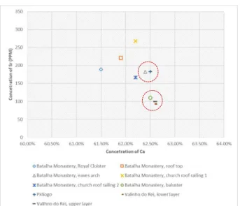
Due to the same coordination number and similar ion radius that strontium and calcium cations have, the Sr2+
may substitute a Ca2+ ions in calcite. Stones formed in the
same geological environment would have similar value of such substitution. From the Ca-Sr element alignment scatter plot, it is seen that the plot which represents Batalha Monastery baluster is very close to the plots of samples from the Valinho do Rei quarry, suggesting that the limestone of this baluster may come from the Valinho do Rei quarry. Similarly, the stone of Batalha Monastery eaves arch was suggested to be coming from the Pidiogo quarry.
IV. CONCLUSION
The proposed multi-analytical techniques were used for establishing the quarries of origin for the Batalha Monastery. Since considerable historical evidence can be drawn by the study of material provenance, these techniques have supplied a powerful tool to collect and analyze valuable information of special interest to conservation research.
The results of this work suggested that two parts of the monastery were constructed from the stones of Pidiogo quarry and Valinho do Rei quarry respectively. To verify the provenance of other parts of monastery, same analysis should be repeated on stones from the restoration quarries. In further research, ICP-MS is strongly advised to provide trace-element information in stones, for the confirmation of provenance.
V. ACKNOWLEDGEMENT
The research presented in this paper was carried out mainly using data collected at Universidade de Évora and Politecnico di Torino, as part of H2020-MSCA-ITN-2017, ED-ARCHMAT (ESR1). This project has received funding from the European Union’s Horizon 2020 research and innovation programme under the Marie Skłodowska-Curie grant agreement No 766311.
REFERENCES
[1] Da Silva, J. C. V. (2007). The Monastery of Batalha. Scala Books.
[2] Gemeda, B. T., Lahoz, R., Caldeira, A. T., & Schiavon,
Fig. 6. TGA curve of stone from Batalha Monastery, Baluster.
N. (2018). Efficacy of laser cleaning in the removal of biological patina on the volcanic scoria of the rock-hewn churches of Lalibela, Ethiopia. Environmental
earth sciences, 77(2), 36.
[3] Schiavon, N., Candeias, A., Ferreira, T., Da Conceiçao Lopes, M., Carneiro, A., Calligaro, T., & Mirao, J. (2012). A combined multi‐analytical approach for the study of Roman glass from south‐ west Iberia: synchrotron μ‐XRF, external‐PIXE/PIGE and BSEM–EDS. Archaeometry, 54(6), 974-996. [4] Aires-Barros, L. (2001). As rochas dos monumentos
portugueses: tipologias e patologias. Instituto
Português do Património Arquitectónico.
[5] Edital n.º 03/2017/g.A.P. Sítio de interesse municipal da pedreira histórica de valinho do rei e sítio de interesse municipal da pedreira histórica de pidiogo – criação de zona especial de proteção (zep).
[6] Soares, Clara Moura. O restauro do mosteiro da batalha: pedreiras históricas, estaleiro de obras e mestres canteiros. Magno Ed., 2001.
[7] Hubbard, C. R., Evans, E. H., & Smith, D. K. (1976). The reference intensity ratio, I/Ic, for computer simulated powder patterns. Journal of Applied
Crystallography, 9(2), 169-174.
[8] Lasne, J., Noblet, A., Szopa, C., Navarro-González, R., Cabane, M., Poch, O., ... & Coll, P. (2016). Oxidants at the surface of mars: a review in light of
recent exploration results. Astrobiology, 16(12), 977-996.
[9] Beltrame, M., Liberato, M., Mirão, J., Santos, H., Barrulas, P., Branco, F., ... & Schiavon, N. (2019). Islamic and post Islamic ceramics from the town of Santarém (Portugal): The continuity of ceramic technology in a transforming society. Journal of
archaeological science: Reports, 23, 910-928.
[10] Flügel, E. (2004). Fossils in thin section: it is not that difficult. In Microfacies of Carbonate Rocks (pp. 399-574). Springer, Berlin, Heidelberg.
[11] Flügel, E. (2013). Microfacies of carbonate rocks:
analysis, interpretation and application. Springer
Science & Business Media.
[12] Flügel, E. (2004). Microfacies data: matrix and grains. In Microfacies of Carbonate Rocks (pp. 73-176). Springer, Berlin, Heidelberg.
[13] Solak, C., Taslı, K., & Koç, H. (2017). Biostratigraphy and facies analysis of the Upper Cretaceous–Danian? platform carbonate succession in the Kuyucak area, western Central Taurides, S Turkey. Cretaceous Research, 79, 43-63.