Original article
Forensic Anthropology
Marcos Paulo Salles Machado
1,2, Andrei de Souza Santos
*3, Casimiro
Abreu Possante de Almeida
2, Carlos Durão
5,6, Eugénia Cunha
1,4,
Eduardo Daruge Júnior
11 Department of Social Dentistry, Piracicaba Dental School, State University of Campinas,
FOP-UNICAMP, Brazil
2 Forensic Anthropology Service of the Legal Medicine Institute, Police of Rio de Janeiro State, Brazil 3 Department of Archaeology, National Museum, UFRJ, Rio de Janeiro, Brazil
4 Life Sciences Department/ Forensic Sciences Center, University of Coimbra, INMLCF.IP.
Portuguese National Institute of Legal Medicine, South Branch, Portugal
5 National Institute of Legal Medicine and Forensic Sciences, Coimbra, Portugal 6 Orthopedic Department- Hospital Vila Franca de Xira Lisbon, Portugal.
* Corresponding author: Trav. Batista, 436, São Gonçalo, RJ, Brazil 24415-410
E-mail: andrei_santos@id.uff.br
Received 11 February 2019; Received in revised form 13 August 2019; Accepted 1 October 2019. Available online 04 May 2020
ABSTRACT
Forensic Anthropology plays an essential role in forensic investigations. The application of its concepts, from the crime scene to the laboratory, is critical to avoid that traces be ignored or lost during a criminal investigation on human skeletal remains. A Forensic Anthropology Office was created in the State of Rio de Janeiro at
the end of 2010 with the purpose to help in criminal investigations. In 2011 and 2012, the Anthropology Office received 66 examination requisitions, comprising 74 human skeletal remains. The biological profile established after the study of the skeletal remains showed that the samples were constituted mainly of male subjects (80%), white (32%), and young adults ranging from ages 21-50 (54%), considered as the risk group. The injuries most frequently observed were blunt trauma (33,3%), followed by gunshot (31,4%). The most susceptible body regions were head and neck, affected in 42% of cases. Gunshots represented 52,1% of injuries found on the head and neck. Only 18,8% of the human remains under analysis contained more than 95% of all skeletal bones. Likewise, only 7 (10,1%) of 47 completely skeletonized remains had more than 50% of his bones recovered. The delays in locating human remains and their continuing decomposition poses a marked reduction in the number of bones recovered, confirming the necessity of a trace collection carried out by trained professionals. Lastly, 80% of incoming cases came from 18 of the 160 districts of Rio de Janeiro, indicating a significant prevalence of human remains found in conflict areas.
Keywords: Forensic Anthropology; Forensic Dentistry; Forensic Medicine; Forensic Sciences.
Introduction
Rio de Janeiro is a large city, with around 6 million inhabitants and an area of 1,200 km2, according to the 2010 census carried out by the Brazilian Institute of Geography and Statistics (IBGE, Portuguese abbreviation). The tremendous social differences provide, for some, ideal ground for violence to germinate. The great land extension, presence of rivers, mounds, lakes, mangroves, and forests, as well as outcast areas where the State has only recently attempted to stand ground, may function as clandestine graves; the tropical weather and rich and abundant fauna accelerate decomposition, decreasing the time-lapse and viability for a conventional necropsy.
In the State of Rio de Janeiro, the Forensic Anthropology Office (SAFO, Portuguese abbreviation) was established in October 2010 within Afrânio Peixoto Legal Medicine Institute (IML-AP, Portuguese abbreviation), linked to the Technical-Scientific Police Department branch of the Civil Police of Rio de Janeiro State.
Therefore, as in most Brazilian States, the forensic examination is subordinated to the Public Security Secretary, that is, constitutes a police unit6,10-12.
SAFO´s establishment generated demand for specialized professionals, which contributed to gather all field professionals in the country, and inspired the creation of the Brazilian Association of Forensic Anthropology (ABRAF, Portuguese abbreviation) in September 2012, encouraged and supported by the Forensic Anthropology Society of Europe (FASE) and, similar to the latter, aims to contribute to the specialize, homogenize and publicize FA concepts13, 14.
The demand mentioned above for professionals to act in forensic institutions should be supplied by Universities, leading to the creation of careers and development of research that will offer back scientific basis and support in the future. Forensic and academic institutions are two sides of the same coin2, 6.
Proper qualification and the adoption of incentive policies towards the scientific police contribute to the investigation process and helps decrease impunity, while its inappropriate functioning paves the way for a rise in violence. Rodrigo Garrido alerts to the difficulties generated by the distancing between forensic and academic institutions and states that their approximation and strengthening support and add consistency to court decisions, being fundamental to the confrontation of violence.
This process depends on the administrative and financial autonomy of forensic investigation organs4, 6, 7. The Nina Rodrigues Legal Medicine Institute, in Bahia, and the Center of Legal Medicine (CEMEL), in Ribeirão Preto, are well-succeeded examples of ongoing partnerships between institutions2, 6, 8.
Forensic Anthropology is the science that brings together concepts of Physical Anthropology and Forensic Sciences10. Its routine includes the search for trace evidence during the examination of skeletal remains, aiming to identify the victim, analysis of traumatic injuries that may have occurred and reconstitute all events leading to death, including an estimate of the time of death, or postmortem interval (PMI)5, 11, 13, 15.
The application of FA concepts is not restricted to skeletonized remains12 and is
applicable in cases of age estimation of live subjects without documents; identification and age estimation of criminals and crime victims of video recorded felonies, such as child pornography13,15; verification of age of majority of criminals is also a part of the anthropological analyses group16; and in the event of mass disasters, as well as in crimes against humanity and human rights violations1, 11, 15, 17, mainly when clandestine graves are located.
The complete and detailed examination of the crime scene represents an element of prime importance to crime-solving. Every time an investigation involves buried corpses, it is mandatory that the search for remains be made using archeological excavation techniques, carried out by trained professionals, in a way that no trace may be lost or destroyed1, 3, 5, 10, 11, 13, 15. The employment of such excavation techniques tends to be very helpful in cases involving narcotraffic victims, serial crimes, massacres, political crimes, homicides, and others1 3, 5 9.
It is doubtless that FA is a multidisciplinary science that embraces anthropologists, archeologists, medical doctors, dentists, biologists, biomedical professionals, among others, acting individually or as a team, provided they have been adequately trained11. Efforts are being directed towards enabling the SAFO to deploy to the field of human remains detection so that it can collaborate more efficiently with crime elucidation. As stated by Cattaneo, today FA lies way beyond calipers and osteometric tables13.
In Brazil, FA lacks support to research to identify the physical peculiarities of the local population, and thus validate the techniques used in forensic examinations. The first step is to create an osteological collection that reflects the current population. For example, the Piracicaba Dental School (FOP-UNICAMP Portuguese abbreviation) is developing, with the technical support of the ABRAF, a skeletal collection consisting of 320 complete and cataloged specimens from the current, local population, so far.
Aware of the contribution FA offers to criminal case elucidation, and, consequently, to decrease crime3, this study aimed to collect and organize data produced by the SAFO. The secondary objectives were to present its examination protocol and to identify the steps of action, where much effort is still necessary to increase the quantity and quality of traces recovered, so that the investigation efficiency may be improved.
Materials and Methods
The research was approved by the Ethics Committee on Research of FOP-UNICAMP, under registration number 040/2014, and authorized by the Technical-Scientific Police Headquarters (DGPTC, Portuguese abbreviation) from the State of Rio de Janeiro. The study comprised a retrospective analysis of information derived from individual examination reports generated by the SAFO of IML-AP in the years of 2011 and 2012.
Data were extracted from each report about the site of corpse location; time elapsed from death; biological profile: sex, age, ancestry, stature, laterality and additional elements that might contribute to identification; and trauma analysis, including search for cause of death and for any other traces that may help clarify its circumstances.
Two SAFO forensic examiners collected all data. Over these two years, 66 examination requisitions were received, and each generated a report. Of these 66 requisitions: 6 corresponded to non-human material; other 3 represented material without forensic implications, 2 consisted of anatomical material discarded from study institutes, and 1 consisted of archeological material; and another one corresponding to a complementary examination requisition for a previously located skeleton, already accounted for statistically.
The remaining 56 requisitions comprised skeletal remains of a total of 74 different individuals since in 9 cases there were bones from more than one person commingled.
Five out of 74 skeletal remains did not enter the SAFO laboratory because they had been positively identified by the Forensic Dentistry Office or through Papiloscopy, due to the presence of soft tissue remains. Therefore 69 skeletal remains were examined, and the resulting data compose the present study. All examination requisitions informed the site of skeletal recovery, so all 74 cases were accounted for statistically.
Results and Discussion
Taphonomy
Quantity of bones recovered
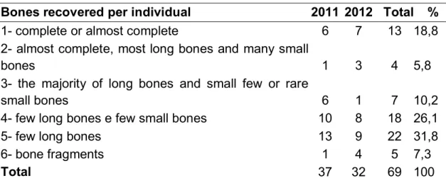
Out of 56 requisitions, 47 (83%) presented skeletal remains of single individuals, while, in other 9 (17%), the commingling of skeletal remains took place. These nine requisitions comprised a total of 27 individuals, although the single request with the highest number of subjects investigated presented skeletal remains of six different people. As previously mentioned, laboratory analyses were centered on 69 skeletal remains because 5 of 74 had been identified before entering the SAFO. Less than 18,8% of skeletal remains recovered comprised more than 95% of body bones (Table 1).
Table 1. Descriptive statistics of recovered bones.
Bones recovered per individual 2011 2012 Total %
1- complete or almost complete 6 7 13 18,8
2- almost complete, most long bones and many small
bones 1 3 4 5,8
3- the majority of long bones and small few or rare
small bones 6 1 7 10,2
4- few long bones e few small bones 10 8 18 26,1
5- few long bones 13 9 22 31,8
6- bone fragments 1 4 5 7,3
Total 37 32 69 100
Stages of decomposition of recovered material (taphonomy)
It was observed that 38 (55,1%) remains examined showed signs of degradation, indicating they had been exposed to the environment for a long time before being located. Besides the skeletal remains, the laboratory received partially saponified corpses, partially mummified corpses, and even at advanced stages of decomposition, namely bloat and liquefying stages (Table 2).
A total of 47 (68,1%) of 69 skeletal remains were fully skeletonized, that is, presented no soft tissue remains or cartilages. Of these, only 7 (10,1%) had more than half of the bones recovered, proving that the longer it takes for remains to be located, and with its subsequent decomposition, there is a marked reduction in the number of bones recovered.
Table 2. Descriptive statistics of body taphonomy. Stage of tissue decomposition at the moment of
recovery 2011 2012 TOTAL %
1- Degrading bones 18 20 38 55.1
2- Skeletonized 8 1 9 13
3- Partially skeletonized 9 1 10 14.5
4- Putrid the presence of soft tissue 1 2 3 4.3
5- Calcined 0 4 4 5.8
6- Mumified/Skeletonized 0 4 4 5.8
7- Saponified/Skeletonized 0 1 1 1.5
Total 36 33 69 100
Partially skeletonized the presence of cartilages, little soft tissue, putrid odor.
Skeletonized the presence of entire bones, absence of cartilages or soft tissue, and lack of putrid odor.
Of the 69 skeletal remains, 65 underwent sexual dimorphism analysis, and the anthropological examination concluded that 55 (80%) displayed male characteristics, and 10 (14%) displayed female characteristics. All skeletal remains that comprised at least the skull, pelvis, femur or humerus allowed for gender estimation. Only four skeletal remains (6%) did not present at least one of the cited bones and were, therefore, not considered for the examination.
Pelvic bones represented the first choice for analysis of sexual dimorphism, followed by the skull. Morphological characteristics were analyzed and organized into a decision table with twelve anatomical items to be examined on the pelvis18 and fourteen on the skull. Landmarks were selected from the study by Buikstra, Ubelaker, and Walker15, 19 21. Each feature was observed and classified according to the characteristics found, as male, female, or undetermined. The method for sex estimation based on pelvic metric characteristics, known as DSP2, was also applied15,
22.
Age analysis
As far as age was concerned, it was observed that fetuses or individuals with ages ranging from 0 to 10 years were not received. 10% of the sample consisted of individuals with age ranging from 11 to 20 years, 34% were included in the age ranging from 21 to 30 years, 20% were in the range between 31 to 50 years and, finally, 20% were included in the group above 50 years. Another 16% did not contain the bones needed for the exam. Individuals with age ranging from 21 to 30 years, represents the group with the highest associated risk, followed by the age range of 31 to 50 years.
As for age estimation, methods used in young adults were based on dental development tables developed by Nicodemo, Moraes e Medici Filho23; on Ubelaker´s chart cited by Couto24, and on the epiphyseal plate, calcification described by Buikstra and Ubelaker21. When it came to adult skeletons, the analysis was based on the stage of development of the pubic symphysis, proposed by Suchey, Brooks15, 24; the first costal arch by DiGangi et al.25, 26; the fourth costal arch27, and the auricular surface28. Lamendin´s analysis was applied as often as possible15, 29, 30.
Ancestry analysis
In 31 (45%) of 69 cases, ancestry research could not be carried out because of missing skulls or due to their fragmentation. From the 38 human skeletal remains that allowed for an ancestry analysis, it was concluded that 22 (32%) displayed predominantly European characteristics, 14 (20%) showed predominantly African features, and 2 (3%) displayed mostly Asian characteristics.
The skull represents the main point of interest while investigating ancestry. The face, especially the nose area, were morphologically analyzed, as advised by
Bass31,32, Rhine33, Gill33 36, and Krogman and Mehmet37. The morphological analysis
of the face is known as the most assertive means to evaluate geographical origin. While examining the skull, a decision table of 17 anatomical characteristics was observed. The six craniometric indices proposed by Arbenz were also applied23. The postcranial skeleton was used only to confirm the findings of the skull, where tibiofemoral and radiohumeral indexes cited by França were applied38.
Stature analysis
Stature estimation was based on the Trotter & Gleser table cited by Couto24, and on the table proposed by Mellega [39]. The latter represents a study conducted on the Brazilian population, offering good results when reproduced. Height is a parameter that should be evaluated with restriction because, in Brazil, height is only measured for military service purposes around the age of 18. As often as possible, it is suggested that ratios be used based on photographs of the missing person and that additionally, measurements of siblings, should there be any, be taken as a frame of comparison.
Functional laterality analysis
This analysis was introduced to the formal examination protocol at the end of 2011 after being considered a relevant determinant that may aid in identification. Since then, 46 skeletal remains were examined, although only ten could undergo the analysis to determine the side of dominance because they comprised bones of the appendicular skeleton of both left and right sides. One of the exams was inconclusive, and the other nine suggested individuals were right-handed.
The protocol followed was the same that has been used and supported by CEMEL, in Ribeirão Preto, for more than 10 years8. It is based on a decision table that
analyzes and compares eight anatomical features on both sides of the upper appendicular skeleton.
Forensic Facial Approximation (FFR) and Craniofacial Superimposition
Between 2011 and 2012, two exams with craniofacial superimposition and three exams of FFR were carried out. Both craniofacial superimposition exams demonstrated coincidence between facial and craniometric landmarks, as did facial characteristics match. Later, DNA tests confirmed the results obtained by approximation.
The FFR was carried out digitally and still cannot function as a means of attracting the missing person´s family members because its results cannot be released to the general public. In one case where the family waited for the DNA result to confirm the identity of skeletal remains, the FFR was applied, and the family recognized the result presented as similar to the missing person´s appearance.
The techniques of overlapping images and forensic facial approximation were used eventually by the SAFO and are not part of the routine protocol. The entire process is usually concluded in a single day40.
Identification
In its first two years functioning as a formal unit, SAFO voided one case of corpse identification that had been wrongly recognized by family members. In another case, the SAFO was responsible for the inclusion of skeletal remains as a possible match after pointing out coincidences between information obtained from family members and the victim´s biological profile. A DNA test confirmed SAFO´s findings.
The SAFO positively identified a case where the biological profile, the FFR, OI matched, and the anterior teeth could be observed, including the gaps between them. It should be noted that at least 29 of 69 skeletal remains examined by the SAFO presented characteristics that could contribute to the identification process, such as dental fillings, fractures and antemortem pathologies, characteristic anatomical variations such as septal and sternal foramina, bifid ribs, metopic suture, sacral bifid spine, sutural bones, ankylosis, unerupted teeth, and osteomas. Again, it is necessary
to bring attention to the fact that a missing person´s database could highly improve the statistics of positive identifications.
Analysis of trauma injury
In the first stage of trauma injury investigation, the triple distinction was made between antemortem, perimortem, and postmortem. From the 69 skeletal remains analyzed, 44 (63,8%) showed some skeletal trauma, namely: blunt force, burning, sharp force, gunshot, or an association between these. In 25 (36,2%) of 69 skeletal remains, no trauma injury was observed.
The most frequently observed injury was blunt trauma (33,3%), followed by gunshot (31,4%), sharp-blunt trauma (21,6%), and burning action (13,7%). Six out of 44 skeletal remains presented an association between more than one type of injury.
The head-neck region was affected in 42% of the 69 cases, representing the most frequently affected location, and having been subjected to all four types of trauma injury evaluated. The upper arms were affected in 13% of cases; the chest-abdomen region was affected in 27,5% of cases. Finally, the hip region was affected in 13% of cases and the lower members in 14,5%. In some cases, the five body regions were affected.
The highest correlation found between a type of trauma, and a specific body part was the gunshot action to the skull-neck region, corresponding to 27,3% of the cases. Of all gunshot injuries, 52,1% were located in the skull-neck area.
Correlation between the site of location and trauma
Of all cases investigated, 80% were located in one of 18 out of 160 districts found in the city of Rio de Janeiro, indicating a substantial prevalence concentrated over few areas and also showing that some specific regions display above average according to violence numbers.
An association between recovery sites and type of trauma injury could also be identified, indicating that criminals acting within a specific district display a modus operandi, as 15% of victims who suffered multiple sharp-blunt injuries, in this case, the scattering of body parts, were found in the surroundings of Ilha do Governador district, while gunshot injuries to the head and neck prevailed (25%) in Realengo district.
The biological profile established by skeletal remains investigation evidenced that the sample was mainly comprised of men (80%), white (32%), young-adult with age range between 21-50 (54%), representing the risk group in 11,59% of studied cases.
The authors have presented the methods used in the examinations and the results obtained after two years of service. Results are still limited due to the reduced sample size, but the presentation of the method employed is significantly relevant because it exposes techniques that have proved efficient.
Final Considerations
We emphasize that the methodology presented in this work were used in the creation of the forensic anthropology section, in 2010, and all cases at the time culminated in success. Currently, the techniques used for the diagnosis of ancestry are based on Hefner (2009). For sex assessment, we use DSP2, Klales et al. (2012)41, and Walker (2008).
Also, we understand that there is no metric methodology, in the literature, that can replace morphological methods. In this sense, we prioritize its uses due to the practicality, reliability, and success rate in obtaining data.
Conclusion
It was possible to point out the existence of a risk group, made up of young-adult, white males. It is noteworthy that a high number of skeletal remains were located in a few regions, and that patterns of trauma can be associated with specific districts, thus establishing a correlation between types of homicide and certain parts of town. Gunshot to the head and neck was the most common combination of trauma and body injury.
The establishment of biological profiles and trauma injury analysis must be carried out by trained and experienced professionals in the forensic anthropology field, which can optimize the trace investigation.
Acknowledgments
The authors are very grateful to Rodrigo Ivo Matoso, Gilberto Paiva de Carvalho e Marina Gratão for helpful comments on, and critical editing of this
manuscript. We want to thank Cristiane Correia Pereira Machado for her support and for offering invaluable advice.
Conflict of interest
There are no known conflicts of interest associated with this publication, and there has been no significant financial support for this work that could have influenced its outcome.
References
1. Fondebrider L (2002) Reflections on the scientific documentation of human rights
violations. RICR 84:885 891.
2.
the optics of the forensic anthropology in Brazil. Ciência e Saúde Coletiva 14(5):1855 1863.
3. Dos Santos AB (2012) Estudos arqueológicos a serviço de contextos criminais:
arqueologia forense. Rev Criminol e Ciências Penitenciárias 2:1 13.
4. Coimbra CMB, Brasil VV (2006) Exumando, identificando os mortos e
desaparecidos políticos: uma contribuição do GTNM/RJ para o resgate da Memória.
5. Dirkmaat DC, Cabo LL, Ousley SD, Symes SA (2008) New perspectives in forensic
anthropology. Yearb Phys Anthropol Suppl 47:33 52. doi: 10.1002/ajpa.20948
6. Giovanelli A, Grazinoli R (2011) A perícia criminal no Brasil como instância
legitimado de práticas policiais inquisitoriais. Rev LEVS 5 24.
7. Iscan MY (1988) Rise of Forensic Anthropology. Yearb Phys Anthropol 31:203 230. 8. Soares ATC, Guimarães MA (2008) Dois anos de antropologia forense no centro de
medicina legal (CEMEL) da faculdade de medicina de Ribeirão Preto-USP. Med Ribeirão Preto 41:7 11.
9. Cunha E, Pinheiro J (2005) A linguagem das fraturas: a perspectiva da Antropologia
Forense. Antropol Port 22/23:223 243.
10. an MY, Quatrehomme G (1999) Medicolegal anthropology in France. Forensic
Sci Int 100:17 35.
11. Vaz M, Benfica FS (2008) The experience of the forensic anthropology service of
e45 e49. doi: 10.1016/j.forsciint.2008.05.004
12. Cunha E, Cattaneo C (2006) Forensic Anthropology and Forensic Pathology The
State of the Art. In: Schimitt A, Cunha E, Pinheiro J (eds) Forensic Anthropol. Med. Complement. Sci. From Recover. to Cause Death. Humana Press Inc., Totowa, pp 39 53
13. Cattaneo C (2007) Forensic anthropology: developments of a classical discipline in
14. Elbio H, Olivera S (2000) Forensic anthropology in Latin America. Forensic Sci Int
109:15 30.
15. Cunha E (2014) A antropologia forense passo a passo. Enferm. Forense. Lidel,
Lisboa, pp 280 288
16. Cunha E, Baccino E, Martrille L, et al. (2009) The problem of aging human remains
and living individuals: a review. Forensic Sci Int 193:1 13. doi: 10.1016/j.forsciint.2009.09.008
17. Lessa A (2010) Perícias forenses e justiça criminal sob a ótica da antropologia
forense no Brasil. 153 172.
18. Bruzek J (2002) A method for visual determination of sex , using the human hip
bone. 168:157 168. doi: 10.1002/ajpa.10012
19. Walker PL (2008) Sexing skulls using discriminant function analysis of visually
assessed traits. Am J Phys Anthropol 136:39 50. doi: 10.1002/ajpa.20776
20. Anderson BE (1990) Ventral arc of the os pubis: anatomical and developmental
considerations. Am J Phys Anthropol 83:449 458.
21. Buikstra JE, Ubelaker DH (1994) Standards for data collection from human skeletal
remains, 10th ed. Arkansas
22. Murail P, Bruzek J, Houët F, Cunha E (2005) DSP: A tool for probabilistic sex
diagnosis using worldwide variability in hip-bone measurements. Bull mémoires la 176.
23. Arbenz GO (1988) Medicina Legal e Antropologia Forense. Atheneu, Rio de Janeiro
São Paulo
24. Couto RC (2011) Pericias em medicina e odontologia legal. Belo Horizonte
25. DiGangi E a, Bethard JD, Kimmerle EH, Konigsberg LW (2009) A new method for
estimating age-at-death from the first rib. Am J Phys Anthropol 138:164 76. doi: 10.1002/ajpa.20916
26. Kunos
its possible utility for human age-at-death estimation. 323:303 323.
27. Loth SR, Iscan MY, Scheuerman EH (1994) Intercostal variation at the sternal end
of the rib. 65:135 143.
28. Buckberry JL, Chamberlain a T (2002) Age estimation from the auricular surface of
the ilium: a revised method. Am J Phys Anthropol 119:231 9. doi: 10.1002/ajpa.10130
29. Lopes JR, Borges S, Queiroz S, et al. (2014) Age estimation by teeth periodontosis
a
Oral Sci 13:10 14.
30. Prince D a, Konigsberg LW (2008) New formulae for estimating age-at-death in the
. J Forensic Sci 53:578 87. doi: 10.1111/j.1556-4029.2008.00713.x
31. Bass WM (2004) A laboratory and field manual, 5th ed. Missouri Archaeological
Society
32. Hefner JT (2009) Cranial nonmetric variation and estimating ancestry*. J Forensic
Sci 54:985 95. doi: 10.1111/j.1556-4029.2009.01118.x
33. Gill GW, Rhine S (2004) Methods for forensic anthropology Maxwell Museum os
anthropological papers. Anthropological Papers, New Mexico
34. Gill GW (1998) Craniofacial criteria in the skeletal attribution of race. Forensic
35. Pickering R, Bachman D (2009) The use of forensic anthropology, 2nd ed. CRC
Press
36. Adams B (2007) Forensic Anthropology.
37. Krogman WM, Iscan MY (1986) The human skeleton in forensic medicine, second.
Springfield, Illinois
38. França GV de (2011) Medicina Legal, 9th ed. Guanabara Koogan, Rio de Janeiro 39. Mellega R (2004) Validação das principais técnicas de determinação da estatura
existentes e aplicadas em amostras de cadáveres brasileiros. Universidade Estadual de Campinas
40. Moraes CA da C, Dias PEM, Melani RFH (2013) Demonstration of protocol for
computer-aided forensic facial reconstruction with free software and photogrammetry. J Res Dent 2:77 90.
41. Klales AR, Ousley SD, Vollner JM. 2012. A Revised Method of Sexing the Human
rensic Sci 149:104 114