DOI: http://dx.doi.org/10.18363/rbo.v77.2020.e1484 Original Article/Endodontics
Evaluation of single visit endodontic treatment and
non-surgical retreatment with foraminal enlargment
of teeth with apical periodontitis
Marcelle Louise Sposito Bourreau,1 Marcos Roberto dos Santos Frozoni,2 Marília Jesus Batista de Brito Mota,2 Alexandre Augusto Zaia,1 Carolina Oliveira de Lima,3 Maíra Prado,4 Adriana de Jesus Soares1
1Department of Restorative Dentistry, Division of Endodontics, Dental School of Piracicaba, State University of Campinas, Campinas, SP, Brazil 2Division of Endodontics, Dental School and Center for Dental Research São Leopoldo Mandic, Campinas, SP, Brazil
3Department of Dental Clinic, School of Dentistry, Federal University of Rio de Janeiro, Rio de Janeiro, RJ, Brazil 4Division of Dental Materials, Dental School of Veiga de Almeida University, Rio de Janeiro, Brazil
• Conflicts of interest: none declared.
AbstrAct
Objective: to evaluate the root canal treatment (RCT) and non-surgical root canal retreatment (NSRCR), associated with foraminal enlargement, performed on a single-visit. Material and Methods: 125 teeth with apical periodontitis and follow-up period ranging from 6 to 12 months were included. The success was considered by the absence of signs and symptoms and complete or incomplete periapical repair. Logistic regression analyses were used to identify factors associated with the repair (p<0.05). Results: RCT showed 71.58% of complete healing and 23.16% of acceptable healing. NSRCR showed 80% of complete healing and 20% of acceptable healing. Age, gender, type of treatment and preoperative pain were not statistically significant for the healing process (p>0.05). Premolars showed the greatest chance of periapical repair. Pulp Canal Sealer showed a greater chance of periapical repair when compared to Sealapex (p<0.05). Conclusion: RCT and NSRCR using a foraminal enlargement protocol provided a favorable prognosis of periapical healing.
Keywords: Periapical periodontitis; Root canal preparation; Tooth apex.
Introduction
R
oot canal treatment consists of the combination of mechanical instrumentation of the root canal system, its chemical debridement and filling with an inert material designed to maintain or restore the health of the periradicular tissue.1Current instrumentation and irrigation techniques are not completely effective in the elimination of debris and bacteria from the apical third due to the complex canal morphology, the narrow canal space, inadequate flushing of irrigants, and variation in the diameter and curvature of the root canals.1 Furthermore, studies have observed the presence of bacteria in the apical foramen, with colonies extending to the extraradicular region in certain cases.2 These bacteria, if not eliminated, can survive owing to the constant supply of nutrition from the periapical area. Thus, to obtain adequate root canal disinfection and ensure a favorable environment for periapical healing, cleaning and shaping of the foramina region may be necessary.2
Root canal treatment has shown a high success rate, greater than 97%.3 When primary root canal treatment fails, retreatment or apical surgery is often indicated. The tooth survival rate of non-surgical retreatment cases at 5 years is reported to be 89%. Ng et al.,4 in meta-analyses, observed that the achievement of patency at the canal terminus and the extension of canal cleaning as close as possible to its apical terminus were conditions found to improve the periapical healing.
Previous studies showed advantages when foraminal
enlargement was performed during root canal treatment. It included better removal of infected dentin and debris, significantly reducing the bacterial load and endotoxin levels in the canal system, and enhancing the flushing action of irrigants in the apical region.1,5,6
Although the foraminal enlargement shows several advantages, the possibility of postoperative pain is still controversial.7,8 Postoperative pain is defined as the unpleasant sensation of any degree of pain that occurs after the initiation of a root canal treatment and has been reported in 25%–40% of all endodontic patients, including those with vital and nonvital pulp.9 Authors advocate that foraminal enlargement might lead to a higher incidence of postoperative pain due to direct mechanical irritation of periapical tissues and/or extrusion of debris during preparation of the area.2 However, Silva et al.9 compared the incidence of postoperative pain
following foraminal enlargement with the pain experienced following a conventional canal preparation technique in anterior teeth with necrosis and apical periodontitis and observed that both techniques resulted in the same postoperative pain and necessity for analgesic medication. They suggest that the use of foraminal enlargement should be performed for endodontic treatment previsibility without increasing postoperative pain.
In respect to the treatment outcome, the results are also controversial. Authors observed a decrease in the success rate with an increase in the apical preparation size.10 Saini et
al.1 evaluated the effect of the apical preparation size on the outcome of primary endodontic treatment in mandibular first
molars and observed that the proportion of successfully healed cases increased with an increase in the apical preparation size. To the best of our knowledge, no study evaluated the effect of apical enlargement on the success in non-surgical root canal retreatment.
The objective of this study was to evaluate the primary root canal treatment and non-surgical root canal retreatment associated with apical enlargement performed on a single visit.
Material and Methods
Ethical approval, inclusion and exclusion criteria
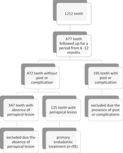
The present study was approved by the Ethics Committee (no. 082/2015). The population of this study was patients attended at a private clinic from November 2010 to June 2014. The inclusion criteria were primary endodontic treatments and non-surgical endodontic retreatments in all dental groups that presented previous periapical lesions. The exclusion criteria were immature teeth, teeth with internal and/or external root resorption; teeth with a history of dental trauma; periodontal disease; treatments not completed in a single session; teeth where the patency of the apical foramen was not achieved; teeth without prior periapical lesion; teeth that presented the postoperative insertion of prosthetic post and teeth that presented pre- and trans-operative complications such as perforations and instrument fractures (Figure 1).
Primary root canal treatment and non-surgical root canal retreatment
All treatments were performed in a single session by a specialist in endodontics with 14 years of experience. All patients were inquired to rate preoperative pain before the root canal treatment started. The same protocol was employed for primary root canal treatment and non-surgical root canal retreatment.
Patients were anesthetized (Lidocaine 2% with adrenaline 1: 100,000, DFL®, Rio de Janeiro, Brazil). Caries and restorations were removed and a standard access opening was performed. Then, teeth were isolated with a rubber dam (Madeitex, São Paulo, Brazil) and clamps (SSWhite Duflex, Rio de Janeiro, Brazil). The root canals were instrumented by a crown-down technique that consisted of the middle-cervical preparation using a Hero 20/.06 instrument (HERO 642, MicroMega®, Besançon, France) and the use of Gates-Glidden burs # 4 to # 2 (Dentsply-Maillefer®, Ballaigues, Switzerland) in a crown-apex direction.
For apical preparation, a K-file # 10 (Hi-5, Miltex®, Pennsylvania, USA) was inserted and patency was performed. The root canal length (RCL) was determined with an electronic apical locator (Novapex, Forum Engineering Technologies®, Israel), with the instrument of patency in the zero length. The initial anatomical file (IAF) was defined by the instrument that best fit the shape of the root canal.
The working length (WL) was established as 1 mm beyond to the RCL, in order to overprepare the apical foramen area, keeping this area clean and free of debris. Then, instrumentation and shaping were done with the rotating instruments, in the sequence 10/.04, 15/.05, 20/.06 and 25/.06, according to the manufacturer’s recommendation (Mtwo system, VDW®, Munich, Germany). After shaping, a foraminal refinement with manual files (K-file CC+, VDW®, Munich, Germany) was performed until the foramen’s final diameter was 3 diameters above IAF.
The chemical auxiliary substance used to prepare the root canals was 2% chlorhexidine gel (2% CHX) (VisNature, Santa Catarina, Brazil), inserted into the root canals with a 3 mL hypodermic syringe and a 20 x 5.5 needle. For root canal irrigation, the physiological saline solution was inserted into the root canal with a 5 mL hypodermic syringe and a 20 x 5.5 needle under pressure at each instrument change. The auxiliary chemical substance was reinserted after irrigation with saline solution, and previously to the use of the instruments.
The diameter of the gutta-percha cone (Konne®, Minas Gerais, Brazil) was established as 2 times above the final diameter of the apical foramen. The root canal was filled with 2% CHX and the cone shaped (submitted to apical pressure) against the walls of the root canal until an optimal locking was obtained at a distance of approximately 2 mm below to
the RCL, and radiographically checked.
After preparation and gutta-percha calibration, the smear layer was removed with 3 successive changes of EDTA 17% (ethylenediamine tetra-acetic acid, Formula & Ação, São Paulo, Brazil), associated with ultrasonic activation (Sonic Four Plus, Gnatus, São Paulo, Brazil) with the E-1 Irrisonic tip (Helse Dental Technology, São Paulo, Brazil) for 10 seconds until final irrigation with saline solution. Root canals were dried with the aid of a silicone cannula (Capillary Tips / Ultradent®, South Jordan, Utah, USA) followed by absorbent paper points (Endopoints®, Rio de Janeiro, Brazil).
Pulp Canal Sealer EWT (SybronEndo®, Orange, California, USA), a zinc-oxide and eugenol based sealer, or Sealapex (SybronEndo®, California, USA), a resin-based sealer, was used for root canal filling. A thermoplastic technique was used. The sealer was inserted into the root canal within the gutta-percha cone that was positioned at the locking site. Then, the thermoplastic and vertical hydraulic compression was performed.
The cervical third of the root canal was sealed with Coltosol (Vigodent®, Rio de Janeiro, Brazil) and coronary access was restored with composite. An adequate adjustment of the occlusion and final periapical radiography were performed.
Postoperative pain evaluation
After 24 hours of the procedure, all patients were contacted by the operator, by telephone, to check the postoperative status. In cases of symptoms, patients were advised to return to the clinic for control.
In cases where a medication for pain control was required, the medication of choice was Nimesulide 100 mg, 1 tablet every 12 hours for up to 3 days. The medicated patients were contacted every 24 hours to check their symptoms.
The postoperative pain evaluation was classified as no pain: no use of the medication; Mild pain: patient made use of 1 dose of medication; Moderate pain: patient made use of 2 doses of medication; Severe pain: patient returned to the office for reassessment.
Follow-up evaluation
The present retrospective observational study investigated the results of endodontic treatments for a period of 6 months to 1 year, evaluated by the presence/absence of clinical signs and symptoms, and the presence/absence of the periapical repair.11,12 The study also evaluated factors associated with periapical repair after endodontic treatment.12
Clinical assessment
The parameters used for clinical evaluation were: absence or presence (failure) of clinical signs and symptoms (pain, swelling and fistula).
Radiographic assessment
All radiographs were performed using a digital X-ray sensor (CMOS Suarez Sensor) of 27.5 x 37.7 x 7.3 mm of external dimensions and the active surface of 22 x 30 mm was used, which generated images of 900 x 1200 pixels and 4096 gray levels (12 bits) (Suarez Brazil Group, São Paulo, Brazil). All radiographs were performed using an intra-buccal positioner that provided the images by the parallelism technique (Hawe X-Ray Sensor Holder System / Kerr). The exposure time was 0.02 seconds (Seletronic, Dabi Atlante®, Ribeirão Preto, Brazil).
For radiographic evaluation, the images (final and follow-up radiographs) were transferred to PowerPoint (Microsoft®, USA). Images were analyzed by three independent examiners (experienced endodontists), blinded to treatment procedures used. They were calibrated using the following criteria modified from Ng et al.4 In the case of disagreement among
examiners, the highest number of equal answers defined the result.
The radiographic healing was classified as Ng et al.4
• Complete: Absence of radiographic signs of apical lesion and presence of a normal periodontal ligament space width. • Incomplete: Initial apical lesion exhibited reduction in size and no return to normal periodontal ligament space width.
• Failure: Pre-existing periapical lesion increased in or remained the same size.
To determine accuracy, the same observers evaluated the images a second time, after 1 month.13 For multirradicular teeth, the root with the largest apical lesion was evaluated. Figure 2illustrates the categories used in the radiographic evaluation.
Determination of outcome
The criteria of success were modified from Ng et al.4 (2011):
• Successful: Absence of clinical signs and symptoms and complete or incomplete radiographic healing (the percentage of cases with incomplete healing is added to the percentage of cases with complete healing).
• Unsuccessful: Presence of clinical signs and symptoms and/or pre-existing injury increased in size.
Statistical analysis
Statistical analysis was performed with SPSS software (20.0, Chicago. Inc. 2006).
Cohen’s kappa coefficients were calculated to assess both intra- and interobserver agreement on radiographic examination. The 95% confidence interval was estimated using bias corrected bootstrap estimates. Good agreement was taken as >0.8, substantial as 0.61–0.8 and moderate as 0.4–0.6.4
absolute and percentage distribution of the variables (conditions examined).
The incidence of postoperative pain and discomfort was recorded and expressed in percentages. The data were statistically analyzed using the Chi-square test.
All parameters evaluated were considered independent variables, to observe their relationships with the result (repair). Logistic regression analyses were used to identify factors associated with the repair. Variables that were statistically significant in the univariate analysis were submitted to multivariate logistic regression analysis. In all analyses the significance level of 5% was adopted.
Results
The final sample consisted of clinical and radiographic findings of 125 teeth with the presence of periapical lesion of 114 patients aged 15-75 years. 95 teeth had a diagnosis of pulp necrosis and were submitted to primary root canal treatment and 30 teeth had previous endodontic treatment and were submitted to non-surgical root canal retreatment. In respect to age, gender, dental location, teeth type, type
of treatment, preoperative pain and sealer, the data were expressed in Table 1.
The intra-observer and inter-observer Kappa coefficients were applied in both analyses and ranged from 0.65 to 0.85, demonstrating a near perfect substantial calibration of the observers among themselves and between them.
The distribution of periapical repair status in relation to the factors evaluated was showed in Table 2. Of the 125 teeth evaluated, 92 (73.6%) showed complete repair (Figure 2, illustrating complete healing (1,2)), 28 (22.4%) showed incomplete repair (Figure 2, incomplete healing (3,4)) and 5 (4.0%) showed disease (failure), Figure 2 (5,6).
The factors age, gender, dental location, type of treatment and preoperative pain were not statistically significant for the repair process. The variables teeth type and endodontic sealer were statistically significant in the univariate analysis.
The multivariate logistic regression analysis (Table 3) showed that in relation to teeth type, premolars showed a greater chance of periapical repair in relation to anterior teeth, while molars did not show statistically significant differences
Figure 2. Radiographic findings (i: initial radiograph; f: final radiograph and o: outcome radiograph) illustrating complete healing (1,2); incomplete healing (3,4) and failure (5,6).
in relation to anterior teeth, although they showed a slightly greater chance of healing (premolars > molars > anterior teeth). The sealer used showed a significant influence on the periapical repair process. The Pulp Canal Sealer showed a greater chance of periapical repair in relation to Sealapex.
Discussion
The evaluation of the success rate in the present study was carried out retrospectively, as reported previously.12 A negative aspect of this type of study is the inability to randomize and standardize the methods and the limitation of the analysis to data collection.14 However, all cases assessed in the current study were standardized with the same treatment technique and their selection included the presence of periapical lesions with a follow-up period range from 6 months to 1 year.
The follow-up period of 6 months to 1 year was established because there is a greater chance of return by the patient with little postoperative time. After 1 year, the controls may become more difficult.15 According to Orstavik,15 more than 88% of the roots that present a reduction of the lesion at 4 years showed this favorable result within 1 year and the rate of return at 1 year was 2 times higher than in 4 years, suggesting that the rate of return may be higher if the result is determined within 1 year.
The foraminal enlargement promotes a great extrusion of debris during preparation of the root canal,2 however Teixeira
et al.16 found no differences in bacterial extrusion and in the
increase in the apical preparation size (#25 and #40), when comparing endodontic treatment with or without foraminal enlargement. In addition, apical extrusion should not solely be the decisive factor in the selection of a specific methodology since there are also other parameters that determine the clinical success of root canal treatment.17 On the other hand, it is important to prevent all types of damage and irritation to surrounding tissues with simple modifications in irrigation methodologies by selecting side-vented needles18 and instruments that produce less extrusion of debris,18 which has been done in the present study.
The teeth evaluated in the present study were instrumented with apical enlargement, considering the high prevalence of bacterial biofilm reported in the literature in the apical third of root canals of teeth with apical lesion.19 Moreover, with the instrumentation limited below or in the apical constriction, some areas may not be reached by the instruments and irrigants.5 Also, according to Silva et al.6 Factors evaluated N % Age 15-45 59 47.2 46-75 66 52.8 Gender Female 81 64.8 Male 44 35.2 Dental location Upper 66 52.8 Lower 59 47.2 Teeth type Anterior 30 24 Premolar 57 45.6 Molar 38 30.4 Type of treatment Primary treatment 95 76 Non-surgical root canal
retreatment 30 24 Preoperative Pain Absence 119 95.2 Presence 6 4.8 Sealer Sealapex 51 40.8
Pulp Canal Sealer 74 59.2
Table 1. Frequency of patients and teeth evaluated.
Complete n (%) Incomplete n (%) Failuren (%) Age 15-45 44 (35.2) 13 (10.4) 2 (1.6) 46-75 48 (38.4) 15 (12.0) 3 (2.4) Gender Female 64 (51.2) 14 (11.2) 3 (2.4) Male 28 (22.4) 14 (11.2) 2 (1.6) Dental location Upper 49 (39.2) 13 (10.4) 4 (3.2) Lower 43 (34.4) 15 (12.0) 1 (0.8) Teeth type Anterior 15 (12.0) 11 (8.8) 4 (3.2) Premolar 50 (40.0) 7 (5.6) 0 (0.0) Molar 27 (21.6) 10 (8.0) 1 (0.8) Type of treatment Primary treatment 68 (54.4) 22 (17.6) 5 (4.0) Non-surgical root canal retreatment 24 (19.2) 6 (4.8) 0 (0.0) Preoperative Pain Absence 87 (69.6) 27 (21.6) 5 (4.0) Presence 5 (4.0) 1 (0.8) 0 (0.0) Sealer Sealapex 32 (25.6) 16 (12.8) 3 (2.4) Pulp Canal Sealer 60 (48.0) 12 (9.6) 2 (16)
Table 2. Factors evaluated in respect to healing.
Factors associated with
repair
OR CI95% P
Teeth type AnteriorPremolar Molar
1 0.18
0.49 0.06-0.570.16-1.49 0.004*0.208 Sealer SealapexPulp Canal
Sealer 2.631 1.05-6.63 0.04*
Table 3. Multivariate logistic regression analysis
OR = Odds Ratio, CI = Confidence Interval * Statistically significant (p <0.05)
46.7% of the cementum canal walls were touched when the instrumentation was performed in the apical foramen while 53.3% were touched when it was performed 1 mm beyond the apical foramen. An anterior study also have shown that more favorable results were obtained when the cementum canal and apical foramen were widened more than the diameter of the instrument of patency.20 Therefore, the apical enlargement and the cementum canal cleaning can promote a more predictable endodontic treatment due to the removal of a larger amount of contaminated cement and reabsorption gaps that harbor microorganisms, promoting a more favorable condition for the repair and the penetration of irrigants.20
A classic study21 observed that the foraminal enlargement performed beyond the apical constriction of teeth with chronic periapical lesions obturated short of apical foramen provided an invagination of the connective tissue to the interior of the root canal, suggesting the disinfection of the cementum canal and apical foramen. The formation of such tissue and the biological periapical repair were observed when the apical overinstrumentation was performed. In these cases, pulpal and periapical tissues disorganized by instrumentation beyond the apical foramen are reconstituted by the proliferation of connective tissue of the periodontal ligament. Another factor discussed is that instrumentation 1 mm beyond the apical foramen may promote greater decentralization of the original anatomy of the apical foramen, however this deformation does not affect the quality of root obturation.6
In respect to postoperative pain in overinstrumented cases, in the present study no patient presented clinical symptoms. In the first 24 postoperative hours, only 3 teeth (2.4%; 3/125) presented mild pain without the use of medication, caused by occlusal trauma or gingival inflammation due the use of the rubber dam clamp. Previous studies7,9 who varied the apical limit of instrumentation (zero and -1mm), with the use of sodium hypochlorite as irrigant, had higher postoperative pain rates when compared with the present study. Cruz Junior
et al.7 found 31.11% of mild pain and 17.78% of moderate pain
and Silva et al.9 found about 10% of mild pain and 15% of moderate pain in the postoperative period of 24 hours. The difference in the results also can be associated with the use of 2% chlorhexidine gel as chemo-auxiliary substance in the present study due to its broad-spectrum antimicrobial action and to lower toxicity to the periapical tissues.
In the present study, type of treatment was not statistically significant for the repair process. The success rate in primary endodontic treatment was 75.6% and for non-surgical root canal retreatment was 80.0%. Considering all teeth evaluated, the complete success rate was 73.6% and the acceptable success rate was 96% in endodontically treated teeth followed up from 6 months to 1 year. These values are in accordance with those in previous studies.4,22 However, these values differ from those
in other study that observed superior11 or inferior23 rates. There are reports in the literature about factors inherent to the patient (gender, age, and general medical health) and their effects on endodontic treatment outcome. The results obtained in the present study were similar to those reported previously where the gender and age of patients did not significantly influence the periapical repair.4,12,22,24
Factors associated with teeth, such as dental location in the arch, type of treatment performed, preoperative pain, and teeth type may also influence the periapical repair.24 In the current study, dental location, type of treatment and previous pain did not significantly influence periapical repair, findings that corroborate with previous studies.4,11,12,23
Contrary to our findings, Orstavik et al.25 reported that
the effect on success rates and pulpal responses to endodontic procedures are less favorable in the superior teeth. Regarding the type of treatment, differently from the present study, it was observed that retreatment had a lower success rate than primary treatments.26 Problems in the root canals negotiation, unfavorable anatomy modified by the previously performed treatment and a highly resistant microflora can explain it.26
Our study showed a greater chance of repair in premolars, followed by molars and finally anterior teeth corroborating with Chandra26 that reported a higher success rate in teeth with 2 roots than in unirradicular teeth. This finding can be attributed to the fact that relatively narrow root canals in the bi and multi-root teeth are instrumented completely more easily than the wider canals of the unirradicular teeth.27 Ng
et al.24 and Chandra26 did not show statistically significant
differences among different teeth types.
The presence of sealer in the periapical region confirms that the apical foramen was patent and sealed. Despite the transitory irritability that sealer may cause in the periapical tissues, it is important to consider that unsealed areas in the periapical region may serve as niches for microorganisms, which may initiate or perpetuate endodontic failure.9 In the present study, the presence of sealer in the periapical area did not prevent the repair, nor did it influence the good results of root canal filling with no correlation to endodontic failures, fact that corroborates with previous studies.27,28
According to Gomes-Filho et al.,29 all endodontic sealers are similarly aggressive to tissues in the first days of contact, more probably due to surgical trauma than toxicity, and the inflammatory reaction becomes lighter until the thirtieth day. Cotton et al.22 and Ng et al.24 reported that the type of sealer does not interfere with the endodontic treatment outcome, contradicting our results and Orstavik et al.25 study. In the present study, Pulp Canal Sealer showed a greater chance of periapical healing in relation to Sealapex. This can be explained by the fact that calcium hydroxide-based sealer causes a larger area of necrosis in the first few days following
14. Borén DL, Jonasson P, Kvist T. Long-term survival of endodontically treated teeth at a public dental specialist clinic. J Endod. 2015;41(2):176-81.
15. Orstavik D. Time-course and risk analyses of the development and healing of chronic apical periodontitis in man. Int Endod J. 1996;29(3):150-5.
16. Teixeira JMS, Cunha FM, Jesus RO, Silva EJNL, Fidel SR, Sassone LM. Influence of working lenght and apical preparation size on apical bacterial extrusion during reciprocating instrumentation. Int Endod J. 2015;48(7):648-53.
17. Tanalp J, Güngör T. Apical extrusion of debris: a literature review of an inherent occurrence during root canal treatment. Int Endod J. 2014;47(3):211–21.
18. Caviedes-Bucheli J, Castellanos F, Vasquez N, Ulate E, Munoz HR. The influence of two reciprocating single-file and two rotator-file systems on the apical extrusion of debris and its biological relationship with symptomatic apical periodontitis. a systematic review and meta-analysis. Int Endod J. 2015;49(3):255-70.
19. Subramanian K, Mickel AK. Molecular analysis of persistent periradicular lesions and root ends reveals a diverse microbial profile. J Endod. 2009;35(7):950-7. 20. Fornari VJ, Silva-Souza YTC, Vanni JR, Pécora JD, Versiani MA, Sousa-Neto MD. Histological evaluation of the effectiveness of increased apical enlargement for cleaning the apical third of curved canals. Int Endod J. 2010;43(11):988-94. 21. Souza-Filho FJ, Benatti O, de Almeida OP. Influence of the enlargement of the apical foramen in periapical repair of contamined teeth of dog. Oral Surgery. 1987;64(4):480-4.
22. Cotton TP, Schindler WG, Schwartz SA, Watson WR, Hargreaves KM. A retrospective study comparing clinical outcomes after obturation with Resilon/ Epiphany or Gutta Percha/Kerr sealer. J Endod. 2008;34(7):789-97.
23. Benenati FW, Khajotia SS. A radiographic recall evaluation of 894 endodontic cases treated in a dental school setting. J Endod. 2002;28(5):391-5.
24. Ng YL, Mann V, Rahbaran S, Lewsey J, Gulabivala. Outcome of primary root canal treatment: systematic review of the literature – Part 2. Influence of clinical factors. Int Endod J. 2008;41(1):6-31.
25. Orstavick D, Qvist V, Stoltze K. A multivariate analysis of the outcome of endodontic treatment. Eur J Oral Sci. 2004;112(3):224-30.
26. Chandra A. Discuss the factors that affect the outcome of endodontic treatment. Aust Endod J. 2009;35(2):98-107.
27. Ricucci D, Rôças IN, Alves FN, Loghin S, Siqueira JF Jr. Apically extruded sealers: fate and influence on treatment outcome. J Endod. 2016;42(2):243-9. 28. Eyuboglu TF, Olcay K, Özcan M. A clinical study on single-visit root canal retreatments on consecutive 173 patients: frequency of periapical complications and clinical success rate. Clin Oral Investig. 2017;21(5):1761-8.
29. Gomes-Filho JE, Gomes BPFA, Zaia AA, Ferraz CR, Souza-Filho FJ. Evaluation of the biocompatibility of root canal sealers using subcutaneous implants. J. Appl. Oral Sci. 2007;15(3):186-94.
References
1. Saini HR, Tewari S, Sangwan P, Duhan J, Gupta A. Effect of different apical preparation sizes on outcome of primary endodontic treatment: a randomized controlled trial. J Endod. 2012;38:(10)1309–15.
2. Saini HR, Sangwan P, Sangwan A. Pain following foraminal enlargement in mandibular molars with necrosis and apical periodontitis: a randomized controlled trial. Int Endod J. 2016;49(12):1116-23.
3. He J, White RK, White CA, Schweitzer JL, Woodmansey KF. Clinical and patient-centered outcomes of nonsurgical root canal retreatment in first molars using contemporary techniques. J Endod. 2017;43(2):231-7.
4. Ng YL, Mann V, Gulabivala K. A prospective study of the factors affecting outcomes of nonsurgical root canal treatment: part 1: periapical health. Int Endod J. 2011;44(7):583-609.
5. Card SJ, Sigurdsson A, Orstavik D, Trope M. The effectiveness of increased apical enlargement in reducing intracanal bacteria. J Endod. 2002;28(11):779–83. 6. Silva EJNL, Ferreira VM, Silva CC, Herrera DR, De-Deus G, Gomes BP. Influence of apical enlargement and complementary canal preparation with the Self-Adjusting File on endotoxin reduction in retreatment cases. Int Endod J. 2017;50(7):646-51.
7. Cruz Junior JA, Coelho MS, Kato AS, Vivacqua-Gomes N, Fontana CE, Rocha DG et al. The effect of foraminal enlargement of necrotic teeth with the reciproc system on postoperative pain: a prospective and randomized clinical trial. J Endod. 2016;42(1):8-11.
8. Yaylali IE, Teke A, Tunca YM. The effect of foraminal enlargement of necrotic teeth with a continuous rotary system on postoperative pain: a randomized controlled trial. J Endod. 2017;43(3):359-3.
9. Silva EJNL, Menaged K, Ajuz N, Monteiro MR, Coutinho-Filho TS. Postoperative pain after foraminal enlargement in anterior teeth with necrosis and apical periodontitis: a prospective and randomized clinical trial. J Endod. 2013;39(2):173-6.
10. Hoskinson SE, Ng YL, Hoskinson AE, Moles DR, Gulabivala K. A retrospective comparison of outcome of root canal treatment using two different protocols. Oral Surg Oral Med Oral Pathol Oral Radiol Endod. 2002;93(6):705–15.
11. Paredes-Vieyra J, Enriquez JJ. Success rate of single versus two visit root canal treatment of teeth with apical periodontitis – a randomized controlled trial. J Endod. 2012;38(9):1164-9.
12. Pirani C, Chersoni S, Montebugnoli L, Prati C. Long-term outcome of non-surgical root canal treatment: a retrospective analysis. Odontology 2015;103(2):185-93.
13. Ilguy D, Ilguy M, Fisekçioglu E, Ersan N, Tanalp J, Dolekpglu S. Assessment of root canal treatment outcomes performed by Turkish dental students: results after two years. J Dent Educ. 2013;77(4):502-9.
Submitted: 08/31/2019 / Accepted for publication: 02/19/2020 Corresponding author
Carolina Oliveira de Lima
E-mail: c.oliveiradelima@yahoo.com.br
Mini Curriculum and Author’s Contribution
1. Marcelle Louise Sposito Bourreau – DDS; MSc. Contributed to the experimental design. ORCID: 0000-0003-0597-8780
2. Marcos Roberto dos Santos Frozoni – DDS; PhD. Contributed to the idea and critical review of the work. ORCID: 0000-0001-8001-4063 3. Marília Jesus Batista de Brito Mota – DDS; PhD. Contributed to the experimental design. ORCID: 0000-0002-0379-3742
4. Alexandre Augusto Zaia - DDS; PhD. Contributed substantially to analysis. ORCID: 0000-0003-1354-1466
5. Carolina Oliveira de Lima – DDS; MSc. Contributed to interpretation of data and wrote the manuscript. ORCID: 0000-0003-2132-4373 6. Maíra do Prado – DDS; PhD. Contributed to critical review of the work. ORCID: 0000-0002-9350-9716
7. Adriana de Jesus Soares – DDS; PhD. Contributed to idea, hypothesis and design study. ORCID: 0000-0002-8078-1606
clinical application due to high alkaline pH. In addition, it showed low cytotoxicity in the fresh state and its increase after prey due to the considerable release of toxic substances from the disintegration of the sealer and its instability in aqueous media.29 On the other hand, the Pulp Canal Sealer EWT showed a better and faster tissue organization29 that may explain the greater chance of periapical repair.
Conclusion
The root canal treatment and retreatment with apical enlargement performed on a single visit provided a favorable prognosis of periapical repair in the period of 6 months to 1 year, which was verified a complete success rate of 73.6% and an acceptable success rate of 96%. Factors such as teeth type and sealer were significant in the periapical repair process.