Alexandre Miguel Barata Dias
Licenciado em Bioquímica
Characterization of MEX3A RNA-binding protein in mouse
stomach
Dissertação para obtenção do Grau de Mestre em
Genética Molecular e Biomedicina
Orientadora: Raquel Almeida, Doutora, i3S/UP
Co-orientador: Bruno Pereira, Doutor, i3S/UP
Júri
:
Presidente: Prof. Doutor José Paulo Sampaio
Arguente: Prof. Doutora Catarina Brito
Alexandre Miguel Barata Dias
Licenciado em Bioquímica
Characterization of MEX3A RNA-binding protein in mouse
stomach
Dissertação para obtenção do Grau de Mestre em Genética
Molecular e Biomedicina pela Universidade Nova de Lisboa,
Faculdade de Ciências e Tecnologias
Orientadora:
Raquel Almeida, Doutor, i3S/IPATIMUP
Co-orientador:
Bruno Pereira, Doutor, i3S/IPATIMUP
Characterization of MEX3A RNA-binding protein in mouse stomach
Copyright © Alexandre Miguel Barata Dias, Faculdade de Ciências e Tecnologia,
Universidade Nova de Lisboa
Preface
Since a long time ago, humans are interested in discovering how live organisms function, the
questions that raise more curiosity, are the ones about ourselves. Science started with
rudimentary methods, just discovering our bodies anatomy, big structures as organs and how
was everything related, beginning just with imagination and curiosity.
With the technology development, it was possible to start observing living beings in a smaller
scale, until there past unnoticed, and consequently it became necessary to develop technology
to understand what was being seen.
Considering scientific research evolution as a function, I believe humans find themselves in the exponential
part of science evolution. Due to the demand for curiosity satisfaction, the current technology is more advanced
than ever and, nowadays, its possible to investigate viscerally in all areas. The answers to these questions
raised more questions that lead to more scientific research, becoming a cycle inherent to human condition,
responsible for knowledge acquisition, society development and amelioration of human race. The
consequences of human curiosity are more evident nowadays, as we find ourselves at a point in history where
Acknowledgments
Queria começar por agradecer à minha orientadora, Raquel Almeida, pela oportunidade de estágio, pelo apoio durante a parte prática e na escrita da dissertação, pelo conhecimento transmitido pela disponibilidade constant e principalmente pela paciência.
Quero agradecer muito, ao Bruno Pereira, pois para além da relação de co-orientador, principalmente, pela relação de amizade. Muito obrigado Bruno, por tudo o que me ensinaste, pelas mil vezes que te repetiste sem perderes a paciência, por me ensinares a ser mais perfecionista e a pensar mais além, não deixes de ser como és!
Queria agradecer ao pessoal do DAC, principalmente à Lu, ao Ricardo, à Rita, à Sara, às Filipas e à por todo o conhecimento que me passaram e esclarecimentos (fossem dúvidas existenciais ou não) tirados, pela paciência e apoio. Ao Ricardo quero também agradecer pelos momentos de descontração e companhia. À Filipa Ponte, quero agradecer a amizade e a grande companhia. Quero agradecer em especial à Lu, por todo o tempo perdido comigo
e me ter tratado como o “irmão” mais novo. Pela paciência infinita, pela ajuda indispensável que me deu, pelas
discussões, quer fossem filosóficas ou académicas, pelos conselhos dados e pela pessoa espetacular que é.
Ao pessoal do i3S um obrigado a todos pela simpatia em geral. Quero agradecer à Ritinha, pela quantidade de pulmão queimado comigo, pelo apoio, pelos momentos espetaculares que passei com ela e por ter entrado na minha vida, para ficar. Ao Jonny boy, pelos momentos parvos, amizade e companhia que me fez durante este ano, também por saber que levo um amigo. À Joana Gomes, pela companhia dada e pelo stress todo! Ao señor Nuno Mendes, quero agradecer pela ajuda que me deu, pela companhia que fez e por ter estado sempre disponível. Ao senhor Luís pela companhia e momentos engraçados.
Quero agradecer do fundo do coração à Ana Spencer por ser o meu anjo da guarda, e uma amiga para sempre! Ao Guerra e a Ana pela companhia e bons momentos passados. Ao David Nunes por tudo, por ser como um irmão e pela cedência do seu mequintoshi que ajudou na construção desta tese. Ao David Torrado, também como um irmão, e ser o mais parvo, fazendo sempre boa companhia. À Inês Vicente, pela sua ajuda e por se encontrar sempre disponível para mim, e ser uma das melhores amigas que tenho. Ao Abreu e João Fonte pela companhia neste ano que me irá acompanhar para a vida. À Maria Pomar por ser das melhores pessoas da minha vida, e me ter feito tanta companhia enquanto escrevi a dissertação. À Marta Pereira, por ser a pessoa mais linda que conheço, e estar sempre aqui quando preciso, a dar-me todo o seu apoio e tudo de si. Também agradeço a todos os outros meus amigos que me ajudaram e acompanharam no decorrer deste estágio, em especial ao Miguel e senhor Hugo.
Resumo
A proteína de ligação ao RNA MEX3A, está associada à diferenciação de células estaminais no intestino
de rato, enquanto que no estômago, a sua função encontra-se por elucidar. É co-expressa com a proteína
LGR5, em uma subpopulação de células estaminais caraterizada pela sua divisão lenta, que se encontra
em criptas intestinais de camundongos. A proteína LGR5, marca diferentes tipos de células estaminais, tanto no antro como no corpo, essenciais para a recuperação da homeostase ou de uma lesão.
Aqui é relatado uma relação entre a MEX3A e a regulação de células estaminais gástricas, através da sinalização da via Wnt/β-catenin. Os camundongos Mex3a-/-, possuem uma manifestação fenotipípica no epitélio gástrico. As manifestações mais acentuadas, são uma redução da espessura da mucosa
gástrica e no número de células Ki67+. Organóides derivados de um ratinho Mex3a-/-, apresentaram uma taxa de crescimento mais lenta e dimensões reduzidas, em comparação com organóides
derivados do um ratinho Mex3a+/- irmão. Recorrendo a organóides derivados de um ratinho
Lgr5+/EGFP, observou-se expressão de LGR5-EGFP, sugerindo a presença de células estaminais. Quando células de cancro gástrico, cresceram em meio suplementado com fatores de crescimentos agonistas da via Wnt/β-catenina, a expressão de MEX3A aumentou nas linhas celulares MKN45, MKN28, NCI-N87 e SNU-638. O marcador de células estaminais SOX2, foi utilizado para avaliar o caráter estaminal da generalidade das células cancerígenas. Nas linhas celulares MKN45 e AGS, a expressão de SOX2 aumentou quando as células cresceram em meio suplementado.
Devido às manifestações fenotípicas dos ratinhos
Mex3a
-/-,
à taxa de crescimento reduzida
dos organóides derivados do ratinho knockout e à sobre-expressão de MEX3A nas células
cancerígenas, aparenta existir uma relação entre a MEX3A e a regulação das células
estaminais gástricas através da via Wnt/β
-catenin.
Abstract
The MEX3A RNA-binding protein, is associated with stem cell differentiation in mouse intestine, whereas in stomach, MEX3A function is still unknown. It is co-expressed with LGR5 in a sub-population of slowly-dividing stem cells in mice intestinal crypts. LGR5 protein, marks different types of stem cells, both in antrum and corpus essential for homeostasis or injury recuperation.
Here it is reported a link between MEX3A and gastric stem cells regulation through the Wnt/β-catenin
pathway signaling. Mex3a-/- mice, exhibit a phenotypic manifestation in gastric epithelium, namely a
reduction of the gastric mucosa thickness and the number of Ki67+ cells. Organoids derived from
Mex3a-/- mouse, have a slower growth rate and reduced dimensions than the organoids derived from
the Mex3a+/-sibling. Using organoids derived from Lgr5+/EGFP mouse, it was observed a LGR5-EGFP
expression, suggesting the presence of stem cells. When gastric cancer cells were grown in medium supplemented with Wnt/β-catenin pathway agonist growth factors, MEX3A expression increased in MKN45, MKN28, NCI-N87 and SNU-638 cell lines. The stem cell marker SOX2 was used to evaluate the general stem character of the cancer cells pool. In 4 of the 5 cellular lines studied, MEX3A expression increased when cells were grown with supplemented medium. In MKN45 and AGS cell lines, SOX2 increased when cells were grown in supplemented medium.
Together, these studies suggest a relation between MEX3A and gastric stem cells regulation through the
Wnt/β-catenin pathway, because of the phenotypic manifestations in Mex3a-/- mice, the slower growth rate of Mex3a-/- derived organoids and the MEX3A overexpression when cells are grown
in a medium supplemented with an activator and agonist of the Wnt/β-catenin pathway.
List of contents
Contents
Preface ... V
Acknowledgments ...VI
Resumo ...VII
Abstract ... IX
1.
Introduction ... 1
1.1.
Murine and Human stomach... 1
1.1.1.
Differences between human and murine stomach ... 1
1.1.2.
Mouse cellular lineages and respective molecular markers ... 3
1.1.2.1.
Pit and neck mucous cells... 3
1.1.2.2.
Proliferative cells ... 3
1.1.2.3.
Neuroendocrine cells ... 3
1.1.2.4.
Parietal cells ... 4
1.1.2.5.
Chief or zymogenic cells ... 4
1.1.2.6.
Gastric stem cells
………
... 4
1.1.3.
Molecular pathways of murine stomach embryogenesis ... 6
1.1.4.
Molecular pathways of murine stomach homeostasis ... 7
1.2.
Research study models ... 9
1.2.1.
Animal models for research... 9
1.2.2.
Two dimension,
in vitro
, mammalian cell culture models ... 10
1.2.3.
Three dimensional,
ex vivo,
cell culture models ... 10
1.2.4.
Organoid culture history ... 12
1.2.5.
Gastric organoids,
ex vivo
, 3D culture systems; ... 13
1.3.
Genetic Regulation. ... 16
1.3.1.
RNA-binding proteins; ... 16
1.3.2.
Mex-3 RNA binding family member A; ... 17
1.4.
Objetive ... 18
2.
Methods and Materials ... 19
2.1.
Mice ... 19
2.2.
Isolation of gastric glands ... 19
2.3.
Organoid culture ... 19
2.4.
Gastric cell lines ... 20
2.5.
Preparation of conditioned media ... 21
2.8.
In situ hybridization ... 23
2.9.
Protein Extraction ... 23
2.10.
Western Blotting ... 23
2.11.
Transient and stable transfections with Mex3a expression vector ... 24
3.
Results ... 25
3.1.
Mex3a
mRNA expression pattern in the stomach ... 25
3.2.
Mex3a
knockout mice general characterization ... 25
3.3.
Stomachs of mex3a knockout mice exhibit an epithelial phenotype ... 27
3.4.
Gastric organoids: culture establishment and optimization ... 28
3.5.
Characterization of gastric organoids from knockout mice ... 30
3.6.
MEX3A and SOX2 expression in AGS cells ... 32
4.
Discussion ... 34
5.
Conclusion and Future Perspectives ... 36
6.
References ... 38
Electronic references ... 48
Bibliography ... 48
List of Figures
Figure 1. 1 Representation of the different anatomical parts of the human and mouse stomachs 1
Figure 1. 2
Schematic representation of corpus and antral glands with their cellular lineages
...
2Figure 1. 3
Schematic representation of stem cells differentiation in gastric cellular lineages.
...
5Figure 1. 4
Distribution of cells types and niche pathways in the gastric corpus gland
...
8Figure 1. 5
Schematic representation of the canonical Wnt/β
-Catenin pathway
……
...
……….
9Figure 1. 6
Schematic representation of organoids derived from ESCs or IPSCs
...
13Figure 1. 7
Representative images of intestinal and antral organoids
...
14Figure 1. 8 Western blot analysis of several molecular markers of gastric and intestinal lineages
...
14Figure 1. 9
A schematic diagram representing two distinct organoid culture methods
...
15Figure 3. 1
Mex3a
mRNA expression in gastric corpus and antrum
...
25Figure 3. 2
Mex3a
wild-type, heterozygous and knockout mice analysis and characterization
...
26Figure 3. 3
Characterization of stomach tissue from 14-day-old mice
...
27Figure 3. 4
Images set of stomach tissue and gastric organoids
...
29Figure 3. 5
Mex3a
-/-and
Mex3a
+/-mice derived organoids
...
30Figure 3. 6 Gastric organoids from Mex3a+/-, Mex3a-/- and double mutant Mex3a-/-:Lgr5+/Egfp
...
31Figure 3. 7
Western blot analysis of SNU-638, MKN45, AGS and MKN28 cell lines
...
32Figure 3. 8
Western blot analysis of several cell lines
...
33Figure 7. 1 Characterization of corpus tissue from a 23-day-old WT mouse and 25-day-old KO and
Het mice
...
49Figure 7. 2
Labelling of 3µm tissue sections from 14-day-old mice by IHC for forestomach
and esophagus characterization
...
50Figure 7. 3
Mex3a
-/-;
Lgr5
+/EGFPmouse derived organoids
... 50Figure 7. 4
Representative images of gastric organoids and glands isolation
...
51Figure 7. 5
Scheme of cellular lines assay, of growth in supplemented medium
...
52Figure 7. 6 AGS, MKN45 MKN28 and SNU-638 cells grown for three days in supplemented and
normal medium
...
52List of abbreviations
MEX
–
Muscle Excess;
PAS
–
Periodate-schiff;
ECL
–
Enterochromatin-like;
GIF1
–
Gastric Intrisc Factor 1;
LGR
–
Leucine-Rich repeat containing G protein coupled receptor;
ZNFR3
–
Zinc and Ring Finger 3;
RNF43
–
RING Finger protein 43;
SRY2 -
Sex Determining Region Y box 2;
CCkbr
–
Cholecystokinin B receptor;
Vil1
–
Villin1;
IFNγ –
pro-inflammatory cytokine interferon gamma;
CDX1, 2 and 4
–
Caudal Type homeobox1, 2 and 4;
PDX1
–
Pancreatic and Duodenal homeobox1;
FGF4
–
Fibroblast Growth Factor 4;
FGF10 -
Fibroblast Growth Factor 10;
BMP
–
Bone Morphogenetic Protein;
SHH
–
Sonic Hedghog;
EGF-
Epidermal Growth Factor;
GSK3-
Glycogen Synthase Kinase 3;
APC-
Adenomatous Polyposis Coli;
FDA-
Food and Drug Administration;
RSPO -
Roof Plate-Specific Spondin;
MUC5AC
–
Mucin5ac;
MUC6
–
Mucin6;
CHGRA
–
Chromogranin A;
TNFRS19-
TNF Receptor Superfamily Member 19;
RBP
–
RNA-Binding Protein;
RNP
–
Ribonucleoprotein;
RBD
–
RNA-Binding Domain;
ARM
–
Arginine-Rich Motif;
PUF
–
Pumilio/FBF;
KH
–
K-Homology;
1.
Introduction
1.1. Murine and Human stomach
1.1.1. Differences between human and murine stomach
The stomach is part of the gastrointestinal tract with the main function of secreting digestive
enzymes and gastric acid, assisting the digestion and preparation of the ingested material to
be absorbed in the intestine. It has a similar function in the majority of mammalians.
The mice stomachs, are divided in three parts, the forestomach, a non-glandular part, the corpus or fundus
and the pyloric antrum both composed by glands. Forestomach is a thin, elastic and grey colored tissue,
which represents approximately two thirds of the total volume of the stomach. It is composed by a stratified
squamous and keratinized epithelium. It stores the food supply, and depending on the energy requirement,
releases food for progressive digestion and absorption. The forestomach allows mice to maintain a
continuously state of digestion regarding the energy demand (Lee et al., 1982; Gärtner, 2001). In humans
the forestomach is absent, and in contrast there is a region called proximal peri-oesophageal cardia, located
in the transition zone of esophagus to corpus (Choi et al., 2014). The other third of the stomach is the
glandular portion, and both parts, corpus and antrum, exhibit an inner coating of secretory active epithelium composed by small units, named glands ( Lee et al., 1982).
Oes
Fu
For
Car
Cor
Oes
Cor
P S
Ant
Duo
Duo
Ant
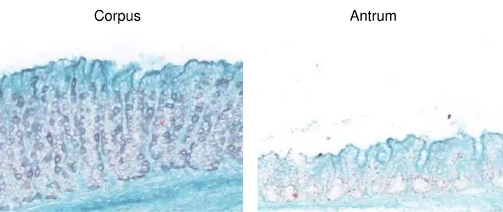
Figure 1. 1 Representation of the different anatomical parts of the human (left) and mouse (right) stomachs.
Oes -Oeshopagus; Fu –Fundus; For – Forestomach; Car – Cardia; Cor –Corpus; Ant –Antrum; Duo –
Duodenum; PS – Pyloric Sphincter;
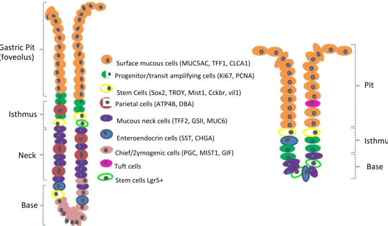
production of hormones (Scanlon and Sanders, 2015). The differences between both glands in mouse
beyond the different dimensions are the presence of different cellular lineages.
Mouse corpus/zymogenic/oxyntic gland
Mouse antral/pyloric gland
Gastric Pit
(foveolus)
Isthmus
Neck
Base
Surface mucous cells (MUC5AC, TFF1, CLCA1)
Progenitor/transit amplifying cells (Ki67, PCNA)
Stem Cells (Sox2, TROY, Mist1, Cckbr, vil1)
Parietal cells (ATP4B, DBA)
Mucous neck cells (TFF2, GSII, MUC6)
Enteroendocrin cells (SST, CHGA)
Chief/Zymogenic cells (PGC, MIST1, GIF)
Tuft cells
Stem cells Lgr5+
Pit
Isthmus
Base
Figure 1. 2 Schematic representation of corpus and antral glands with their cellular lineages. Corpus glands are larger in
lengthand fission size. In parentheses are described different molecular markers for the same lineage except to stem cells that each marker label different stem cells. The left and right bars depict the different regions of the glands
1.1.2.
Mouse cellular lineages and respective molecular markers
1.1.2.1.
Pit and neck mucous cells
Gastric glands harbour several cellular lineages with distinct functions. In homeostasis, these
cells are driven from tissue resident stem cells (Mills and Shivdasani, 2011; Leushacke et al.,
2013; Arnold et al., 2011). Adult stem cells ensure the gastric epithelial turnover within a 7-10
day rate (Barker et al., 2010a).
In the distal part of the glands, in the region called pit, are found mostly, the pit mucous cells, characterized by mucous production. Pit cells have a life span of approximately 3 days (Karam and Leblond, 1993a). The most accepted biomarker for this cellular lineage is the glycoprotein mucin5ac. There are also mucous producing cells in the neck region, these ones are distinguished by the expression of mucin6 (Schlaermann et al., 2016). Mucous is characterized by the presence of several mucins, the periodate-Schiff (PAS) reaction can be used to stain these mucins and therefore mark pit and neck mucous cells (Thornton et al., 1989).
1.1.2.2.
Proliferative cells
The isthmus is characterized by the abundance of proliferative cells. From this region occurs a bidirectional migration of progenitor cells towards the pit or the base of the glands (Ramsey et al., 2007; Lee et al., 1982). These cells possess a turnover time of 3 days or less (Karam and Leblond, 1993b). A common molecular marker used in proliferative cells identification is the KI67 protein, which is associated with ribosomal RNA transcription and cellular proliferation (Bullwinkel et al., 2006).
1.1.2.3.
Neuroendocrine cells
Endocrine cells are found along the gastrointestinal tract, which are responsible by regulation of other cells
function, through hormones and peptides secretion (Rehfeld, 1998). Endocrine cells are located mostly in
the gland base, and exhibit a turn-over time of approximately 3 months (Karam and Leblond, 1993c). At
least six different types of endocrine cells were identified in the gastric mucosa. The following endocrine
cells are the well-described, enterochromatin-like cells (ECL cells) that secrete histamine, Delta cells also
called D-cells distinct by somatostatin secretion, gastrin-secreting cells or G-cells responsible by the gastrin
hormone production, A-cells that synthesize glucagon, enterochromaffin cells which secret serotonin and
X-cells also known as A-like X-cells which can be distinguished from the others by Ghrelin production (Grube
and Forssmann, 1979; Cockburn et al., 2013; Enrico et al., 1975). Since chromograninA expression is
1.1.2.4.
Parietal cells
Parietal cells are found dispersed through the neck and base regions of corpus glands. These cells are responsible for hydrochloric acid production and secretion, through a coordinate action of endocrine, paracrine and neuroendocrine pathways (Mills and Shivdasani, 2011; Rehfeld, 1998).
These cells are the ones with the larger dimensions found in the stomach and have a low turnover rate of approximately 54 days. They are originated in the isthmus, and migrate bidirectional inward to the neck and eventually to the base, or outward to the pit (Karam, 1993). Parietal cells exhibit an unique intracellular structure denominated Canaliculus, that has the function to increase the cell superficial area, assisting in the secretions. In their membrane there is a characteristic enzyme denominated
hydrogen potassium ATPase (H+/K+ ATPase), responsible for the production of H+ ions concentration
characteristically of gastric juice, approximately 3 million times superior than the concentration found in blood (Martinsen et al., 2005). Due to this unusual characteristic, H+/K+ ATPase is the most used molecular marker for parietal cells identification.
1.1.2.5.
Chief or zymogenic cells
These cells occupy the corpus glands base, their main function is the secretion of proteolytic enzymes such as pepsinogen, that assist in the organic material degradation (Jun and Shinichiro, 1998). Chief cells possess the slowest turnover of all gastric cell lineages, being estimated in order of half of a year (Karam and Leblond, 1993a). These cell are the only ones derived from neck cells, passing through a pre-zymogenic stage before maturation, whereas the other lineages derive directly from isthmal cells (Karam and Leblond, 1993c). For chief cells immunostaining, there are at least two molecular markers used, the transcription factor Mist 1, associated with proteins secretion, and the Gastric Intrisic Factor 1 (GIF1) a glycoprotein necessary to B12 vitamin absorption (Lennerz et al., 2010), (Howard et al., 1996), (Alpers and Russell-jones, 2013).
1.1.2.6.
Gastric stem cells
Adult stem cells reside within organs and tissues, fueling their growth and maintaining their regeneration throughout adult life. These cells exhibit multipotency, possessing the ability to produce one or more cellular lineages. In adult homeostasis, stem cell fate decisions are controlled by extrinsic signals of their microenvironment or niche (Watt et al., 2000). These cells are located in the isthmus and base of corpus glands, or in the base of antral glands (Barker et al., 2010a).
The turnover time varies depending on the stem cell type. Adult stem cells molecular markers are not
well-characterized. The best well-characterized biomarker of adult, quiescent and homeostatic, stem cells, in
several tissues, including the antrum and the corpus, is the membrane protein Leucine-rich
rich repeat containing G protein-coupled receptor (LGR) family. Until recently it was thought to be present only in stem cells in the base of antral glands (Barker et al., 2010a),. More recently Leushhacke et al. demonstrated that it also exists in the region of the isthmus in the body (Leushacke et al., 2017). When activated by molecules of the Roof Plate-Specific Spondin (Rspondin) family, the complex LGR-Rspo inactivates an E3 ubiquitin ligase zinc and ring finger 3 (ZNRF3) and ring finger protein 43 (RNF43), which are Wnt/β-catenin pathway antagonists, activating the pathway activity (Kretzschmar and Clevers, 2017; Kazanskaya et al., 2004).
It has been proven that a specific cell type that expresses the transcription factor Sex Determining Region Y box 2 (SRY2 also known as SOX2), label a different and more rare population of cells than LGR5. These cells have the potency to originate whole monoclonal glands (Arnold et al., 2011). Mist1, is also a transcription factor, that marks a type of quiescent stem cells in the gastric corpus isthmus (Hayakawa et al., 2016). Both SOX2 and Mist1 do not label exclusively stem cells (Stange et al., 2013). Hayakwa et al., demonstrated that the cholecystokinin B receptor (Cckbr or CCK2R) protein
also identifies a type of quiescent stem cells that are able to transform in LGR5+ stem cells (Hayakawa
et al., 2014). Qiao et al. shown that a subpopulation of reserve stem cells, localized in the neck and base of antral glands, is marked by the expression of Villin (Vil1). These cells are more rare than
LGR5+ cells, divide slowly and form monoclonal glands only when mice were treated with
pro-inflammatory cytokine interferon-gamma (IFNγ), suggesting these cells do not contribute to epithelium
homeostasis (Qiao et al., 2007). More studies will be needed to definitely establish whether Villin+,
LGR5+, SOX2+, Cckbr+ and mist1+ cells are different types of stem cells that act independently to maintain the stomach homeostasis, or whether these cells are hierarchically related to each other.
Figure 1. 3 Schematic representation of stem cells differentiation in gastric cellular lineages. Stem cells divide to give
1.1.3. Molecular pathways of murine stomach embryogenesis
Although it is not known precisely how gastric development occurs, several molecular
mechanisms and signaling pathways associated with differentiation, patterning and growth of
the gastrointestinal tract have been elucidated (Lewis and Tam, 2006; Jensen et al., 2000;
Kolterud et al., 2009). There is no exact model that explains how all steps occur, but there
are small templates that put together some pieces of the puzzle.
The formation and patterning of the gut is an ancestral process, which probably dates back to
the first organisms of the animal kingdom to exist. These processes, have probably, been
controlled by genes with similar functions in the last 500 million years. In mice, the
gastrointestinal tract develops from the embryonic gut. The epithelium, is derived from
endoderm, and mesenchyme is originated through the migration and condensation of
mesoderm around endoderm (Wells and Melton, 1999; Ramalho-santos et al., 2000).
There are several well-characterized transcription factors associated with control and
regulation of mouse embryogenesis, such as Caudal type homeobox 1, 2 and 4 (CDX1,2 and
4), pancreatic and duodenal homeobox 1 (Pdx1), Fibroblast Growth Factor 4 (FGF4), hox
proteins and β
-Catenin (Wells and Melton, 1999), (Lewis and Tam, 2006).
During embryonic development, inhibition of the Wnt/β-Catenin pathway occurs, due to the expression of the transcription factor BARX Homeobox 1 (negative regulator of secreted frizzled-related protein 1 and 2) in the mesenchyme, promoting a gastric specification of the epithelium rather than intestinal (B. Kim et al., 2005), (Rattner et al., 1997), (Verzi and Shivdasani, 2008).
McCraken et al. have shown that
unlike the intestine, the Wnt/β
-Catenin and Fbroblast
Growth Factor 10 (FGF10) pathways act epistatically with the Bone Morphogenetic Protein
(BMP) pathway, but independently, to conduct morphogenesis of gut tube structures. The
retinoic acid pathway, is required for the differentiation of the gut tube into a posterior foregut
fate, namely the antrum (McCracken et al., 2014).
1.1.4. Molecular pathways of murine stomach homeostasis
After the decision to specify the endoderm in
differentiation of several cell lineages begins.
mesenchyme-epithelium interactions, and
retain development (Wells and Melton, 1999).
gastric epithelium occurs, the proliferation and These processes are dependent on time-related several pathways in common with embryonic
In the adulthood, homeostasis of the stomach is dependent on tissue-resident stem cells. Some of the signaling pathways that ensure stomach homeostasis are the Wnt/β-Catenin pathway (Radulescu et al., 2012; Barker et al., 2010a), Notch (Demitrack and Samuelson, 2016; Demitrack et al., 2015), Epidermal
Growth Factor (Fukuda and Yasugi 2005), FGF10 (Nyeng et al., 2007), gastrin (Jain and Samuelson, 2006), BMP (Bartfeld and Koo, 2017) and Hedgehog (Ramalho-santos et al., 2000; Lees et al., 2005).
In the adult corpus epithelium, Shh is secreted by the parietal cells, and regulates gastrin production by the endocrine G cells. When the Shh produced in the parietal cells is inhibited, hyperproliferation of the foveolar epithelium occurs (Brink et al., 2001), (Konstantinou et al., 2016). The BMP pathway is associated with the stimulation of stem cell differentiation, and when inhibited, hyperproliferation and
poor compartmentalization occurs (Kaestner et al., 1997). In intestine, a BMP gradient is established with low expression in the crypts and high expression in the villi, where the cellular differentiation is higher, contributing to restrict the stem cells to the crypts. This gradient is achieved due to the activity of Shh that is highly concentrated in the mesenchyme that surrounds villi (Shyer et al., 2015). In the
stomach, the expression pattern of bmp4 differs from that of the intestine, because Shh is secreted only by parietal cells in the neck region, leading to a high concentration of Bmp4 in this region, where stem cells are scarcer (Brink et al., 2001). This suggests that Shh secretion by parietal cells shapes the negative area for stem cells microenvironment, which can be established in the isthmus and base
Figure 1. 4 Distribution of cells types and niche pathways in the gastric corpus gland. On the left side are
represented theabundance of each cell lineage. The circles between the two glands represent gradients of niche pathways activity. The Shh high concentration in the neck region leads to the mesenchymal BMP expression. There is only indirect data for the proposed WNT gradients represented here (Bartfeld and Koo, 2017).
A similar phenotype is achieved when overexpression of the transforming growth factor-
α (an
EGFR ligand) (TGF-
α) is induced, leading also to hyperproliferation of the foveolar epithelium
and loss of parietal cells (Goldenring et al., 1996; Sharp et al., 1995).
Notch signaling is restricted to the isthmus, and is essential to the maintenance of the stem cell
compartment. Overactivation of Notch, causes cellular hyperproliferation, generating corpus adenomas and
antral polyps possibly due to the downstream activity of the mammalian Target of Rapamycin (mTOR)
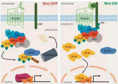
Figure 1. 5 Schematic representation of the canonical Wnt/β-Catenin pathway, which is activated by the Wnt
ligands (right)and inactivated in the absence of these (Kretzschmar and Clevers, 2017).
In the intestine, the Wnt/β
-Catenin pathway is the best characterized pathway and has a
central importance in stem cells, whereas in the stomach, its role is not fully understood. The
canonical Wnt/β
-Catenin pathway is represented in the figure below.
TROY and LGR5, two adult gastric stem cell markers, are targets of the Wnt/β-Catenin pathway (Stange et
al., 2013), (Barker et al., 2010a). When over-activated in stem cells via deletion of either glycogen synthase kinase 3 (GSK3) or Adenomatous Polyposis Coli (APC), the Wnt/β-Catenin pathway induces hyperproliferation and formation of polyps in the antrum and corpus (Radulescu et al., 2012).
1.2.
Research study models
1.2.1.
Animal models for research
Probably, due to the inexistence of a study model similar, in such a way, to the human being,
its very complicated the extrapolation of the knowledge acquired of studies, to be directly
applied in humans. Animal models are the ultimate barrier to overcome before human trials.
Approximately 90% of new drugs tested in clinical trials fail to obtain Food and Drug
Administration (FDA) approval (Gurwitz, 2001).
Animal models, although imperfect, are an irreplaceable tool in biomedical research. There are several
animal models used in scientific research, such as mouse, rat, xenopus and zebrafish. For differentstudies,
Mural et al., 2002). Despite this genomic resemblance, regarding to anatomy and physiology, are alsosimilar to the human being. They are characterized by a relative short lifespan (1 mouse year equals approximately to 30 human years). Mice reproduce relatively quickly, possessing a gestation time is around ten weeks. To enlight a role of a gene, mice are a relatively simple animal to generate transgenic or knockout models. Taking these advantages, Mus musculus was the main animal model opted to this study (Yourgenome, 2017; Spencer, 2012).
The bigger pitfalls from mice models regarding to genetic research, is the insufficient representation of the natural genome diversity, due to usage of a single strain of inbred mice (Gurwitz, 2001).
1.2.2. Two dimension,
in vitro
, mammalian cell culture models
Due to the complexity of animal models, the study at a cellular and molecular level becomes
quite difficult, requiring the use of simpler study models (Shamir and Ewald, 2014). As such,
another model largely used in several areas such as neuroscience, cardiology or cancer
research is the 2 dimension (2D) culture of mammalian cells (Goodspeed et al., 2016)
.
Briefly, this method is based on the growth of cells in vitro, under controlled biological, chemical and physical conditions, suspended or adhered (Carrel and Burrows, 1911). There are mammalian 2D cell
culture systems, that consist in the establishment of human tumor-derived or cancer cell lines, primary cell lines and embryonic stem cell lines (Sharma et al., 2010; Martin, 1981). In general, due to their lower complexity, in scientific research these culture systems are mainly used for studies of the molecular mechanisms associated with various biological processes. Some of these processes are the ability of self-renewal of stem cells, cell differentiation and the carcinogenic process (Chowdhury et al., 2010; Takahashi and Yamanaka, 2006; Pan et al., 2011).
Although most cell culture studies have been conducted on 2D systems to date, there are
several limitations inherent to this method. The standard procedure of screening compounds
starts with the 2D cell culture-based tests, followed by animal model tests, to clinical trials.
Until 2010, in cancer drug research, only about 10% of the new compounds tested,
progressed successfully for clinical trials (Hait, 2010).
1.2.3. Three dimensional,
ex vivo,
cell culture models
In vivo, cells are surrounded in the three spatial dimensions by a totally different microenvironment than that
provided by 2D cultures (Edmondson et al., 2014). Cells are in organized structural arrangements, and a specific chemical, biochemical and physical environment provided by the extracellular matrix (ECM). In
the need emerged for the development of more complex cell culture systems that would
mimic biological systems at a higher level of complexity (Shamir and Ewald, 2014). In order
to bridge the gap of information extrapolated from 2D culture systems and animal models, 3D
cell culture systems were created (Pampaloni et al., 2007; Haycock, 2010).
The concept of 3 dimensional (3D) cultures systems was first thought due to the pioneer work of Mina J. Bissell and her team. They were the first trying to elucidate how ECM influences the maintenance of tissue specificity, cellular shape and functions (Bissell et al., 1982). The discovery of different tissues interactions, and how they could modulate the genes expressions program of cells, consequently contributing to cell differentiation or stem cell maintenance was a crucial step for 3D systems development (Fuchs et al., 2004; Mazzoleni et al., 2009). It was shown that the microenvironment has a dramatic influence on the cellular behavior and 2D culture systems do not consider this important factor. Nowadays, it is known that cellular exposure to spatial restrictions established by a 3-dimensional scaffold defines how cells react to these signals from the surrounding microenvironment. Due to the niche, cells alter their gene expression resulting in the modulation of differentiation, proliferation, apoptosis, shape, movement and structure formation (J. Bin Kim et al., 2004; Li et al., 1987; Schmeichel and Bissell, 2003).
Briefly, 3D culture systems differ from 2D cultures in the recreation of an organic and complex scaffold, where cells are grown. In these cultures, cells are exposed to a more native
microenvironment, providing different stimuli and making possible a cellular behavior more similar as the one that occurs in vivo, thus resulting in distinct cellular arrangements and structures formation (Vinci et al., 2012). Normally, these models are composed by porous substrates that can support differently cellular proliferation, organization, and differentiation (J. Lee et al., 2008). It is now feasible
to perform studies, that address cell-ECM interactions, and enhanced studies about cell-cell interactions (Pampaloni et al., 2007; Li et al., 1987; Mazzoleni et al., 2009). There are currently more than 100 different biomaterials to be used in extracellular matrices depending on the study. It is
possible to use different types of matrices, namely, biological scaffolds, polymeric hard scaffolds and micropatterned surface microplates. Biological matrices composition, commonly include, but are not limited to, fibronectin, collagen, laminin, and gelatin (Ravi et al., 2014).
It is possible to make 3D culture systems without a solid scaffold, for which there are three
different models: hanging drop microplates, spheroid microplates containing Ultra-Low
Attachment (ULA) coating and microfluidic 3D cell culture.
The paradigm has been changing, and the spheroids, although essential, are more related to
the study of cancer.
Currently there are 3D culture systems that enable the maintenance of
basic characteristics of a
normal adult tissue indefinitely (Barker et al., 2010a).
1.2.4. Organoid culture history
Organoids, also called “mini organs”, are a 3D culture method that allows growing the small units of animal organs, using an artificial media and resulting in self-organized structures, which mimic the natural physiological structures. The establishment of these culture systems, is only possible due to self-organizing, self-renewal and multipotency capacities of adult stem cells (Shaker and Rubin, 2012). In 2007, the intestinal stem cell marker, LGR5 (also known as Gpr49) was identified (Barker et al., 2007). This elucidation, made possible the development of an organoid culture method, for the first
time, in 2009 (Sato et al., 2009). Murine intestinal LGR5+ stem cells, were single sorted and cultured
with a combination of their, previously described, natural and essential niche growth factors, Epidermal Growth Factor (EGF) an EGFR ligand, to promote cell proliferation (Dignass and Sturm, 2001); R-Spondin, a Wnt/β-Catenin pathway agonist (K.-A. Kim, 2005); Noggin, a BMP pathway antagonist (Haramis et al., 2004) and a 3D solid matrix, rich in laminin (because laminin, α1 and α2 is enriched at the crypt base) named matrigel (Sasaki et al., 2002; Sato et al., 2011; Hughes et al., 2010). Stem cell proliferation and expansion was observed, together with the formation of structures that contained differentiated cells, corresponding to all cell lineages that exist in the murine gut. Moreover, these cellular structures even self-organized into domains that resembled intestinal crypts. These organoids were long-lived, appear to posess unlimited expansion ability, could be split and re-seeded, frozen and thawed (Sato et al., 2009).
Figure 1. 6 Schematic representation of organoids derived from ESCs or IPSCs. ESCs or IPSCs can be conduct into all thegerm layers. Next to this specification (endoderm, mesoderm or ectoderm), cells can generate different types of organoids according the modulation of differentiation. These organoids formation faithfully mimic the in vivo embryonic development steps (Huch and Koo, 2015).
There are several organoid applications, namely study of stem cell behavior, stem cell niche
components, differentiation factors, tissue homeostasis, genetic screening, drug screening,
modeling of disease and pathogen
–
epithelia interactions, embryonic development, lineage
specification, among others (Fatehullah et al., 2016). Organoids can be grown as well, using
an air-liquid interface system (Nadauld et al., 2014).
1.2.5. Gastric organoids,
ex vivo
, 3D culture systems
Due to the identification of LGR5+ adult stem cells in pyloric antrum, the development of a long-lived,
in vitro, 3D culture system of antral gastric organoids was conducted from single sorted LGR5+ stem
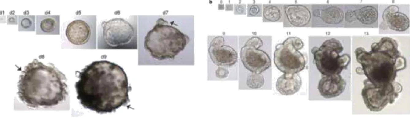
Figure 1. 7 Representative images of intestinal and antral organoids.On the left is a representative example of a unique growingantral organoid derived from a single lgr5+ stem cell. Arrows show the gland-like domain buds starting to form at day 5. Magnifications: days 1–4, 403 magnification; days 5–6, 203 magnification; days 7–8, 103 magnification; and day 9, 53 magnification. (Barker et al., 2010). On the right panel is a representative example of a unique growing intestinal organoid derived from a single lgr5-GFPhi cell. Numbers above the images are the days of growth. magnifications: days 0–4, x40; days 5–7, x20; days 8–11, x10; days 12 and 13, x4 (Sato et al. 2009).
When cultured in normal conditions, organoids expressed various gastric epithelial markers, such as Mucin 6 (MUC6) marking mucous neck cells, pepsinogen C and gastric intrinsic factor labeling chief cells but no expression of enteroendocrine or pit cell markers was observed. To overcome this limitation, the authors reduced the Wnt3a concentration in the culture media, resulting in the formation of differentiated cells, evidenced by the expression of TFF2 in progenitor neck cells
(Quante et al., 2010), scattered immature ChromograninA+ positive enteroendocrine cells and
Mucin 5AC (MUC5AC), a typical pit mucous cell marker (Barker et al., 2010).
Figure 1. 8 Western blot analysis of several molecular markers of gastric and intestinal lineages. Comparison
between stomach proteins and antral gastric organoids proteins. Molecular markers characteristics of small intestine lineages were used as control (Barker et al. 2010a).
In 2013, corpus organoids were grown for the first time, using the same conditions than antral organoids, from entire glands and isolated single chief cells that express TNF Receptor Superfamily Member 19
(TNFRS19 also known as TROY), and Mist1. Interestingly, TROY+ parietal cells did not have the ability to
form long-term organoids culture. The organoids driven from these TROY+ stem cells, had no
1993). The corpus glands were cultured as described by (Barker et al., 2010a), grown in an
insert with pores, and transferred to a well with IMSCs plated with medium, at the bottom of
the well. With this co-culture system, parietal cells were formed and maintained for several
passages. Without ISMCs co-culture Schumacher et al. organoids formed parietal cells in the
initial seeding, but their number decreased over culture time (Schumacher et al., 2015).
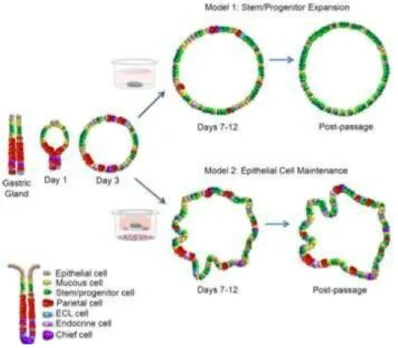
Figure 1. 9 A schematic diagram representing two distinct organoid culture methods. Model 1 (above) represents a system
whereby stem/progenitor cells are expanded, the method described by Barker et al. Model 2 (below) represents a culture system of maintained epithelial cells due to a co-culture with immortalized mesenchymal cells (Schumacher et al. 2015).
To grow human stomach driven organoids for the first time, it was necessary the addition of nicotinamide (an apoptosis inhibitor) to the culture medium (Avalos et al., 2005), to promote an initial organoid formation. It was also necessary the addition of a Transforming growth factor β
inhibitor, to increase the culture lifespan to a maximum of half a year, by inhibition of exaggerate proliferation (Zhang, 2010). Using the same culture conditions, it was possible to form human antral organoids at least for 3 months, the corpus organoids were maintained for 1 year. These organoids expressed all differentiation markers except for parietal cells marker and Enterochromaffin-like (ECL) cells (Bartfeld et al., 2015). In 2016, Schlaermann et al. made a
quasi-immortal gastric proliferative organoids culture, that was possible to be transformed in
differentiate organoids by withdrawal of Wnt3a and Rspo1 (Schlaermann et al., 2016).
Gastric organoids derived from pluripotent stem cells (PSCs), induced pluripotent stem cells (iPSCs) and
embryonic stem cells (ESCs) were also grown (McCracken et al., 2014). An interesting particularity,
maintain epithelium and mesenchyme, but have limited expansion capacities (Wells and
Spence, 2014; Spence et al., 2011).
1.3.
Genetic Regulation
1.3.1.
RNA-binding proteins
DNA is the molecule that encodes genetic predispositions related with the growth, development, functioning and reproduction of all known living organisms. The temporal and spatial regulation of DNA, allows the increase or decrease of different genes expression, in distinct tissues of the same organism. It is due to the existence of genetic regulation, which is obtained, from a single genome, a functional multicellular organism. The regulation and control of genes is achieved through several mechanisms that act at the level of transcription, RNA processing and post-translational modifications. These mechanisms are inherently associated with regulatory biomolecules, in general constituted by RNA or proteins (Lewin, 2000).
One way by which genetic expression regulation can occur, is with the usage of RNA-binding proteins (RBPs) (Jansen et al., 1995). RBPs orchestrate all aspects of RNA fate and functions since its biosynthesis to an eventual degradation, becoming essential in several biological processes such as sex determination, metabolism, neurogenesis, stem-cell proliferation, erythropoiesis and imperative in embryogenesis. Defects in their normal function can be associated with a variety of human pathologies including cancer (Kuersten and Goodwin, 2003), (Gebauer and Hentze, 2004; Wurth and Gebauer, 2015; Castello et al., 2013).
When RBPs interact with RNA, ribonucleoprotein (RNP) complexes are formed, although all RBPs bind to
RNA, they do that with different specificities and affinities depending on the RNA sequence. To RBPs network, achieve gene expression regulation through mRNA stability, cellular localization, function and translatability control, the presence of several regulatory elements in the mRNA chain, to assist and conduct regulation, are needed (Glisovic et al., 2008). These RNA cis-regulatory elements, are frequently located in the 5’ or 3’ untranslated regions, and can be recognized as binding sites for the trans-acting factors RBPs by specific domains (Preiss and Hentze, 2012). The three prime untranslated region (3’ -UTR), is the part of the RNA sequence between the stop codon and the start of the poly(A) tail, and has a great variety of regulatory functions (Moore, 2005). There are several known sequences in this region,
On the other hand, RBPs possess numerous RNA binding domains (RBDs), also known as RNA recognition motif (RRM). One RBP may have multiple RBDs, and also domains that interact with other proteins (Glisovic et al., 2008). The well-described domains are the K-Homology (KH) type I and II, RGG (Arg-Gly-Gly) box, Sm domain, DEAD/DEAH box, zinc finger (mostly C-x8-X-x5-X-x3-H type) motif, dsRNA-binding domain, the Arginine-Rich Motif (ARM), cold-shock domain, Pumilio/FBF (PUF or Pum-HD) and the Piwi/Argonaute/ Zwille (PAZ) domain (Glisovic et al., 2008; Burd and Dreyfuss, 1994).
1.3.2. Mex-3 RNA binding family member A;
MEX3A RNA binding protein, is codified by
Mex
3a gene, in humans, is located in
chromosome 1q22 and possesses a size of 9986pb. The citoplasmatic and nuclear protein
MEX3A, has a molecular weight of 54KDa with a 520 amino acids size chain. MEX3A has
three RNA interaction domains, being two of these, KH sequences, and the other a
carboxy-terminal zfRING finger domain (C3HC4 type) which binds two zinc cations.
In humans, this gene belongs to a conserved across evolution family, denominated MEX, that its composed by four homologues genes, ME3XA, B, C and D (Buchet-Poyau et al., 2007). MEX genes orthologues, are presente in several other species, as like Caenorhabditis elegans and Mus musculus
(Draper et al., 1996). Biological relevance of MEX family is still unknown, and is starting to be explored. In C. elegans, MEX-3 act as a translational repressor that regulates blastomere identity during an early embryogenesis stage (Draper et al., 1996). In adult worm, MEX-3 has been associated with germ line totipotency maintenance (Ciosk and Priess, 2006). MEX3B, was associated with innate immune response and fertility in mice (Yang et al., 2016). MEX3C, was related with the decay of HLA-A allotypes, in human embryonic kidney 293 cells (Cano et al., 2012). In human gastric cell lines, MEX3A expression inhibition, was related with cells proliferation capacity reduction (Jiang et al., 2012). Pereira et al., have proved that MEX3A has direct implications in epithelial intestinal cells differentiation, by CDX2 expression decrease, putatively as a translation repressor. CDX2 plays a crucial role in intestinal cell fate specification, both during normal development and in tumorigenic processes involving intestinal reprogramming, associating MEX3A with stemness maintenance (Pereira et al., 2013). Recently, Barriga et al. unveiled that MEX3A is co-expressed with LGR5 protein,
in a subpopulation of slow proliferation LGR5+ intestinal stem cells (Barriga et al., 2017). Although
1.4. Objetive
The main objective was the characterization of the protein Mex3a role in mice stomach using three
study models, a mex3a knockout mice (mex3a-/-), gastric cancer cell lines and organoids culture.
The animal model allows a direct visualization and characterization of the effects caused by
the lack of the mex3a protein in the gastric epithelium.
Gastric organoid culture was first established and optimized, to be possible the
characterization of growth dynamics and differentiation of gastric organoids driven from
wild-type and knockout mice. This method supplements the animal model by enabling, in an agile
approach, several tests that could influence the gastric units.
2. Methods and Materials
2.1. Mice
C57BL/6 mice of both sexes, with ages ranging from 14-70 days, were used. Lgr5-EGFP-IRES-CreERT2 (Lgr5) (Barker et al, 2007), Mex3a knockout (Mex3a-/-) (Jackson Labs #010531).
2.2. Isolation of gastric glands
C57BL/6 mice with 3-10 weeks were sacrificed according to ethical procedures of i3S Animal Facility, using isoflurane followed by cervical dislocation. Mice abdomen was washed with 70% EtOH, an incision into the abdominal cavity just to the external genitalia was made and extended to the rib cage
by cutting the abdominal musculature on both sides. The stomach was gently pulled out of the abdominal cavity, separated from the intestinal tract by cutting at the level of the duodenum and at the level of the esophagus. The stomach was opened through the greater curvature and the contents were
removed by vigorous washing with ice cold PBS. The corpus and antrum regions were separated under a magnifying lamp, cut into 5mm fragments and placed in 15mL falcon tube with 10 mL PBS on ice. The fragments were rinsed with ice-cold PBS 3x with vigorous agitation. Then, the fragments were
incubated for 3h in 10mL EDTA 10mM with agitation at 4°C to detach the epithelial layer from the mesenchymal one. From this step onward the procedure was carried out under a laminar flow chamber. The supernatant was discarded and 10mL fresh PBS added. Tissue chunks were pipetted
approximately 40 times up and down with a FBS-coated Pasteur pipette to disrupt the tissue and separate the antral/corpus glands from the basal layer. The gland suspension was transferred to a new 50mL falcon tube using a 70µm strainer to remove large chunks of tissue. The remaining glands
were resuspended in 5mL fresh PBS and the procedure was repeated twice more. The filtered solution (20mL) containing the glands was centrifuged at 150g, 4°C for 10 min. The majority of the supernatant was removed, leaving approximately 1mL to resuspend the pellet, then the solution was transferred to an eppendorf and centrifuged again at 150g, 4°C for 10 min. The supernatant was fully discarded whereas the pellet containing the gastric glands was further used to establish the organoids.
2.3. Organoid culture
humidified atmosphere for 15-30min. After polymerization has been achieved, 500µL of gastric corpus and antrum organoid medium was added to each well and refreshed every 2 days. Antrum organoid medium was composed of Advanced DMEM/F12 (Thermo Fisher) supplemented with 50% Wnt3a conditioned medium (ATCC®, CRL-2647™), 10% Rspondin-1 conditioned medium (kind gift from Calvin Kuo), 10% Noggin conditioned medium (kind gift from Gijs van den Brink), 10% Fetal Bovine Serum (Biowest), 10mM Hepes (Thermo Fisher), 1X glutamax (Thermo Fisher), 10µM Y-27632
(Sigma), 1mM N-acetilcystein (Sigma), 0.02% (v/v) primocinTM (InvivoGen). Corpus organoid medium
was similar to the previous one with the addition of 10nM gastrin (Sigma). Organoid plate passage was performed every 7-8 days (depending on the rate of growth). Organoid passage was carried out by medium aspiration, followed by the addition of 1mL cold PBS to each well. Matrix disruption and detachment was achieved by pipetting up and down the PBS, approximately 20x times. The resulting homogeneous suspension was transferred to a 15mL conical tube (Sarstedt). A centrifugation was made for 10min at 4°C and 800g. Supernatant was discarded and 300µL cold PBS added. Organoid mechanical disruption was achieved by pipetting up and down with a micropipette (P200) until visual disappearance of big pieces, approximately 40x times. The suspension was washed with 5mL PBS and centrifuged for 10min at 4°C and 800g. The supernatant was removed leaving 1mL to resuspend the pellet. The suspension was transferred to an eppendorf tube and centrifuged for 10min at 4°C and 800g. At this point the supernatant was fully removed and the pellet was resuspended in 0.02% (v/v) primocin cultrex solution which allowed a dilution of the initial material from 2 wells to 3. The disrupted organoid suspension was plated in a 24 well plate and incubated at 37°C with 5% (v/v) CO2 and 99%
humidified atmosphere for 15-30min and cultured as previously described.
2.4. Gastric cell lines
Gastric cancer cell lines AGS (ATCC®, CRL-1739™), MKN45 (Riken, RCB1001), MKN28 (Riken,
RCB1002), SNU-638 (Korean Cell line bank,) and NCI-N87 (ATCC, CRL-5822™) were grown in Roswell Park Memorial Institute (RPMI) 1640, GlutaMAX™ Supplement, HEPES (Thermo Fisher Scientific) medium supplemented with 10% (v/v) heat inactivated fetal bovine serum (Biowest) and 100 units/mL of penicillin,
100µg/mL of streptomycin, or 1% (v/v) Penicillin/Streptomycin (Thermo Fisher Scientific). Cells were
maintained in an incubator at 37°C with 5% (v/v) CO2 and 99% humidified atmosphere in 75cm2 culture
flasks (Sarstedt). For cell passage, the medium was aspirated and the cells were washed with 5mL of pre
heated PBS. Three mL trypsin (Thermo Fisher Scientific) were added and flasks were incubated at 37°C for 5-10min. To confirm that cells were detached from the flask surface, the flasks were observed in an inverted
discarded and the cells were resuspended in culture medium and cultured in new T75 flasks. This process was repeated every 3-5 days and depending on the cellular density, different dilutions were applied.
2.5. Preparation of conditioned media
For Wnt3a conditioned medium production, mouse transgenic fibroblasts cells (ATCC®,
CRL-2647™), were cultured in simple Dulbecco's Modified Eagle's Medium glutamine supplemented
(Thermo Fisher Scientific) on a T75 flask. The cells were grown for 2 days, then passed to two T75 flasks one with simple Dulbecco's Modified Eagle's Medium (DMEM) to maintain the cellular line in culture, and another with Advanced DMEM F12 (Thermo Fisher Scientific). The one with advanced DMEM F12, extracellular medium was collected and substituted at the end of 4 days and cells were grown for more 3 days. In the final of the seventh day, the medium was collected, mixed with the fourth day medium and filtered using a PES Syringe filter with 220nm (Frilabo) pore size, becoming ready to use. The remaining cells were discarded.
2.6. Histology and histochemistry
After collection, mouse stomachs were opened and fixed at least 24h with 10% (v/v) neutral
buffered formalin (PanReac) at RT, and processed for 12h in a Microm STP-120 spin tissue
processor (Thermo Fisher, USA) to achieve paraffination.
For histochemistry procedures, 3µm tissue sections were obtained followed by deparaffination with 2x xylene (Fisher Scientific UK) for 10 min, and hydration through a series of ethanol solutions (100%, 100%, and 70%) for 3 min each and finally a bath in tap water for 5 min. Modified Mayer’s hematoxylin
(Thermo Scientific) staining was achieved by 1 min incubation followed by a 5 min current water washing. Eosin counterstaining was carried out by 2/3 min incubation with an alcoholic eosin solution (Thermo Scientific) preceding one wash in 95% ethanol for 3 min. For dehydration step a series of three increasing % (v/v) ethanol solutions were used (95%, 100% and 100%), 3 min each step, ending with 2x xylene solution incubation, for 5 min long. For cover slips mounting medium (Thermo Scientific) was used and air dried for 30min at least.
For periodic-acid Schiff staining, incubation with periodic acid (Thermo Scientific) was performed for
2.7. Immunohistochemistry and immunofluorescence
IHC was performed on deparaffinated and rehydrated 3µm tissue sections, using two
different detection systems, Avidin-Biotin complex (ABC) and peroxidase polymer systems.
Antigen retrieval was carried out by heating slides in an IHC-Tek Epitope Retrieval Steamer Set (±100°C) for 40 min either in a modified citrate buffer pH 6(Thermo Scientific) or Tris/EDTA buffer pH 8, (Thermo Scientific). After that, the slides were cooled down for 20min at RT, and washed 1x in
Tris-buffered saline (GRISP) with 0.05% (v/v) TWEEN® 20 (Sigma) for 5min. Next endogeneous peroxidase inhibition was performed by incubation in a 3% (v/v) hydrogen peroxide solution (EMsure), for 10min at room temperature (RT), and slides were washed twice in 0.05% (v/v) Tris-buffered saline Tween (TBS-T). For blocking step, unnecessary for peroxidase polymer system, incubation for 30min at RT was made with normal serum of the same animal species as the secondary antibodies were produced (1:5, DAKO). Approximately 100µL of primary antibody solutions per slide were incubated overnight at 4°C. Every dilution was made in antibody diluent OP Quanto (Thermo Scientific).
The ABC system was used with the following primary antibodies: mouse monoclonal anti-ATPase (1:500, Santa Cruz Biotechnology, sc-374094), mouse monoclonal anti-ChgrA (1:50, Santa Cruz Biotechnology, sc-393941), rabbit monoclonal anti-Ki67 (1:1000, Abcam, ab16667). The biotinylated secondary antibodies were rabbit polyclonal anti-mouse (1:100, DAKO, E0354) and swine polyclonal anti-rabbit (1:100, DAKO, E0353). Before secondary antibody incubation, a solution with avidin (1:100,
Vector Laboratories) and biotin (1:100, Vector Laboratories) was prepared at least 30min before usage. Secondary antibody incubation was carried out for 30min at RT and next the slides were washed 2x times in 0.05% (v/v) TBS-T. For signal amplification, an incubation with the complex solution of Avidin/Biotin, was carried out for 30min at RT followed by 2x washes in 0.05% (v/v) TBS-T.
The peroxidase polymer system was used with the primary antibody rabbit monoclonal
anti-Sox2 (1:50, cell marque, 371R-16). Secondary antibody solution with polymer (DAKO,
Denmark) was added to the slides, just enough to cover the tissue, and incubated for 30min
at RT. Revelation step of both systems was performed by adding diaminobenzidine (1:50,
DAKO) to slides for 5-10 min, following washing for 5 min in tap water.
After these procedures, slides were counterstained with hematoxylin and mounted with
mounting medium.
Immunofluorescence was performed with the mouse monoclonal antibody anti-β-catenin (1:100, Santa Cruz
Biotechnology, sc-7963). The procedure was similar to immunohistochemistry until primary antibody
incubation after which the technique was carried out in the dark. The secondary antibody used was a goat
in 0.05% TBS-T. Cover slips were mounted using VECTASHIELD Mounting Medium
containing DAPI (Vector Laboratories) for nuclear staining. The slides were isolated with
varnish. Tissues immunostainings were repeated on at least four different animal cases.
Only representative immunostainings were included in the manuscript.
2.8. In situ hybridization
All in situ hybridizations were carried out using RNAscope® 2.5 kit (Advanced Cell Diagnostics) and the protocol was according to the manufacturer instructions. Hybridizations were performed in 5µm tissue sections fixed with 4% (v/v) paraformaldehyde in Superfrost™ Plus (Thermo Fisher) slides.
Probes were anti-Mex3a and anti-Olfm4 mRNA (Advanced Cell Diagnostics). All assay
reagents belong to the RNAscope® 2.5 kit.
2.9. Protein Extraction
Protein extraction was performed in cultured cells, using a lysis solution (90% (v/v) NP-40 lysis buffer, 1% (v/v) 100mM Phenylmethanesulfonyl fluoride (Sigma, 174.2g/mol), 4% (v/v) 25X cOmpleteTM
(ROCHE), 4% (v/v) 500mM NaF (Sigma) and 1% 100mM Na
2VO
4(Sigma)). On ice, cells
pellet were resuspended in a proportion of 1mL of lysis solution per 10
8cells and incubated
for 20min, shaking the tubes every 5min. Then digested cells were centrifuged at 17000g for
15min at 4°C, followed by supernatant recoil. Protein quantification was performed using the
Pierce
TMBCA Protein Assay Kit following the manufacturer’s instructions.
2.10.
Western Blotting
To protein sample preparation, 30µg of extracted protein was transferred to an eppendorf tube, added dH20 to fulfill 12µl of volume. After that, 4µl of loading solution (90% (v/v) 4x Laemmli solution, 5% (v/v)
bromophenol blue loading buffer (Sigma, 669.96g/mol) and 5% (v/v) 2-marcaptoethanol (Sigma)) was added and incubated for 5min at 95°C. Next 0.75mm thick acrylamide gel was loaded with 16µl of
protein sample per well, along with Precision Plus ProteinTM Dual Color standards (Bio-Rad) molecular