DECLARAÇÃO Nome: Ana Rita Soares Pereira
Endereço Eletrónico: [email protected] Telemóvel: 934519087
Número do cartão de cidadão: 14819793
Título da Dissertação: Neural basis of pantomime and intransitive gestures: an fMRI study Orientação: Professora Doutora Adriana Sampaio, Professora Doutora Angela Bartolo Ano de Conclusão: 2018
Designação do Mestrado: Mestrado Integrado em Psicologia
É AUTORIZADA A REPRODUÇÃO INTEGRAL DESTA DISSERTAÇÃO APENAS PARA EFEITOS DE INVESTIGAÇÃO, MEDIANTE DECLARAÇÃO ESCRITA DO INTERESSADO, QUE A TAL SE
COMPROMETE;
Universidade do Minho, de junho de 2018
Table of Contents Acknowledgements ... iv Resumo ... v Abstract ... vi Introduction ... 7 Method ... 12 Participants ... 12 Cognitive assessment ... 12 Gestures’ assessment ... 13 fMRI experiment ... 14 Stimuli ... 14 fMRI task... 14
fMRI data acquisition ... 15
Procedure ... 15
fMRI Analysis ... 16
Results ... 16
Cognitive results ... 16
Pantomime Gestures and Intranstive Gestures ... 16
Pantomime Gestures and Intransitive Gestures ... 18
Meaningful Gestures and Meaningless Gestures ... 18
Discussion ... 20
References ... 25
List of tables Table 1. Pantomimes and intransitive gestures used in the gestures’ assessment ... 13
Table 2. Activations for Pantomime Gestures > Meaningless Gestures ... 16
Table 3. Activations for Intransitive Gestures > Meaningless Gestures ... 17
Table 4. Activations for Meaningless Gestures > Meaningful Gestures ... 18
List of Figures
Figure 1. Model of limb praxis ... 8 Figure 2. Cognitive model of limb praxis ... 8 Figure 3. Examples for intransitive gestures and pantomimes for the gestures’ assessment.. ... 13 Figure 4. Procedure for the experimental task ... 15 Figure 5. Axial and coronal views of the activation of the right medial temporal gyrus during the recognition of pantomimes contrasted with meaningless gestures. ... 17 Figure 6. Axial and coronal views of the activation of the left superior frontal gyrus during the recognition of instransitive gestures contrasted with meaningless gestures. ... 17 Figure 7. Axial and coronal views of the activation of the bilateral middle occipital gyrus during the recognition of meaningless gestures contrasted with meaningful gestures ... 19
iv Acknowledgements
To Professor Adriana Sampaio, for all the support over the years and for always challenge and encourage me to go further in my learning. It would not have been possible without all the knowledge you provided me. To Professor Angela Bartolo, for teaching me all I know about gestures and for all the dedication and constant availability. To both, for making me grow up as a psychologist and researcher. To Professor Sónia Sousa, for all the time and patience in analysing the data, I learned a lot from you. To Sara Cruz and Alberto Gonzalez for helping with the pilot study when I was in Lille.
To my family. To my mother, for all the support and efforts so that I would always be happy and achieve my goals. To my father, for being my hero and always an inspiration to me. To both, for making all of this possible and make me the person I am today.
To my second family, my friends. To Adriana, Eduardo, Rui, Luísa and Cátia, my “Meliantes”, for five years of friendship, support and loyalty. It was so much easier and funny with you by my side. And a special acknowledgment to my dear friend Eduardo, for never letting me give up on my dreams. To Patrícia, Raquel, Sofia, Ivone and Silvana, my “squad”, for always being there.
To Natália, Catarina, Rita and Patrícia, my “Lilloises”, for always sharing my madness and for all the adventures, making all the moments unforgettable.
To Rui, for the unconditional friendship and for never letting me down.
To Pedro, for all the incentives and motivation. You are my “safe harbor” and I cannot thank you enough for being always by my side. And for being rude, of course.
v Bases Neuronais de Gestos Mímicos e Gestos Intransitivos: Um Estudo de Ressonância Magnética
Funcional Resumo
Estudos anteriores de neuroimagem encontraram resultados inconsistentes relativamente às redes cerebrais recrutadas para o processamento de gestos mímicos e intransitivos. Assim, comparamos as ativações cerebrais envolvidas no reconhecimento de gestos mímicos, gestos intransitivos e gestos sem sentido, usando um evento relacionado de ressonância magnética funcional, em 12 participantes saudáveis. Os resultados mostraram que os gestos mímicos, quando comparados com gestos sem sentido, estão associados a um aumento da atividade BOLD na circunvolução temporal medial direita, cuneus esquerdo e cerebelo, e no precuneus esquerdo quando comparado com os gestos intransitivos. Para os gestos intransitivos, quando comparados com gestos sem sentido, encontramos ativação na circunvolução frontal superior esquerda, na circunvolução precentral esquerda, na circunvolução temporal média esquerda, na circunvolução supramarginal e na insula direita, e na circunvolução frontal medial esquerda quando comparados com os gestos mímicos. Estes resultados sugerem que gestos mímicos recrutam áreas cerebrais ligadas à informação semântica, dar sentido ao objeto e armazenar a forma apropriada da sua utilização, representação motora do uso do objeto e orientação atencional, enquanto gestos intransitivos estão associados a ativações em áreas relacionada com cognição social. Gestos sem sentidos parecem recrutar áreas ligadas à perceção de movimento.
Palavras-chave: gestos, gestos mímicos, gestos intransitivos, gestos sem sentido, ressonância magnética funcional
vi Neural Basis of Pantomime and Intransitive Gestures: an fMRI Study
Abstract
Previous neuroimaging studies have found mixed and inconsistent results regarding dissociative brain networks recruited for processing pantomimes and intransitive gestures. We compared brain activation involved in the recognition of pantomime (e.g., “brushing teeth”), intransitive (e.g., “waving goodbye”) and meaningless gestures in 12 healthy subjects by using event-related fMRI. Overall, we found that recognition of pantomimes when compared to meaningless gestures was associated with an increased BOLD-activity of the right middle temporal gyrus, left cuneus and left uvula, and of the left precuneus when compared with intransitive gestures. The brain activations for intransitive gestures when
compared to meaningless gestures included the left superior frontal gyrus, the left precentral gyrus, the left middle temporal gyrus, the left supramarginal gyrus and the right insula, and the left medial frontal gyrus when compared with pantomimes. These findings point out that pantomimes recruit brain areas linked to semantic information, giving a meaning and storing the appropriate way of using an object, motor representation of the tool use and attentional orientation, whereas intransitive gestures were associated with activations in areas linked to social cognition. Finally, the meaningless gestures recruit brain-areas linked to motion perception.
RUNNING HEAD: NEURAL BASIS OF PANTOMIME AND INTRANSITIVE GESTURES 7 Neural basis of pantomime and intransitive gestures: an fMRI study
Communication is a central part in the human life. Most of the time, the human social interaction includes the co-occurrence of verbal (e.g., tone of voice) and non-verbal cues (e.g., facial expression, posture or gesture; Mehrabian & Wiener, 1967). Non-verbal communication, used by both humans and animals, has a significant role in the regulation of interpersonal relationships. In humans, non-verbal signals are important for verbal communication (e.g., to obtain feedback on what is being said) (Argyle, 1969). Furthermore, non-verbal cues, such as gestures or only facial expressions, have been described as having more impact and being more effective than verbal cues in communicating interpersonal attitudes (Argyle, Alkema, & Gilmour, 1971; Argyle, Salter, Nicholson, Williams, & Burgess, 1970), i.e., subjects tend to use more non-verbal cues to make their judgements about what they are hearing.
An important non-verbal communication is derived from the use of gestures, that can be
voluntary/intentional or involuntary (Bartolo & Stieglitz Ham, 2016). In the literature, the gestures have been mainly divided in two categories as meaningful and meaningless gestures (Bartolo & Stieglitz Ham, 2016). The meaningless gestures are those that do not convey any meaning, that is, arbitrary gestures with no semantic content (e.g., “put the fist under the chin”). The meaningful gestures have been distinguished into object-related, transitive gestures and pantomimes, and non object-related, i.e., intransitive gestures. Transitive gestures refer to the actual object manipulation (e.g., “demonstrating the use of a toothbrush”), while pantomimes are gestures that describe the object use (e.g., the mime of the use of a toothbrush). On the other hand, intransitive gestures are those that present
communicative features, and this category can be expressive or symbolic, but not oriented towards objects (e.g., “waving goodbye”), and may vary across social cultures (Bartolo & Stieglitz Ham, 2016; Stieglitz Ham, Bartolo, Corley, Swanson, & Rajendran, 2010). Finally, all gestures can be performed near and far from the body (Bartolo & Stieglitz Ham, 2016; Goldenberg & Strauss, 2002).
Several studies have been unravelling the neural structures involved in gestures processing and production, using both brain lesion and neuroimaging studies. Over the years, the models of praxis processing have been developed to investigate difficulties in producing gestures in left-brain damaged patients (e.g., limb apraxia; Bartolo & Stieglitz Ham, 2016). The first cognitive model, based on studies and reports of apraxic subjects, was published by Gonzalez Rothi, Ochipa and Heilman (1991, 1997; see Figure 1) which proposed a distinct semantic route for meaningful gestures and a nonsemantic route for meaningless gestures. However, some authors highlighted the existence of relevant confusions within the model between cognitive and anatomical categories.
NEURAL BASIS OF PANTOMIME AND INTRANSITIVE GESTURES 8
Figure 1. Model of limb praxis (Gonzalez Rothi, Ochipa, & Heilman, 1991). Adapted from “A Cognitive Neuropsychological Model of Limb Praxis” by L. Gonzalez Rothi, C. Ochipa, & K. Heilman, 1991, Cognitive Neuropsychology, 8:6, p.457.
In 2000, Cubelli, Marchetti, Boscolo and Sala proposed a modified version of the Gonzalez and Rothi’s model that was entirely based on cognitive concepts (see Figure 2).
Figure 2. Cognitive model of limb praxis (modified from the model of limb praxis from Gonzalez Rothi, Ochipa and Heilman, 1991, 2000). Adapted from “Cognition in Action: Testing a Model of Limb Apraxia” by R. Cubelli, C. Marchetti, G. Boscolo, & S. Sala, 2000, Brain and Cognition, 44, p.147.
NEURAL BASIS OF PANTOMIME AND INTRANSITIVE GESTURES 9 For decades, the pantomime and intransitive gestures were considered as being processed and relying on similar cognitive mechanisms (Rogers, Bennetto, McEvoy, & Pennington, 1996; Smith & Bryson, 2007). However, recent studies have suggested different cognitive mechanisms for both type of gestures, i.e., pantomime and intransitive (Bartolo, Cubelli, DellaSala, & Drei, 2003; Stamenova, Roy, & Black, 2010; Stieglitz Ham et al., 2010). In particular, a case report of a left-brain damaged patient that consistently showed deficits in producing pantomimes in different modalities of
presentation in the absence of any other deficits in the production of the other meaningful gestures was an example illustrating different cognitive mechanisms for processing both types of gestures (Bartolo et al., 2003). A deeper analysis of this dissociation led in some cases to assume that the production of pantomimes was associated to control strategies, as working memory (Bartolo et al., 2003) or motor imagery disturbances (Buxbaum, Kyle, & Menon, 2005). In another study, the opposite profile was shown by an individual with autism spectrum disorder, who showed preserved abilities to perform pantomimes and deficits in the execution of intransitive gestures (Stieglitz Ham et al., 2010). The authors also found that this individual had an intact cognitive profile but difficulties in social-communicative skills, suggesting that the social cognitive deficits might be associated with the observed difficulties in the production of intransitive gestures (Stieglitz Ham et al., 2010). A similar profile was also reported by Stamenova and colleagues (2010) in 4 right brain damage patients among the 80 stroke tested (42 with left-brain damage and 38 right-brain damage patients).
Neuroimaging studies reported inconsistent findings regarding the neural structures involved in the processing of intransitive and pantomime gestures (Króliczak & Frey, 2009). Króliczak and Frey (2009) asked participants to see a video of a gesture, that could be intransitive or pantomime, and to mentally plan that gesture. Subsequently, they were asked to execute the gesture. Results showed activations in similar brain regions during the planning phase of both pantomimes and intransitive gestures. Specifically, a higher BOLD-response of left hemisphere, that included the caudal ventral premotor cortex (cPMV), the dorsal premotor cortex (PMd), the intraparietal sulcus (IPS), the supramarginal gyrus (SMG), the superior parietal gyrus (SPG), the caudal middle temporal gyrus (cMTG) and the rostral middle frontal gyrus (rMFG) for all the gestures during the planning phase, whereas the IPS, SMG, SPG, and the PMd cortices were more activated for pantomimes than for intransitive gestures. Finally, during the execution phase, a higher activation in the contralateral
sensory-motor regions, namely the postcentral gyrus, IPL and SPG was found for pantomimes, whereas a small activation in the left posterior cingulate gyrus (PCC) was observed for intransitive gestures. The involvement of the PM was also found by Bohlhalter, Hattori, Wheaton, Fridman and Shamim (2009),
NEURAL BASIS OF PANTOMIME AND INTRANSITIVE GESTURES 10 in which they asked participants to plan and imitate intransitive and pantomime gestures. For both types of gestures and in the planning phase, results showed significant activations in the posterior parietal cortex (PPC) association areas, i.e., mainly in the inferior and superior parietal lobes, including the left precuneus, and in the premotor cortex (PMC), i.e., mainly in the left inferior and middle frontal gyrus. Furthermore, strong BOLD signals were also found in the anterior cingulate cortex,
supplementary motor area (SMA), cerebellum bilaterally and posterior temporal regions. For the execution phase, there was a widespread bilateral activation of the PPC and PMC, when compared with the planning phase. The results also showed that, in the planning phase, the intransitive gestures were more left lateralized than pantomimes, but no significant differences between pantomimes and
intransitive gestures were observed during the execution phase. The authors concluded that planning both pantomime and intransitive gestures recruit mainly the left hemisphere. This left-hemisphere lateralization found for both types of gestures was consistent with other studies. In another fMRI study, carried out by Lotze et al. (2006), asked participants to watch three types of gestures, namely isolated hand movements (e.g., “using a key”) – pantomimes –, body-referred movements (e.g., “brushing teeth”) – pantomimes and expressive gestures (with emotionally neutral, e.g., “thinking deeply”; negatively, e.g., “threatening with the index finger”; and positively, e.g., “waving friendly”) – and intransitive gestures. Although the results observed for isolated hand movements when compared with body-referred movements were not significant, an activations of the bilateral posterior superior temporal sulcus (STS), adjacent left posterior superior temporal gyrus (STG), left angular gyrus, left
supramarginal gyrus and the left BA 45 (pars triangularis, part of the inferior frontal gyrus and part of Broca's area) were found for the reversed contrast (body-referred movements> hand movements). The brain regions activated for expressive gestures included the bilateral STS and STG, bilateral temporal pole, medial PFC, left supramarginal and angular gyrus, bilateral IFG (triangular inferior part), bilateral vlPFC, bilateral amygdala, pre-supplementary motor area (pre-SMA) and lingual gyrus, when compared with isolated hand movements. For the same gestures but compared with body-referred movements, a significant activation was observed in the left vlPFC only.
Villarreal and colleagues (2008) asked participants to perform a recognition task, including pantomime and intransitive gestures as well as meaningless gestures as a control task. The main effect of gestures categorization was associated with an activation of the right occipitotemporal cortex and the left dorsolateral prefrontal cortex (dlPFC) for the pantomime gestures when contrasted with the control videos. The same analysis was carried out for intransitive gestures and activation of the left inferior frontal gyrus (IFG), namely in the PMV, and the left dlPFC was found. When the intransitive gestures
NEURAL BASIS OF PANTOMIME AND INTRANSITIVE GESTURES 11 were contrasted with the pantomime gestures, the authors found activation in the posterior third of the left IFG (pars opercularis, PMV) and pars triangularis (BA 45) as well as in the bilateral dlPFC. The reverse analysis, yielded non-significant results.
Other studies were performed to explore the neural substrates of different type of gestures in comparison not only with meaningful gestures but also with meaningless gestures. Rumiati and colleagues (2005), using PET, investigated the brain regions involved in the observation and imitation of meaningful and meaningless gestures. During the task, there was a significant positive correlation of regional cerebral blood flow with the amount of meaningful actions was observed in the left inferior temporal gyrus for the meaningful gestures and also a significant positive correlation with the amount of meaningless gestures was found in the right parietooccipital junction for the meaningless gestures. Also in the imitation task, whereas for the meaningful gestures a significant activation in the left inferior temporal gyrus, the left parahippocampal gyrus and the left angular gyrus was observed, there was an increased activation in the bilateral superior parietal cortex, right parieto-occipital junction, right occipital–temporal junction and in the left superior temporal gyrus for the meaningless gestures condition. Furthermore, the primary sensorimotor cortex, the supplementary motor area, and the ventral premotor cortex were recruited for processing both types of gestures. In another study carried out with left brain damage patients, individuals were asked to imitate meaningless gestures (specific hand and finger postures) (Goldenberg & Karnath, 2006). Results showed that deficits in the imitation of finger postures was associated to lesions in the inferior frontal gyrus (IFG) and the insula with subcortical extension to the putamen and caudate nucleus, whereas disturbed imitation of hand postures was associated with posterior lesions affecting the junction of middle temporal and middle occipital gyrus with the inferior parietal lobule (IPL) [i.e., the temporo-parieto-occipital (TPO) junction] with extension to the underlying temporo-parietal white matter. Thus, these authors suggested that distinct neural substrates were underlying the imitation of fingers postures, performed far from the body, and hand postures, performed toward the body. For these last gestures, a conceptual
representation of body schema is necessary (that might rely on posterior regions; Chaminade, Meltzoff, & Decety, 2005).
To our best knowledge, the results of these studies are mixed and inconsistent regarding dissociate brain networks recruited for processing intransitive and pantomimes gestures. However, as proposed by Bartolo and Stieglitz Ham (2016), several distinct approaches to study gestures
processing may have biased the results, namely the inclusion of gestures performed far from the body in most of the studies. Therefore, the aim of the present study is to explore the neural subtracts for
NEURAL BASIS OF PANTOMIME AND INTRANSITIVE GESTURES 12 processing different type of gestures (intransitive gestures, pantomime gestures and meaningless gestures) in healthy participants. Thus, we expect to find an increased activation of brain areas associated with object interaction, as the parietal and frontal areas for pantomime gestures, whereas for intransitive gestures, we hypothesize an increased activation of social cognition brain related areas, including the frontal and temporal regions. We also expect a higher activation on the left hemisphere for the both type of gestures, more pronounced in the intransitive gestures than in pantomimes. For that, we assessed with fMRI a group of 12 healthy subjects in a recognition task.
Method Participants
Twelve healthy volunteers (6 males), 1 left-handed (-60% in the Edinburgh Handedness
Inventory; Oldfield, 1971), without history of neurological or psychiatric disorder, and Portuguese native speakers participated in a functional magnetic resonance imaging (fMRI) study. Ages were between 20 and 31 years old (Mage = 23,05; SD = 3,59). Handedness was assessed by the Edinburgh
Handedness Inventory (Oldfield, 1971). After receiving the ethical approval from the Commission of Ethics of Minho University, participants were recruited through the credit system from School of Psychology in University of Minho.
Cognitive assessment
In order to assess social cognition we used the Theory of Mind (ToM) task (Sebastian et al., 2012). This task consisted in 30 cartoons, each with three frames that depict a story of two persons, and one final screen in which the participant had to choose between two images according to the end that they find most appropriate for the story. There were three conditions in the task, namely 10 cartoons of cognitive, 10 of affective and 10 of physical conditions. For the cognitive condition, an inference based on the intentions and beliefs was required; in the affective condition, an inference about how one story character would react to their companion’s affective state was required and finally, for the physical condition it was required an understanding of cause and effect.
Phonemic fluency skills was evaluated by the Phonemic fluency subtest from the Verbal Fluency Tests (Cavaco et al., 2013). The test has three trials of 1 minute each, in which the participant must produce orally as many words as possible beginning with specific letters (M, R and P).
Motor imagery was assessed with an adaptation of a mental chronometry paradigm (Decety & Michel, 1989). In this task, the time the participant took to write 3 sentences and the time it took to imagine writing the same sentences was recorded and compared. The participants started writing at a specific point in a paper the sentences: "Eu sou português" (I am Portuguese), “Eu sou português e
NEURAL BASIS OF PANTOMIME AND INTRANSITIVE GESTURES 13 vivo em Portugal” (I am Portuguese and I live in Portugal) and “Eu sou português e vivo em Portugal com a minha família” (I am Portuguese and I live in Portugal with my family).
Gestures’ assessment
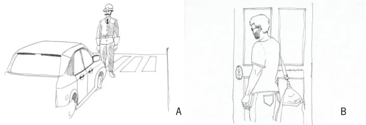
An adaptation of a gesture protocol previously described for Portuguese participants (Viana, 2015) was used in this study. First, we assessed pantomimes with a visual task, in which drawings with a person in different scenarios were presented and participants were asked to mime the utilization of the object needed in that specific context. Second, intransitive gestures were assessed with drawing pictures depicting different scenarios, and participants were asked to perform the gesture that a person in the picture would do. For each type of gesture, there were one training item and 6 test items (see Figure 3 and Table 1).
Figure 3. Examples for intransitive gestures and pantomimes for the gestures’ assessment. A) Example for the intransitive gesture task, the participant was expected to execute the gesture of “stop”. B) Example for the pantomime gestures task, the participant was expected to mime the use of keys. Table 1
Pantomimes and intransitive gestures used in the gestures’ assessment
Pantomimes Intransitive Gestures
Training item: Scissors Training item: Hands up
Spoon Stop
Gun Give a punch
Comb Waving goodbye
Hatchet Sign of tapping one’s nose
Watering can Sign of removing an annoying fly
Key Military salute
NEURAL BASIS OF PANTOMIME AND INTRANSITIVE GESTURES 14 fMRI experiment
Stimuli
In order to select the stimuli to the experimental fMRI task, we conducted a pilot study with 16 subjects (Mage = 29,38; SD = 4,83) and 83 videos. Briefly, we asked the participants to observe silent video clips of 4s duration with 4s inter-stimulus interval. Each video depicted a unique actor performing a single gesture of two categories: meaningless (near from the body and far from the body) and
meaningful (intransitive gestures near from the body, intransitive gestures far from the body, pantomimes near from the body and pantomimes far from the body). During the presentation, participants had to answer the following question: “Is this gesture meaningful?”, by pressing a button manually for “yes” or “no” on the keyboard. The participants were advisedto try to answer as fast as possible and as accurate as possible.
According to the accuracy in the responses, from the 83 videos we selected 48. Then, we used these 48 videos with the right and left hand. Thus, we selected total of 96 stimuli for the experimental task: 48 meaningless gestures, 24 near and 24 far from the body; 48 meaningful gestures, 24
intransitive gestures (12 near and 12 far from the body) and 24 pantomimes (12 near and 12 far from the body).
fMRI task
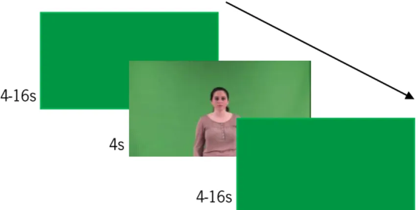
In order to assess the brain correlates of gestures recognition, an event-related functional magnetic resonance imaging (fMRI) design was used. The stimuli consisted of silent video clips of 4s duration with 4-16s inter-stimulus interval, with 191 null events (white cross on a green background), and were presented in an optimized sequence with optseq. Each video depicted a unique actor performing a single gesture belonging to two possible categories (the order of the gestures was counterbalanced across subjects): 48 meaningless gestures, (24 near and 24 far from the body); and 48 meaningful gestures (24 intransitive gestures - 12 near and 12 far from the body, and 24
pantomimes - 12 near and 12 far from the body) (See Fig. 3). Therefore, this study had seven conditions, namely (1) meaningless gestures near the body; (2) meaningless gestures far from the body; (3) intransitive gestures near the body; (4) intransitive gestures far from the body; (5) pantomimes near the body; (6) pantomimes far from the body; (7) null event.
During the presentation of the video clips, the participants were required to observe the gestures and answer to the following question: “Is this gesture meaningful?”. The participants answered by pressing a three-button keypad marked with “yes” or “no” (the order of the buttons was counterbalanced across subjects). The percentage of correct answers for pantomime gestures was 85%
NEURAL BASIS OF PANTOMIME AND INTRANSITIVE GESTURES 15 (SD = 13,81), for the intransitive gestures was 91% (SD = 11,93) and for the meaningless gestures was 94% (SD = 5,56).
Figure 4. Procedure for the experimental task fMRI data acquisition
Magnetic Resonance (MR) images were obtained in a clinical approved 3T MRI (Siemens). The T1-weighted 3D volumetric acquisition was obtained with a 3D MPRAGE (Magnetization Prepared Rapid Gradient Echo) sequence performed with the following protocol: time of repetition (TR)/ time of
inversion (TI)/ time of echo (TE) = 2700 ms/1000 ms/2,33 ms/, flip angle (FA)= 7º, field of view (FoV)=240x256 mm2, 240 sagittal slices and isotropic voxel size = 0.8x0.8x0.8mm3. MPRAGE images
were used as auxiliary for the spatial normalization of the functional imaging data. For the functional acquisition, a 2D echo planar imaging (EPI) blood-oxygen-level dependent (BOLD) sensitive sequence with the following parameters was used: TR/TE = 2000ms/29ms, FA=90º, FOV=256 mm2, voxel size=3x3x3 mm,3 41 ascending interleaved axial slices with no gap, 535 slices.
Procedure
The participants who agreed to participate in the study signed an informed consent and filled a sociodemographic and a checklist questionnaire that assessed the conditions to be fulfilled to be enrolled in the fMRI settings. After that, participants entered the scanner to carry out the fMRI study. Then, after the scanner, participants performed the cognitive and the gestures’ assessment protocols described above. This session lasted approximately 20 minutes and was carried out in a silent room at the School of Psychology in University of Minho. In order to prevent possible influences from the assessments, we decided to administer the cognitive and gestures’ assessment after the recognition fMRI task.
4-16s
4-16s 4s
NEURAL BASIS OF PANTOMIME AND INTRANSITIVE GESTURES 16 fMRI Analysis
The fMRI data were analysed using the Statistical Parametric Mapping software (SPM12, Wellcome Trust Centre for Neuroimaging, London, UK) running on MATLAB version R2015a (Mathworks Inc., Natick, Mass, USA). After pre-processing the data, a quality assessment was
performed on MATLAB version R2015a (Mathworks Inc., Natick, Mass, USA) with the software Artifact Detection Tools (ART), excluding slices with artifacts. By using statistical maps for the single-subject analyses, we performed a random-effect second-level analysis (one-sample t-test) where we contrasted the brain activations to the pantomime gestures vs. meaningless gestures, intransitive gestures vs. meaningless gestures, meaningless gestures vs. meaningful gestures and pantomime gestures vs. intransitive gestures, as well as the inverse contrasts, with a voxel level intensity threshold of p < 0.0001 (uncorrected). We used the spatial coordinates in MNI with the softwares Automated Anatomical Labeling (AAL) atlas.
Results
Results will be presented in four different sections. First cognitive assessment results will be presented, then the brain activation results for the contrasts between pantomime gestures, intransitive gestures and meaningless gestures; contrasts between pantomimes gestures and intransitive gestures and, finally, the contrast between meaningful and meaningful gestures.
Cognitive results
Scores for theory of mind task were between 22 and 30 (M = 27,67; SD = 2,01), of verbal fluency task were between 13 and 51 (M = 34,42; SD = 9,98), of motor imagery task were between 0,59 and 9,64 (M = 3,83; SD = 2,39) and for the production of gestures task were between 8 and 12 (M = 10,92; SD = 1,19).
Pantomime Gestures and Intranstive Gestures
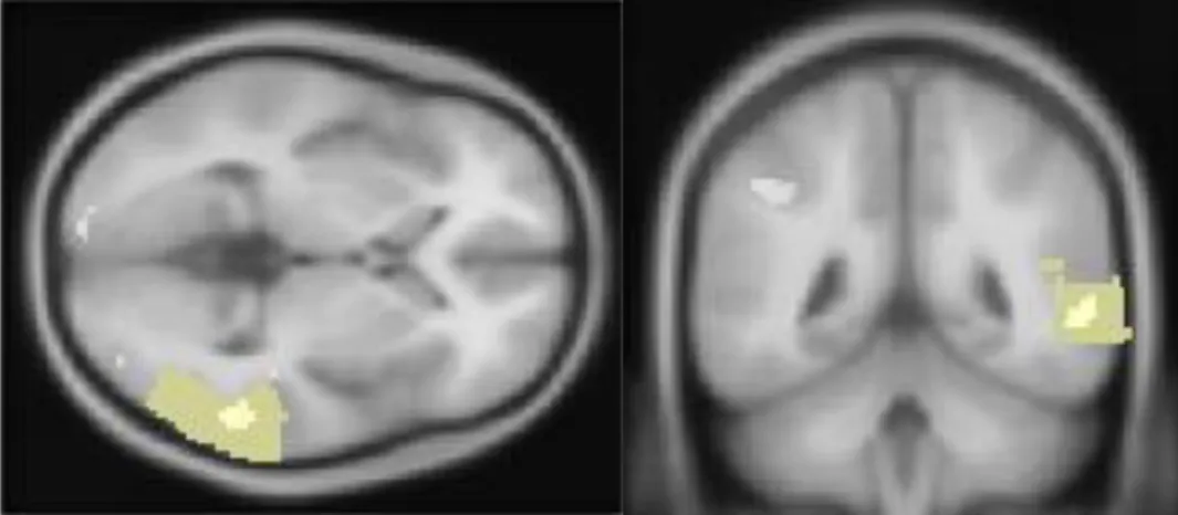
When pantomime gestures were contrasted with meaningless gestures, brain activations (p < .0001) of the right middle temporal gyrus (x = 58, y = 50, z = 4, Zvalue = 3.80), the left cuneus (x = -18, y = -98, z = 10, Zvalue = 4.51), and the left uvula (x = -16, y = -78, z = -32, Zvalue = 3.74) were observed (see Table 2 and Figure 5).
NEURAL BASIS OF PANTOMIME AND INTRANSITIVE GESTURES 17
Figure 5. Axial and coronal views of the activation of the right medial temporal gyrus during the recognition of pantomimes contrasted with meaningless gestures (p < .0001, uncorrected).
Table 2
Activations for Pantomime Gestures > Meaningless Gestures
H Anatomical Region p x y z Clusters Z-value*
R Middle Temporal Gyrus 0.000 58 -50 4 4 3.80
L Cuneus (Occipital Lobe) 0.000 -18 -98 10 62 4.51
L Uvula (Cerebellum, Posterior Lobe) 0.000 -16 -78 -32 1 3.74
Note: *p < .0001
When intransitive gestures were contrasted with meaningless gestures, we observed significant brain activations (p < .0001) in the left superior frontal gyrus (x = -10, y = 48, z = 44, Zvalue = 3.82), the left precentral gyrus (x = -56, y = 12, z = 8, Zvalue = 3.84), the left middle temporal gyrus (x = -62, y = -40, z = -4, Zvalue = 4.31), the left supramarginal gyrus (x = -60, y = -46, z = 36, Zvalue = 4.83), the left lingual gyrus (x = -2, y = -84, z = -2, Zvalue = 4.57) and the right insula (x = 42, y = -24, z = -2, Zvalue = 4.13) – see Table 3 and Figure 6.
Figure 6. Axial and coronal views of the activation of the left superior frontal gyrus during the recognition of intransitive gestures contrasted with meaningless gestures (p < .0001, uncorrected).
NEURAL BASIS OF PANTOMIME AND INTRANSITIVE GESTURES 18 Table 3
Activations for Intransitive Gestures > Meaningless Gestures
H Anatomical Region p x y z Clusters Z-value*
L Superior Frontal Gyrus 0.000 -10 48 44 2 3.82
L Precentral Gyrus 0.000 -56 12 8 3 3.84
L Middle Temporal Gyrus 0.000 -62 -40 -4 9 4.31
L Supramarginal Gyrus (Parietal Lobe) 0.000 -60 -46 36 26 4.83
L Lingual Gyrus (Occipital Lobe) 0.000 -2 -84 -2 91 4.57
R Insula 0.000 42 -24 -2 7 4.13
Note: *p < .0001
Pantomime Gestures and Intransitive Gestures
The contrast between pantomime and intransitive gestures yielded significant activations (p < .0001) in the left precuneus (x = -8, y = -62, z = 62, Zvalue = 4.06). In the opposite contrast
(intransitive gestures > pantomime gestures) significant activation in the left medial frontal gyrus (x = -6, y = 48, z = 42, Zvalue = 3.82) was observed.
Meaningful Gestures and Meaningless Gestures
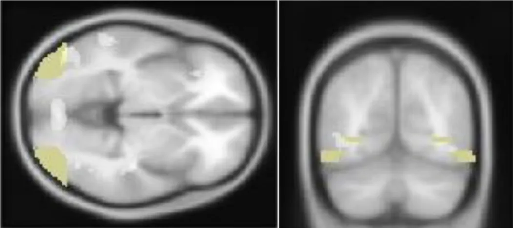
Significant activation of the right inferior parietal lobule (x = 32, y = -36, z = 44, Zvalue = 4.88) and the bilateral middle occipital gyrus (x = -50, y = -74, z = -2, Zvalue = 3.90; x = 42, y = -78, z = 12, Zvalue = 3.79) were observed when contrasting meaningless with meaningful gestures – see Table 4.
Table 4
Activations for Meaningless Gestures > Meaningful Gestures
H Anatomical Region p x y z Clusters Z-value*
R Inferior Parietal Lobule 0.000 32 -36 44 57 4.88
L Middle Occipital Gyrus 0.000 -50 -74 -2 19 3.90
R 0.000 42 -78 12 2 3.79
Note: *p < .0001
When meaningful were contrasted with meaningless gestures, significant brain activations of the left superior frontal gyrus (x = -12, y = 34, z = 52, Zvalue = 3.78), the bilateral middle frontal gyrus (x = 54, y = 30, z = 18, Zvalue = 4.22; x = -48, y = 18, z = 40, Zvalue = 3.76), the bilateral inferior frontal gyrus (x = 52, y = 42, z = 10, Zvalue = 4.42; x = -32, y = 30, z = -6, Zvalue = 4.07), the left middle temporal gyrus (x = -60, y = -38, z = -4, Zvalue = 3.89), the left inferior parietal lobule (x = -52, y = -46, z = 54, Zvalue = 4.30), the left supramarginal gyrus (x = -58, y = -54, z = 34, Zvalue = 4.10; x = -64, y
NEURAL BASIS OF PANTOMIME AND INTRANSITIVE GESTURES 19
= -50, z = 26, Zvalue = 3.92; x = -58, y = -46, z = 34, Zvalue = 3.87), the right lingual gyrus (x = 0, y = -84, z = 97, Zvalue = 4.59), the left cuneus (x = -16, y = -94, z = 6, Zvalue = 3.86), the right uvula (x = 14, y = -78, z = -34, Zvalue = 4.16) and the right insula (x = 42, y = -24, z = -4, Zvalue = 4.27) was observed (see Table 5 and Figure 7).
Figure 7. Axial and coronal views of the activation of the bilateral middle occipital gyrus during the recognition of meaningless gestures contrasted with meaningful gestures (p < .0001, uncorrected)
Table 5
Activations for Meaningful Gestures > Meaningless Gestures
H Anatomical Region p x y z Clusters Z-value*
L Superior Frontal Gyrus 0.000 -12 34 52 1 3.78
R Middle Frontal Gyrus 0.000 54 30 18 16 4.22
L Middle Frontal Gyrus 0.000 -48 18 40 1 3.76
R Inferior Frontal Gyrus 0.000 52 42 10 23 4.42
L Inferior Frontal Gyrus 0.000 -32 30 -6 5 4.07
L Middle Temporal Gyrus 0.000 -60 -38 -4 3 3.89
L Inferior Parietal Lobule 0.000 -52 -46 54 87 4.30
L Supramarginal Gyrus (Parietal Lobe) 0.000 -58 -54 34 6 4.10
0.000 -64 -50 26 9 3.92
0.000 -58 -46 34 4 3.87
R Lingual Gyrus (Occipital Lobe) 0.000 0 -84 -4 97 4.59
L Cuneus (Occipital Lobe) 0.000 -16 -94 6 5 3.86
R Uvula (Cerebellum, Posterior Lobe) 0.000 14 -78 -34 13 4.16
R Insula 0.000 42 -24 -4 12 4.27
NEURAL BASIS OF PANTOMIME AND INTRANSITIVE GESTURES 20 Discussion
Several studies have been conducted around gestures processing, the results have been inconsistent regarding dissociate or overlapping brain networks for processing these gestures and, as proposed by Bartolo and Stieglitz Ham (2016), several distinct approaches to study gestures
processing may bias the results. Therefore main aim of present study was to explore the neural substrates underlying recognition of different types of gestures in healthy participants, including intransitive gestures, pantomime gestures and meaningless gestures. Overall, we found activations in the right middle temporal gyrus, the left cuneus, and the left uvula for pantomimes (pantomimes > meaningless gestures), and in the left precuneus (pantomimes > intransitive gestures). For intransitive gestures, we found activations in the left superior frontal gyrus, the left precentral gyrus, the left middle temporal gyrus, the left supramarginal gyrus, the left lingual gyrus and the right insula (intransitive gestures > meaningless gestures), and in the left medial frontal gyrus (intransitive gestures >
pantomimes). Finally, for meaningless gestures, we found activations in the right inferior parietal lobule and the bilateral middle occipital gyrus (meaningless gestures > meaningful gestures). We expected that pantomimes would activate brain areas associated with object interaction, including the parietal and frontal areas, and frontal and temporal areas for the intransitive gestures, i.e., brain areas associated with social cognition. We also expected a higher activation of the left hemisphere for the both types of gestures, but more pronounced for intransitive than pantomimes gestures.
For the recognition of pantomimes the predictions were confirmed for the parietal areas but not for the frontal areas. When we contrasted pantomimes with meaningless gestures, we found activations in the right middle temporal gyrus, the left cuneus, and the left uvula. The role of the middle temporal gyrus was previously described for pantomimes (Króliczak & Frey, 2009). In fact, brain lesion data and functional neuroimaging studies support the role of the MTG in semantic and conceptual information
storage (Démonet et al., 1992; Martin & Chao, 2001;Mummery et al., 2000;Noonan, Jefferies,
Visser, & Ralph, 2013; Vandenberghe, Price, Wise, Josephs, & Frackowiak, 1996; Visser, Jefferies, Embleton, & Ralph, 2012; Wei et al., 2012; Whitney, Kirk, O'Sullivan, Ralph, & Jefferies,
2011).Therefore, lesions in this area were associated with deficits in lexical semantic (Damasio, Tranel, Grabowski, Adolphs, & Damasio, 2004) and it has been linked to ideational apraxia and conceptual deficits of tool use (e.g. use a comb to cut a piece of cake, Buxbaum 2001; Damasio et al., 2004; Goldenberg & Hagmann, 1998). Additionally, studies suggest that the MTG is also associated with processing familiar tools (vs. unfamiliar tools; Senior et al., 2000; Weisberg, Turennout, & Martin, 2006; Whatmough, Chertkow, Murtha, & Hanratty, 2002), as well as in tasks such as naming and
NEURAL BASIS OF PANTOMIME AND INTRANSITIVE GESTURES 21
categorization of tools, which suggests that MTG is associated with information storage such as tool name, shape and tool function (Chao, Haxby, & Martin, 1999; Jonhnson-Frey, Newman-Norlund, & Grafton, 2004) and other characteristics such as weight and fragility (Glover, 2004). Moreover, we also found an activation of the left cuneus in pantomimes, a brain region usually activated in basic visual processing (Vanni, Tanskanen, Seppä, Uutela, & Hari, 2001). Vanni et al. (2001) found activation of the anteromedial cuneus at the beginning of the cortical response to visual stimuli, stating that this area interacts with the primary visual cortex (V1) and modifies information transferred via V1 to extrastriate cortices. Additionally, the authors suggested that this early activation could reflect an automatic activation of the attention and visuomotor related networks (Vanni et al., 2001). Other studies showed similar results regarding the involvement of the cuneus in attentional processes (Chica, Bartolomeo, & Lupiáñez, 2013; Peelen, Heslenfeld, & Theeuwes, 2004). Overall, these results suggest that for the recognition of pantomime gestures the MGT seems to be a repository of semantic and conceptual information that is required for the object manipulation, thereby giving a meaning to the object and storing the appropriate and correct way of using an object. Additionally, other areas seem to be recruited to the motor representation of the use of the tools and for the attentional orientation for this recognition.
Furthermore, when comparing pantomimes with intransitive gestures, we found a significant activation of the left precuneus, a central area involved in visuospatial imagery (Hanakawa et al., 2003; Knauff, Fangmeier, Ruff, & Johnson-Laird, 2003; Simon, Mangin, Cohen, Le Bihan, & Dehaene, 2002; Wenderoth, Debaere, Sunaert, & Swinnen, 2005) episodic memory retrieval (Addis, McIntosh,
Moscovitch, Crawley, & McAndrews, 2004; Gilboa, Winocur, Grady, Hevenor, & Moscovitch, 2004; Lundstrom, Ingvar, & Petersson, 2005; Lundstrom et al., 2003) and self-processing operations (Den Ouden, Frith, Frith, & Blakemore, 2005; Kircher et al., 2000; Kircher et al., 2002; Vogeley et al., 2004).
Lastly, we found activation in the left uvula, in the posterior lobe of the cerebellum. Functional neuroimaging studies suggest that cerebellum is associated to tool use gestures (Choi et al., 2001; Emmorey, McCullough, Mehta, Ponto, & Grabowski, 2011; Higuchi, Imamizu, & Kawato, 2007; Johnson-Frey, 2004; Króliczak et al., 2009). This area also seems to be involved in motor control functions, patients with cerebellar dysfunction continue to be able to motor activities but without precision, producing erratic, uncoordinated and incorrectly timed movements (Wright, Skaggs, & Nielsen, 2016), thus, they affirmed that the basic function of the cerebellum is to calibrate the details of a movement (Wright et al., 2016).
NEURAL BASIS OF PANTOMIME AND INTRANSITIVE GESTURES 22 In our study, we found activations which are consistent with the results of previous studies (Króliczak and Frey, 2009; Bohlhalter et al., 2009). Króliczak and Frey (2009) also found activation in the middle temporal gyrus and Bohlhalter et al. (2009) in the left precuneus and in the bilateral cerebellum. On the other hand, our results are inconsistent with the studies of Lotze et al. (2006) and Villarreal et al. (2008).
When we assessed the brain regions associated with recognition of intransitive gestures (comparing with meaningless gestures), we found activation in the left superior frontal gyrus, the left precentral gyrus, the left middle temporal gyrus, the left supramarginal gyrus, the left lingual gyrus and
the right insula. Furthermore, significant an activation of theleft medial frontal gyrus was reported when intransitive were contrasted with pantomime gestures. This confirms our hypothesis that intransitive gestures would activate frontal and temporal areas.
These frontal, parietal and temporal regions are part of a large scale resting state network, i.e., the Default Mode Network (DMN), integrating different subsystems with high overlapping with social-cognition brain regions (Andrews-Hanna, Smallwood, & Nathan Spreng, 2014; Li, Mai, & Liu, 2014; Mars et al., 2012). One of these subsystems is the dorsal medial system, linked to thinking about others that integrates several areas such as the temporo-parietal junction (TPJ; Andrews-Hanna et al., 2014; Li, Mai, & Liu, 2014) and is recruited for theory of mind tasks (i.e., to reflect about and to attribute mental states to the others and to the self; Andrews-Hanna et al., 2014; Spreng, Mar, & Kim,
2008). In our study we found an activation of the left middle temporal gyrus and the left supramarginal
gyrus when we contrasted intransitive gestures with meaningless gestures. We also found activation in the insula, another area associated with theory of mind and empathy (Das, Calhoun, & Malhi, 2012; Sebastian et al., 2012). Moreover, a meta-analysis (Van Overwalle, 2009) based on over 200 fMRI studies highlighted the role of TPJ and medial prefrontal cortex (mPFC) in social cognition. The TPJ is strongly activated when temporary states (e.g., goals, intentions) are inferred. This brain region has an important role in identifying the direction and goal of behaviours that are visually available. The medial prefrontal cortex is engaged by the understanding of humans as an organism with social and
psychological traits. It takes part in the integration of information, social or not, through time (Van Overwalle, 2009). Our findings also document activation of frontal areas, namely of the left superior frontal gyrus and the left precentral gyrus when intransitive gestures were contrasted with meaningless gestures, as well as the left medial frontal gyrus when intransitive gestures were contrasted with pantomimes.
NEURAL BASIS OF PANTOMIME AND INTRANSITIVE GESTURES 23
Therefore, considering the results of our study, it is likely that intransitive gestures are associated with overlapping brain regions described for the DMN (Default Mode Network) and social cognition (Gallese, Keysers, & Rizzolatti, 2004). This results are consistent with the results of previous studies of Lotze et al. (2006) that also found activations in the left supramarginal gyrus and in the left lingual gyrus and of Króliczak and Frey (2009) that found activations in the left supramarginal gyrus, in the rostral middle frontal gyrus and in the caudal middle temporal gyrus. On the other hand, our results do not have any activations in common with Bohlhalter et al. (2009) and Villarreal et al. (2008) studies.
Finally, when we contrasted meaningless gestures with meaningful gestures, we found a pattern of brain activation that included the right inferior parietal lobule and bilateral middle occipital gyrus. In the opposite contrast (meaningful gestures > meaningless gestures) significant activations in the left superior frontal gyrus, bilateral middle frontal gyrus, bilateral inferior frontal gyrus, left middle temporal gyrus, left inferior parietal lobule, left supramarginal gyrus, right lingual gyrus, left cuneus,
right uvula and right insula were observed. Based on a previous work by Goldenberg and Karnath
(2006), participants with left brain damage have impaired imitation of meaningless gestures. These impairments are associated with lesions that affect the junction of the middle temporal and middle occipital gyrus with the inferior parietal lobule (IPL) [i.e., the temporo-parieto-occipital (TPO) junction] with extension to the underlying temporo-parietal white matter (Goldenberg & Karnath, 2006). In our study, we found activation in the bilateral middle occipital gyrus and in the right inferior parietal lobule, which is consistent with the results of this previous study (Goldenberg & Karnath, 2006). This
activation of the TPO junction was also found in the study by Rumiati (Rumiati et al., 2005), for the meaningless gestures, but, concerning meaningful gestures, the results were not consistent.
Taken together, our current findings suggest that pantomime gestures recruit brain areas linked to semantic information, motor representation of the tool use and attentional processes whereas recognition of intransitive gestures are associated with a pattern of brain activation in areas linked to social cognition. Finally, the meaningless gestures seem to recruit brain areas linked to motion perception. Furthermore, our results suggests a left hemisphere lateralization for the both type of gestures.
The main contribution of this study is the better comprehension/understanding of the processes underlying the recognition of the intransitive gestures, pantomimes and meaningless gestures, trying to control the limitations of previous studies. However, some limitations to this study can be pointed out. The size of the sample would be the first to be mentioned, since increasing the size of this sample would allow a more significant generalization. Also, in our study we used a recognition task that may
NEURAL BASIS OF PANTOMIME AND INTRANSITIVE GESTURES 24 help in explaining the different results from the literature, as most of the studies use planning and execution tasks. Finally, the fact that we did not used training trials may have contributed to the lack of familiarization with the task, which may have led to a greater difficulty of the first trials. Taking this into account, future investigations should increase the sample size, use a training task and also perform a behaviour tasks with the gestures used in the fMRI. It would be interesting to test the same task in near and far from the body conditions.
NEURAL BASIS OF PANTOMIME AND INTRANSITIVE GESTURES 25 References
Addis, D. R., McIntosh, A. R., Moscovitch, M., Crawley, A. P., & McAndrews, M. P. (2004).
Characterizing spatial and temporal features of autobiographical memory retrieval networks: a partial least squares approach. Neuroimage, 23, 1460-1471.
doi:10.1016/j.neuroimage.2004.08.007
Andrews‐Hanna, J. R., Smallwood, J., & Spreng, R. N. (2014). The default network and self‐generated thought: component processes, dynamic control, and clinical relevance. Annals of the New York Academy of Sciences, 1316, 29-52. doi:10.1111/nyas.12360
Argyle, M. (1969). SociaI interaction. New York, Atherton Press.
Argyle, M., Salter, V., Nicholson, H., Williams, M. & Burgess, P. (1970). The communication of inferior and superior attitudes by verbal and non-verbal signals. British Journal of Clinical Psychology, 9, 222 - 231. doi:10.1111/j.2044-8260.1970.tb00668.x
Argyle, M., Alkema, F., & Gilmour, R. (1971). The Communication of friendly and hostile attitudes by verbal and non-verbal signs. European Journal of Social Psychology, 1, 385-402.
doi:10.1002/ejsp.2420010307
Bartolo, A., Cubeççi, R., DellaSala, S., & Drei, S. (2003) Pantomimes are special gestures which rely on working memory. Brain and Cognition, 53, 483–494. doi:10.1016/S0278-2626(03)00209-4 Bartolo, A. & Stieglitz Ham, H. (2016). A cognitive overview of limb apraxia. Current Neurology and
Neuroscience Reports, 16, 75. doi:10.1007/s11910-016-0675-0
Bohlhalter, S., Hattori, N., Wheaton, L., Fridman, E., Shamim, E. A., Garraux, G. & Hallett, M. (2009). Gesture subtype dependent left lateralization of praxis planning: An event-related fmri study. Cerebral Cortex. 19, 1256-1262. doi:10.1093/cercor/bhn168
Buxbaum, L. J. (2001). Ideomotor apraxia: a call to action. Neurocase, 7, 445-458. doi:10.1093/neucas/7.6.445
Buxbaum, L. J., Kyle, K. M. & Menon, R. (2005). On beyond mirror neurons: Internal representations subserving imitation and recognition of skilled object-related actions in humans. Cognitive Brain Research, 25, 226–239. doi:10.1016/j.cogbrainres.2005.05.014
Cavaco, S., Gonçalves, A., Pinto, C., Almeida, E., Gomes, F., Moreira, I., … & Teixeira-Pinto, A. (2013). Semantic fluency and phonemic fluency: regression-based norms for the portuguese population. Archives of Clinical Neuropsychology, 28, 262–271. doi:10.1093/arclin/act001
NEURAL BASIS OF PANTOMIME AND INTRANSITIVE GESTURES 26 Chaminade, T., Meltzoff, A. N., & Decety, J. (2005). An fMRI study of imitation: action representation
and body schema. Neuropsychologia, 43, 115-127. doi:10.1016/j.neuropsychologia.2004.04.026
Chao, L. L., Haxby, J. V., & Martin, A. (1999). Attribute-based neural substrates in temporal cortex for perceiving and knowing about objects. Nature neuroscience, 2, 913. doi:10.1038/13217 Chica, A. B., Bartolomeo, P., & Lupiáñez, J. (2013). Two cognitive and neural systems for endogenous
and exogenous spatial attention. Behavioural Brain Research, 237, 107-123. doi:10.1016/j.bbr.2012.09.027
Choi, S., Na, D. L., Kang, E., Lee, K., Lee, S., & Na, D. (2001). Functional magnetic resonance imaging during pantomiming tool-use gestures. Experimental Brain Research, 139, 311-317.
doi:10.1007/s002210100777
Cubelli R., Marchetti C., Boscolo G. & Sala, S. D. (2000). Cognition in action: testing a model of limb apraxia. Brain and Cognition, 44, 144–165. doi:10.1006/brcg.2000.1226
Das, P., Calhoun, V., & Malhi, G. S. (2012). Mentalizing in male schizophrenia patients is compromised by virtue of dysfunctional connectivity between task-positive and task-negative
networks. Schizophrenia Research, 140, 51-58. doi:10.1016/j.schres.2012.06.023 Decety, J. & Michel, F. (1989). Comparative analysis of actual and mental movement times in two
graphic tasks. Brain and Cognition, 11, 87-97. doi:10.1016/0278-2626(89)90007-9
Démonet, J. F., Chollet, F., Ramsay, S., Cardebat, D., Nespoulous, J. L., Wise, R., … & Frackowiak, R. (1992). The anatomy of phonological and semantic processing in normal subjects. Brain, 115, 1753-1768. doi:10.1093/brain/115.6.1753
Den Ouden, H. E., Frith, U., Frith, C., & Blakemore, S. J. (2005). Thinking about intentions. Neuroimage, 28, 787-796. doi:10.1016/j.neuroimage.2005.05.001
Emmorey, K., McCullough, S., Mehta, S., Ponto, L. L., & Grabowski, T. J. (2011). Sign language and pantomime production differentially engage frontal and parietal cortices. Language and Cognitive Processes, 26, 878-901. doi:10.1080/01690965.2010.492643
Gallagher, H., L., & Frith, C., D. (2004). Dissociable neural pathways for the perception and recognition of expressive and instrumental gestures. Neuropsychologia, 42, 1725–1736.
doi:10.1016/j.neuropsychologia.2004.05.006
Gallese, V., Keysers, C., & Rizzolatti, G. (2004). A unifying view of the basis of social cognition. Trends in Cognitive Sciences, 8, 396-403. doi:10.1016/j.tics.2004.07.002
NEURAL BASIS OF PANTOMIME AND INTRANSITIVE GESTURES 27 Gilboa, A., Winocur, G., Grady, C. L., Hevenor, S. J., & Moscovitch, M. (2004). Remembering our past:
functional neuroanatomy of recollection of recent and very remote personal events. Cerebral Cortex, 14, 1214-1225. doi:10.1093/cercor/bhh082
Glover, S. (2004). Separate visual representations in the planning and control of action. Behavioral and Brain Sciences, 27, 3-24. doi:10.1017/S0140525X04000020
Goldenberg, G., & Hagmann, S. (1998). Tool use and mechanical problem solving in apraxia. Neuropsychologia, 36, 581-589. doi:10.1016/S0028-3932(97)00165-6
Goldenberg, G. & Karnath, H. (2006). The neural basis of imitation is body part specific. Journal of Neuroscience, 26, 6282-6287. doi:10.1523/JNEUROSCI.0638-06.2006
Goldenberg, G. & Strauss, S. (2002). Hemisphere asymmetries for imitation of novel gestures. Neurology, 59, 893–897. doi:10.1212/WNL.59.6.893
Gonzalez Rothi, L., Ochipa, C. & Heilman, K. M. (1991). A Cognitive neuropsychological model of limb praxis. Cognitive Neuropsychology, 8, 443-458. doi:0.1080/02643299108253382
Gonzalez Rothi, L., Ochipa, C., & Heilman, K. M. (1997). A cognitive neuropsychological model of limb praxis and apraxia. Apraxia: The Neuropsychology of Action, 29-49.
doi:10.1080/02643299108253382
Hanakawa, T., Immisch, I., Toma, K., Dimyan, M. A., Van Gelderen, P., & Hallett, M. (2003). Functional properties of brain areas associated with motor execution and imagery. Journal of Neurophysiology, 89, 989-1002. doi:10.1152/jn.00132.2002
Higuchi, S., Imamizu, H., & Kawato, M. (2007). Cerebellar activity evoked by common tool-use execution and imagery tasks: an fMRI study. Cortex, 43, 350-358. doi:10.1016/S0010-9452(08)70460-X
Johnson-Frey, S. H. (2004). The neural bases of complex tool use in humans. Trends in Cognitive Sciences, 8, 71-78. doi:10.1016/j.tics.2003.12.002
Johnson-Frey, S. H., Newman-Norlund, R., & Grafton, S. T. (2004). A distributed left hemisphere network active during planning of everyday tool use skills. Cerebral Cortex, 15, 681-695. doi:10.1093/cercor/bhh169
Kircher, T. T., Senior, C., Phillips, M. L., Benson, P. J., Bullmore, E. T., Brammer, … & David, A. S. (2000). Towards a functional neuroanatomy of self processing: effects of faces and words. Cognitive Brain Research, 10(1-2), 133-144. doi:10.1016/S0926-6410(00)00036-7
NEURAL BASIS OF PANTOMIME AND INTRANSITIVE GESTURES 28 Kircher, T. T., Brammer, M., Bullmore, E., Simmons, A., Bartels, M., & David, A. S. (2002). The neural
correlates of intentional and incidental self processing. Neuropsychologia, 40, 683-692. doi:10.1016/S0028-3932(01)00138-5
Knauff, M., Fangmeier, T., Ruff, C. C., & Johnson-Laird, P. N. (2003). Reasoning, models, and images: Behavioral measures and cortical activity. Journal of Cognitive Neuroscience, 15, 559-573. doi:10.1162/089892903321662949
Króliczak, G., & Frey, S. H. (2009). A common network in the left cerebral hemisphere represents planning of tool use pantomimes and familiar intransitive gestures at the hand-independent level. Cerebral Cortex. 19, 2396-2410. doi:10.1093/cercor/bhn261
Li, W., Mai, X., & Liu, C. (2014). The default mode network and social understanding of others: what do brain connectivity studies tell us. Frontiers in Human Neuroscience, 8, 74.
doi:10.3389/fnhum.2014.00074
Lotze, M., Heymans, U., Birbaumer, N., Veit, R., Erb, M., Flor, H. & Halsband, U. (2006). Differential cerebral activation during observation of expressive gestures and motor acts. Neuropsychologia, 44, 1787–1795. doi:10.1016/j.neuropsychologia.2006.03.016
Lundstrom, B. N., Petersson, K. M., Andersson, J., Johansson, M., Fransson, P., & Ingvar, M. (2003). Isolating the retrieval of imagined pictures during episodic memory: activation of the left precuneus and left prefrontal cortex. Neuroimage, 20, 1934-1943. doi:10.1016/S1053-8119(03)00467-1
Lundstrom, B. N., Ingvar, M., & Petersson, K. M. (2005). The role of precuneus and left inferior frontal cortex during source memory episodic retrieval. Neuroimage, 27, 824-834.
doi:10.1016/j.neuroimage.2005.05.008
Martin, A., & Chao, L. L. (2001). Semantic memory and the brain: structure and processes. Current Opinion in Neurobiology, 11, 194-201. doi:10.1016/S0959-4388(00)00196-3
Mars, R. B., Neubert, F. X., Noonan, M. P., Sallet, J., Toni, I., & Rushworth, M. F. (2012). On the relationship between the “default mode network” and the “social brain”. Frontiers in Human Neuroscience, 6, 189. doi:10.3389/fnhum.2012.00189
Mehrabian, A. & Wiener, M. (1967). Decoding of inconsistent communication. Journal of Personality and Social Psychology, 6, 109-114. doi:10.1037/h0024532
Mummery, C. J., Patterson, K., Price, C. J., Ashburner, J., Frackowiak, R. S., & Hodges, J. R. (2000). A voxel‐based morphometry study of semantic dementia: relationship between temporal lobe
NEURAL BASIS OF PANTOMIME AND INTRANSITIVE GESTURES 29 atrophy and semantic memory. Annals of Neurology, 47, 36-45.
doi:10.1002/1531-8249(200001)47:1<36::AID-ANA8>3.0.CO;2-L
Noonan, K. A., Jefferies, E., Visser, M., & Lambon Ralph, M. A. (2013). Going beyond inferior prefrontal involvement in semantic control: evidence for the additional contribution of dorsal angular gyrus and posterior middle temporal cortex. Journal of Cognitive Neuroscience, 25, 1824-1850. doi:10.1162/jocn_a_00442
Oldfield, R. C. (1971). The assessment and analysis of handedness: the Edinburgh inventory. Neuropsychologia, 9, 97-113. doi:10.1016/0028-3932(71)90067-4
Peelen, M. V., Heslenfeld, D. J., & Theeuwes, J. (2004). Endogenous and exogenous attention shifts are mediated by the same large-scale neural network. Neuroimage, 22, 822-830.
doi:10.1016/j.neuroimage.2004.01.044
Rogers, S. J., Bennetto, L., McEvoy, R., & Pennington, B. F. (1996). Imitation and pantomime in high‐functioning adolescents with autism spectrum disorders. Child Development, 67, 2060-2073. doi: 10.1111/j.1467-8624.1996.tb01843.x
Rumiati, R. I., Weiss, P. H., Tessari, A., Assmus, A., Zilles, K., Herzog, H., & Fink, G. R. (2005). Common and differential neural mechanisms supporting imitation of meaningful and meaningless actions. Journal of Cognitive Neuroscience, 17, 1420-1431.
doi:10.1162/0898929054985374
Sebastian, C. L., Fontaine, N. M. G., Bird, G., Blakemore, S., Brito, S. A., McCrory, E. J. P., & Viding, E. (2012). Neural processing associated with cognitive and affective theory of mind in adolescents and adults. SCAN, 7, 53-63. doi:10.1093/scan/nsr023
Senior, C., Barnes, J., Giampietroc, V., Simmons, A., Bullmore, E. T., Brammer, M., & David, A. S. (2000). The functional neuroanatomy of implicit-motion perception or ‘representational momentum’. Current Biology, 10, 16-22. doi:10.1016/S0960-9822(99)00259-6
Simon, O., Mangin, J. F., Cohen, L., Le Bihan, D., & Dehaene, S. (2002). Topographical layout of hand, eye, calculation, and language-related areas in the human parietal lobe. Neuron, 33, 475-487. doi:10.1016/S0896-6273(02)00575-5
Smith, I. M., & Bryson, S. E. (2007). Gesture imitation in autism: II. Symbolic gestures and pantomimed object use. Cognitive Neuropsychology, 24, 679-700.
NEURAL BASIS OF PANTOMIME AND INTRANSITIVE GESTURES 30 Spreng, R. N., Mar, R. A., & Kim, A. S. (2009). The common neural basis of autobiographical memory,
prospection, navigation, theory of mind, and the default mode: a quantitative
meta-analysis. Journal of Cognitive Neuroscience, 21, 489-510. doi:10.1162/jocn.2008.21029. Stamenova, V., Roy, E. A. & Black, S. E. (2010). Associations and dissociations of transitive and
intransitive gestures in left and right hemisphere stroke patients. Brain and Cognition, 72, 483-490. doi:10.1016/j.bandc.2010.01.004
Stieglitz Ham, H. S., Bartolo, A., Corley, M., Swanson, S., & Rajendran, G. (2010). Case report:
Selective deficit in the production of intransitive gestures in an individual with autism. Cortex, 46, 407-409. doi:10.1016/j.cortex.2009.06.005
Vandenberghe, R., Price, C., Wise, R., Josephs, O., & Frackowiak, R. S. J. (1996). Functional anatomy of a common semantic system for words and pictures. Nature, 383, 254.
doi:10.1038/383254a0
Van Overwalle, F. (2009). Social cognition and the brain: a meta‐analysis. Human Brain Mapping, 30, 829-858. doi:10.1002/hbm.20547
Vanni, S., Tanskanen, T., Seppä, M., Uutela, K., & Hari, R. (2001). Coinciding early activation of the human primary visual cortex and anteromedial cuneus. Proceedings of the National Academy of Sciences, 98, 2776-2780. doi:10.1073/pnas.041600898
Viana, J. A. R. (2015). The role of social cognition and motor imagery in gesture processing: A patients' study. Tese de Dissertação de Mestrado. Braga: Universidade do Minho.
Villarreal, M., Fridman, E., Amengual, A., Falasco, G., Gerscovich, E., Ulloa, E. & Leiguarda, R. (2008). The neural substrate of gesture recognition. Neuropsychologia, 46, 2371-2382.
doi:10.1016/j.neuropsychologia.2008.03.004
Visser, M., Jefferies, E., Embleton, K. V., & Lambon Ralph, M. A. (2012). Both the middle temporal gyrus and the ventral anterior temporal area are crucial for multimodal semantic processing: distortion-corrected fMRI evidence for a double gradient of information convergence in the temporal lobes. Journal of Cognitive Neuroscience, 24, 1766-1778. doi:10.1162/jocn_a_00244 Vogeley, K., May, M., Ritzl, A., Falkai, P., Zilles, K., & Fink, G. R. (2004). Neural correlates of
first-person perspective as one constituent of human self-consciousness. Journal of Cognitive Neuroscience, 16, 817-827. doi:10.1162/089892904970799
Wei, T., Liang, X., He, Y., Zang, Y., Han, Z., Caramazza, A., & Bi, Y. (2012). Predicting conceptual processing capacity from spontaneous neuronal activity of the left middle temporal gyrus. Journal of Neuroscience, 32, 481-489. doi:10.1523/JNEUROSCI.1953-11.2012
NEURAL BASIS OF PANTOMIME AND INTRANSITIVE GESTURES 31 Wenderoth, N., Debaere, F., Sunaert, S., & Swinnen, S. P. (2005). The role of anterior cingulate cortex
and precuneus in the coordination of motor behaviour. European Journal of Neuroscience, 22, 235-246. doi:10.1111/j.1460-9568.2005.04176.x
Weisberg, J., Van Turennout, M., & Martin, A. (2006). A neural system for learning about object function. Cerebral Cortex, 17, 513-521. doi:10.1093/cercor/bhj176
Whatmough, C., Chertkow, H., Murtha, S., & Hanratty, K. (2002). Dissociable brain regions process object meaning and object structure during picture naming. Neuropsychologia, 40, 174-186. doi:10.1016/S0028-3932(01)00083-5
Whitney, C., Kirk, M., O'sullivan, J., Lambon Ralph, M. A., & Jefferies, E. (2010). The neural
organization of semantic control: TMS evidence for a distributed network in left inferior frontal and posterior middle temporal gyrus. Cerebral Cortex, 21, 1066-1075.
doi:10.1093/cercor/bhq180
Wright, M., Skaggs, W., & Nielsen, F. A. (2016). The cerebellum. WikiJournal of Medicine, 3, 1. doi:10.15347/wjm/2016.001