Ana Cristina Costa Veloso
T
HE ROLE OF
N-
GLYCANS IN THE STRUCTURE AND
FUNCTION OF THE
C.
ELEGANS
NERVOUS SYSTEM
Dissertação de Mestrado
Mestrado em Ciências da Saúde
Trabalho efetuado sob a orientação do:
Prof. Doutor João Carlos Sousa
e da:
Doutora Andreia Teixeira Castro
iii
Funding
This work has been funded by ICVS/3B’s associated lab, under the internal pilot grant "Tracking cell-cell communication in neural cells by sugar labeling."
“The area dividing the brain and the soul is affected in many ways by experience – Some lose all mind and become soul: insane. Some lose all soul and become mind: intellectual. Some lose both and become: accepted.” Charles Buckowski
vii
Agradecimentos
Estes dois anos de mestrado, de trabalho, nos quais todas as experiências e aprendizagens não teriam sido possíveis sem a ajuda e o apoio de várias pessoas, às quais não podia deixar de agradecer.
Primeiro ao meu orientador, João por me deixar embarcar nesta aventura que me ensinou muito mais do que poderia esperar, que me fez crescer não só em ciência, mas também como pessoa. Muito obrigada por todas as conversas, pelo esforço para me fazer crescer.
À minha co-orientadora, Andreia Castro, por me guiar num modelo desconhecido para mim, pela constante disponibilidade para me tirar qualquer dúvida, ensinar protocolos e me ouvir, sem a tua ajuda não teria conseguido fazer esta tese nem percebido os meus resultados.
Às pessoas das C. elegans, Jalles, Liliana, Marta, Carlos e Neide por toda a ajuda, há pelo menos uma parte deste trabalho em que algum de vocês me ajudou, ensinou tirou dúvidas e me ajudou a interpretar, sempre com um sorriso e toda a paciência do mundo. Foram elementos essenciais deste trabalho. Um obrigada especial à Jalles e Liliana pelos momentos de descontração e riso na salinha quando nada funcionava, a troca de histórias sem dúvida ajudou-me a manter a sanidade. Às pessoas do grupo, João Costa, Fernanda, Sandro, Catarina e Ashley por me receberem no grupo e me ensinarem a navegar no laboratório.
Sem dúvida ao Mastergang, Leonor, Bárbara, Eduardo, Margarida, Mendanha, esta experiência foi sem dúvida especial também por a ter partilhado convosco. Todos os almoços, lanches e momentos de descontração ajudaram me muito. Não podia passar sem um comentário especial a duas meninas que foi um prazer conhecer e que me aturaram mais, Joana e Sara obrigada pela paciência, pelas conversas e distrações. Sara por seres a prova de que nós conseguimos, Joana por sempre arranjares forma de nos fazeres rir, pelo vosso carinho, obrigada.
À Diana que foi o um grande apoio, obrigada pela tua amizade durante esta experiência. Lidaste com tudo risos, nervos e mau humor, e estiveste sempre lá para me ajudar, sempre disponível e foi um prazer ter o mesmo papel contigo.
Um obrigado a todos os NeRDs
À minha família por sempre me apoiarem, por me deixarem descarregar e estarem sempre presentes. Aos meus pais por me ensinarem que nada se consegue sem trabalho, é sem dúvida a lição que mais apliquei durante estes dois anos. Obrigada por me darem todas as oportunidades que nunca tiveram e me darem a certeza de que sempre o vão fazer. À minha maninha, Bia, por
viii
seres a voz constante de que estão a fazer o que escolhi, por me chateares todos os dias. Pelo amor incondicional muito obrigada.
ix
The role of N-glycans in the structure and function of the
C. elegans
nervous system
Abstract
Glycans are the most diverse and complex organic compounds synthetized by living organisms. This diversity makes them complex regulatory and signaling molecules due to their high specificity. Their synthesis, generally termed glycosylation, is highly conserved throughout evolution in terms of enzymatic machinery; also the role of glycans is conserved, which stands for the relevance of these molecules for general cellular processes.
Particularly in the case of the nervous system, a temporal expression of specific glycans that have an impact in neuronal development has been described, with abnormal composition often leading to lethal phenotypes. Furthermore, glycans are key intermediates in maintaining homeostasis, modulating processes such as axon guidance, transporter localization at the synapse and vesicular release. One particularly interesting group of glycans, in the context of the nervous system, are the N-glycans class that were shown to impact both nervous system development and function, from structural stability to synaptic transmission.
Taking this into consideration, in this thesis we studied the role of N-glycans, focusing in the structure and function of the nervous system, through the elimination of the GNT-I enzyme in C. elegans.
This targeted approach allowed us to find that disturbing N-glycans synthesis impacts the nervous system leading to structural and functional alterations. While motor neurons show no visible structural abnormalities, subtle changes in motor function were observed, namely an increase in average speed when the N-glycosylation enzymatic machinery is disturbed. Most interestingly, we observed that sensorial neurons present foci along their dendritic projections and that this is translated into deficits in assays that target the function of these specific neurons. Specifically, eliminating GNT-I enzymes elicited different behavioral outcomes in attractive and repulsive chemotaxis assays. Furthermore, it changes the response to a repulsive stimulus.
This study is the first to address the role of N-glycans in the nervous system of the C. elegans model and reinforces the role of this class of glycans in nervous system function. Furthermore, the approach used and the data collected bring a novel perspective on the potential of using an invertebrate species to measure in vivo, and in adult animals with fully assembled nervous system, how glycosylation affects neuronal function; this may later be translated to mammalian models where the impact of disturbing glycosylation is often lethal.
xi
O papel de N-glicanos na estrutura e função do sistema nervoso de
C. elegans
Resumo
Os glicanos são os compostos orgânicos mais diversos e complexos sintetizados por organismos. Esta diversidade permite que sejam moléculas regulatórias e de sinalização devido à sua elevada especificidade. A glicosilação, o processo de síntese dos glicanos, é altamente conservada em termos evolutivos o que mostra a sua relevância para os processos biológicos.
No caso do sistema nervoso, a regulação da expressão temporal de glicanos específicos foi descrita como sendo essencial para o desenvolvimento neuronal, e alterações no padrão de expressão frequentemente resultam em fenótipos letais. Adicionalmente, os glicanos são intermediários essenciais na manutenção da homeostasia do sistema nervoso adulto modulando processos como navegação axonal, localização de proteínas na sinapse e libertação vesicular.
Um grupo de glicanos particularmente interessante no contexto do sistema nervoso são os N-glicanos, que foram implicados no desenvolvimento e estabilidade estrutural de neurónios e na neurotransmissão sináptica.
Nesta tese estudamos o papel dos N-glicanos na estrutura e função do sistema nervoso através da eliminação da enzima GNT-I em C. elegans. Esta abordagem permitiu-nos determinar que eliminar N-glicanos tem um impacto no sistema nervoso destes animais, nomeadamente conduzindo a alterações estruturais e funcionais. Especificamente verificamos que eliminar a enzima GNT-I resulta em alterações estruturais nos neurónios sensoriais, que apresentam foci ao longo das suas projeções dendríticas, e isto resulta em deficiências em testes comportamentais dependentes especificamente destes neurónios. Uma análise mais específica destas alterações mostrou que estes animais têm respostas comportamentais diferentes a um impulso repulsivo. Este trabalho mostra que os N-glicanos têm um papel no sistema nervoso de organismos invertebrados como a C. elegans, e que estudos funcionais neste modelo poderão ajudar a identificar os mecanismos moleculares e possivelmente transferir esta informação para a função desta classe de glicanos em mamíferos.
xiii
Funding ... iii
Agradecimentos ... vii
Abstract ... ix
Resumo ... xi
Table of Contents ... xiii
Abbreviation list ... xvii
List of figures ... xix
List of tables ... xxi
1 Introduction ... 1
1.1 Glycans and Glycosylation ... 3
1.1.1 Proteoglycans ... 7
1.1.2 Glycophosphatidylinositol (GPI) ... 8
1.1.3 Glycosphingolipids ... 8
1.1.4 Glycoproteins ... 9
1.2 Evolutionary view of glycosylation ... 13
1.3 Glycans and the nervous system ... 14
1.3.1 Glycosylation and the ECM ... 15
1.3.2 Glycans and signaling/ cellular communication ... 16
1.3.3 Glycosylation: a conserved post translational modification ... 17
1.3.4 N-glycans in the nervous system ... 18
2 Aims ... 23
3 Materials and methods ... 27
3.1 C. elegans strains ... 29 3.2 Backcrossing ... 29 3.3 Genotyping ... 30 3.3.1 DNA extraction ... 30 3.3.2 PCR ... 31 3.4 Crossings ... 32 3.5 Microscopy Analysis ... 33 3.5.1 Animal preparation ... 33 3.5.2 Fluorescence microscopy ... 33 3.5.3 Confocal microscopy ... 34
Table of Contents
xiv
3.6 DiD staining of amphid neurons ... 34
3.7 qRT-PCR of the GNT-I genes ... 34
3.7.1 RNA extraction ... 34
3.7.2 cDNA synthesis ... 35
3.7.3 qRT-PCR ... 35
3.8 Behavioral analysis of motor function ... 35
3.8.1 Strains preparation ... 35 3.8.2 Motility assays ... 36 3.8.3 Tracking assay ... 36 3.9 Chemotaxis assays ... 36 3.9.1 Animal preparation ... 37 3.9.2 Plate preparation ... 37
3.9.3 Attraction chemotaxis assay ... 37
3.9.4 Repulsion Chemotaxis Assay ... 38
3.10 Dry drop assay ... 38
3.10.1 Animal preparation ... 38
3.10.2 Plate preparation ... 38
3.10.3 Dry drop assay... 39
3.11 Alignment of GNT-I protein sequences ... 39
3.12 Glycosylation sites prediction ... 39
3.13 Statistical analysis ... 40
4 Results ... 41
4.1 Analysis of the glycosylation sites in C. elegans neurotransmitter receptors ... 43
4.2 Evaluation of the sequence of the GNT-I proteins ... 44
4.3 GABAergic neurons evaluation ... 46
4.3.1 Structural analysis ... 46
4.3.2 Evaluation of motor function ... 47
4.4 Structural evaluation of amphid head neurons ... 48
4.5 Analysis of glutamatergic neurons ... 49
4.5.1 Structural evaluation ... 49
4.5.2 Evaluation of the sensorial function ... 56
4.5.3 Developmental analysis of glutamatergic neurons ... 58
4.6 Assessment of AWB/AWC neuronal network ... 60
xv
5 Discussion ... 65
5.1 GNT-I is conserved throughout evolution ... 67
5.2 GNT-I impacts neuronal structure ... 69
5.3 GNT-I affects neuronal function ... 70
5.4 The role of GNT-I dependent N-glycans in the nervous system ... 72
6 Conclusions / Future Perspectives ... 75
7 References ... 79
xvii
Abbreviation list
AMPAR-α-amino-3-hydroxy-5-methyl-4-isoxazolepropionic acid receptor CI - Chemotaxis index
CGD - Congenital Glycosylation Disorder CSPG - Chondroitin sulphate
Dol-P - Dolichol-phosphate ECM - Extracellular matrix ER - Endoplasmic reticulum
ERAD - ER associated protein degradation system
GABA - γ-Aminobutyric acid GAG - Glycosaminoglycan GlcNAc - N-acetyl glucosamine
GNT-I - α-1,3-mannosyl-glycoprotein 2-β-N-acetylglucosaminyltransferase
GPI - Glycophosphatidylinositol HSPG - Heparan sulphate IAA - Isoamyl alcohol
JCGDB - Glycoprotein Database LTD - Long term depression LTP - Long term potentiation
NCAM - Neural cell adhesion molecule NMJ - Neuromuscular junction
MS - Mass spectrometry OST - Oligosaccharyltransferase PA - Pseudomonas aeruginosa PCR - Polymerase chain reaction PNN - Perineuronal net
PSA - Polysialic acid KO - Knockout
SEM - Standard error of the mean WT - Wild type
xix
List of figures
Figure 1 - Simplified scheme of the biosynthesis of monosaccharides ... 5
Figure 2 - Synthesis of the different glycan classes. ... 6
Figure 3 - Representation the most relevant classes of glycans (glycoproteins and glycolipids).. ... 7
Figure 4 - Core structure of N-glycans classes.. ... 10
Figure 5 - Synthesis of the different N-glycans classes. ... 11
Figure 6 - Representation of perineuronal nets. ... 15
Figure 7 - Eukaryotes phylogenetic tree ... 18
Figure 8 - Schematic representation of the backcrossing protocol . ... 30
Figure 9 - Representation of the primer design strategy employed to genotype the GNT-I mutants. .... 31
Figure 10 - Discrimination of steps in the crossing protocol ... 33
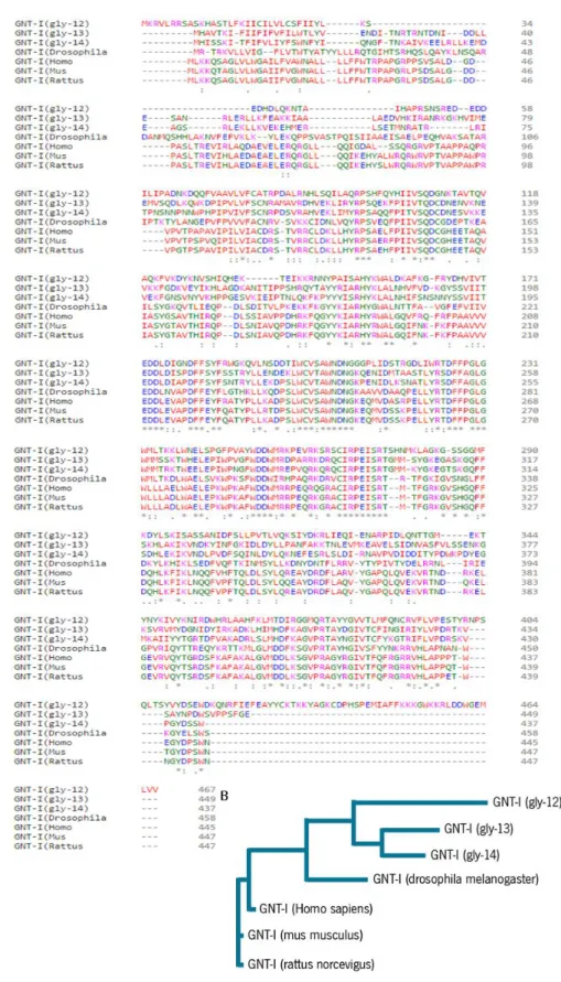
Figure 11 - Sequence alignment of GNT-I proteins.. ... 45
Figure 12 - Fluorescence microscopy evaluation of GABAergic neurons ... 46
Figure 13 - Evaluation of motor function. ... 47
Figure 14 - DiD assay. ... 48
Figure 15 - Confocal evaluation of AWB and AWC structure. ... 51
Figure 16 - Confocal evaluation of AWB neurons structure. ... 52
Figure 17 - Confocal evaluation of AWC neurons structure. ... 55
Figure 18 - Functional analysis of AWC neurons through attractive compound chemotaxis. ... 57
Figure 19 - Functional evaluation of AWB neurons function through repulsion chemotaxis assay.. ... 58
Figure 20 - Evaluation of AWB/AWC neurons structures through animal development ... 59
Figure 21 - Schematic of AWB/AWC neuronal network. ... 60
Figure 22 - Dry drop assay. ... 62
xxi
List of tables
Table 1 - Representation of the most common monosaccharides ... 4
Table 2 - Different types of alterations in the biosynthesis of glycans that have been described to originate congenital disorders of glycosylation. ... 12
Table 3 - List of strains used, respective phenotype and indication of purpose they were used for. ... 29
Table 4 - Primers for genotyping of GNT-I strains. ... 32
Table 5 - Primers for GNT-I genes (gly-12 to 14) RT-PCR ... 35
Table 6 - Brief overview of the number of glycosylation sites for the main neurotransmitter classes .... 43
Table 7 - Statistical analysis of motility and tracking assays ... 48
Table 8 - Statistical analysis of the foci phenotype and number of foci in AWB/AWC neurons. ... 53
Table 9 - Statistical analysis of foci phenotype and number of foci in AWB neurons. ... 53
Table 10 - Statistical analysis of the phenotype on AWC neurons. ... 56
Table 11 - Statistical analysis of attractive chemotaxis assays.. ... 57
Table 12 - Statistical analysis of repulsive chemotaxis assay. ... 58
Table 13 - Statistical analysis of the AWB/AWC neurons phenotype during the animals development. 60 Table 14 - Statistical analysis of the parameters evaluated in the dry drop assay. ... 63
Table 15 - Statistical analysis of qRT-PCR for gly-13 and gly-14 genes. ... 64
Table 16 - Summary of the results from the different analysis performed for the mutants for the GNT-I enzyme. ... 73
Table S 1 - Glycosylation sites of C. elegans neurotransmitters receptors. ... 91
Table S 2 - Blastp information for the GNT-I proteins of the alignment.. ... 135
3
Introduction
Glycans are the most diverse and complex organic compounds synthetized by living organisms. This diversity makes them complex regulatory and signaling molecules due to their high specificity. Making them essential, but also, in certain situations, detrimental, for a myriad of cellular processes. Particularly in the case of the nervous system it has been described a temporal expression of specific glycans that impact on neuronal development, with abnormal composition often leading to lethal phenotypes. Furthermore, these are key intermediates in maintaining homeostasis, modulating processes such as axon guidance, transporter localization at the synapse and vesicular release (Wright et al., 2012, Scott and Panin, 2014, Hall et al., 2015). We will next provide an overview of glycans composition and structure, and of the glycosylation process, with a particular focus on the nervous system.
1.1 Glycans and Glycosylation
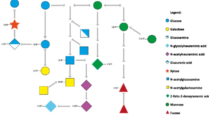
Glycans consist of a series of monosaccharides that are bound through glycosidic linkages, which are a type of covalent bonds that results of a condensation reaction between a hydroxyl group and a hemiacetal; the anomeric carbon of the sugar is the central carbon of the hemiacetal (Bertozzi and Rabuka, 2009). Glycans can be associated to proteins or lipids resulting in glycoproteins and glycolipids, respectively. The most common monosaccharides are glucose, fucose, mannose, galactose, xylose and sialic acid. Some less common monosaccharides appear only in particular species, e.g. deoxyhexoses, other than fucose, occur only in bacteria and plants (see table 1) (He and Liu, 2002, Varki and Sharon, 2009). The source of these monosaccharides may be external, entering the cell through specific transporters, or internal, such as salvage from degraded glycoconjugates or interconversion between different sugars, as is the case of glucose that can originate all the other monosaccharides (fig.1) (Freeze and Elbein, 2009). The variability and specificity of the glycans structures results not only from (1) all the possible combinations of the different sugars that give rise to glycans, but also (2) the oxygen that participates in the glycosidic bond, (3) the existence of tissue specific enzymes, where they are bound - either protein residues or lipids and (4) protein conformation which may hinder glycans addition.
4
Table 1 - Representation of the most common monosaccharides as well as examples of monosaccharides specific to some species with their respective chemical structure. For each common monosaccharide there is a defined abbreviation as well as a symbol.
5
Figure 1 - Simplified scheme of the biosynthesis of monosaccharides and interconversion between them. As can be observed from the figure several types of sugars that can be part of glycans structure may be synthetized from a common metabolite which is glucose.
Glycans are involved in several cellular processes: protein folding, quality control surveillance in the endoplasmic reticulum (ER), secretion, immune surveillance, inflammatory response, hormone action and tumor metastasis (Moremen et al., 2012). Glycans also play several key roles in organism development and homeostasis, from cellular adhesion to signaling, affecting cellular structure through the communication of the glycans in the cell membrane with the extracellular matrix (ECM).
As referred above, a role in cellular signaling was attributed to glycans, but due to technical restraints only recently it was possible to attribute specific functions to the different classes (further developed bellow) of glycans, and also to characterize how specific modifications in the glycosylation pattern may affect protein levels, function and localization.
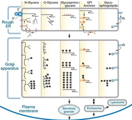
Glycosylation, the synthesis of glycans, is a highly specific process, varying with the cell type and even with the cellular stimuli. Although the sequence of enzymes that act to form specific classes of glycans are generally maintained, there are specific isoforms for different tissues and also tissue specific glycan epitopes (sugar structures present in several glycans e.g. Lewis X). This process occurs in the cellular organelles ER and Golgi, and the sequence of active enzymes define the class and function of the glycan structure that will be formed as well as the aminoacidic sequence or lipid that they are bound to (fig.2).
6
This synthesis starts in the ER and at this point it is important for protein quality control, where the addition of specific residues facilitates the binding to calnexin and calreticulin, and the interaction with these chaperones facilitates folding (Vembar and Brodsky, 2008). After leaving the calnexin/calreticulin cycle, proteins that obtain their native conformation will proceed in the secretory pathway where other sugars can be added. When the proteins do not fold correctly, they are recognized by the ER associated protein degradation system (ERAD), and sent to the proteasome for degradation (Vembar and Brodsky, 2008). The correctly folded proteins proceed to the Golgi for the addition of the final ramifications that determine the subclass that will be present in each glycosylation site; ultimately, this final structure will affect protein localization and function. Although this is the main glycan synthesis pathway, glycosylation enzymes were found in the nucleus and cytoplasm, and some glycans can be formed there, such as O-GlcNAc which is neither elongated or modified (Hart and Akimoto, 2009).
Figure 2 - Synthesis of the different glycan classes. Glycosylation occurs mainly in the ER and Golgi, the structures complexity and maturation increases along the secretory pathway. From Varki (2009).
These pathways are highly complex and regulated and, in most cases if one of the enzymes of the pathway does not act, the ones downstream in the biosynthetic pathway will not be able to bind to the glycan, resulting in a truncated structure. As an example, the fucosyltransferase 8 has to act, forming the fucose
7 nucleus in order for following fucosyltransferases (3 and 4) to recognize the substrate and add additional fucose residues (Paschinger et al., 2005).
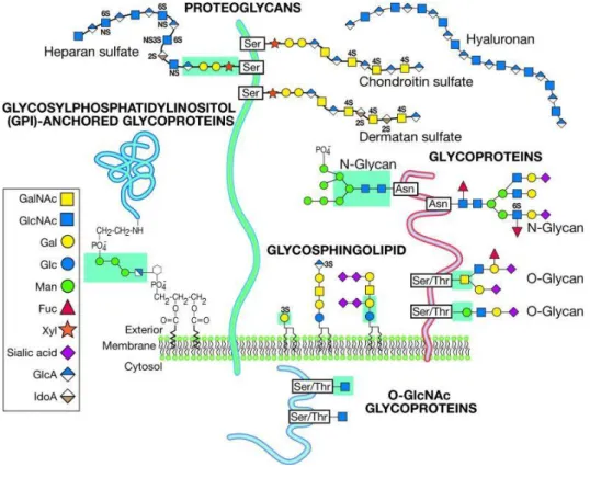
There are several classes of glycans, with the most relevant being proteoglycans, glycoproteins (N and O-glycans), glycophosphatidylinositol (GPI) and glycosphingolipids (fig.3). Glycans can also have a role as free sugars, not bound to proteins or lipids. Although the focus of this thesis are N-glycans a short overview of all the classes as well as their roles will next be presented.
Figure 3 - Representation the most relevant classes of glycans (glycoproteins and glycolipids). From Varki and Sharon (2009).
1.1.1 Proteoglycans
Proteoglycans are constituted by a core protein with one or more glycosaminoglycan (GAGs) chains (considerably larger than the sugars of glycoproteins, ~80 sugar residues compared to the 10-12 of glycoproteins). Proteoglycans are characterized by their sugar composition; they are linear polysaccharides composed by disaccharide units. GAGs can be separated according to their function/localization: ECM components, membrane or secretory vesicles (Bernfield et al., 1999, Yamaguchi, 1999). Inside these groups they are divided into families presenting a great variability resulting from the core proteins, the type and number of GAGs chains (1 to 100), and the cell type where they are being expressed; furthermore, there are also ‘part time’ chains that are not always attached
8
(Varki, 2009). These structures have several roles, being the most significant its structural role, conferred by the interactions between proteoglycans in the ECM and proteoglycans in the membrane. In the nervous system, chondroitin and dermatan sulphates proteoglycans (CSPGs and HSPGs) have been described to inhibit axonal growth (Bulow and Hobert, 2006). Other proteoglycans, like tenascin and brevican were shown to impair long term potentiation (LTP) in hippocampal mouse neurons, and also impact the perineuronal nets (PNNs), affecting synapse formation (Geissler et al., 2013). Mutations on this class of glycans also lead to phenotypes in the nervous system of simpler organisms such as Caenorhabditis Elegans, where the animals present defects in axon patterning and cell migration showing the importance of these molecules in the ECM (Bulow and Hobert, 2004).
Sugars are not only bound to proteins but also to lipids such is the case of GPI anchors and glycosphingolipids.
1.1.2 Glycophosphatidylinositol (GPI)
GPI membrane anchors are structures that anchor proteins to the cell membrane leaving the glycan motifs in the outer leaf. This structure is bound to different types of proteins such as hydrolytic enzymes, adhesion and regulatory proteins, and receptors (Paulick and Bertozzi, 2008). Its synthesis occurs in the ER and Golgi. Alternative mRNA splicing can lead to the formation of a soluble GPI anchor; one example is neural cell adhesion molecule (NCAM) that has a GPI when expressed in the brain but it is soluble in the muscle (Chatterjee and Mayor, 2001).
1.1.3 Glycosphingolipids
Glycosphingolipids are localized in the membrane and are the most abundant class in the vertebrate brain (Schengrund, 2015). The basic structure is constituted by a ceramide bound to a galactose; in turn, ceramide is formed by sphingosine (long chain alcohol) bound to lipids. The lipids anchor the sugar to the membrane and the glycans are presented to the outside portion (Schnaar, 2004). They can be divided in neutral, sialylated (gangliosides) and sulphated. The expression pattern of the structures is determined by the different expression and localization of the enzymes. In the membrane, they are not equally distributed but are concentrated in lipid rafts increasing the glycan-glycan and glycan-protein interactions that are dependent on their density (Schnaar, 2004). Glycosphingolipids help convey information from the outside to the inside of the cell, and have roles in cell to cell interactions and in the modulation of the activity of proteins in the same membrane.
9 This type of glycans is highly conserved in the brain. In oligodendrocytes they constitute more than 20% of the membrane lipids, playing an important role in myelination. Furthermore, gangliosides impact neural development and function due to altered expression during neuro and astrocyte-genesis (Schnaar et al., 2014, Schengrund, 2015).
1.1.4 Glycoproteins
Glycoproteins can be divided in two main classes, O-glycans and glycans. O-glycans present a N-acetylglucosamine (GlcNAc) bound to a serine or threonine. The majority are mucins, which are highly glycosylated proteins, that are usually transmembranar and are involved in cell adhesion, signal transduction and gel forming (Van Klinken et al., 1995). They have an important role in hydrating and protecting epithelial cells. In a disease context, specific epitopes have been found in cancer cells (Brockhausen, 1999). Of note, not all are present in the membrane, smaller soluble mucins can be also secreted. In these structures, the variability rises from the number of sites in the variable number tandem repeat regions of the gene.
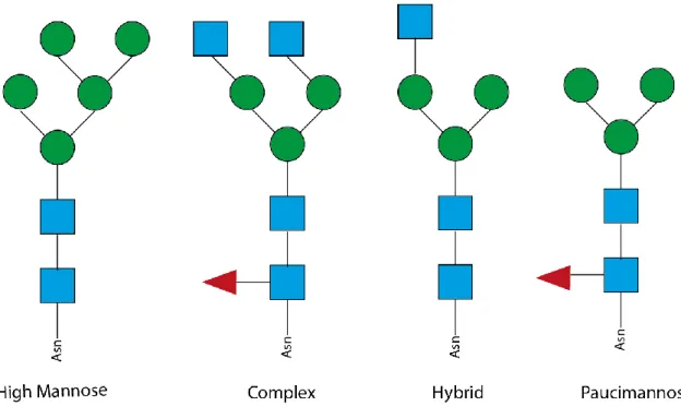
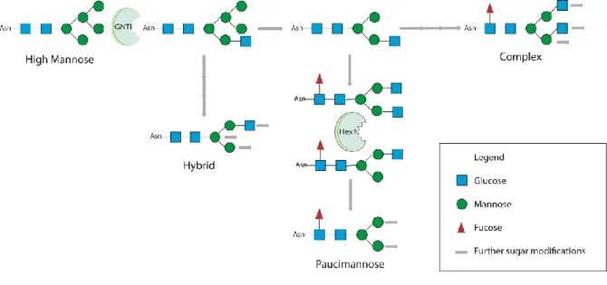
N-glycans are highly variable structures that share a common core structure (Manα1–6(Manα1– 3)Manβ1–4GlcNAcβ1–4GlcNAcβ1-Asn-X-Ser/Thr) and vary in the terminal residues that will define their subclass: (1) oligomannose/ high mannose (2) complex, (3) hybrid and (4) paucimannose (fig.4) (Stanley et al., 2009).
10
Figure 4 - Core structure of N-glycans classes. All four subclasses share a common backbone, differing in the ramifications, high mannose have mainly mannose residues; complex and paucimannose have the fucose nucleus but the complex subclass displays more ramifications; the hybrid, as the name suggests, are an intermediate (regarding complexity) between high mannose and complex N-glycans.
N-glycans are bound to sequons Asn-X-Ser/Thr from which two thirds are thought to be glycosylated, or Asn-X-Cys, being the cysteine in its reduced form. The probability of a site being glycosylated depends on the identity of the X amino acid and on conformational restraints. Its synthesis starts with the transference of a GlcNAc-P to a lipid-like molecule dolichol-phosphate (Dol-P). This occurs in the cytoplasmic side of the ER, where sugars are added until Man5GlcNAc2-P-P-Dol, at this point this structure is transferred to the luminal side of the ER through the action of a flippase (Rush, 2015). Mannose and glucose are added from Dol-P-Man/Glc, that also needs to be transferred to the luminal side, until this structure reaches 14 sugars (Stanley et al., 2009). Then it is transferred as a block to a N-glycosylation site of a protein, throughout the action of an enzymatic complex oligosaccharyltranferase (OST), constituted by 9 different subunits and three multisubunits. Mammalians have 3 OSTs that differ in their kinetic properties and capacity to transfer structures with less than 14 sugars, as it happens in congenital disorders of glycosylation where smaller structures are transferred to the lumen of the ER (Hennet and Cabalzar, 2015).
In the lumen of the ER, N-glycans have been described to have a role in the regulation of protein folding, where they regulate the cycles of the chaperones calnexin and calreticulin, by cycles of glycosylation and
11 deglycosylation (Vembar and Brodsky, 2008, Moremen et al., 2012). The proteins that pass the quality control are processed, trimmed, and before leaving the ER, glycans suffer the action of α-mannosidaseI. N-glycans that enter the Golgi, where they suffer further sequential enzymatic alterations, usually have 8-9 mannose residues (fig.5). The action of α-mannosidase I originates Man5GlcNAc, the substrate needed for the formation of hybrid, complex and paucimannose N-glycans (Moremen et al., 2012). On the other hand, if they do not proceed in this pathway they give rise to oligomannosidic N-glycans. The first enzyme of the hybrid and complex glycans biosynthesis is α-1,3-mannosyl-glycoprotein 2-β-N-acetylglucosaminyltransferase (GNT-I) that acts in the medial Golgi (Schachter, 2009, 2010). From here the two classes are separated: if mannosidase II acts it will give rise to complex N-glycans, and if not to the hybrid class. If an hexosaminidase acts after the mannosidase II it gives rise to another class, the paucimannosidic glycans, which are found in large amounts in invertebrates and plants and have only recently been described in vertebrates (Zhang et al., 2003, Berninsone, 2006, Dahmen et al., 2015).
Figure 5 - Synthesis of the different glycans classes highlighting the essential steps that initiate the synthesis of different N-glycan classes (hybrid, paucimannose and complex).
The maturation of the various classes occurs in the trans Golgi and occurs in three different forms: (1) additions to the core structure, like the fucose nucleus, where a fucose residue is added to the N-acetylglucosamine close to the asparagine; (2) elongation of the branching N-N-acetylglucosamine, where galactose or GlcNAc are added to the GlcNAc residue to elongate the branches; and (3) decoration, which consist in the addition of sialic acids and fucose to these elongated branches (Stanley et al., 2009).
12
Besides the variability created by the classes and maturation of these glycans there are other factors that increase the variability of these structures, such as specific site heterogeneity where the same protein may have different glycans or several sequons (Cipollo et al., 2005, Parker et al., 2013). The glycan formed also depends on the sugar availability, the localization of the enzyme in the Golgi - as some of them compete for the same substrate - and also protein conformation which might affect the enzymes that can bind to the substrate.
The use of enzyme inhibitors and the development of mutants for this pathway enzymes brought insights into the role of N-glycans, which have been specifically described as having a role in protein quality control, susceptibility to proteases, antigenicity, immunity, neuronal cell migration, inflammation and in tumor progression (Liedtke et al., 2001, Stanley et al., 2009).
The importance of glycosylation is showed by the disorders that result of alterations in various factors/steps of its synthesis, congenital disorders of glycosylation (CGDs) (Hennet and Cabalzar, 2015) (table 2).
Table 2 - Different types of alterations in the biosynthesis of glycans that have been described to originate congenital disorders of glycosylation. An example is presented for the most common alterations (Hennet and Cabalzar, 2015).
Alteration Example Result Disease
Loss of enzymes ER glycosyltranferases
Loss of glycan chains, affecting folding, secretion and
protein stability
Muscle-eye-brain disease.
Alteration in donor
substrates Dol-P-sugar
Phenotype will vary with the severity of the mutation
Total loss Embryonic muscle-eye-brain disease or death.
Partial loss Mild intellectual disabilities such is the
case of PMM2. Mutations in the
transporters of the substrates to the ER
lumen and Golgi
Glc and UDP-GalNAc transporter –
SLC35D1
Affect the chondroitin sulphates synthesis Schneckenbecken dysplasia (skeletal disease) Localization of the glycosyltranferases Mediated by conserved oligomeric Golgi (COG)
Affect the proper maturation of glycans structures
Neurologic impairments liver
dysfunction and infantile lethality.
13 If before it could only be attributed functions to a general class of glycans, recent technical advances allowed a characterization of the different glycosylation patterns present on a protein and even determine in which glycosylation site a specific glycan is bound. This has helped to define the function of a specific glycan structure. In fact, in vitro studies, studying specific proteins where glycosylation sites are mutated helped to understand the role of a specific glycan in a specific site (Hall et al., 2014, Lichnerova et al., 2015, Kaniakova et al., 2016). And from these studies new data has emerged, for example it seems that complex and hybrid N-glycans have different roles in the same protein if present in different sites of the protein (Hall et al., 2015, 2016). The development of new mass spectrometry (MS) techniques has helped to understand the impact of alterations in whole cell glycosylation and also to study conservation of glycosylation across species (Ji et al., 2015, Zacchi and Schulz, 2016).
1.2 Evolutionary view of glycosylation
Glycosylation is present in all living organisms and has a strong impact in their survival, not only in protein quality control and their localization, but due to the presence of the glycocalyx (dense coat of glycans that surrounds all cells), it also impacts on cellular motility, pathogen recognition, cellular adhesion and structure (Varki, 2006).
All glycan classes are present in eukaryotes, the core structure is shared as well as some steps of their extension. The differences are found in the terminal alterations, with the trimming varying with strain and phylum, and the sugars that are turned to the outside vary also giving rise to a specific glycocalyx (Corfield and Berry, 2015).
Not only the structures are conserved, but also the general functions of each class. One example of the differences between species is in the abundancy of the different subclasses, for example complex and hybrid N-glycans are highly expressed in mammals while paucimannosidic have only recently been described, but in low concentrations (Moremen et al., 2012, Dahmen et al., 2015). In nematodes this ratio is inverted, where paucimannosidic are highly expressed and the complex and hybrid are only present in small quantities. Despite these differences in the expression levels of all the classes, the enzymatic machinery is conserved in all organisms (Berninsone, 2006).
This stands for N-glycans, but also for other classes in which there are similarities and differences. As an example, in O-glycosylation, O-GalNAc is the most common in vertebrates but in plants it does not occur, instead there are others like O-Gal, and bacteria for instance have a Galβ1-O-Tyr core. Also, in GAGs that were thought to be only present in higher order animals, have been described in insects and molluscs although not as highly sulphated as in mammals (Varki et al., 2009).
14
Even looking to terminal modifications and not glycans classes there are some that are highly conserved such as sialic acids, which are also present in species such as Drosophila melanogaster, that have homologs for the mammalian enzymes.
Thus, the variability and specificities of glycans structure and function introduced by evolution across species is a relevant aspect that should be taken in consideration when addressing the role of glycans in specific tissues and/or cell types.
1.3 Glycans and the nervous system
The nervous system is constituted by different types of cells that have specific requirements when it comes to structure/adhesion and signaling, processes for which, as described above, glycans are essential. The nervous system needs a fine regulation of all cellular processes, from cell adhesion to synaptic transmission, and these are influenced and regulated by several processes such as, post translational modifications, that play an important role in this regulation as it can be cell type specific (Peters and Connor, 2014).
In the nervous system, neurons are the main cells responsible for processing information and response to stimuli. These cells are asymmetrically polarized which allows directional transmission of information, through the propagation of action potentials that will elicit a response at the synapse level, depending on the type of neuron and synapse (excitatory or inhibitory outcome). The synapse is a highly regulated and complex structure and at this level there are several factors that need to be tightly controlled, from the formation of vesicles and secretion of neurotransmitters and ions, to the localization of receptors in the cellular membrane. Another important aspect is axonal transport, which ensures that everything needed for a proper synaptic transmission is present at the synapse. In order to respond to different stimuli and produce a correct response, neurons have to be highly plastic cells, being able to alter their number of projections and their size according with the stimuli (Peters and Connor, 2014).
Besides neurons, other cell types play a role in nervous system formation and homeostasis, namely glia, that is further divided in astrocytes, microglia and oligodendrocytes, each with a specific role. Astrocytes participate in the tripartite synapse, are the only deposit of glycogen in the nervous system and also respond to injury influencing neuronal survival and homeostasis. Microglia are the immune cells of the nervous system and their expression pattern can be altered in response to different stimuli from an anti-inflammatory to a more pro-anti-inflammatory profile (Crain et al., 2013).
Glycans are essential components in the interplay between all these cell types of the nervous system. For instance, nervous system cells have specific glycocalyx and also secrete different molecules to the ECM.
15 For example, glia cells produce CSPGs, while neurons secret only a small amount of CSPGs and gangliosides; in turn, HSPGs are produced by both and act as growth regulators. This layer of glycoconjugates is in the extracellular side of the membrane and has an important role in recognition, barrier formation and patterning (Dityatev and Schachner, 2003).
1.3.1 Glycosylation and the ECM
The ECM plays an important role in the nervous system, the most relevant being the modulation of cellular stability and structure. Furthermore, it has a high impact in signal transmission due to its presence in the synaptic cleft, and given that its composition can influence the passage of neurotransmitters and their binding to the receptors (Dityatev and Schachner, 2003, Dityatev et al., 2010).
As mentioned above, the perineuronal nets or PNNs are specific matrix structures surrounding some synapses (fig.6). These structures are heterogeneous in structure and composition varying according to the type of neuron (Dityatev and Schachner, 2003) and brain area (Yamaguchi, 1999). It has been proposed that a ternary complex (hyaluran-lectican-tenascin-R) constitutes the core structure of PNNs. Both hyaluran and tenascin-R are lectican ligands, and the CSPGs and HSPGs (sugar portion bound to lectins) that are present varies with the brain area. The relevance of CSPGs and HSPGs, that are a major component of the PNNs, is illustrated by their role in axonal growth and migration as well as synaptic plasticity; as an e.g., removal of chondroitin was described to decrease LTP (Dityatev and Schachner, 2003, Dani and Broadie, 2012, Missler et al., 2012)
Figure 6 - Representation of perineuronal nets from Ramón y Cajal (a) confocal images of PNNs in the CA1 region of the hippocampus from WT (b) and Tenascin R deficient animals (c), representation of the core structure formed by hyaluran-lectican-tenascin R in the PNNs (d). From Dityatev and Schachner (2003).
Although not only neurons secret the molecules that form the ECM in the nervous system, these seem to be the major contributors as shown in a co-culture assay where neurons were able to initiate PNN
16
formation; however when using neurons with mutations in these proteins, the neurons lose the ability to bind to astrocytes (Geissler et al., 2013). Furthermore, PNNs associated neurons seem to be less affected by neurodegeneration and the removal of specific components has different impacts on this neuroprotective effect: without tenascin R the effect is lost, aggrecan has a partial effect and brevican does not affect the protective effect (Suttkus et al., 2014).
In addition to their role as modulators of cell-cell and cell-matrix interactions, glycans have also been described to affect major cellular pathways, as they can act as an on/off switch. For instance, the deglycosylation of Akt allows its phosphorylation that will initiate a pathway that results in the synthesis and secretion of hyaluronic acid (Dawson, 2014).
Another important component of the ECM is dystroglycan, a protein of the dystroglycan-glycoprotein complex. Dystroglycan is a membrane protein that links the cytoskeleton to the ECM and its glycosylation is essential for its function. It is required for axonal migration by binding to axon guidance cues acting as a regulator, namely it determines Slit localization, this glycoprotein binds to Robo and both have a conserved role in repulsive axon guidance (Wright et al., 2012, Goddeeris et al., 2013).
The ECM composition is essential from development to adulthood as it has important roles in modulating proper cellular development, (cellular positioning, axonal growth and guidance, cellular adhesion) and in adulthood, in maintaining cellular stability and capacity to respond to stimuli such as the modulation of synaptic plasticity (Maness and Schachner, 2007).
1.3.2 Glycans and signaling/cellular communication
The term glycosignaling describes the facilitation of signaling that results from the concentration of membrane glycans in lipid rafts. The glycoconjugates in the lipid rafts recruit signaling molecules and can both inhibit and induce signal transduction. They have been described to affect major cellular pathways, such as Notch which is only active if glycosylated (Dawson, 2014).
One of the most well described glycosylated protein in the nervous system is NCAM and the L1 family of adhesion molecules. Polyasialylated (PSA)-NCAM is important for development and it is highly expressed in areas of neurogenesis (neurogenic niches), being more expressed during development, and in areas of synaptic plasticity (S. Liedtke et al., 2001). Moreover, PSA decreases adhesion and it also has the neural glycosylation epitope HNK-1 that is involved in cell movement, and in neural and glia cell migration. L1 domains can recruit proteins in the membrane, such as ion channels and pumps. It is also needed for cell differentiation. In addition, L1 can cluster increasing cell adhesion to ECM proteins through integrins, potentiating cell motility and migration (Maness and Schachner, 2007). The relevance of this
17 family of proteins is not exclusive to mammals as mutations in C. elegans homolog protein, Sax-7, leads to defective neural placement with defects in axonal guidance and cell body placement. The role for neural development and cellular migration is maintained throughout evolution, highlighting the relevance of glycosylation for development and homeostasis of the nervous system (Sasakura et al., 2005, Pocock et al., 2008). As stems from the above described, this is a good example of how the glycosylation of a single protein can influence differently several cellular processes (adhesion/plasticity and protein target to the membrane).
Besides the direct role in communication through the modulation of binding of the ligands to the receptors, glycosylation also has an impact in protein localization, as it has been shown that alterations in this process impacts protein targeting to the membrane, as well as their proper conformation. Several studies have described the importance of glycosylation, particularly the N-glycans class, for the targeting of synaptic proteins, which are glycosylated and alterations in this process result in different phenotypes (Scott and Panin, 2014). This regulation of protein targeting can impact proper protein function. Specifically, in the context of the nervous system, neurotransmitters receptors, mainly glutamate (Kaniakova et al., 2016) and γ-Aminobutyric acid (GABA) (Geng et al., 2012) have a high number of predicted N-glycosylation sites (supplementary table S1). Several authors have focused on dissecting the importance of each site, which ones are glycosylated and what type of glycans are present at each site (Hall et al., 2015, 2016). In fact correct protein targeting is modulated by specific glycans in a specific protein site as mutating glycosylation sites one by one shows that not all have the same role/impact in protein localization and function (Takeuchi et al., 2015).
One other example of how glycans influence nervous system cells communication stems from the fact that PNNs composition, that regulate synaptic plasticity, can modulate α-amino-3-hydroxy-5-methyl-4-isoxazolepropionic acid receptor (AMPAR) mobility in the membrane but also inhibit axonal growth and sprouting (Dou and Levine, 1995). The matrix regulates the synaptic structure and morphology. It affects both glutamatergic LTP and long term depression (LTD), where different molecules will have different effects. For example, reelin potentiate synaptic response in the hippocampus while, for example tenascin R regulates the efficacy of GABA and the number of perisomatic inhibitory contacts in CA1 area of the hippocampus (Dityatev and Schachner, 2003).
1.3.3 Glycosylation: a conserved post translational modification
As mentioned before glycosylation is a highly conserved process and one good example is the nervous system where there have been described a conservation of roles and relevance of several ECM and
18
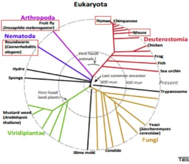
signaling glycosylated molecules across species (Corfield and Berry, 2015) (fig.7). In general, all classes of glycans are present in all eukaryotes and so are their biosynthetic pathways. As an organism complexity increases so does the complexity of this process, leading to an increase of complexity of the structures as a result of the increase of protein isoforms and different forms of maturation. In more complex organisms there are cell type specificity, specific isoforms and glycoepitopes.
Figure 7 - Eukaryotes phylogenetic tree highlighting the species discussed in this work. Adapted from Varki (2009)
The relevance of glycosylation in the nervous system is shown by its conservation throughout evolution as homologs/orthologues seem to play the same role in very evolutionarily distant organisms, such as mammals, Drosophila melanogaster and C. elegans (Cipollo et al., 2002, Park and Zhang, 2011, Corfield and Berry, 2015). In general, the number of members of the different classes seems to increase with the species complexity. Even though there are some differences in terms of degree of variability, complexity of the structures and the expression levels of the different classes, the core structures are maintained as well as their roles in the cell (Varki, 2009). One case of this are the heparin sulphates where for example, alterations in collagen results in neuronal migratory defects not only in mammals but also in drosophila and C. elegans (Bulow and Hobert, 2006).
1.3.4 N-glycans in the nervous system
The N-glycans class is of particular relevance in the nervous system. They play an important role in neuronal development, as the expression of specific glycoepitopes and glycoproteins changes with
19 different developmental stages. During neuronal development there is a tight regulation of glycosylation, namely, certain glycoepitopes are restricted to specific developmental ages, e.g. N-glycans epitopes, lewis X, disappear after cellular differentiation while they initially constituted 20% of the total glycans (Yagi et al., 2012); another example is nestin expression which is restricted to undifferentiated cells (Gattazzo et al., 2014).
Considering the processes were glycans have been involved, both physiological and pathological, with particular relevance for the regulation of the nervous system. Glycans have been described to impact several processes that are essential for synaptic transmission. Some examples are vesicle release, where for synaptic vesicle protein 2 and synaptophysin N-glycans impact their localization and folding; neurotransmitter receptors, where for acetylcholine receptors it regulates desensitization, and for ionotropic glutamate receptors affects maximal currents and desensitization of AMPA and kainite receptors (Han et al., 2004, Scott and Panin, 2014). Glycans impact neuronal and synaptic structure and function (Scott and Panin, 2014). Here, N-glycans have been described as active intermediates in several functions, signaling (synaptic proteins), synaptic constitution through the modulation of protein localization and levels and synaptic currents by modulating the opening of the channels (Hall et al., 2014). The most well described is their role in the ECM, in the case of synapses matrix, that is responsible for the maintenance of the synaptic structure also being capable of influencing its function through the modulation of protein mobility in the membrane.
As mentioned before, N-glycans are divided into four subclasses: hybrid, high mannosidic/oligomannose, complex and paucimannosidic that are differentially expressed in eukaryotes. The first three, are highly expressed in vertebrates (Varki, 2009) while the fourth has only recently been described in the mammalian brain and only in a short post-natal period. On the other hand, in other eukaryotes, such as nematodes and plants there is an inversion of the levels of these subclasses.
When analyzing N-glycans biosynthesis, there are enzymes that are essential for a specific class or group of classes such is the case of GNT-I, which mutant will affect all N-glycans classes except high mannose. The mutant for this enzyme has been studied in three different models, in C. elegans, drosophila and mouse (Chen et al., 2003, Schachter, 2010). Where in the case of mouse and drosophila nervous system alterations were described, this was not explored in the C. elegans mutant. The absence of this enzyme should result in an elimination of all hybrid, complex and paucimannosidic N-glycans. Alterations in the levels of these glycans have already been described through mass spectrometry of these mutants, and as expected they result in an increase of the high mannosidic and a significant decrease in all the others (Zhu et al., 2004). This enzyme was shown as detrimental for mouse development, where a full knockout
20
(KO) leads to embryonic death and a neuronal specific KO resulted in a severe phenotype, with significantly reduced body mass, lifespan and alterations in the hybrid and complex N-glycans, resulting in increased apoptosis and astrogliosis (Ye and Marth, 2004). In drosophila, a specific analysis into the neuromuscular junction (NMJ) revealed an overgrown structure affecting synaptic function, increased capacity to vesicle release upon stimulation, alterations in the levels of synaptic proteins, such as GluRIIB and transsynaptic signaling pathways, such as Wnt, and synaptic scaffolds DLG (Parkinson et al., 2013). Of notice, the role of these glycans in the nervous system is maintained throughout evolution, although it appears that the importance increases with the complexity of the nervous system. The elimination of the same enzyme leads to increasingly severe phenotypes from C. elegans to Drosophila melanogaster and Mus musculus. Contrary to what usually happens with other classes of glycans and to mammals and drosophila, that only have one GNT-I gene, C. elegans has three genes that have GNT-I activity, gly-12, 13 and 14, so compensatory mechanisms may be present (Chen et al., 1999, Varki, 2009).
For paucimannose N-glycans, three enzymes have been described as essential to its synthesis, GNT-I, mannosidase II and hexosaminidase I (Zhang et al., 2003). Hexosaminidase I is the most specific for paucimmanose N-glycans, since it is the one that separates the paucimannosidic from complex glycans, so mutations in this enzyme should only affect the paucimannosidic class, and in theory lead to an increase in the hybrid and complex class. This class of glycans has only been described in plants and some insect species and until very recently there was no report of being present in mammals. The only evidence suggests a role in cellular differentiation in a short post natal period, where the end of its expression in the subventricular zone coincides with an increase of cellular differentiation in the nervous system (Dahmen et al., 2015).
Taking into account the conservation of glycosylation throughout development and the higher expression of a different N-glycans class, the paucimannose, C. elegans is a good model for the study of N-glycans for because it allows the analysis of the role of hybrid and paucimannosidic N-glycans in the nervous system structure and function and most importantly it has a simple nervous system. In fact, the C. elegans nervous system is constituted by 302 neurons, it is completely mapped (identification and position of each neuronal cell body and respective dendrites are well established), which allows the study of specific classes of neurons and the evaluation of their structure as well as their phenotypical outcome. A specific group of neurons can be analyzed, studying its structure (animals are transparent), synaptic outputs and most importantly because their function is also specifically described, understand the phenotypic output (e.g. behavior) that an alteration may induce.
21 Thus considering the above described, several questions may be explored on the role of N-glycans specifically in the C. elegans neuronal network. For instance, what are the effects of eliminating these classes of N-glycans in the C. elegans nervous system? Are their roles conserved? Do these molecular alterations lead to functional phenotypes? Does this result in different impacts in different groups of neurons? Are there compensatory mechanisms between the different GNT-I coding genes? Are their expression levels altered in the single mutants? These questions will be explored in this thesis.
25
Aims
Glycans have emerged has important regulators of most of cellular processes being essential mediators of development and homeostasis. Understanding the role of the different and diverse structures existent in each class of glycans is of relevance.
One particular interesting group of glycans in the context of the nervous system is the N-glycans class. This class has been described as important for the development of the nervous system but also its function, namely impacting processes from structural stability to synaptic transmission.
Considering this, the general goal of this thesis is to understand how N-glycans impact the nervous system structure and function. Specifically, by using an invertebrate model, the nematode C. elegans, with this work we aim at:
1. Understanding the similarities and differences of N-glycosylation between vertebrates and invertebrates in the nervous system.
2. Exploring the impact of eliminating the GNT-I enzyme in neuronal structure. 3. Assessing the functional impact of eliminating N-glycans.
29
Materials and methods
3.1
C. elegans
strains
Strains were maintained under standard conditions on nematode growth media (NGM-agar, bacto-peptone, 1mM NaCl, 1mM CaCl2, 1mM MgSO4, 1mM cholesterol and 5mM KH2PO4) plates seeded with
Escherichia coli (OP50) (Stiernagle, 2006). All experiments were conducted at 20ºC except otherwise specified.
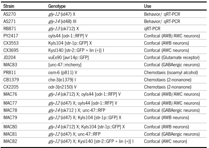
All the glycosylation enzyme mutants, fluorescently tagged neuron and chemotaxis control strains used were obtained from the Caenorhabditis Genetics Center and are described in table 3. Glycosylation enzyme mutants with fluorescently labeled neurons (table 3) were obtained as previously described in Fay (2013).
Table 3 - List of strains used, respective phenotype and indication of purpose they were used for.
Strain Genotype Use
AS270 gly-12 (id47) X Behavior/ qRT-PCR AS271 gly-14 (id48) III Behavior/ qRT-PCR
RB871 gly-13 (ok712) X qRT-PCR
PY2417 oyIs44 [odr-1::RFP] V Confocal (AWB/AWC neurons) CX3553 Kyls104 [str-1p::GFP] X Confocal (AWB neurons) CX3695 Kysl140 [str-2::GFP + lin (+)] I Confocal (AWC neurons) JD204 vuEx90 [avr14p::GFP] Confocal (Glutamate receptor) MAC83 [unc-47::mcherry] Confocal (GABAergic neurons) PR811 osm-6 (p811) V Chemotaxis (isoamyl alcohol) CB1379 che-3(e1379) I Chemotaxis (2-nonanone) CX2205 odr-3(n2150) V Chemotaxis (2-nonanone) MAC76 gly-14 (ok712) X; oyIs44 [odr-1::RFP] V Confocal (AWB/AWC neurons) MAC77 gly-12 (id47) X; oyIs44 [odr-1::RFP] V Confocal (AWB/AWC neurons) MAC78 gly-14 (ok712 ) X; unc-47::RFP Confocal (GABAergic neurons) MAC79 gly-12 (id47) X; Kyls104 [str-1p::GFP] X Confocal (AWB neurons) MAC80 gly-14 (ok712) X; Kyls104 [str-1p::GFP] X Confocal (AWB neurons) MAC81 gly-12 (id47) X; unc-47::RFP Confocal (GABAergic neurons) MAC82 gly-12 (id47) X; Kysl140 [str-2::GFP + lin (+)] I Confocal (AWC neuron)
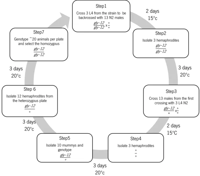
3.2 Backcrossing
To eliminate possible random undesired mutations occurring outside the genome sequence of interest, all GNT-I strains were backcrossed at least 8 times. Briefly, GNT-I mutants L4 were crossed with N2
30
males, then the resulting heterozygous males with N2 hermaphrodites (Fay, 2013). The animals were genotyped and the homozygous selected for the following backcross round (see fig.8 for detail).
Figure 8 - Schematic representation of the backcrossing protocol followed to eliminate possible mutations other than the target mutation.
3.3 Genotyping
3.3.1 DNA extractionTwo methods were employed to extract C. elegans genomic DNA considering the number of animals used.
For single worm, the animal was placed in 5µl of worm lysis solution [dH20, 1x Taq buffer (100mM Tris,
500mM KCl, 15mM MgCl2) and 0.1mg/ml proteinase K], then the tubes were placed at -80ºC for 30min,
followed by a temperature cycle to allow enzyme function and inactivation (65ºC for 1h and 95ºC during 15min).
Step1
Cross 3 L4 from the strain to be backrossed with 13 N2 males
gly−12 gly−12 x ++ Step2 Isolate 3 hemaphrodites gly−12 gly−12 Step3
Cross 13 males from the first crossing with 3 L4 N2 gly−12 + x++ Step4 Isolate 3 hemaphrodites + + Step5
Isolate 10 mummys and genotype
gly−12 +
Step 6
Isolate 12 hemaphrodites from the heterozygous plate
gly−12
+
Step7
Genotype ~20 animals per plate and select the homozygous
gly−12 gly−12 2 days 15ºc 3 days 20ºc 2 days 15ºC 3 days 20ºc 3 days 20ºc 3 days 20ºc
31 For ~20 animals the Phire Animal Tissue Direct PCR Kit (BioLabs, Hertfordshire, UK) was used. The animals were placed in 20µl of dH20 and centrifuged at 10000rpm for 5min, and the supernatant was
removed to eliminate some of the OP50 bacteria. Next 10µl of kit dilution buffer was added to the pellet as well as 0,5µl of DNA release. The mix was left to incubate at room temperature for 5min and then placed at 98ºC for 2min. The samples were stored at 4ºC until the polymerase chain reaction (PCR) was performed.
3.3.2 PCR
In order to genotype the mutant strains for the GNT-I enzymes (AS270, RB871 and AS271; table 3) primers were designed for each strain using the PrimerBlast tool (Ye et al., 2012). The strategy employed for primer design implicated different size bands for the mutant gene and the wild-type (WT) one. For this, one forward primer was designed before the deletion region, another inside the deleted region, and a reverse primer after the deletion (fig.9). The PCR cycles were optimized through the performance of a standard temperature gradient (55-60ºC) to determine the annealing temperature. It was performed using a standard PCR mix (1x Taq Buffer; 1.5mM MgCl2; 0.2mM dNTPs; 0.2µM Primer F1; 0.2µM Primer F2;
0.2µM Primer R1; 5U/µl Taq polymerase) (Thermo-Scientific, Waltham, Massachusetts, USA). The PCR cycle consisted of 2min at 95ºC followed by 35 cycles (1min at 95ºC, 45sec at the annealing temperature and 1min at 72ºC), and a final cycle of 5min at 72ºC.
Specific primers, band sizes and annealing temperatures are discriminated in table 4. For the genotyping of the gly-13 mutant strain some changes were made, namely the extension time was increased to 2min as well as the final extension to 10min.
32
Table 4 - Primers for genotyping of GNT-I strains, the band size expected for both WT and mutant animals and the annealing temperature used.
Mutant Primer name Primer sequence Band size (bp) Annealing Temperature (ºC) gly-12 (id47) Gly-12F1 5’ TTACTCTCCACACCAGCTCA 3’ WT-794 MUT-538 58 Gly-12F2 5’ TCTCTCTTCTCCCTGTCACC 3’ Gly-12R1 5’ AGCTCATTACCTTCCAGTCAC 3’ gly-13 (ok712) Gly-13F1 5’ GTAGGGCAATCACATCTCGAA 3’ WT-674 MUT-419 59 Gly-13F2 5’ ATAAACCGCAGTTTGACGTG 3’ Gly-13R1 5’ CGAATCTAGTCGACTCTGGC 3’ gly-14 (id48) Gly-14F1 5’ ACGCAGTTTGTAGACAGTGG 3’ WT-382 MUT-648 58 Gly-14F2 5’ AGCCTTTCCATGTGCTCTTT 3’ Gly-14R1 5’ GAACTGATCTACACGCCGAA 3’
3.4 Crossings
To evaluate neuron structure, it was necessary to cross the mutant strains with animals expressing fluorescent protein in specific neurons [str-1p::GFP (AWB neurons), str-2::GFP (AWC neurons), odr-1::RFP (AWB/AWC neurons) and unc-47::RFP (GABA neurons)]. For this purpose, the mutant strains [gly-12 (id47) and gly-14 (id48)] were crossed with the fluorescently labeled neuron strains (odr-1::RFP, str1p::GFP, str2::GFP, unc-47::mcherry). This was accomplished by crossing mutant hermaphrodite animals with males expressing fluorescent proteins in heterozygosity in the neurons specified above. These heterozygous fluorescent male animals were obtained through crossing with N2 males (Fay, 2013) (see fig.10 for detail). The genotype was confirmed by PCR for the mutation and through fluorescence observation. A list of the crossed strains is discriminated in table 3.
33
3.5 Microscopy Analysis
3.5.1 Animal preparation
The crossings of the GNT-I mutants with the fluorescent strains (MAC76 to MAC82) were synchronized by placing 10 adult hermaphrodites in 60mm plates for 2h, a period of time during which the animals lay eggs. These animals were synchronized with a maximum of 2h difference between them; the adults were then removed. These plates were placed at 20ºC for 3 days (adult day 1 stage), day at which we analyzed the neurons. The animals were placed in an agar pads with levamisole (3mM) for microscopy analysis. In all observations, control images of the WT (N2) worms were taken as well as a homozygous WT resulting from the crossing; this confirms that the crossing protocol did not induce any alteration in the animals and will confirm that any alteration is the result of the target mutation.
3.5.2 Fluorescence microscopy
The crossings for the fluorescently labelled GABAergic motor neurons were observed in an inverted fluorescence microscope Olympus IX53 (Olympus, Tokyo, Japan).
Step 1
Cross 13 males N2 with 3 L4 fluorescent strain + +x F+ F+ Step 2
Isolate the 3 hemaphrodites +;F+
+;F+
Step 3
Cross 13 F+ males with 3 L4 from the mutant strain
+;F+ +;F+X
gly−12; +
gly−12; +
Step 4
Isolate the 3 hemaphrodites
gly−12; +
gly−12; +
Step 5
Isolate 3 double heterozygous hemaphrodites
gly−12; F+ +; +
Step 6
Select the plate with more fluorescent animals and isolate
24
Step 7
Genotype the 24 plates, select homozygous
gly−12; F +
gly−12; F+
Figure 10 - Discrimination of steps in the crossing protocol necessary to obtain strains with fluorescently tagged neurons in the GNT-I mutant strains. 2 days 15ºC 3 days 20ºC 2 days 15ºC 3 days 20ºC 3 days 20ºC 3 days 20ºC
34
3.5.3 Confocal microscopy
The crossings for the fluorescently labelled AWB and AWC neurons were observed in a confocal microscope, Olympus FV 1000 (Olympus) to analyze neuronal structure. For the crossings with odr-1::RFP, str-1p::GFP and str2p::GFP, all images were taken in 512 by 512, in the 60x objective with 1.6 zoom and Kallman was performed twice to increase the quality of the image; furthermore a step-size of 0.24 was made to allow a 3D reconstruction of the images necessary for neuronal structure analysis.
3.6 DiD staining of amphid neurons
Synchronized 3 days old animals were washed with M9 twice and then placed in a 96 wells plate in a solution of lipophilic tracer DiD (Invitrogen, California, USA) in M9 (0.01mg/ml). The animals were placed in agitation for 2h and then were placed in an NGM plate for 1h to wash off the excess dye from the digestive tube. Afterwards the animals were prepared for fluorescence microscopy analysis has described above.
3.7 qRT-PCR of the GNT-I genes
3.7.1 RNA extractionGNT-I mutants and N2 animals were synchronized by bleaching and at day 3, they were washed 3 times in M9 to remove bacteria. The animals were pelleted and TRIzol reagent (Invitrogen, California, USA) was added, the tubes were vortexed to break the animals body and 3 cycles of freeze/thaw (liquid nitrogen/ water bath 37ºC) were performed. After 10min at room temperature the samples were centrifuged at 14000rpm for 10min. The supernatant was transferred to a new tube and chloroform was added, then the samples were centrifuged as before and the aqueous phase was removed to a new tube. Isopropanol was added to precipitate the RNA and the samples were centrifuged again. The samples were washed with ethanol 75% and centrifuged at 1000rpm for 5min. The supernatant was discarded and the pellet was air dried. Lastly, the pellet was dissolved in 10µl of nuclease free water. The samples were then quantified and the quality of the RNA was determined in the spectrophotometer nanodrop ND-1000 (Thermo-Scientific) by ratios of absorbance at 260nm/280nm to assess the purity and quality of the RNA, and 260nm/230nm to assess possible contamination with protein and phenols; the samples were used only if the ratios obtained were between 1.8 and 2.
35 3.7.2 cDNA synthesis
Using reverse transcriptase total RNA was converted into cDNA using iScript cDNA Synthesis Kit (Bio-Rad, Hercules, California, USA) following manufactures instructions. Briefly a mix with iScript reaction mix, iScript reverse transcriptase, water and 1µg of RNA was prepared and then a temperature cycle is performed as follows: 5min at 25ºC, 3min at 42ºC and 5min at 85ºC.
3.7.3 qRT-PCR
The primers for the amplification of GNT-I genes (gly-12 to 14) were designed using PrimerBlast tool; primers are listed on table 5. The qRT-PCR was performed using SsoFast EvaGreen Supermix (Bio-Rad), and analyzed using CFX96 Touch Real-Time PCR Detection System (Bio-Rad). Before determining gene expression, we performed a temperature gradient to determine the best annealing temperature and we also assessed primer efficiency by performing a cDNA concentration gradient. Even though we tested two different pairs of primers for the gly-12 gene neither of them presented an acceptable efficiency, and we were only able to evaluated expression levels for the gly-13 and gly-14 genes. The expression levels of the GNT-I genes were normalized considering the expression levels of the housekeeping gene rpb-2. Results are presented in terms of fold change of mRNA levels between N2 animals and the GNT-I mutants.
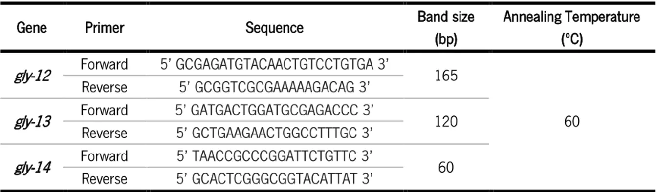
Table 5 - Primers for GNT-I genes (gly-12 to 14) RT-PCR, the band size expected and the annealing temperature.
Gene Primer Sequence Band size
(bp)
Annealing Temperature (ºC)
gly-12 Forward 5’ GCGAGATGTACAACTGTCCTGTGA 3’ 165
60 Reverse 5’ GCGGTCGCGAAAAAGACAG 3’
gly-13 Forward 5’ GATGACTGGATGCGAGACCC 3’ 120 Reverse 5’ GCTGAAGAACTGGCCTTTGC 3’
gly-14 Forward 5’ TAACCGCCCGGATTCTGTTC 3’ 60 Reverse 5’ GCACTCGGGCGGTACATTAT 3’
3.8 Behavioral analysis of motor function
In order to evaluate the motor function of GNT-I mutants, motility and tracking assays were performed; all assays were performed at least 4 independent times.
3.8.1 Strains preparation
The animals were synchronized by placing 10 adult hermaphrodites in 60mm plates (3 per strain) for 2h, time during which the animals lay eggs. These animals were synchronized with a maximum of 2h