Syllabic patterns in typical and atypical phonological development: ultrasonographic analysis
Texto
Imagem


Documentos relacionados
Lopes (2015), reitera que o processo de modelagem de negócios proposto pelo trabalho de Osterwalder (2004), busca através de um processo simulado, “(...) conceber uma abordagem de
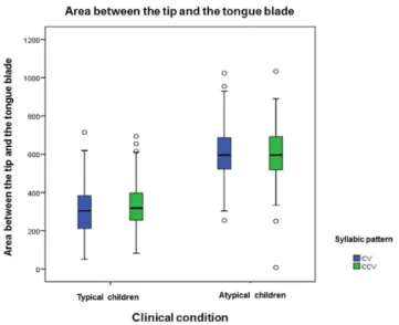
Os resultados mostraram que as crianças atípicas produzem as sílabas estudadas com maior área entre a ponta e lâmina de língua se comparadas às produções do grupo de crianças
Purpose: To compare performance between children with typical speech acquisition, phonological disorders, and childhood apraxia of speech for the variables overall
Given these considerations, the aim of the present study was to describe the typical mobility methods and devices used by Brazilian children with CP in their home, school, and
H2: Ultrasound measures (ratio between tip and blade of the tongue, between tip and dorsum of tongue and between blade and dorsum of tongue) and acoustic (duration) of target
74 Tabela 4: Quantificação de fenóis totais (μg equiv. de ácido gálico/mg de extrato), taninos (μg equiv. ácido tânico/mg de extrato), e determinação de atividade
to analyze and to compare the mean contours of tongue and articulatory gestures in the production of [j] in adults and children with typical development and atypical
The present study aimed to compare the ultrastructural alterations (dilated intercellular space) of the esophageal mucosa in patients with UGIE-proven typical GERD and
