1
“You may never know what results come of your actions, but if you do nothing, there will be no results.”
3
Acknowledgements
This work would not be complete without this section. Although the following sentences are written with deep feeling, no words could really express how thankful I am to all of tireless people who participated in this project.
First of all, to Therapist Adelaide Dias, who were responsible for this huge interest and passion I actually have for swallowing disorders, and for giving me the motivation I need to creation this research.
To Professor Miguel Coimbra in regard to all the guidance and help he gave to me with friendship, understanding and patience, as well as, to his encouragement words and sharing of knowledge and wisdom.
To Unidade de Saúde Local de Matosinhos – Hospital Pedro Hispano, mainly to Dr. Delfim Duarte, the director of ENT service, for authorizing the implementation of this research and for had provided the material necessary for its execution.
Specially thanks to Dr. Nuno Oliveira, doctor from ENT service, for his tireless availability, sympathy, patience and understanding. Without his precious collaboration this work would not be possible to do. Thank him also for its professionalism and sharing of knowledge.
I also express my gratitude to all volunteer participants, which made this happen and for having a fundamental contribution to the advancement of scientific research in this field of knowledge.
To my friend Guilherme and my cousin Claudia for their monitoring in English and for making me unlock my own English skills to develop this work.
To my work friends that help me a lot being my “guinea pigs” drinking water thickened, even though they had feel it was nasty. Thank them also for their real professional and emotional support and friendship, with getting tired.
4
To Rita, my housemate, who always believed in me and motivated me every day with strength and happiness. Thank her for her friendship and for making me laugh and to go on when nerves and fear seemed to take hold on me. Without her, this journey would have probably been difficult and less pleasant in the strenuous moments.
To all my friends for supported me with patience and friendship and for understanding my temporary unavailability in particular moments. To some of them, I also express my gratitude for their help in specific issues I found during this work.
To my family, especially my parents, for always believing in me and in what I do in personal and professional field. I am greatly honored for they giving me constantly love, support and dedication thorough my whole life. To them, I dedicate all this work.
I am completed blessed for having these incredible people in my life. Thank you all!
5
Abstract
Despite of the uncertainties in the interpretation of swallowing sounds, Cervical Auscultation (CA) has been used as a complementary method in assessment of swallowing disorders.
The aims of this study is to describe the acoustic characteristics of swallowing sounds collected through an electronic stethoscope, using Cervical Auscultation method, in healthy adults, and to identify the anatomophysiological origin of sound components.
They were involved 20 subjects aged over 45 years old in good health. CA and Fibreoptic Endoscopic Evaluation of Swallowing (FEES) were performed simultaneously. Sounds were collected using an eletronic stethoscope into a computer device. For each subject it was tasted the swallowing of saliva, free gulp of thin liquid, 10ml of thin liquid, 5ml of pudding, 10ml of pudding and solid. A total of 116 swallowing sounds were analysed in terms of number of sound components and its order of appearing, duration of swallowing process and variation in frequency domain.
They were identified three sounds during swallowing in 59 records. Lub-Dub (LD) and Breath were the most observed sound component. Solid consistency presents the longest time, with a mean result of 15562+-2590 milliseconds, considering the total duration of anatomophysiological event, including Oral Transit Time (OTT). Without OTT, free gulp of thin liquid has the highest mean duration. The longest swallowing sound corresponds to 10ml of pudding. All subjects swallow saliva and 10ml of thin liquid in one gulp. The number of gulps increases with the raise of volume of the bolus. Considering the association between sounds and anatomophisyiological events, LD has the most significant result. In frequency range between 517 and 3617 Hz were observed the most significant difference between the consistencies. In a pilot study included in this thesis, there were found significant differences between duration of swallowing sounds from healthy and dysphagic individuals.
There is still no evidence that CA could be independently used. We can conclude that swallowing sounds contains acoustic characteristics that may allow reliable classification, but further researches are needed to achieve this goal.
Key words: Deglutition, Deglutition Disorders, Cervical Auscultation, Swallowing
6
7
Resumo
Apesar das incertezas existentes na interpretação dos sons da deglutição, a Auscultação Cervical (CA) tem vindo a ser usada como método complementar na avaliação das perturbações da deglutição.
Este estudo pretende descrever as características acústicas dos sons da deglutição recolhidos através de um estetoscópio eletrónico, utilizando o método da CA, em adultos saudáveis, bem como identificar a origem anatomofisiológica dos mesmos sons.
Participaram neste estudo 20 indivíduos com mais de 45 anos, em bom estado de saúde. A AC foi concretizada simultaneamente à realização da Videoendoscopia da Deglutição. Os sons foram recolhidos para um computador através de um estetoscópio eletrónico. Para cada participante foi testada a deglutição de saliva, deglutição em gole livre de liquido, 10ml de liquido fino, 5ml e 10 ml de pudim e sólido. Foram analisados 116 sons em relação ao número de constituintes identificados e ordem de aparecimento, duração do processo de deglutição e variação dos sons no domínio das frequências.
Foram identificados três sons da deglutição em 59 gravações. Lub-Dub (LD) e Breath foram os componentes sonoros mais observados. Considerando a duração total dos eventos anatomofisiológicos, a consistência sólido apresentou o tempo mais longo, com uma média de duração de 15562+-2590 milésimos de segundos, incluindo o Tempo de Trânsito Oral (TTO). Excluindo o TTO, a deglutição livre de líquido apresentou a duração média mais elevada. O som de deglutição mais comprido foi identificado na deglutição de 10ml de pudim. Todos os participantes deglutiram apenas num gole saliva e 10 ml de pudim. O número de goles aumenta com o aumento do volume de alimento. Considerando a associação entre o sons e os eventos anatomofisiológicos, LD foi o componente sonoro com resultado mais significativo. Relativamente às frequências, foram observadas diferenças mais significativas entre 517 e 3617 Hz. No estudo piloto incluído nesta teses, foram observadas diferenças significativas entre as durações medias dos sons da deglutição de indivíduos saudáveis e com disfagia.
Ainda não há evidencia de que a AC possa ser utilizada de forma independente. Conclui-se que os sons da deglutição contêm características acústicas possíveis de uma classificação confiável, no entanto, são necessários mais estudos para atingir esse objetivo.
Palavras-chave: Deglutição, Disfagia, Auscultação Cervical, Sons da deglutição,
8
9
Preamble
Graduated in Speech and Language Pathology, since 2011, at Escola Superior de Tecnologias da Saúde do Porto (ESTSP), Politecnico do Porto, the author integrated a master’s degree in Medical Informatics at Faculdade de Medicina da Universidade do Porto e Faculdade de Ciências da Universidade do Porto, in 2014.
The fact of working in a Continuing Care Unit Speech, evaluating, re-evaluating and treating individuals with swallowing disorders (resulting from stroke, brain injury or other neurologic conditions), created specific needs of knowledge in this field.
Due to difficulties felt in accessing objective exams to confirm the results of clinical assessment of swallowing disorders, stethoscope is the easier and accessible tool at that moment. However, CA is a hard skill to master depending on professional experience and it only has been used as a complementary method to reinforce an initial examination. Although some sounds could be clearly distinctive from each other, uncertainties still remain about the interpretation of the swallowing sounds. A digitally stethoscope could help in studying the acoustic characteristics of the swallowing sounds.
These issues after evolved to the need to know more about CA, by which the research process has been started and a huge interest to contribute to the study of swallowing sounds has grown up to make CA a stronger method, improving its use by health professional.
10
11
Table of Contents
Acknowledgements ... 3 Abstract ... 5 Resumo ... 7 Preamble ... 9 Table of Contents ... 11 List of figures ... 15 List of tables ... 17List of abbreviations and acronyms ... 19
1. Introduction ... 21 1.1. Research problem ... 21 1.2. Research question ... 22 1.3. Objectives ... 22 1.4. Thesis structure ... 23 2. Normal Swallowing ... 25
2.1. Anatomy and Physiology of Swallowing ... 25
2.2. Stages of swallowing ... 26
3. Dysphagia ... 29
3.1. Classification of dysphagia according to the cause ... 29
3.2. Classification of dysphagia considering changes in deglutition phases ... 29
3.3. Classification of disorders in Pharyngeal Dysphagia of neurological ... 30
4. Stroke, swallowing disorders and their impact on quality of life ... 31
5. Swallowing Assessment ... 33
5.1. Clinical evaluation of swallowing ... 33
5.1.1. Selecting consistencies and volumes of the bolus ... 35
5.2. Cervical Auscultation ... 35
5.2.1. Traditional stethoscope versus digital stethoscope ... 36
12
5.4. Objective assessment of swallowing ... 38
5.4.1. Videofluoroscopy Swallowing Study ... 38
5.4.2. Fibre-Optic Endoscopic Evaluation of Swallowing Safety ... 38
5.5. Factors that influence a normal swallowing ... 39
6. Role of Speech-Language Pathologist in swallowing disorders ... 41
7. Acoustic Analysis of sounds ... 43
8. State of Art ... 45
8.1. Methods and materials ... 45
8.1.1. Selection Criteria ... 45
8.1.2. Strategy of research ... 46
8.1.3. Selected Studies ... 46
8.1.4. Studies characteristics ... 46
8.2. Results and discussion ... 47
8.2.1. Health condition, age and gender ... 47
8.2.2. Equipment and procedures ... 48
8.2.3. Volumes and consistencies of food ... 49
8.2.4. Acoustic analyses ... 50
9. Study design and Methodology ... 59
9.1. Participants ... 59 9.2. Local Characterization ... 60 9.3. Resources ... 60 9.3.1. Human Resources ... 60 9.3.2. Instruments collection ... 60 9.3.3. Material resources ... 60 9.4. Procedures ... 61
9.4.1. Ethical procedures for data collection ... 61
9.4.2. Procedures for data collection ... 61
9.4.3. Acoustic analysis ... 63
9.4.4. Statistic analysis ... 65
10. Results ... 67
10.1. Sample description ... 67
10.2. Anatomophysiological events and sound components analysis ... 67
10.3. Duration of anatomophysiological events and sounds from CA ... 69
10.4. Association between anatomophysiological events and each sound component heard 72 10.4.1. Acoustic characteristics of swallowing sounds ... 73
10.4.2. Duration of swallowing sounds in Dysphagic subjects ... 74
11. Discussion ... 77
12. Conclusion ... 83
13
12.2. Main recommendations ... 84
13. Future Work ... 85
14. References ... 87
15. Appendix ... 93
15.1. Appendix 1 - Guideline for Fiberoptic Endoscopic Examinations of Swallowing and Cervical Auscultation exams ... 93
15.2. Appendix 2 – Query used on the online library PubMed with the following keywords ... 95
14
15
List of figures
Figure 1: Representation of three phases of the normal swallowing ... 26
Figure 2: Placement of the stethoscope for cervical auscultation ... 35
Figure 3: Scheme of the selected articles from query. ... 47
Figure 4: Scheme of deglutition waveform ... 50
Figure 5: Profile of pharyngeal swallowing sound in normal subjects ... 51
Figure 6: Acoustic analysis of 10 ml of water in a healthy subject ... 52
Figure 7: Example of vídeo and audio synchronization process. ... 63
Figure 8: Example of acoustic analyzed using Audacity software with label tracks correlating anatomophysiological events and sound componentes. ... 65
Figure 9: Images from FEES. ... 67
Figure 10: Number of sound components identified in the whole sample (116 swallowing records) analyzed in percentage terms, in healthy subjects. ... 68
Figure 11: Percentage of sound componentes identified in the whole sample ... 68
Figure 12: Boxpolt for outcomes about swallowing sounds durations according to different volumes and consistencies of the bolus. ... 71
Figure 13: Percentage of sound components observed in anatomophysiological events .... 73
Figure 14: Average FFT spectra of swallowing sounds' behavior in terms of amplitude and frequency, according to consistencies of the bolus in healthy participants. ... 73
Figure 15: Boxplot correlating swallowing sounds from dysphagic and healthy adults. ... 74
16
17
List of tables
Table 1: Description of Clinical Signals of Changes in Swallowing Biomechanic ... 34
Table 2: Results regarding to mean duration of swallowing sounds in Healthy Subjects according to the consistency and volume of the bolus. ... 54
Table 3: Resume of all articles' results ... 55
Table 4: Found values of Peak Frequencies of swallowing sounds in healthy subjects ... 57
Table 5: Mean Duration of swallowing sounds in dysphagic individuals ... 58
Table 6: Definition of anatomophysiological events and sound components ... 64
Table 7: Frequency and percentage of sound components heard in the each swallowing according to the volumes and consistencies tested in healthy volunteers. ... 69
Table 8: Global variables that describe the anatomophysiological events and the acoustic signal of swallowing sounds in healthy subjects. ... 69
Table 9: Outcomes from SPSS showing the resutlts of Shapiro-Wilk test. ... 70
Table 10: Mean duration of anatomophysiological events and sound components according to different volumes and consistencies of the bolus in healthy participants. ... 71
Table 11: Association between FEES and CA ... 72
Table 13: Total duration of swallowing sounds of 5ml of pudding consistency from dysphagic participants, represented in msec. ... 74 Table 14: Resume of SPSS outcomes about DSS from Dyspagic and Healthy participants 75
18
19
List of abbreviations and acronyms
1B First Burst
2B Second burst
AC Aucultação Cervical
BTS Bolus Transport Signal
C Click
CA Cervical Auscultation
CN Cranial Nerve
CSE Clinical (bedside) Swallow Examinations
D Deglutition of Younger
DA Duration of apnea
DA Deglutition
DS Duration of swallowing
DSS Duration of swallowing sounds
DSS-OTT Duration of swallowing sounds without Oral Transit Time ENT Ear, Nose and Throat
FEES Fiberoptic Endoscopic Evaluation of Swallowing
FFT Fast Fourier Transform
I Interval
LAS Laryngeal Ascension Sound
LB Lub-Dub
M Men
M Misc
msec milliseconds
O Older
OFF Offset Time
ON Onset Time
OTT Oral Transit Time
PC Preclicl
SC Sound Component
SLP Speech-Language Pathologist
T Total duration
Introduction 20
UES Upper Esophageal Sphincter
VFSS Videofluoroscopy Swallowing Study
W Women
Y Younger
PI Peak Intensity
PF Peak Freqency
Introduction 21
1. Introduction
This initial chapter is the first approach to this study, where will be exposed the research problem and its question, the objectives purposed to be studied and the contributions this work can bring not only to the scientific research but also to clinical practicing, mainly in Speech and Language Therapy field.
1.1. Research problem
Swallowing is a complex function of the body that requires the coordination of different structures of the oral cavity, pharynx, larynx and esophagus. It consists in the food passage from the oral cavity to stomach and involves important biological processes that ensure airway protection (Manuscript, 2009; Sarraf Shirazi & Moussavi, 2011; Valim et al., 2007)
However, some ambient and biological factors can disturb this coordination and swallowing becomes unsafe.
Dysphagia may cause a huge incapability, and in most extreme cases might lead to death, for being a risk factor of Aspiration Pneumonia, chronic lung disease, malnourishment or dehydration (Huang et al., 2014; Santamato et al., 2009; Valim et al., 2007).
It is important to make an early diagnosis of this pathology, mainly the signs of deglutition’s pharyngeal phase, once that it is in this phase that it is possible to see all main mechanisms related with airway protection (Canongia & Alves, 2010; Sarraf Shirazi & Moussavi, 2011).
Currently, there are several ways to evaluate deglutition’s capability that can be used in individuals that might present dysphagia.
Clinical (bedside) Swallow Examinations (CSE) is the most frequently used swallowing test in the first contact between patient and health professionals. Although it is ineffective in detecting silent aspiration, it is safe, simple and easily repeatable (Logeman, 1998).
Speech and Language Pathologists (SLP) usually use Cervical Auscultation (CA) to complete their dysphagia’s clinical evaluation. This technique allows swallowing and breath sounds analysis by using a stethoscope (Borr et al., 2007; Moussavi, 2005).
To make an objective assessment there are exams using image as a resource to detect swallowing disturbs, such as silent aspiration phenomena. Videofluoroscopic Swallowing Study (VFSS) and Fiberoptic Endoscopic Evaluation of Swallowing (FEES) are considered the most effective methods to assess dysphagia (Ramsey, Smithard, & Kalra, 2003; Sarraf Shirazi et al. 2012).
Introduction 22
Although less used in swallowing evaluation, there are other complementary exams such as Electromyography (Aboofazeli & Moussavi, 2006) and Pharyngeal Manometry (Shirazi & Moussavi, 2011). There also has been growing interest by the researchers in Sonoe Doppler (Santos et al, 2006; Soria & Furkim, 2016)and Accelerometry (Dudik et al., 2015; Lee et al., 2011) alternative methods of evaluating dysphagia.
In the last years, doctors and researchers are using new techniques based in sound analysis in order to develop alternative and specific techniques of swallowing evaluation (Balasubramanium & Bhat, 2012), such as CA. This method is based in sound analyzes from which it has to be decided if it corresponds to a dysphagia or a normal swallow. Therefore, it must contain objective information that allows classifying in an effective way.
CA has been used as a complementary method in assessment of swallowing disorders, but there are no protocols to conduit the procedures or any objective information about the signal meaning. Professionals interpret the sounds they heard according to their experience in this field, even though they unaware the true origin of the sound.
Despite the objective exams to evaluate swallowing skills, they not always are available on the health care institutions and could be expensive to the patient. CA has advantage of being less expensive, easier to carry and to use without drastically modifying the examination and the swallowing process. Therefore, CA might improve clinical practicing in swallowing disorders, if it could be used as an effective and safe method.
Swallowing sounds occur in milliseconds, requiring practice for a human ear to distinguish a normal from a pathological sound using a traditional stethoscope. A digitally stethoscope could help in studying the acoustic characteristics of the swallowing and make CA a stronger method, improving its use by health professional.
Many researches have been studying the characteristics of swallowing sounds in healthy and dysphagic people. However, there is still a disagreement between them regarding the duration of sounds, the number of sound components and their anatomophysiological origin.
1.2. Research question
The research question for this work is whether there is standardization in swallowing sounds in healthy people, allowing the use of Cervical Auscultation as a potential clinical method to detect swallowing disorders.
1.3. Objectives
The aims of this research is to study acoustic characteristics of swallowing sounds in healthy subjects collected through an electronic stethoscope, using CA method, and identify the anatomophysiological origin of sound components to try improve clinical assessment of swallowing disorders. More specifically, this study intends to:
§ Describe swallowing sounds in healthy people according to volumes and consistencies of the bolus in terms of number of sound components and its order of
Introduction
23
appearing, duration of swallowing process and variation in frequency domain.
§ Identify the origin of swallowing sounds, checking if all the anatomical and physiological events involved in the pharyngeal phase of swallowing, which are observable through FEES, have acoustic representation when collected by digital CA. § Check if CA could be used as a secured method on swallowing assessment.
1.4. Thesis structure
The thesis is organised in fifteen chapters:
§ Chapter one: corresponds to this first approach to this work.
§ Chapter two: defined a normal Swallowing and describes the main concepts about the anatomy and physiology of wallowing.
§ Chapter three: defines dysphagia, presenting classifications done according to its cause or changes in deglutition phases, and classifications of disorders in pharyngeal dysphagia of neurological.
§ Chapter four: indicates the prevalence of stroke and its relation with dysphagia, as well as, their impact on quality of life.
§ Chapter five: presents swallowing assessment methods.
§ Chapter six: describes the role of Speech-Language Pathologist in swallowing disorders.
§ Chapter seven: describes some main concepts related with acoustic analysis of sounds.
§ Chapter eight: presents results from previous studies in regard to swallowing sound and cervical auscultation using a digital stethoscope.
§ Chapter nine: presents the study design and implementation. § Chapter ten: reports the results of the implementation.
§ Chapter eleven: analyse the results, comparing them with others that were found in state of art, and describes some limitations of this work.
§ Chapter twelve: presents the main findings and some recommendations. § Chapter thirteen: purposes to a future work.
§ Chapter fourteen: presents references used thorough this work. § Chapter fifteen: presents the appendix.
Introduction 24
Normal Swallowing 25
2. Normal Swallowing
Swallowing is a complex behaviour with reflex and voluntary actions, involving different structures. An healthy adult swallows approximately 600 times a day, about 35 times per hour while awake and 6 times per hour while sleeping (Manuscript, 2009).
Health professional can only make a corrected swallowing evaluation if they have knowledge of all involved structures and about the physiology of this process. It is important to get the right information about the patients so they can posteriorly plan individuals’ rehabilitation in an adjusted and individualized way.
2.1. Anatomy and Physiology of Swallowing
Different anatomical structures are involved in swallowing events. Bone structures with a distinct participation in this process are maxilla, mandible, cervical spine and hyoid bone. From involved articulations, stands out larynx cartilages: 3 unpaired cartilages (cricoids, thyroid, epiglottis) and 3 pairs of smaller cartilages (arytenoids, corniculate, cuneiform) (Jotz et al., 2009; Leslie et al., 2007; Manuscript, 2009).
Muscle structures are mentioned as tongue and suprahyoid muscles, such as digastric, stylohyoid and infrahyoid muscle. Tongue has both oral and pharyngeal surfaces. Faucial pillars separate oral cavity from pharynx, where there is a tier of constrictors muscles that are originated in hyoid bone and cranium. Cricopharyngeus muscles are connected to the sides of cricoids cartilage and it closes Upper Esophageal Sphincter (UES), compressing it against posterior part of cricoids cartilage. Between tongue’s pharyngeal surface and epiglottis there are the valleculae. In larynx there are the true and the false vocal folds. Adjacent to the larynx, there are two spaces named pyriform recesses (Jotz et al., 2009; Leslie et al., 2007; Manuscript, 2009).
Deglutition demands neuromotor control with participation of cerebral cortex, cerebral trunk and six pairs of Cranial Nerves (CN) that determine sensory and motor impulses, controlling approximately 31 muscles. Involving CN, they all contain both sensory and
Normal Swallowing 26
motor fibers, with exception of CN XII that just contains motor fibers
CN V (Trigeminal) has three divisions (the ophthalmic, maxillary and mandibular nerves). It is responsible for sensation to nasopharynx mucosa, soft palate, hard palate, gums, upper and lower teeth, lips, lower eyelid, sensitivity of the anterior 2/3 of tongue (except sensory taste receptors), cheeks mucosa, floor of the mouth and Temporomandibular Joint. Controls the muscles of mastication (masseter, temporal, pterygoid, mylohyoid, and digastric) and tensor muscle of soft palate. CN VII (Facial) controls the sensation in oropharynx and taste to anterior 2/3 of tongue. Controls facial muscles, stylohyoid, platysma and posterior belly of digastric. CN IX (Glossopharyngeal) is the nerve of ordinary sensation (pressure, touch, temperature and pain) to the pharynx, faucial pillars and palatine tonsil and controls the taste of posterior part of the tongue. It controls stylopharingeus that is involved in pharynx movimentes. CN X (Vagus) is responsible for movements of pharynx muscles (except stylopharingeus) muscle of soft palate (except tensor muscle) and larynx movements that are necessary for airway protection. It is important for sensation to oropharynx, larynx, epiglottis and valleculae mucosa. CN XII (Hypoglossal) controls all tongue movements (Manuscript, 2009; Rainbow, 2001).
2.2. Stages of swallowing
A normal swallowing is generally divided into three phases known as Oral, Pharyngeal and Esophageal (Aboofazeli & Moussavi, 2006; Palmer & Hilamae, 2003). The first phase has its beginning with food entry in mouth. When bolus is ready, tongue exerts pressure against pallet, pushing it towards oropharynx, and Pharyngeal phase starts. This process is now automatic; food stimulates sensory receptors, which trigger the swallowing reflex. In this phase, bolus is transported from oropharynx to pharynx, reaches the epiglottis and then larynx goes up. Esophageal phase starts with bolus passage to esophagus and then food is carried into the stomach by its peristaltic contractions (Canongia & Alves, 2010; Jotz et al., 2009; Sarraf Shirazi & Moussavi, 2011). Figure 1 shows a representation of
swallowing phases.
Figure 1: Representation of three phases of the normal swallowing: (1) Oral phase; (2) Pharyngeal phase; (3)Esophageal phase. Adapt. (Humbert, Christopherson, & Lokhande, 2015)
Normal Swallowing
27
Some authors consider four swallowing stages, dividing Oral Phase in Oral Preparatory Stage and Oral Propulsive Stage. Oral Preparatory Stage corresponds to the moment when bolus is introduced in mouth and starts bolus preparation with the adequate consistency that allows its passage towards stomach. Oral Propulsive Stage has its beginning with bolus transfer from anterior oral cavity to oropharynx propelled by the tongue movements (Hammoudi et al., 2014; Jotz et al., 2009; Hilamae & Palmer, 1999; Manuscript, 2009; Palmer et al., 1992)
However, it is known that there are differences in bolus manipulation, according to introduced food in mouth, and Process Model of Feeding was established to describe the mechanism of eating and swallowing of solid food. All swallowing stages will be explained in detail according to this model (Manuscript, 2009).
Pharyngeal phase involves the most important and complex anatomical and physiological events related to airway protection, which occur rapidly sequential manner. Soft palate elevates against lateral and posterior pharyngeal walls, closing nasopharynx, and then soft palate rises to prevent nasal regurgitation. Tongue base moves back pushing bolus against pharyngeal walls. Pharyngeal constrictor muscles (upper, medium and lower) are activated to transport bolus towards the Pharynx by pharyngeal peristaltic contraction (Manuscript, 2009).
In this stage it is possible to verify the mechanisms of airway protection, which prevent episodes of foreign body aspiration in the trachea before or during swallowing. The vocal folds close to seal the glottis (space between the vocal folds) and the arytenoids tilt forward to contact the epiglottic creating a rapid moment of apnea. Hyoid bone and larynx are pulled upward and forward by contraction of suprahyoid and thyrohyoid muscle. Bolus moves into esophagus after UES is opened (Manuscript, 2009) and then the Esophageal stage begins.
In sum, swallowing is one of the most complex mechanisms that occur in human body (Sarraf Shirazi et al., 2012; Yadollahi & Moussavi, 2007). During all this process, it is possible to observe a sequence of phenomena that they only last milliseconds, which duration and coordination are crucial for a normal and safe swallowing. Any delay or change in this sequence might cause a Oropharyngeal Aspiration and thus dysphagia (Aboofazeli & Moussavi, 2006; Hammoudi et al., 2014; Moussavi, 2005; Sarraf Shirazi & Moussavi, 2011; Valim et al., 2007)
Dysphagia 29
3. Dysphagia
Dysphagia refers to a symptom related with changes that occur during deglutition’s phase that might block or hamper a safe, efficient and comfortable ingestion of food. It may occur in any age group, from new-borns till seniors, due to many different causes (Jotz et al., 2009).
In adults, the most common cause of dysphagia are neurological problems such as Stoke, Parkinson, Alzheimer, Severe Myasthenia, Amyotrophic Lateral Sclerosis, Traumatic Brain Injury and Cerebral Palsy. Dysphagia is also associated to aging process, neck and head cancer, brain tumors and gastroenterological disorders (Escoura, 1998; Jotz et al., 2009).
Classification of dysphagia can be done according to its cause or the phase of swallowing where disorder happens.
3.1. Classification of dysphagia according to the cause
§ Mechanical Dysphagia: normally associated to structural changes caused by head and neck cancer, traumas, infections, among others (Jotz et al., 2009).§ Neurogenic Dysphagia: it is related to the central or peripheral nervous system (Jotz et al., 2009).
3.2. Classification of dysphagia considering changes in
deglutition phases
§ Oral Dysphagia: when there is commitment in the occurred events of Oral Preparatory phase and Oral Propulsive phase. It might be present in cases, such as Oral-Motor Apraxia, Tongue unilateral paralysis and also in maladjusted dental prosthesis (Jotz et al., 2009).
§ Pharyngeal Dysphagia: when there is commitment in Pharyngeal phase in one or more phenomena present, such as in cases like pharynx and larynx paralysis or partial laryngectomy (Jotz et al., 2009).
Stroke, swallowing disorders and their impact on quality of life 30
in Oral Propulsive phase and Pharyngeal phase, in most part of cases it is possible to observe changes of both phases, specially in neurological diseases (Jotz et al., 2009).
§ Esophageal Dysphagia: it occurs when there are mechanical changes and/or dysmotility (e.g. Achalasia). Tumors in the region of the lower esophageal sphincter or gastric cardia can give rise to “pseudo-achalasia”. The diagnosis of this condition is made after medical evaluation and objective tests such as Esophageal Biopsies (Kuo et al., 2012). In contrast with what happens on dysphagia of other types, SLP cannot intervene in Esophageal.
3.3. Classification of disorders in Pharyngeal
Dysphagia of neurological
§ Light Dysphagia: when bolus control and transport is delayed and slow. In these cases, normally are loss of food through lips, tongue’s incoordination, c, absence of cough, absence of significant changes in larynx mobility, absence of change of vocal quality after deglutition and negative CA (Escoura, 1998).
§ Mild or Moderate Dysphagia: when bolus control and transport is delayed and slow, with signs of Oropharyngeal Aspiration. In general it is possible to observe los of food through lips, tongue’s incoordination, when bolus control and transport is delayed and slow, absence of cough, presence of cough before, during or after deglutition, larynx mobility decrease, vocal quality changes after deglutition and CA sometimes modified (Escoura, 1998).
§ Severe Dysphagia: when there is a considerable presence of Oropharyngeal Aspiration and absence or fail in bolus complete deglutition. It is possible to observe a delay or absence in deglutition’s reflex, Larynx mobility decrease, absence of cough, presence of cough during or after deglutition, vocal quality changes after deglutition, a evident breath change, modified CA and incomplete deglutition (Escoura, 1998).
Stroke, swallowing disorders and their impact on quality of life 31
4. Stroke, swallowing disorders and
their impact on quality of life
Stroke is still one of the main mortality causes in Europe (Carvalhido & Pontes, 2009; CIES Observatório das Desigualdades, 2012; Sousa-Uva & Dias, n.d.).
In Portugal, there isn’t yet much information about this issue. According to literature’s most referenced Community based study, involving people over 50 years old, from Coimbra’s county, it is estimated that Stroke prevalence in Portugal might be about 8% (male gender: 10,2%, female gender: 6,6%) (Carvalhido & Pontes, 2009). A more recent study, made in 2013, suggests that Stroke gross prevalence for the resident people in mainland Portugal is 1.9% (Sousa-Uva & Dias, n.d.).
The highest prevalence of stroke was observed in Male gender people with ages between 65 and 74 years old (Sousa-Uva & Dias, n.d.). In the US, studies showed that 34% of people hospitalized for stroke were younger than 65 years (Hall, Levant, & DeFrances, 2012).
Stroke is considered the main cause of dependence and inability of Portuguese population, despite all increasing positive developments in prevention and rehabilitation areas (Carvalhido & Pontes, 2009)
Mortality rate caused by cardiovascular diseases has decreased between 2007 and 2011, from 79,9 deaths per 100 000 inhabitants to 61,9 (Direção-Geral da Saúde, 2013). With all advances in health area, it is possible to verify that there is a growing number of people that survived to this condition, being more frequent the presence of sequelae in these people.
All effects caused by a Stroke are unpredictable and diverse. Brain is an extremely complex organ that controls several parts of human body, and all consequences vary according to brain’s affected region. It might appear some changes that can be physical, emotional or behavioural and communication or speech (American Heart Association, 2012). Swallowing disorders are associated to Stroke (Santamato et al., 2009).
Dysphagia is a condition that is present in most individuals with stroke, especially in the acute stage (González-Fernández, Ottenstein, Atanelov, & Christian, 2013), and it is one of the most common causes of morbidity (Falsetti et al., 2009). In some situations patients recover spontaneously and virtually without sequelae. Other ones maintain long-term difficulties that put them at risk of malnutrition, dehydration and develop pneumonia (González-Fernández et al., 2013; Falsetti et al., 2009) and in extreme cases, lead to death (Huang et al., 2014). There is a high incidence of aspiration pneumonia associated with
Stroke, swallowing disorders and their impact on quality of life 32
dysphagia after stroke. It may occur in 43% to 50% of cases during the first year, with a mortality rate of about 45%. It is also associated with recurrent and prolonged hospitalization (Smithard et al., 2007).
In more serious cases, oral feeding is no longer possible and it is necessary to find alternative forms of nutrition and hydration. In on of the articles found it was mentioned that 80% of individuals with long-term dysphagia require alternative means of enteral feeding (Broadley et al., 2003). These disabilities can significantly impact the quality of life of these people (González-Fernández et al., 2013; Huang et al., 2014; Falsetti et al., 2009; Singh, 2006; Smithard et al., 2007).
It is a difficult task to assess quality of life because it depends on a subjective assessment that involves parameters related to personal, social, professional and emotional aspects of a person’s life (Jotz et al., 2009). WHO defines Quality of Life “as individuals perception of their position in life in the context of the culture and value systems in which they live and in relation to their goals, expectations, standards and concerns” (Group, 1998).
Restrictions and swallowing difficulties experienced by these individuals in their daily routines can lead to feelings of frustration, discouragement, shame and embarrassment especially when they are in front of their family, friends and colleagues. These feelings can make them to want to have a meal alone and avoid public places (Jotz et al., 2009). Often there are sensorial changes, such as the change in the sense of taste, and less pleasure in eating.
Ekberg et al., 2002, notes that only 45% of individuals with dysphagia have pleasure in eating, 41% experience anxiety and panic during meals and that more than 1/3 of the individuals avoid eating because of swallowing difficulties (Ekberg et al, 2002).
The evaluation of swallowing disorders is an important process in monitoring of this people, without forgetting the study of quality of life, although it will not be a subject in this study.
Swallowing Assesment
33
5. Swallowing Assessment
Prior to the completion of the evaluation process, the health professional should collect all necessary and available information about the person who will observe, namely its ability to respond to anamnesis issues. The anamnesis is the first step in this process, enabling the detection of signals that may be associated with changes in swallowing and in the perception of expectations of the person and/or caregivers to the feed. Anamnesis is also important because it is the first contact between the healthcare professional and the person with dysphagia, being at this stage that starts the therapeutic ratio.
Evaluation’s decision depends on the underlying pathology of the person, the data collected during anamnesis, and the all factors involved in the intervention context.
Currently, the standard of dysphagia assessment begins with CSE to observation of signs and symptoms (Dudik et al., 2015). This evaluation is very simple, easily reproducible, doesn’t have any risk to the person examined and allows to define disturbances on oral phase and to analyse aspiration risk signs. However, it is not effective in silent aspiration’s detection, because it doesn’t allow direct observation of pharyngeal phase (Ramsey et al., 2003; Santamato et al., 2009).
Over time, they have been developing studies that investigates the reliability and validity of these two forms of assessment (Bergstrom et al., 2013).
Screening test indicates the probability of an individual having dysphagia and the need to be monitored by a SLP for a complete evaluation. It does not define the severity nor the nature of dysphagia and can not be used to plan the intervention (Bergstrom et al., 2013).
CSE may actually occur as the first dysphagia assessment (with no screening test taking place) or may occur after a simple screening test. The purpose of the CSE is to determine the individual's baseline, realizing their real skills, may using a swallowing evaluation protocols, and to plan intervention in dysphagia (Carnaby-Mann & Lenius, 2008; Cichero & Murdoch, 2006; Luker et al., 2010; Bergstrom et al., 2013).
5.1. Clinical evaluation of swallowing
Clinical examination is made perceptually, according to the knowledge of the observer and his professional experience. Prior to food swallowing test, must be observed all the structures involved in stomatognathic functions. It begins by evaluating the oral sensory-motor system, seeking to collect information in regard to the overall look, symmetry and posture, extra sensibility and intra-oral, tone and mobility of oral and laryngeal structures,
Swallowing Assesment 34
as well as oral reflex (pathological and normal). The professional ought to conduct a thorough neurological examination of the cranial nerves, through specific orders to overall assessment of their duties. The evaluation of speech (voice and joint) is also required to complete the process (Donovan et al., 2013).
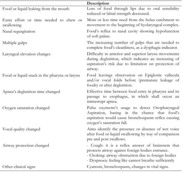
If the individual has conditions to eat certain food or liquid, the assessment continues using different consistencies, selected according to the characteristics of the person identified in previous procedures (Canongia & Alves, 2010). This type of evaluation is often called functional assessment. After the consistency test being defined, and respective order, must be aware of the clinical signs of alternations in the biomechanics of swallowing according to Logeman (1998) (Table 1).
Table 1: Description of Clinical Signals of Changes in Swallowing Biomechanic
Signs Description
§ Food or liquid leaking from the mouth Loss of food through lips due to oral sensibility reduced or labial strength decreased.
§ Extra effort or time needed to chew or
swallowing More or less time need from the bolus catchment to movement to the beginning of hyolaryngeal complex. § Nasal regurgitation Food’s reflux to nasal cavity showing hypofunction
of soft palate.
§ Multiple gulps The increasing number of gulps that are needed to complete food’s cleanliness, as a dysphagia indicator. § Laryngeal elevation changes Difficulty in anterior and superior larynx movements
during deglutition, which indicates an increasing of aspiration’s risk due to limitation on protection of airway.
§ Food or liquid stuck in the pharynx or larynx Food leavings observation on Epiglottic vallecula and/or vocal folds before (premature leakage of foods) or after deglutition.
§ Apnea’s deglutition time changed Effective time between food entry in pharynx and its passage to esophagus, in which shall occur an ininterrupt apnea.
§ Oxygen saturation changed Pulse oxymetre’s usage to detect Oropharyngeal Aspiration, basing in the chance that food’s aspiration would cause bronchospasm reflex causing oxygen’s saturation fall.
§ Vocal quality changed Aims identify the presence or absence of wet voice after food or liquid swallowing by way of comparison pre and post swallows.
§ Airway protection changed - Cough: it is a reflex answer of brainstem that protects airway against foreign bodies entrance. - Choking: airway obstruction due to foreign bodies - Dyspnoea: feeling like cannot breathe sufficiently § Other clinical signs Cyanosis, bronchospasm, changes in vital signs.
To complete the functional assessment of swallowing, SLP must resort to CA (before, during and after the swallowing process). Pulse Oxymetry can also be used to complete evaluation of dysphagia.
SLP can guide their evaluation through informal or formal protocols. There are two types of instruments: those that assess the functional swallowing skills and other assessing
Swallowing Assesment 35
how swallowing influences the quality of life of a person. The existence of several rating scales proofs that there is no optimal method that can be applied equally to all people. Some scales were developed and validated for use in specific circumstances such as in cancer pathologies of the head and neck. Thus, it is important to analyse all the factors in deciding the most appropriate protocol (Vinidh Paleri & Bradley, 2012).
5.1.1. Selecting consistencies and volumes of the bolus
There are different classifications to define the consistencies of food. Most authors consider that the consistency can range from thin, nectar-thick and honey-thick liquid, semi-solid, soft solid consistence and hard solid.
Thin liquids (such as water) rapidly lose its cohesion. Thus, if the person has a serious deterioration of swallowing, the risk of penetration and aspiration increases considerably. In these situations the assessment can be carried out using a slightly thicker consistency as the nectar-thick and honey-thick (Cichero & Murdoch, 2006).
All consistencies are ingested, swallowed and handled in different ways. The volume of food also influences how we control the cake in the mouth and the way we swallow it. Volumes 5 and 10 ml are considered the most appropriate for use in functional swallowing assessment by providing more accurate information to identify the tell-tale signs of laryngeal penetration and tracheal aspiration, and to facilitate the interpretation and preparation of the intervention process (Logeman, 1998). In most studies found in regard to this subject the volumes of 5 and 10 ml are the selections of researchers, making measurements with appropriate spoons or syringes.
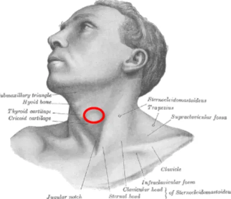
5.2. Cervical Auscultation
This technique stands out for being a non-invasive and simple manipulation procedure in individuals that are seriously affected (Borr et al., 2007). This technique complements clinical evaluation, it gives additional information about pharyngeal phase with no significant influence in the process of swallowing.
Figure 2: Placement of the stethoscope for cervical auscultation. The red circle corresponds to the placement of the stethoscope. Adapt. (Gray, 1985)
Swallowing Assesment 36
You can hear the sounds of breathing and swallowing through a stethoscope, placed on the lateral side of the neck onto the lateral border of larynx and trachea (posteroinferior border of the cricoid cartilage) (Takahashi et al., 1994).
In 1992, first suggestion were made about the existence of three physiological causes of the sounds of swallowing: the first sound corresponds to the elevation of the larynx and bolus flow through the pharynx; the second corresponds to the passage of the cake by the hypopharynx and the movement of the cricopharyngeal sphincter; the last sound is associated with the downward movement of the larynx, after swallowing (Cichero & Murdoch, 2006).
Over the years, there are still studies to be carried to analyze these phenomena, and there is still controversy regarding the source of sounds of swallowing. More recent studies point to the existence of more than three sounds of swallowing.
The majority of studies conclude that the AC can not be used as an isolated method in tracheal aspiration detection, but add that it should not be excluded from the clinical evaluation (Borr et al., 2007; Leslie et al 2004; Santamato et al. 2009; Stroud et al., 2002)
When you hold the acoustic analysis of sounds, consider that the results suggest lack of parameters used to investigate the temporal structure of swallowing sounds. Reinforce the idea that the values are not suitable for use as guidance in defining a diagnosis of dysphagia with particular risk of aspiration and penetration (Borr et al., 2007; Santamato et al., 2009).
Yet there is little evidence regarding the correspondence between the sounds of swallowing and physiological events of the pharyngeal phase which makes the standard changes detection (normal or pathological) more difficult, so you cannot replace the objective tests.
However, CA made through this way still remains as a subjective method (Santamato et al., 2009) because it depends on appraiser’s characteristics and its clinical experience (Rainbow, 2001).
5.2.1. Traditional stethoscope versus digital stethoscope
These days there are two types of stethoscopes on the market that can be used by health professionals: the traditional acoustic and electronic (amplifying and digitizing) stethoscopes (Alaska Native Tribal Health Consortium, 2013; Cruz-Cunha et al. 2016; Leng et al., 2015)
Traditional acoustic stethoscope, created in 1816 by French physician Renne Laennec, is a device that allows listen sounds coming from inside the body. Initially it was made up of a wooden tube and was monaural, meaning that only you could hear was the sound of a single ear. In 1851 Arthur Leard invented the binaural stethoscope with characteristics similar to that currently used in clinical practice. The information provided by acoustic stethoscope is due to three distinct components (Chest Piece, tubing and ear pieces) each with a specific function (Cruz-Cunha et al., 2016; Leng et al., 2015).
The acoustic stethoscope allows the transmission of the sound that is collected from the chest piece through the tubing to the examiner's ear (Cruz-Cunha et al., 2016; Leng et al., 2015).
Swallowing Assesment 37
The chest piece is usually made up of two parts that have to be in direct contact with the person examined to be able to extract sound, these are: the bell (hollow cup) and the diaphragm (disc). The examiner hears the sound that results from converting pressure waves transmitted by the human body (Cruz-Cunha et al., 2016; Leng et al., 2015).
Stethoscope tubing can be defined as a means of sound propagation. It consists of a hollow tube with air that allows the passage of sound energy picked up by the chest piece to the examiner's ear (Cruz-Cunha et al., 2016; Leng et al., 2015).
Ear Pieces closes the circuit propagation of the collected sound. Any change in this process, such as rupture of any of the components can result in decreased sound quality, or completely prevent its transmission (Cruz-Cunha et al., 2016; Leng et al., 2015).
Electronic stethoscope, created by Albert Abrams, amplifies the sound of the human body through advanced technology, overcoming the noise levels that are normally associated with sound recorded by traditional stethoscopes. These devices convert acoustic sound waves through the chest piece obtained by electronic signals propagating in well-defined circuits to process a sound optimization on different frequencies (Alaska Native Tribal Health Consortium, 2013; Cruz-Cunha et al., 2016; Leng et al., 2015)
Electronic stethoscope has also the function of scanning, coding and decoding beep, managing to reduce ambient noise and eliminate other type of contamination of the original sound. The collected sound generally can be recorded to an external device such as a computer or tablet, to be subsequently manipulated (for example, signal processing). This allows the health professional access to this information every time they consider necessary for their clinical practice to confirm diagnoses or control medical conditions (Alaska Native Tribal Health Consortium, 2013; Cruz-Cunha et al., 2016; Leng et al., 2015).
It is understood that the above characteristics are presented as the primary advantages of electronic stethoscopes compared to traditional. As disadvantages it is mentioned that they are generally heavier and more difficult to transport. Furthermore they require batteries to operate properly and are more expensive than the traditional ones. They can also suffer interference from mobile phones or other devices that emit electronic signals (Alaska Native Tribal Health Consortium, 2013; Cruz-Cunha et al., 2016; Leng et al., 2015).
5.3. Pulse Oxymetry
Pulse Oximetry is a method of assessment using a device (pulse oximeter) that is placed on the finger and, through infrared light, measures blood oxygen saturation of the blood. Prior to the evaluation of swallowing are measured baseline values. During the Feeding of the test consistency, the values observed are the oxygen saturation of the blood and compared with the initial values. Variations in saturation may be indicative of aspiration or penetration without clearing the bolus (Rainbow, 2001; Ramsey et al., 2010).
More recent studies are inconclusive regarding the association of these phenomena. Some researchers find relationship but with the wide range of sensitivity and specificity values on comparison with VFSS or FEES (Ramsey et al., 2010).
Swallowing Assesment 38
Although uncertainties remain regarding its effectiveness in detecting aspiration and penetration, this method continues to be part of most protocols established by SLP in the evaluation of swallowing disorders.
In order to objectify the results of the functional assessment of swallowing, especially in inconclusive clinical examinations, supplementary diagnostic tests may be performed in order to identify the actual skills and limits of swallowing.
5.4. Objective assessment of swallowing
According to The American Speech-Language-Hearing Association (ASHA) a clinical-instrumental correct evaluation of swallowing must be such as organic and functional alterations of the structural involved and identify the level of effectiveness of swallowing in different phases. It should also enable the observation of the mechanisms involved in the protection of the lower airways and co-ordination between breathing and swallowing, as well as allow the detection and, if possible, quantify any penetration of the bolus in the tracheal-bronchial passage (Nacci et al., 2008; American Speech-Language-Hearing Association, n.d.)
Currently the two most used objective evaluation methods of swallowing are VFSS and FEES. Both are considered the most effective methods for evaluation of dysphagia (Ramsey et al., 2003; Sarraf Shirazi et al., 2012), using the image to observe the anatomical and physiological phenomena that occur during the process of swallowing.
5.4.1. Videofluoroscopy Swallowing Study
VFSS is a radiological exam that uses modified barium during swallowing. Allows to evaluate the anatomy and physiology of swallowing, and monitor the traffic of the bolus in real time, evidenced all these amendments in case of dysphagia, including disturbed structures and pathological phenomena (such as silent aspiration) (Rainbow, 2001).
It is considered the gold standard, although it presents some disadvantages, due to its not portable, complex and slow process and imply that people are exposed to radiation (Ramsey et al., 2003), which gets harder to make a periodic revaluations and compromise intervention’s effectiveness.
5.4.2. Fibre-Optic Endoscopic Evaluation of Swallowing Safety
FEES is often the first choice when it comes to choosing the objective method for the study of swallowing disorders (American Speech-Language-Hearing Association, 2004).
Using a nasendoscope can functionally evaluate the pharyngeal stage of swallowing. The test involves inserting a flexible endoscope through the nose, getting positioned with the tip slightly below the uvula and above the vocal cords. Some people need local anesthesia during the exam because they show greater discomfort associated with the endoscope (Rainbow, 2001).
During this examination are requested speech tasks as well as vocalizations to assess the anatomy and physiology of the velum, oropharynx, hypopharynx and larynx. The
Swallowing Assesment 39
evaluation of swallowing is performed using different food types (according to the pre-established objectives) containing food coloring. This dye helps differentiate more easily foods secretions, structures and mucous in the oropharynx and larynx (Working). During this examination it is still possible to test maneuvers and swallowing strategies required for effective and safe (American Speech-Language-Hearing Association, n.d.).
The conduct of the examination and analysis of the results from the SLP requires advanced knowledge and specific on this area. According to American Speech-Language-Hearing Association (2004), SLP should be able to: determine the appropriate evaluation protocol; make quick and assertive decisions during the examination; have knowledge about the anatomy and physiology of the larynx; observe secretions; quickly assess swallowing food and liquid; select therapeutic maneuvers and interventions to Improve the swallow (American Speech-Language-Hearing Association, n.d.)
FEES can be performed during hospitalization at the bedside or in an outpatients department, being advantageous for individuals who have some sort of limitation or restriction that prevents them from moving on. Also it can be recorded in video if needed, it is a safe method and tolerated. It can be done with periodicity in order to control patient's evolution (Rainbow, 2001).
However, it has some disadvantages such as the fact that only allow access to the pharyngeal phase of swallowing. It is also dependent on qualified health professional and specialized also equipment. Besides that, it provides little information about pharyngeal and esophageal phases (Ramsey et al., 2003) and presents swallowing whiteout, when the swallowing reflex starts and the endoscope is in contact with the base of the tongue, with the epiglottis and the bolus itself, losing image (American Speech-Language-Hearing Association, 2004; Nacci et al., 2008).
5.5. Factors that influence a normal swallowing
Eating and drinking are basic needs for human being. For the majority, these actions assume big importance for its social character, considering the number of meals in someone else’s company, happening often in public places. Appetite, food presentation, the atmosphere, local hygiene and comfort of, social context, person’s state of mind and their life experiences may influence the way how we eat or drink (Rainbow, 2001).
Deglutition’s physiology may be influenced by the following factors: posture, self-feeding, cognitive influences, bolus size, bolus viscosity, disuse-dysfunction and normal ageing. Therefore, it is necessary to take into account all these factors during evaluation process and dysphagia’s intervention (Rainbow, 2001).
Role of Speech-Language Pathologist in swallowing disorders
41
6. Role of Speech-Language
Pathologist in swallowing disorders
According to American Speech-Language-Hearing Association (n.d.), SPL play a primary role in the evaluation and treatment of swallowing and feeding disorders in infants, children, and adults.This professional have knowledge of anatomy, physiology, and functional aspects of the upper aerodigestive tract (including oral, pharyngeal and cervical esophageal anatomic regions), that are involved in swallowing process and speech. He knows about underlying medical and behavioral etiologies of swallowing and feeding disorders too. Considering cognition, language, and behavioural skills could be affected in these patients, SLP could be more efficient diagnosis and management of swallowing, hence he is expert in communication disorders (Arvedson et al., 2015).
The assessment of swallowing disorders is a crucial moment for developing management strategies. First of all, SLP needs to take a careful history of medical conditions and symptom of the patient. Then, he has to examine the strength and movement of the muscles that are involved in swallowing process. Following a correct procedure, he has to observe the patient feeding looking to posture, behavior, and oral movements during eating and drinking.
The treatment is depending on the cause, symptoms, and type of swallowing problem SLP had detected. Usually, exercises to improve muscle movement are needed. SLP can also give strategies to help the individual to swallow safety more effectively and to select specific consistencies of food and liquid textures most appropriated to the patient. Family and caregivers are included in intervention too. They can make question and expose the doubts as well as to share strategies and receive recommendations to follow out of the therapy context (American Speech-Language-Hearing Association, n.d.)
In fact, the assessment and treatment of swallowing disorders are complex procedures. Therefore, SLP ought to work as a team with families, caregivers, and patients to have success in the intervention (Arvedson et al., 2015; American Speech-Language-Hearing Association, n.d.)
Acoustic Analysis of sounds 43
7. Acoustic Analysis of sounds
Sound can be defined as a vibration traveling through the air as a sound wave. It results from a source of vibration that requires a material medium for its propagation, compressing the surrounding air molecules, squeezing them closer together, and then rarefying them, pulling them farther apart. When these changes in air pressure vibrate our eardrum, nerve signals are sent to the brain and are interpreted as sound (Foreman, 1990; Martins, 2005).
Frequency is the number of times that a wave cycle repeats per unit time, usually per second. It is measured in cycles per second, or hertz (Hz). About human’s range of hearing, the values are from 20 Hz to 20,000 Hz. Although other frequencies exist, they are inaudible to humans. The perception of frequency is called pitch (Plack, 2013).
The maximum displacement of the vibrating particle from the mean position is defined as Amplitude (or intensity). It is measured in decibels and the human ear interprets this strength of a sound wave as volume or loudness (Martins, 2005).
Acoustic analysis has been used as a tool for studying food textures, mainly through the sounds that are generated by mastication. They analyse the frequency and amplitude of the signal of this biological process. Over the last years, swallowing sounds have also been studied regarding their acoustic characteristics by different researchers (Taniwaki & Kohyama, 2012).
State of Art 45
8. State of Art
Currently, the ability to detect dysphagia in the bed remains limited (Santamato et al., 2009).
There are several ways to assess swallowing skills that can be used at an early stage. Cervical Auscultation is one of the techniques proposed to complete the clinical evaluation of dysphagia, although it still presents as subjective method (Balasubramanium & Bhat, 2012; Bergstrom et al., 2013; Jestrovi et al., 2014; Morinière et al., 2008; Santamato, et al., 2009; Spaddoto et al., 2012). It is based on a sound analysis from which characterizes the swallowing process and decides whether the sound is normal or pathological (Borr et al., 2007). Should therefore contain objective information enabling an appropriate classification.
In recent years, health professionals and researchers have been using new techniques based on the sound analysis in order to develop alternatives and specific techniques for evaluation of swallowing.
Some authors consider that the acoustic analysis of the sounds of swallowing can help identify disease patterns (Santamato et al., 2009). Although there are different studies focused on this issue, it has not been defined benchmarks for swallowing sounds, nor irrefutable theories to explain the anatomical and physiological causes and acoustic characteristics of these sounds yet.
8.1. Methods and materials
8.1.1. Selection Criteria
They were included all studies that refer and analyze the swallowing sounds of healthy or dysphagic adults, collected by cervical auscultation using a digital stethoscope or a microphone. In pathological cases, dysphagia must be caused by neurological disease, including stroke mandatorily. Inclusion criteria for selecting articles were: studies involving healthy adults; studies involving adults with dysphagia after stroke; cervical auscultation as a method of collecting swallowing sounds that must be recorder through microphone or a