Wubshet Belay Abagero
Exploring the potentialities of waste plant materials for the production of gold
nanoparticles and multi-metallic composite particles and its application in
wastewater treatment
UNIVERSIDADE DO ALGARVE
FACULDADE DE CIÊNCIAS E TECNOLOGIA
Wubshet Belay Abagero
Exploring the potentialities of waste plant materials for the production of gold
nanoparticles and multi-metallic composite particles and its application in
wastewater treatment
Erasmus Mundus MSc in Chemical Innovation and Regulation
Mestrado Erasmus Mundus em Inovação Química e Regulamentação
Trabalho efetuado sob a orientação de:
Work supervised by:
Prof. Maria Clara Costa
Dr. Anahi Dandlen
UNIVERSIDADE DO ALGARVE
FACULDADE DE CIÊNCIAS E TECNOLOGIA
iii
Exploring the potentialities of waste plant materials for the production of gold
nanoparticles and multi-metallic composite particles and its application in
wastewater treatment
Declaration of Authorship
I declare that I am the author of this work, which is original. The work cites other authors and
works, which are adequately referred in the text and are listed in the bibliography.
Wubshet Belay
Copyright: Wubshet Belay. The University of Algarve have the right to keep and publicize this
work through printed copies in paper of digital form, or any other means of reproduction, to
disseminate it in scientific repositories and to allow its copy and distribution with educational
and/or research objectives, as long as they are non-commercial and give credit to the author and
editor.
iv
Wubshet Belay Abagero
UNIVERSIDADE DO ALGARVE Faculdade de Ciências e Tecnologia
2017
Exploring the potentialities of waste plant materials for the production of gold nanoparticles and multi-metallic composite
particles and its application in wastewater treatment
v
Acknowledgment
First and foremost, I would like to express my gratitude to my supervisors; Prof. Maria Clara Costa and Dr. Anahi Dandlen for the continuous guidance, invaluable suggestions and encouragement throughout the study. Without their guidance at every stage, this thesis could never have been completed. I would also like to thank European Commission, the EMMC-ChIR management team, University of Algarve, University of Bologna, University of Barcelona, Heriot-watt University and CCMAR for giving me this great opportunity. I would like to express appreciation to Dr. Jorge Carlier, Tânia Luz Palma for the great help during my laboratory work. Last but for not least, I want to thank GOD for helping me to complete my thesis in due time.
vi
Abstract
Biosynthesis of metallic nanoparticles has been regarded as a green, environmentally friendly and efficient method for nanoparticles production that avoids the usage of toxic chemicals and generation of hazardous waste during the process. In this study we reported a green, facile and rapid biosynthesis method for the synthesis of gold nanoparticles (AuNPs) and multi-metallic composite particles using raspberry leave extract as both reductant and capping agent in a single-pot process. Various waste plant materials were collected from the Algarve region for extract preparation. The waste plant materials were selected based on the availability and the cost of the material. Reduction potential of each extract was determined using FRAP method. Optimization of the synthesis method (synthesis time and metal precursor concentration) was investigated to get the best synthesis conditions. Standard gold-(III) chloride solution for AuNPs and AMD wastewater from São Domingos mine site, southwest Portugal, for multi-metallic composite particles was used as source of the corresponding metal ions. The UV–Vis spectra showed a SPR peak at 560nm for the AuNPs that were synthesized using optimized synthesis conditions which were 15 min synthesis time, 100 mg/L gold-(III) solution concentration and 1:10 (v/v) extract to standard gold (III) solution ratio at room temperature. TEM images of the precipitates obtained from Au-(III) solution showed different shape and sizes which consist of spherical NPs (51–70 nm), along with few rods (71–80 nm), triangular (61-70 nm) and hexagonal (61-70 nm) particles. TEM images of multi-metallic composite particles revealed there were mostly composed of spherical particles with an approximate average size of 100 nm (range between 70-150 nm). In XRD pattern, the crystalline natures of the synthesized AuNPs showed sharp intense peaks of Bragg reflections corresponding to (111), (200), (220), (311) and (222) planes at 2θ values of 38.20, 44.50, 64.70, 77.70 and 81.20, respectively, displays the crystalline nature of AuNPs and no other peaks were observed due to impurities. These planes are assigned to cubic structure of the AuNPs according to the high-Score Plus software with the ICDD PDF-2 database. The AuNPs were further characterized by Energy dispersive X-ray spectra (EDS) analysis, which gives additional evidence for the reduction of Au-(III) solution to elemental gold thus confirming gold as the only element in the precipitate. The EDS analysis for multi-metallic composite particles suggested that the particles are mainly composed by iron, aluminum and copper, probably as oxides. The stability of AuNPs and multi-metallic composite particles were measured using zeta potential and both particles showed moderate to good stability at different pH range and lower stability at pH 1 and pH 2. The application of the synthesized multi-metallic particles as adsorbent was tested for the removal of phosphate from synthetic wastewater and it showed up to 70.0 ± 3.9 % removal efficiency. In conclusion, in this study suitable, eco-friendly, nontoxic, and single pot synthesis method was developed for the production of AuNPs and multi-metallic composite particles using raspberry leave extract. This study also revealed the potential utilization of acid mine drainage wastewater as a source of multi-metallic composite particles which has proven efficiency to remove phosphate from wastewater via adsorption.
vii
Contents
Declaration of Authorship ... iii
Acknowledgment ... v
Abstract ... vi
List of figures ... x
List of abbreviation ... xii
1. Introduction ... 1
1.1 Nanoparticles: Synthesis methods ... 3
1.2 Use of plant extracts in nanoparticle synthesis ... 6
1.3 use of plant extracts for treatment and for metal recovery from metal bearing wastewaters ... 10
1.4 Characterization of nanoparticles ... 10
1.6 Factors influencing the synthesis of metallic nanoparticles using plant extract ... 13
1.6.1 pH ... 13
1.6.2 Time ... 13
1.6.3 Temperature ... 13
1.6.4 Concentration of metal ion or metal salt ... 13
1.6.5 Metal precursors to extract volume ratio ... 14
1.6.6 The surrounding environment ... 14
1.7 Application of NPs in wastewater treatment ... 14
1.7.1 Phosphate removal from wastewater ... 15
1.7.2 Factors affecting phosphate removal via adsorption ... 16
1.8 Objective of the study ... 16
2. Experimental Part ... 17
2.1 Chemicals and Materials ... 17
2.2 Waste plant materials selection and collection ... 18
2.3 Extract preparation ... 18
2.4 FRAP method for measuring reduction potential ... 19
2.4.1 FRAP reagent preparation ... 19
viii
2.5 AuNPs synthesis using raspberry leave extract... 20
2.6 Synthesis of multi-metallic composite particles ... 21
2.6.1 Collection and characterization of acid mine drainage ... 21
2.6.2 Synthesis of multi-metallic composite particles using raspberry leave extract ... 21
2.7 Characterization of AuNPs and multi-metallic composite particles ... 22
2.7.1 UV-Vis spectrophotometer ... 23
2.7.2 Transmission electron microscopy (TEM) ... 23
2.7.2 Energy-dispersive X-ray spectroscopy (EDS/EDX) ... 23
2.7.3 X-ray diffraction (XRD) ... 23
2.7.4 Zeta potential ... 23
2.8 Application of multi-metallic composite particles as adsorbent for phosphate removal ... 24
2.9 Removal efficiency ... 24
2.10 Statistical analysis ... 25
3. Results and Discussions ... 25
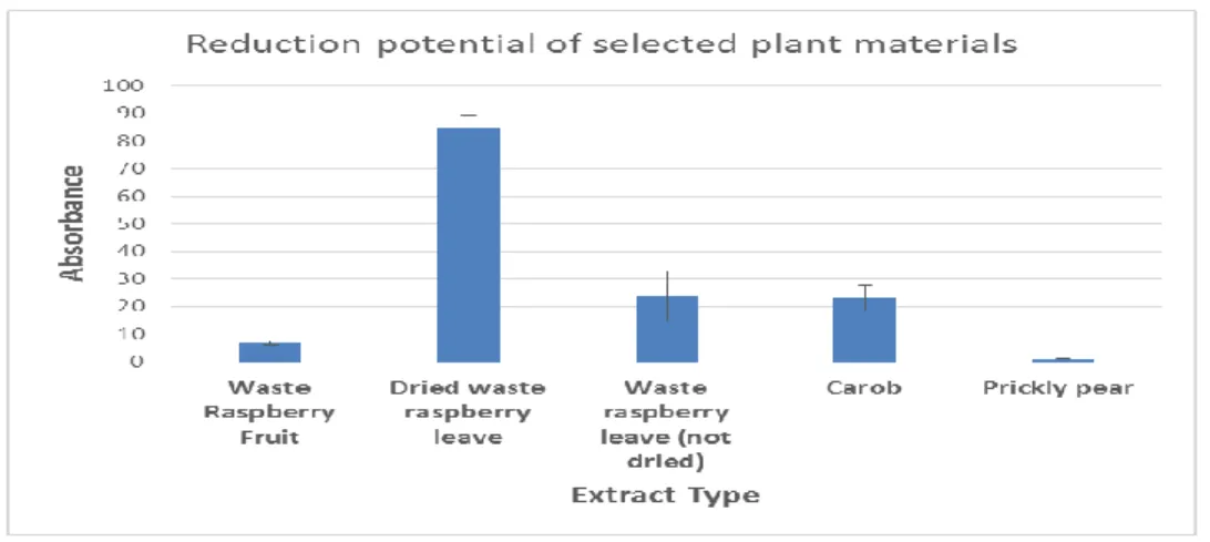
3.1 Reduction potential of selected plant materials ... 25
3.2 Synthesis of gold nanoparticles and multi-metallic composite particles ... 26
3.3 Ultraviolet-visible (UV-Vis) spectroscopy analysis ... 27
3.4 Synthesis of multi-metallic composite particles ... 31
3.4.1 Characteristics of acid mine drainage ... 31
3.4.2 Synthesis of multi-metallic composite particles using dried raspberry leave extract ... 31
3.5 X-ray diffraction (XRD) analysis ... 35
3.6 Transmission electron microscopy (TEM) analysis ... 35
3.6.1 AuNPs ... 35
3.6.2 Multi-metallic nanocomposite particles ... 37
3.7 Energy-dispersive X-ray spectroscopy (EDS/EDX) analysis ... 39
3.8 Zeta Potential analysis ... 40
3.9 Phosphate removal efficiency of multi-metallic composite particles ... 42
4. Conclusion and recommendations ... 44
ix
4.2 Recommendation ... 44 Bibliography... 45 APPENDIX ... 53
x
List of figures
Figure 1 Summary of nanoparticles synthesis method (Amit Kumar Mittal, 2013) ... 5
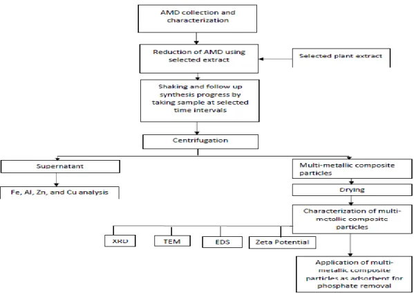
Figure 2 General flow of the experimental procedure ... 17
Figure 3 General flow chart of the extraction process. ... 18
Figure 4: General flow chart for gold (0) synthesis ... 21
Figure 5: General flow chart for synthesis of multi-metallic composite particles ... 22
Figure 6 Reduction potential of selected plant materials. ... 26
Figure 7 Mechanisms of metal nanoparticle synthesis (M+-metal ion) (Amit Kumar Mittal, 2013). ... 26
Figure 8 Color change progress before and after addition of extract to the Au-(III) solution. ... 27
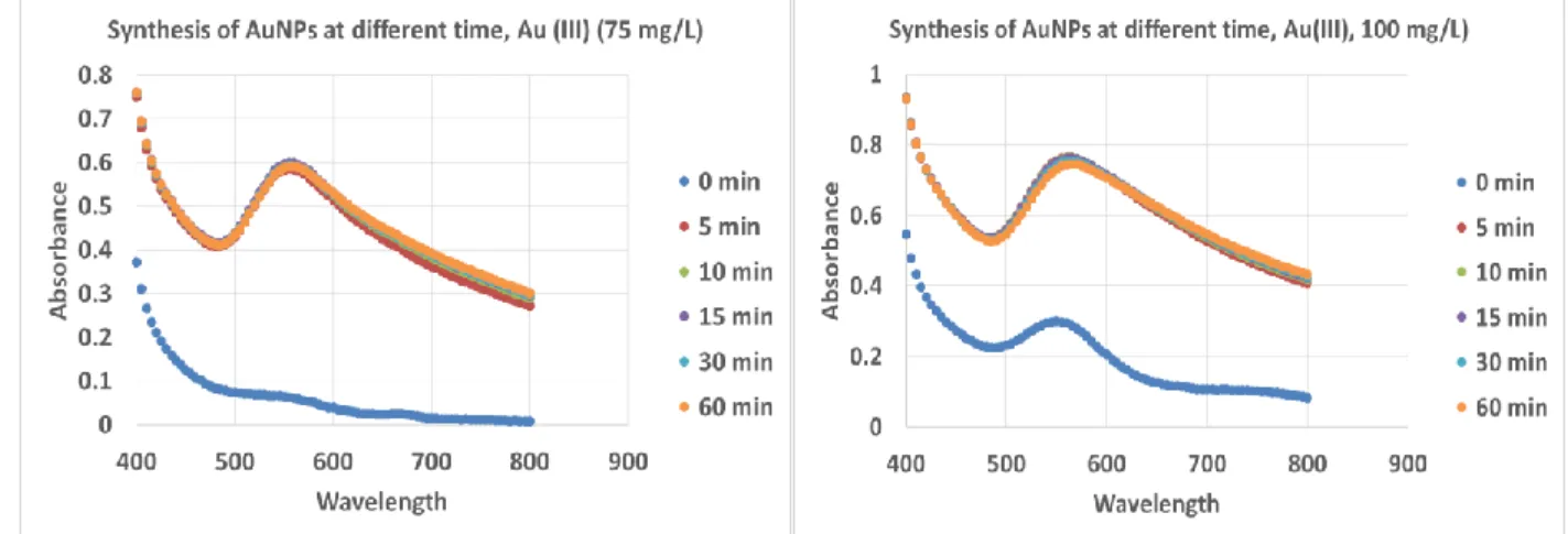
Figure 9 UV-Vis synthesis progresses of AuNPs at different time and concentration. ... 29
Figure 10 UV-Vis comparisons between different synthesis time and concentrations of Au (III) chloride solution. ... 30
Figure 11 Gold film and precipitate on the side wall and bottom of falcon. ... 30
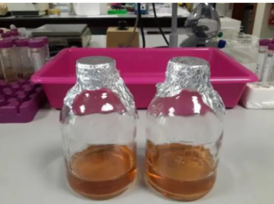
Figure 12 AMD wastewater before (left) and after (right) addition of plant extract ... 32
Figure 13 Initial and final concentrations of metals after extract addition at different time and mixing ratio. (A) 0.5 h, (B) 1h, (C) 2h, (D) 3h, (E) 6h. ... 34
Figure 14 Multi-metallic composite particles. ... 34
Figure 15: XRD analysis of AuNPs. ... 35
Figure 16 TEM images of AuNPs. ... 36
Figure 17: Size distribution of AuNPs. ... 37
Figure 18 Selected electron diffraction areas of AuNPs. ... 37
Figure 19: TEM images of multi-metallic composite particles. ... 38
Figure 20: Selected electron diffraction area of multi-metallic composite particles. ... 38
Figure 21 EDS spectroscopy displays the chemical composition of the gold nanoparticles. ... 39
Figure 22 EDS spectroscopy displays the chemical composition of multi-metallic composite particles. .. 40
Figure 23 Zeta Potential of AuNPs at different pHs. ... 41
Figure 24 Zeta Potential of multi-metallic composite particles at different pHs. ... 42
Figure 26: Phosphate removal efficiency of multi-metallic composite particles using adsorbent concentration. (A) 0.5 g/L, (B) 1g/L ... 43
xi
Table 1 Summary of previous studies on plant mediated synthesis of nanoparticles and their
applications. ... 7
Table 2 Common methods for characterization of nanoparticles. ... 11
Table 3 Preparation of Fe+2 standard aqueous solutions... 19
Table 4: Component 1, Media A………..………..25
Table 5: Component 2, Media B. ... 24
Table 6 Reduction efficiency of dried raspberry leaves extract. ... 31
Table 7 Composition of AMD sample from São Domingo’s mine site in terms of main metals and pH .... 31
Table 8 the stability of colloid particles based on their zeta potential values. ... 40
xii
List of abbreviation
NPs Nanoparticles AuNPs Gold nanoparticles AgNPs Silver nanoparticles FeNPs Iron nanoparticles
TEM Transmission electron microscopy DLS Dynamic light scattering (DLS) UV-Vis Ultraviolet-visible
ED Electron diffraction XRD X-ray diffraction
EDS/EDX Energy dispersive X-ray spectra FRAP Ferric Reducing Ability of Plasma COD Chemical Oxygen Demand ANOVA Analysis of variance AMD Acid Mine Drainage
FAAS Flame atomic absorption spectroscopy
MP-AES Microwave plasma-atomic emission spectroscopy FAAS Flame atomic absorption spectroscopy
SPR Surface plasmon resonance TPTZ Tripyridyltriazine
1
1. Introduction
Nowadays nanoparticles and their technologies are booming rapidly due to their enormous applications namely water and wastewater treatment, pharmaceuticals, electronics, paints, cosmetics, catalysis, biosensor etc.). There is huge production of nanomaterial worldwide (Palaniselvam Kuppusamy M. M., 2016; C. Mystrioti, 2016). However the current manufacturing practice involves many chemicals which are toxic enough to pollute the environment and make the process unsafe during the large scale production of nanomaterial. Thus, exploring green synthesis methods, recovery of resource with less cost for the production of nanomaterials and their application is mandatory (C. Mystrioti, 2016). Hence, it is important to understand the following: what are the available resources, how can we recover resources from waste materials? How nanoparticles can be synthesized greenly without affecting its noble their properties? How can we optimize the application of nanomaterial in various areas such as wastewater treatment? (Palaniselvam Kuppusamy M. M., 2016; S.S. Godipurgea S. Y., 2016).
Nanomaterials are very important developing research area which has been an interest of most of researcher (María Martínez-Cabanas, 2016; P.P.N. Vijay Kumara, 2014; Jasmine Jacob, 2012; Palaniselvam Kuppusamy S. J., 2015). Due to the advantageous distinctive characteristics nanoparticles, their applications are growing promptly on various fields like water and wastewater treatment, biomedical, pharmaceutical, catalysis, drug delivery, antimicrobial, biosensor technology, Catalysts, conductors in various areas such as health sectors, food industry, manufacturing environmental, space industry, optical industries and so on (P. Mohanpuria, 2008; Amit Kumar Mittal, 2013; Niederberger, 2013). The change in the physico-chemical properties of nanoparticles is responsible for the mentioned novel functional attributes of nanoparticles (Niederberger, 2013; Shakeel Ahmed, 2016).
The main factors that nanotechnology depends on are the synthesis and modulation of nanoparticles, which significantly affect their properties. Because of the smaller particle size, various shapes and increased surface area, nanoparticles display very different properties than their bulk materials and are found to be interesting candidates for the numerous applications mentioned above (P.P.N. Vijay Kumara, 2014; P. Mohanpuria, 2008; Niederberger, 2013). Nanomaterials have been used unknowingly for thousands of years; for example, gold nanoparticles that were used to stain drinking glasses also cured certain diseases. Scientists have been progressively able to observe the shape and size dependent physiochemical properties of nanoparticles by using advanced techniques. Recently, the diverse applications of metal nanoparticles have been explored in biomedical, agricultural, environmental, and physiochemical areas (Niederberger, 2013).
Comparing to other nanoparticles, the metallic nanoparticles have been considered as the most promising as they contain remarkable properties due to their large surface area to volume ratio (Niederberger, 2013; Palaniselvam Kuppusamy M. M., 2016). Specific examples of gold nanoparticles applications include delivery of specific drug, such as paclitaxel, methotrexate, and doxorubicin. Gold nanoparticles have been also used for tumor detection, angiogenesis, genetic disease and genetic
2
disorder diagnosis, photo imaging, and photo thermal therapy (Khwaja Salahuddin Siddiqi, 2017). Silver nanoparticles have been used for many antimicrobial purposes, as well as in anticancer, anti-inflammatory, and wound treatment applications (J. Das, 2013; H. Joy Prabu, 2015; B. Ajithaa, 2016; Niederberger, 2013). Due to their biocompatible, nontoxic, self-cleansing, skin-compatible, antimicrobial, and dermatological behaviors, zinc and titanium nanoparticles have been used in biomedical, cosmetic, ultraviolet (UV)-blocking agents, and various cutting-edge processing applications (J. Das, 2013; B. Ajithaa, 2016; H. Joy Prabu, 2015; Niederberger, 2013).
Metallic nanoparticles are commonly used as adsorbent as in polishing steps to remove organic and inorganic contaminants in water and wastewater treatment. The efficiency of conventional adsorbents is usually limited by the surface area or active sites, the lack of selectivity, and the adsorption kinetics. Nano-adsorbents offer significant improvement with their extremely high specific surface area and associated sorption sites, short intraparticle diffusion distance, and tunable pore size and surface chemistry (H. Joy Prabu, 2015; Xiaolei Qu, 2013).
In addition, metallic nanoparticles have been used in the spatial analysis of various biomolecules, including several metabolites, peptides, nucleic acids, lipids, fatty acids, glycosphingolipids, and drug molecules, to visualize these molecules with higher sensitivity and spatial resolution (María Martínez-Cabanas, 2016; Niederberger, 2013; P. Mohanpuria, 2008). Moreover, the unique properties of metallic nanoparticles make them well suited for designing electrochemical sensors and biosensors. For example on its environmental applications, nano-sensors have been developed and used for the detection of algal toxins, mycobacteria, and mercury presence in drinking water. Nano-sensors are also applied for hormonal regulation and for detecting crop pests, viruses, soil nutrient levels, and stress factors. Moreover, nano-sensors for sensing auxin and oxygen distribution have been developed (H. Joy Prabu, 2015; B. Ajithaa, 2016; Khwaja Salahuddin Siddiqi, 2017; Michael Iv, 2015).
Recent investigations in nanotechnologies showed leapfrogging opportunities to develop next-generation water and wastewater treatment systems using nanomaterial (Akbar Soliemanzadeh, 2017; C.P. Devatha, 2016; Jing Liu, 2017). Our current water and wastewater treatment and discharge practices, which heavily rely on conveyance and centralized systems, are not always suitable and sustainable. The highly efficient, modular, and multifunctional processes enabled by nanomaterials are foreseen to provide high performance, affordable wastewater treatment solutions that less relies on large infrastructures. Nanomaterial based wastewater treatment promising not only to overcome the major challenges faced by existing treatment technologies, but also to provide new treatment capabilities that could allow reuse of treated wastewater (A. Jafaripour, 2015; D. Barrie Johnson, 2005; M.A. Martín-Lara, 2014).
Therefore, currently, there is an increasing demand to develop environmentally friendly and sustainable methods for the synthesis of nanomaterials that do not use toxic chemicals in the synthesis protocols so as to reduce the adverse impact on the environment and minimize health risks for human being. Various
3
metallic nanoparticles were successfully synthesized using different plant extracts (Arun Kumar Thalla, 2016; Palaniselvam Kuppusamy M. M., 2016). And also these nanoparticles tested for the application of industrial and domestic wastewater treatment and showed very promising pollutant removal efficiency. The green synthesis methods are advantageous over other conventional methods because they are simple, cost-effective, environmentally friendly, and easily scaled up for industrial scale synthesis (C. Mystrioti, 2016; Palaniselvam Kuppusamy M. M., 2016; Palaniselvam Kuppusamy M. M., 2016).
1.1 Nanoparticles: Synthesis methods
Over the last three decades the synthesis of nanoparticles has been drawn attention in the emerging areas of nanoscience and technology as particles in their nano form show different properties compared to the corresponding bulk material (P. Mohanpuria, 2008; Niederberger, 2013). Usually, metal nanoparticles can be synthesized through several chemical and physical methods. However, many chemicals used are toxic enough to pollute the environment during the large scale production of nanomaterial. Green synthesis methods have been adopted these days to reduce or eliminate the use or generation of toxic substances in the design, manufacture and applications of nanomaterial (H. Joy Prabu, 2015; J. Das, 2013; Khwaja Salahuddin Siddiqi, 2017; María Martínez-Cabanas, 2016; Muhammad Jamil Ahmed, 2015; P.P.N. Vijay Kumara, 2014; Amit Kumar Mittal, 2013; Peter Logeswari, 2015; Silvia Groiss, 2017).
Previous studies showed that various chemical have been explored for the synthesis of NPs (Amit Kumar Mittal, 2013; Jasmine Jacob, 2012; Niederberger, 2013; Shakeel Ahmed, 2016). Nowadays, Extracts of a diverse range of plant species have been successfully used in making NPs. In addition to plant extracts, microorganisms, plant tissue and fruits, plant and marine algae have been used to produce nanoparticles as alternative method to conventional chemical synthesis (Amit Kumar Mittal, 2013; Niederberger, 2013; P. Mohanpuria, 2008). Different types of nanomaterial such as silver, gold, iron, copper, zinc, titanium, magnesium and multi-metallic composite have been produced using this green alternative method. (Amit Kumar Mittal, 2013; Niederberger, 2013; P. Mohanpuria, 2008; Peter Logeswari, 2015). Among various metal nanoparticles, gold, silver, gold/silver composite, iron and multi-metallic composite particles nanoparticles are of particular interest and their green synthesis method still attracting scientists in the field of nanotechnology (Khwaja Salahuddin Siddiqi, 2017; Silvia Groiss, 2017; María Martínez-Cabanas, 2016; S.S. Godipurgea S. Y., 2016; C. Mystrioti, 2016).
Currently, there is an increasing demand to develop environmentally friendly and sustainable methods for the synthesis of nanomaterial that do not use toxic chemicals in the synthesis protocols. A number of approaches are available for the synthesis of nanoparticles, such as thermal decomposition, electrochemical, microwave assisted process and green synthesis. Many of the nanoparticle synthesis or production methods involve the use of hazardous chemicals, low material conversions and high energy requirements. This is the reason why there is a growing demand to develop environmentally friendly and safe processes for NPs synthesis (Khwaja Salahuddin Siddiqi, 2017; María Martínez-Cabanas, 2016; S.S. Godipurgea S. Y., 2016; Silvia Groiss, 2017; B. Ajithaa, 2016; Shakeel Ahmed, 2016; H. Joy Prabu, 2015).
4
Many studies showed that green synthesis methods are advantageous over other conventional methods because they are simple, cost-effective, environmentally friendly, easily scaled up for industrial scale synthesis (Ting Wang, 2014; C. Krishnaraj, 2010; B. Ajithaa, 2016; Jasmine Jacob, 2012; María Martínez-Cabanas, 2016; C. Mystrioti, 2016; C. Krishnaraj, 2010; Arun Kumar Thalla, 2016). In fact a number of bacteria, fungi, and yeast have been well known for the synthesis of nanoparticles (Niederberger, 2013; P. Mohanpuria, 2008). However, it is difficult, time consuming and expensive to use microbial mediated synthesis of nanoparticles industrially because it requires expensive medium (Amit Kumar Mittal, 2013; Niederberger, 2013). Therefore, the exploration of plant extracts as potential alternatives for the synthesis of nanoparticles has gained huge interest. Studies demonstrated that plants with high active ingredients such as water soluble antioxidant polyphenols, alkaloids, flavonoids etc, had reducing, capping or stabilizing ability to reduce metal into their respective nanoparticles (Amit Kumar Mittal, 2013; B. Ajithaa, 2016; Khwaja Salahuddin Siddiqi, 2017; M. Sigamoney, 2016; Muhammad Jamil Ahmed, 2015; María Martínez-Cabanas, 2016; C. Mystrioti, 2016; Arun Kumar Thalla, 2016) .
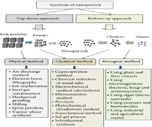
Generally nanoparticles can be produced in two ways, the first one is a “top down” approach and the second one is “bottom up” approach. In “top down” synthesis, nanoparticles are produced by size reduction from their respective suitable starting material. Size reduction can be achieved by various lithographic techniques e.g. grinding, milling, sputtering, thermal or laser ablation, etc. “Top down” method has disadvantage because it will not result perfect nanoparticles in terms of the surface structure and this is the main limitation of the process because the surface chemistry and the other physical properties of nanoparticles are highly dependent on the surface structure. In “bottom up” synthesis, the nanoparticles are built from smaller entities, for example by joining atoms, molecules and smaller particles. In this synthesis, the nanostructured building blocks of the nanoparticles are formed first and then assembled to produce the final particle. In this synthesis approach, nanoparticles can be produced using chemical (chemical reduction) or biological methods, such as the use of plants extract, micro-organisms, by self-assemble of atoms to new nuclei which grow into a particle of nanoscale (Amit Kumar Mittal, 2013; Khwaja Salahuddin Siddiqi, 2017; Niederberger, 2013; P. Mohanpuria, 2008). In chemical synthesis of metallic NPs different organic and inorganic reducing agents, such as sodium borohydride (NaBH4), sodium citrate, ascorbate, elemental hydrogen, Tollen’s reagent, N, N-dimethyl formamide (DMF) and poly (ethylene glycol) block copolymers are used in aqueous or non-aqueous solutions as reducing agents. Capping agents are also used for size stabilization of the nanoparticles. One of the advantages of the chemical methods are a large quantity of nanoparticles can be synthesized in a short period of time. The main disadvantage of these syntheses methods are the fact that the chemicals in use might be toxic, making the production process unsafe and may have an impact on the environment (Shakeel Ahmed, 2016; Niederberger, 2013; P. Mohanpuria, 2008; C. Mystrioti, 2016). This is the reason why biosynthesis draw more attention of scientists in the area of nanoparticles production via green route that does not employ toxic chemicals and also doesn’t create any toxic waste (H. Joy Prabu, 2015; Khwaja Salahuddin Siddiqi, 2017; P.P.N. Vijay Kumara, 2014; Amit Kumar Mittal, 2013; M. Sigamoney, 2016; María Martínez-Cabanas, 2016; Arun Kumar Thalla, 2016). Thus, the advancement of
5
green syntheses of nanoparticles is progressing as a key branch of nanotechnology; where the use of biological entities like microorganisms and plants extracts for the production of nanoparticles could be an alternative to chemical and physical methods (Khwaja Salahuddin Siddiqi, 2017; Amit Kumar Mittal, 2013; Niederberger, 2013; P. Mohanpuria, 2008; S.S. Godipurgea S. Y., 2016). A summary of NPs synthesis method is described in the figure 1 below.
Figure 1 Summary of nanoparticles synthesis method (Amit Kumar Mittal, 2013)
Several methods have been developed for the biological synthesis of nanoparticles from salts of the corresponding metals as a safe and environmentally friendly alternative comparing to chemical synthetic procedures (Priyanka Singh, 2016; Amit Kumar Mittal, 2013; Muhammad Jamil Ahmed, 2015; Ting Wang, 2014; C. Mystrioti, 2016). Synthesis of nanoparticles using microorganisms or plants can possibly eliminate the problem mentioned. The use of plant extracts for the synthesis of nanoparticles could be advantageous over other environmentally friendly biological processes by eliminating the elaborate process of maintaining cell cultures.Biosynthesis methods are useful not only because of the reduced environmental impact and health hazard of the manufacturing process in comparison with some of the physicochemical production methods, but because they can be used to produce large quantities of nanoparticles that are free of contamination and have a well-defined size and morphology (Priyanka
6
Singh, 2016; Xiaolei Zhang, 2011; Khwaja Salahuddin Siddiqi, 2017; María Martínez-Cabanas, 2016; Muhammad Jamil Ahmed, 2015; Palaniselvam Kuppusamy S. J., 2015; Peter Logeswari, 2015).
1.2 Use of plant extracts in nanoparticle synthesis
Typically, a plant extract mediated reduction involves simply mixing the extract with an aqueous solution metal precursor at room temperature or at certain fixed temperature. Generally the synthesis complete within short time. Many researcher synthesized various nanoparticles of silver, gold, Iron and many other metals have been produced this way (Amit Kumar Mittal, 2013; B. Ajithaa, 2016; H. Joy Prabu, 2015; J. Das, 2013; Khwaja Salahuddin Siddiqi, 2017; M. Sigamoney, 2016; Muhammad Jamil Ahmed, 2015; Peter Logeswari, 2015; S.S. Godipurgea S. Y., 2016; C. Mystrioti, 2016). The nature of the plant extract, its concentration, the concentration of the metal salt, the pH, temperature, the addition of extract to the metal solution and contact time are known to affect the rate of production of the nanoparticles, their quantity and other characteristics (Jayachandra Reddy Nakkala, 2016; Amit Kumar Mittal, 2013; H. Joy Prabu, 2015; Peter Logeswari, 2015; Priyanka Singh, 2016; Khan Behlol Ayaz, 2014) . Plant extracts may act both as reducing and stabilizing agent in the synthesis of nanoparticles (Amit Kumar Mittal, 2013). The source of the plant extract is known to influence the characteristics of the nanoparticles. This is because different extracts contain different concentrations and combinations of compounds such as phenolic, alkaloids, flavonoids etc. and the reduction process is relatively complex (Amit Kumar Mittal, 2013; H. Joy Prabu, 2015; Jasmine Jacob, 2012; María Martínez-Cabanas, 2016; Khwaja Salahuddin Siddiqi, 2017; C. Mystrioti, 2016).
Plant extracts are usually composed by different metabolites like terpenoids, phenols, proteins or carbohydrates. These compounds are directly responsible or the extract capacity to carry out the NPs biosynthesis. Each extract contains different concentrations and combination of reducing and stabilizing agents. Therefore, the extract composition determines the characteristics of synthesized NPs (Palaniselvam Kuppusamy M. M., 2016; Rani Mata, 2016; Jayanta Kumar Patra, 2016; Sathishkumar G., 2016; Amit Kumar Mittal, 2013; Akbar Soliemanzadeh, 2017). Comparing to other synthesis procedures, the use of plant extracts for making nanoparticles is simpler. Nowadays plant extract mediated synthesis of metallic NPs, such as AuNPs, AgNPs, FeNPs and multi-metallic composites, is attracting most of scientists’ attention and explored a lot to get the best out of it (Arthanari Saravanakumar, 2015; Sathishkumar G., 2016; Amit Kumar Mittal, 2013; H. Joy Prabu, 2015; Jayachandra Reddy Nakkala, 2016; María Martínez-Cabanas, 2016; Palaniselvam Kuppusamy M. M., 2016). Processes for making nanoparticlesusing plant extracts are readily scalable, less expensive, and take shorter period of time in comparison with the relatively expensive and complex method of microbial synthesis (Amit Kumar Mittal, 2013; Priyanka Singh, 2016; Arthanari Saravanakumar, 2015; P. Mohanpuria, 2008; Khwaja Salahuddin Siddiqi, 2017; Arun Kumar Thalla, 2016).
In comparison to plant extract synthesis, the microbe mediated synthesis methods have several disadvantages such as high cost, need of identification of potential strain, maintenance of aseptic conditions for the profuse growth of microorganisms, chances of infection and contamination. Above all, the microbial synthesis methods are quite time consuming and require about 2-3 days for the growth of
7
a suitable strain and another 1-2 days for the synthesis and purification of NPs. As a result, scientists switched over their interest toward plant extract synthesis methods (Priyanka Singh, 2016; P. Mohanpuria, 2008; Palaniselvam Kuppusamy M. M., 2016; Xiaolei Zhang, 2011; Utkarsha Shedbalkar, 2014).
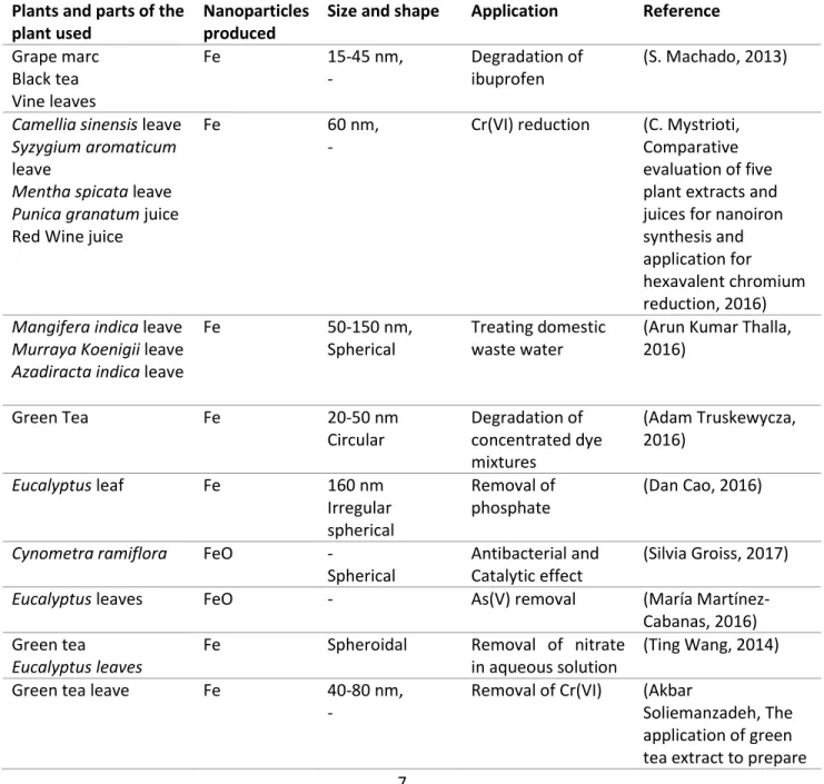
Table 1 Summary of previous studies on plant mediated synthesis of nanoparticles and their applications.
Plants and parts of the plant used
Nanoparticles produced
Size and shape Application Reference Grape marc Black tea Vine leaves Fe 15-45 nm, - Degradation of ibuprofen (S. Machado, 2013)
Camellia sinensis leave Syzygium aromaticum leave
Mentha spicata leave Punica granatum juice Red Wine juice
Fe 60 nm,
-
Cr(VI) reduction (C. Mystrioti, Comparative evaluation of five plant extracts and juices for nanoiron synthesis and application for hexavalent chromium reduction, 2016) Mangifera indica leave
Murraya Koenigii leave Azadiracta indica leave
Fe 50-150 nm,
Spherical
Treating domestic waste water
(Arun Kumar Thalla, 2016) Green Tea Fe 20-50 nm Circular Degradation of concentrated dye mixtures (Adam Truskewycza, 2016) Eucalyptus leaf Fe 160 nm Irregular spherical Removal of phosphate (Dan Cao, 2016)
Cynometra ramiflora FeO -
Spherical
Antibacterial and Catalytic effect
(Silvia Groiss, 2017)
Eucalyptus leaves FeO - As(V) removal (María
Martínez-Cabanas, 2016) Green tea
Eucalyptus leaves
Fe Spheroidal Removal of nitrate
in aqueous solution
(Ting Wang, 2014)
Green tea leave Fe 40-80 nm,
-
Removal of Cr(VI) (Akbar
Soliemanzadeh, The application of green tea extract to prepare
8
bentonite-supported nanoscale zero-valent iron and its
performance on removal of Cr(VI): Effect of relative parameters and soil experiments, 2017) Green tea Oolong tea Black tea Fe 40-50 nm, Spherical Degradation of malachite green (Lanlan Huang, 2014) Onion peel Au 25-70 nm, Spherical Triangular Antibacterial Antioxidant
(Jayanta Kumar Patra, 2016)
Piper longum fruit Au 56 nm
Spherical Antioxidant Catalytic activities (Jayachandra Reddy Nakkala, 2016) Salicornia brachiate Au 25-35 nm face centered cubic Antibacterial Catalytic activities
(Khan Behlol Ayaz, 2014)
Plumeria alba flower Au 15 and 28 nm,
Spherical
Catalytic degradation of organic dyes and inhibit bacterial growth (Rani Mata, 2016) Couroupita guianensis fruit Au 25 nm, Spherical Triangular Hexagonal Face centered cubic
Antioxidant activity (Sathishkumar, 2016)
Capsicum annuum var. grossum pulp
Au 6-37 nm
Triangle Hexagonal Quasi- spherical
Catalytic activity (Chun-Gang Yuan, 2017) Pogestemon benghalensis (B) O. Ktz. leaf Au 10-50 nm Spherical Triangular Photocatalytic activity degradation of methylene blue (Bappi Paul, 2015) Moringa oleifera flower Au 3-5 nm Spherical anti-cancer and catalytic activity (K. Anand, 2015)
9 Hexagonal Triangular Green and red
cabbages Ag, Au and bimetallic (Ag/Au) 20nm (Au) Triangular(Au) Spherical(Au) Spherical (Au/Ag) 25nm (Au/Ag) - (Jasmine Jacob, 2012) Aerial parts of R. hypocrateriformis Ag, Au and Au-Ag alloy 10-50 nm, Spherical Antimicrobial Antioxidant Anticancer activities (S.S. Godipurgea S. Y., 2016)
Cassia tora leave Ag -
Spherical Hexagonal Irregular Antioxidant Antibacterial activities (Arthanari Saravanakumar, 2015) Sesbania grandiflora leave Ag 16 nm, Spherical cubic Antimicrobial activity (B. Ajithaa, 2016)
Acalypha indica leaf Ag 20-30nm
cubic face-centered
antibacterial activity against water borne pathogens (C. Krishnaraj, 2010) Tragia involucrate leave Cymbopogon citronella Solanum verbascifolium leave Tylophora ovata leave
Ag 40-45 nm Spherical face-centered cubic - (H. Joy Prabu, 2015) Sesbania grandiflora leave Ag 10-25, Spherical Antibacterial human pathogens (J. Das, 2013) Onion extracts Ag 5-10 nm Spherical preparation of a modified electrode for determination of ascorbic acid (Mohammad A. Khalilzadeh, 2016)
Skimmia laureola leave Ag 38 nm
Spherical Hexagonal Antibacterial activity to human pathogens (Muhammad Jamil Ahmed, 2015) Ocimum tenuiflorum leave Solanum tricobatum Ag 22-65 nm Irregular shape antimicrobial activity against pathogenic (Peter Logeswari, 2015)
10 leave
Syzygium cumini leave Centella asiatica leave Citrus sinensis peel
bacteria
1.3 Use of plant extracts for treatment and for metal recovery from metal bearing wastewaters
Anthropogenic sources are the main responsible for the generation of Metals bearing wastewaters, which cause huge environmental and health impacts. In particular, various industries are, to a large extent, responsible for the pollution of the environment worldwide (M.A. Martín-Lara, 2014; Moo Joon Shim, 2015; K. Vijayaraghavan a, 2015). Several studies have shown that a large number of active and abandoned mines sites are significant contributors to heavy metal pollution of water bodies and soils (Evgenia Iakovleva, 2015; Young-Soo Han, 2017). This deterioration of environmental conditions is the major contributory factor that hinders sustainable development. Conventional methods such as chemical precipitation, ion exchange and other processes have a number of shortcomings; which are production of large secondary solid waste, high capital, chemical and operating costs (E.Y. Seo, 2017; A. Jafaripour, 2015; Evgenia Iakovleva, 2015; M. Kobya, 2017).Therefore, there is a need to investigate new and sustainable ways of treating and recovery of metals from metal bearing wastewaters. Reduction of metals from metal-bearing wastewaters using plant extracts and application of the recovered metal particles as adsorbents for other wastewaters treatment can be sustainable in terms of avoiding environmental pollution and resource recovery (Olivier Lefebvrea, 2012; D. Barrie Johnson, 2005; K. Vijayaraghavan a, 2015).
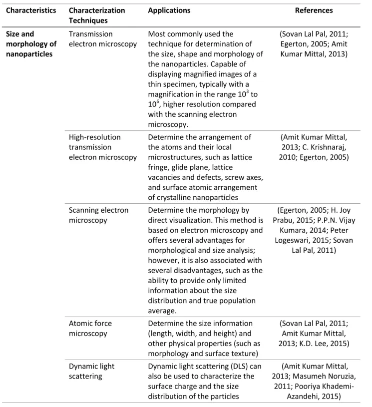
1.4 Characterization of nanoparticles
The characterization of nanoparticles is an essential step in biosynthesis of nanoparticles. The shape, size, morphology, surface area, stability, and their dispersion are some of the properties used to characterize the synthesized nanoparticles. Homogeneity of these properties is important in many applications. The common techniques which are used by most of investigators for characterizing nanoparticles and to control synthesis processes are: Ultraviolet–visible (UV-Vis) spectrophotometry, Dynamic light scattering (DLS), Scanning electron microscopy (SEM), Transmission electron microscopy (TEM), Fourier transform infrared spectroscopy (FTIR), Powder X-ray diffraction (XRD) and Energy dispersive spectroscopy (EDS) (Adam Truskewycza, 2016; Amit Kumar Mittal, 2013; Chun-Gang Yuan, 2017; Chun-Gang Yuan, 2017; Jayanta Kumar Patra, 2016; K. Anand, 2015; Mohammad A. Khalilzadeh, 2016; Silvia Groiss, 2017). Common methods for characterization of nanoparticles are discussed in the table 2 below.
11
Table 2 Common methods for characterization of nanoparticles. Characteristics Characterization Techniques Applications References Size and morphology of nanoparticles Transmission electron microscopy
Most commonly used the technique for determination of the size, shape and morphology of the nanoparticles. Capable of displaying magnified images of a thin specimen, typically with a magnification in the range 103 to 106, higher resolution compared with the scanning electron microscopy.
(Sovan Lal Pal, 2011; Egerton, 2005; Amit Kumar Mittal, 2013)
High-resolution transmission
electron microscopy
Determine the arrangement of the atoms and their local microstructures, such as lattice fringe, glide plane, lattice
vacancies and defects, screw axes, and surface atomic arrangement of crystalline nanoparticles
(Amit Kumar Mittal, 2013; C. Krishnaraj, 2010; Egerton, 2005)
Scanning electron microscopy
Determine the morphology by direct visualization. This method is based on electron microscopy and offers several advantages for morphological and size analysis; however, it is also associated with several disadvantages, such as the ability to provide only limited information about the size distribution and true population average. (Egerton, 2005; H. Joy Prabu, 2015; P.P.N. Vijay Kumara, 2014; Peter Logeswari, 2015; Sovan Lal Pal, 2011) Atomic force microscopy
Determine the size information (length, width, and height) and other physical properties (such as morphology and surface texture)
(Sovan Lal Pal, 2011; Amit Kumar Mittal, 2013; K.D. Lee, 2015) Dynamic light
scattering
Dynamic light scattering (DLS) can also be used to characterize the surface charge and the size distribution of the particles
(Amit Kumar Mittal, 2013; Masumeh Noruzia,
2011; Pooriya Khademi-Azandehi, 2015)
12
suspended in a liquid. This technique is also used by many scientists for the characterization of nanoparticles.
Formation of nanoparticle
Ultraviolet-visible spectrophotometry
It is used to confirm the formation of nanoparticles by measuring Plasmon resonance and evaluating the collective oscillations of conduction band electrons in response to electromagnetic waves. That provide information regarding the size, structure, stabilization, and aggregation of nanoparticles (Arthanari Saravanakumar, 2015; Bappi Paul, 2015; C. Mystrioti, 2016; Chun-Gang Yuan, 2017; Jayanta Kumar Patra, 2016; S.S. Godipurgea S.
Y., 2016; Arun Kumar Thalla, 2016) Crystallinity X-ray diffraction Powerful nondestructive
technique. It provides information on crystal structure, phase, preferred crystal orientation (texture), and other structural parameters, such as average grain size, crystallinity, strain, and crystal defects.
(Sovan Lal Pal, 2011; C. Suryanarayana, 1998; C.
Krishnaraj, 2010; Khan Behlol Ayaz, 2014)
Surface charge Zeta potential Determine the stability and surface charge of the colloidal nanoparticles, as well as the nature of the materials encapsulated inside the nanoparticle or coated on its surface (Pooriya Khademi-Azandehi, 2015) Fourier transform infrared spectroscopy
Used to identify organic functional groups attached to the surface of nanoparticles and the surface chemistry of biogenic
nanoparticles
(Amit Kumar Mittal, 2013; Shakeel Ahmed, 2016; Arun Kumar Thalla, 2016) Other techniques Energy dispersive X-ray spectra (EDS)
Identification of the elemental composition of the nanoparticles
(J. Das, 2013; Jayachandra Reddy Nakkala, 2016; Jayanta
13
1.6 Factors influencing the synthesis of metallic nanoparticles using plant extract
The properties of the synthesized nanoparticle depend on the pH of the synthesis medium, temperature, time, concentration of plant extract and the metal salt (Palaniselvam Kuppusamy M. M., 2016). All of these conditions are discussed below.
1.6.1 pH
pH is an important factor that influences the synthesis of nanoparticles by green methods. A study by reported the variation in pH of a solution is responsible the different size and shapes of nanoparticles formation (Shankar S.S., 2003). Another study has discovered that pH of the medium influences the size and texture of the synthesized nanoparticle which proved nanoparticle size can be controlled by varying the pH of the solution media (V. Armendariz, 2004).
1.6.2 Time
The synthesis time (the reduction time) is another factor that affects the characteristics of nanoparticles. The optimum reduction time produces high absorbance and sharp peak value that indicates higher concentration nanoparticles in the medium. This determines the characteristics of the synthesized NPs namely their size and shape that can be spherical, triangular, hexagonal and rectangular. In studies in which the synthesis of NPs finished within the short period of time the SPR (Surface plasmon resonance) peak get broader as the time goes on, which indicates size and yield increment. The size variations as a function of time may be due to several factors such as aggregation of particles (Bappi Paul, 2015; Chun-Gang Yuan, 2017; Jia Yu, 2016; Jayachandra Reddy Nakkala, 2016).
1.6.3 Temperature
Temperature is also another important factor that affects the synthesis of nanoparticles. In comparison to other synthesis methods, green synthesis requires minimum temperatures or even room temperature is generally enough. A study showed that the temperature of the reaction medium determines the nature of the nanoparticle formed (A. Rai, 2006). Another study also revealed the effect of temperature on the synthesis of nanoparticles. It showed that at high temperatures, it lead to the formation of higher spherical nanoparticles and triangular nanoparticles, whereas at lower temperature mostly increased triangular NPs formation increased (Raju, 2011). Another study reported different UV-Vis spectrophotometer absorbance spectra of gold nanoparticles and silver nanoparticles were obtained at different temperature. The peak sharpness increases with an increase in the reaction temperature. Most likely this might occurred due to an increase in the reduction rate at higher temperatures (Amarendra Dhar Dwivedi, 2010).
1.6.4 Concentration of metal ion or metal salt
The concentration of metal precursors could change the size of the synthesized nanoparticles. Increasing trend of particle size was observed with increasing concentration of metal ion in solution from 0.1 to 5.0 mM (Shashi Prabha Dubeya, 2010). TEM and UV-Visible result revealed that as the concentration of silver nitrate and auric acid increase, larger sized silver and gold nanoparticles were synthesized (Shashi Prabha Dubeya, 2010). A Similar trend was shown in another study: An increase in the concentration of the metal ion from 0.1–5 mM ratio, result in an increase in the particle size observed at the end of the synthesis (30 minutes) (Amarendra Dhar Dwivedi, 2010). In another study, the absorption increased steadily as the Au (III) concentration increased in the reaction mixture from 1 mM to 5 mM. It was
14
concluded that an increase in the concentration of available ions in a solution increases the yield of nanoparticles. While increasing the substrate concentration the large size and aggregation of nanoparticles was occurred due to the occurrence of competition between gold ions and functional groups (Nabeel Ahmad, 2017).
1.6.5 Metal precursors to extract volume ratio
The volume ratio of the solution of metal precursor and extract has an effect on the characteristics of nanoparticles produced. On a study done on the production of iron nanoparticles, the polyphenols extract was mixed with an iron (III) solution at certain volume ratios (i.e. 1:2, 1:1, 2:1). The maximum concentration of iron particles in suspensions was observed at a mixing ratio of iron (III) solution to extract (v/v) equal to 2 (C. Mystrioti, 2016). Another study was conducted with different leaf extract concentration ratio, (v/v) varied from 1:30, 2:30 and 3:30 of silver nitrate and auric acid solutions for the synthesis of silver and gold nanoparticles. As the ratio increased in favor of leaf extract, UV-vis spectra sharpness increased which was correlated with the formation of more nanoparticles (Amarendra Dhar Dwivedi, 2010). A report on a green synthesis of AuNPs revealed the increase of the intensity of the band of AuNPs as a consequence of an increase volume of leaf extract. The peak starts becoming narrower and narrower with further increase in volume of leaf extract, which indicated more nanoparticles formation but no size increment (Nabeel Ahmad, 2017).
1.6.6 The surrounding environment
The surrounding environment where the nanoparticles produced also contributes a lot in determining the characteristics of the synthesized nanoparticles. In various environmental conditions, nanoparticle might become core-shell nanoparticles quickly by absorbing materials or reacting with other materials from the environment through the process of oxidation or corrosion (V. Sarathy, 2008). To mention an example that showed the effect of the environment on the nature of the synthesized nanoparticles, the crystalline nature of the zinc sulphide nanoparticles changed immediately when its environment was changed from a wet to a dry condition (S. V. N. T. Kuchibhatla, 2012).
1.7 Application of NPs in wastewater treatment
Nowadays greenly synthesized nanoparticles are being used in cost effective and ecofriendly water treatment techniques attracting many scientists in the field wastewater. Thus, various NPs have been used for the treatment of different kind of wastewaters with specific characteristics. Iron and iron oxide NPs has been successfully used since very promising results have been reported by many investigators for the removal/degradation of various pollutant from water/wastewater: Degradation of ibuprofen (S. Machado, 2013), Cr (VI) reduction (Akbar Soliemanzadeh, 2017),treating domestic wastewater (C.P. Devatha, 2016), removal of phosphates (Dan Cao, 2016), As(V) removal (María Martínez-Cabanas, 2016), removal of nitrate from aqueous solution (Ting Wang, 2014), degradation of malachite green (Lanlan Huang, 2014) are several examples in which NPs were used.
15
As it is mentioned above, the best characteristic of nanoparticles comes from its small size that provides them their distinctive properties. However, their size makes difficult their use in certain applications such as water treatment due to the extremely complex separation of the material from solution. The immobilization of the synthesized materials onto porous solids is a good procedure to avoid this operational problem and to allow their reuse in several sorption cycles (C.K.S. Pillai, 2009). In addition, the immobilization helps to enhance certain properties of the materials such as their stability and mechanical strength. However, the immobilization process may also imply some disadvantages such as the reduction of NPs sorption capacity by blocking their binding sites or the deceleration of sorption kinetics. The choice of the immobilization matrix determines the physical and chemical properties of the final material; therefore the selection of an appropriate matrix is an important step to develop an adequate sorption procedure. Several compounds such as chitosan, alginate, silica, polyacrylamide or polyvinyl alcohol have been used as immobilization matrixes. (María Martínez-Cabanas, 2016; C.K.S. Pillai, 2009).
1.7.1 Phosphate removal from wastewater
Phosphorus (P) is an essential macroelement for plant and algae growth; however its disproportionate presence as phosphate in aquatic ecosystems can lead to deterioration of water quality. Phosphate is a limiting nutrient in the environment and it is one of the pollutants that cause eutrophication of water bodies. High amounts of phosphate can cause algal blooms and depletion of dissolved oxygen in the water bodies with negative impacts on the aquatic organisms. Human activities such as animal manure and fertilizer application in soil or discharge of industrial, domestic and agricultural wastewater are responsible for the enrichment of surface water with phosphate loadings (Dimitris Mitrogiannis, 2017). To make the problem worse, there are few removal mechanisms under normal operating conditions of wastewater treatment plants. Thus a system must be amended specifically with compounds to bond to or adsorb phosphate (Hossain M. Azam, 2014).
There are various physical, chemical and biological methods that have been proposed for phosphate removal from water or wastewater such as anion exchange, sorption, chemical precipitation, membrane nanofiltration, reverse osmosis, electrodialysis and biological removal through constructed wetland, activated sludge and microalgal systems (Dimitris Mitrogiannis, 2017). However, nowadays the most widely used methods are biological removal and chemical precipitation. Yet, these methods have limitations such as lower removal efficiency, high cost, undesired waste sludge production, pH dependence, temperature dependence, effect the organic load regarding on biological removal (BOD:N:P ratio), and requirement of large space. In addition chemical precipitation requires high input of chemical reagents and also the management of the sludge produced. On the other hand, adsorption by using metallic particles is considered as an efficient, simple and low-cost method for PO4-3-P removal
even at low phosphate concentrations (Hossain M. Azam, 2014; Pei Luo, 2017; Asya Drenkova-Tuhtan, 2017).
Earlier studies have shown promising phosphate removal via adsorption using: zero valent iron (ZVI) (Nathalie Sleiman, 2017), ZnFeZr adsorbent (Asya Drenkova-Tuhtan, 2017), nanosized lanthanum hydrous doped on magnetic graphene nanocomposite (Hamid Rashidi Nodeh, 2017) and iron oxide
16
nanotubes (Minseok Kim, 2016). Therefore, it is important to find efficient alternative adsorbents with affordable price and sustainable production to protect the environment from phosphate pollution.
1.7.2 Factors affecting phosphate removal via adsorption
There are different factors that affect phosphate removal efficiency via adsorption. These are adsorbent particle size, pH of the wastewater, temperature, adsorbent-wastewater (m/v) ratio, which is related to the adsorption capacity, kinetics and separation of the adsorbent from the liquid phase (Dimitris Mitrogiannis, 2017).
1.8 Objective of the study
The main objective of this work was to explore the reduction potential of various waste plant materials for the synthesis of gold nanoparticles and the treatment of acid mine drainage wastewater via removal of metals aiming additional production of multi-metallic composite particles as well as their application as phosphate adsorbents from synthetic wastewater. The following are the specific objectives of the study.
Identification of the potential of extract of waste plant materials for the synthesis of gold nanoparticles and multi-metallic composite particles.
Green synthesis of gold nanoparticles using selected extract of waste plant material
Removal of metals from acid mine drainage wastewater aiming the production of multi-metallic composite particles.
Assess the potential application of the multi-metallic composite particles obtained from acid mine drainage as phosphate adsorbent, aiming phosphate removal from wastewaters.
Generally, this study intends to hit three birds with one stone. First, it will explore the potential of waste plant materials for the synthesis of gold nanoparticles and for the production of a multi-metallic composite particles from acid mine drainage, thus turning wastes into useful materials. Second, it aims to apply the composite obtained to other wastewater treatment. Third, it will reduce the operational, chemical and sludge management cost required by other traditional wastewater treatment systems.
17
2. Experimental Part
2.1 Chemicals and Materials
The following chemicals were used in the present work:
Standard gold (III) chloride solution (Sigma-Aldrich, gold atomic spectroscopy standard concentrate, 1.00 g/L Au standard solution) was used to pre, anhydrous ethanol (96% vol, VWR International Laboratory Chemicals) was used in the extraction process. To conduct ferric reducing ability plasma (FRAP) experiment, iron (III) sulphate, heptahydate, sodium acetate trihydrate, glacial acetic acid, hydrochloric acid (37%, analytical reagent grade, fisher Scientific), tripyridyltriazine (TPTZ), and iron (III) chloride, Hexahydrate were bought from VWR international Laboratory Chemicals. Anhydrous sodium hydroxide, nitric acid
(65%, merck kommanditgesellschaft auf aktien),
pphosphate monodibasic, Potassium phosphate dibasic, ammonium chloride, magnesium sulphate heptahydrate, potassium chloride, and soduim acetate were bought from VWR International Laboratory Chemicals and used to prepare the synthetic wastewater. Distilled and milli-Q water were used as required. Phosphate, Nitrate and COD reagent were bought from Hach Lange and used to determine synthetic wastewater characteristics before and after adsorption.General flow of experimental procedure
18
2.2 Waste plant materials selection and collection
Various waste plant materials were collected from Algarve region. Waste plant materials were selected based on the availability and the cost of the plant material (plant materials with less cost were given priority). Waste raspberry leave (dried and wet), waste raspberry fruit from National Fruit Company and prickly pear and carob were collected in the Algarve region. The fresh leaves of raspberry were washed 3 times with distilled water to remove any dirty and dried in an oven at 40 oC for 48 hours (only for the dried leaves). The plant biomass was crushed using a mechanical crusher to get the powder and the fruit part was crushed manually using crushing equipment.
2.3 Extract preparation
20 g of each selected plant materials mentioned above were used for preparation of the extract. The extract were prepared by boiling the mixture of 20 g of the plant material in 150 ml mixture of water-ethanol (50:50, v/v) used as extracting agent in 250 ml glass bottle in water bath at 60 oC for 30 minutes. The extraction conditions were selected taking into account information from literature values and the sustainability and feasibility of the process (Bappi Paul, 2015; K. Anand, 2015; Sathishkumar G., 2016). The prepared extract was filtered using a Whatmann filter paper nº 1 and stored at 4 oC for future use. The pH of the extract was measured using a pH meter (GLP 21, Crison). The flow chart below (fig. 3) shows the extract process.
19
2.4 FRAP method for measuring reduction potential
Ferric reducing ability plasma (FRAP) method is simple, automated test measuring the ferric reducing ability of plasma, presented as a novel method for assessing “antioxidant power.” Ferric to ferrous ion reduction at low pH causes a colored ferrous-tripyridyltriazine complex to form. FRAP values are obtained by comparing the absorbance change at 593 nm in test reaction mixtures with those containing ferrous ions in known concentration. Absorbance changes are linear over a wide concentration range with reducing compounds (Iris F. F. Benzie, 1996).
FRAP method was used for measuring the reduction potential of selected plant material. For that purpose 1.00 mM Fe+2 stock aqueous solution was prepared by dissolving 0.1390 g of FeSO4.7H2O into
500 ml distilled water and then from that one various iron (II) solutions with concentration in the range of 0.10-1.0 mM (0.10, 0.20, 0.40, 0.60, 0.80, 1.0 mM) were prepared for calibration according to table 3 below. Aliquots of 0.20 mL of each standard solution were pipetted to Eppendorf’s and placed in ice before being used.
Table 3 Preparation of Fe+2 standard aqueous solutions Standard
concentration
FeSO4.7H2O Solution Distilled water
mM (ml) (ml) 0.10 1 9 0.20 2 8 0.40 4 6 0.60 6 4 0.80 8 2 1.0 10 0
2.4.1 FRAP reagent preparation
A buffer solution with a pH of 3.6 was prepared by dissolving 1.55 g of sodium acetate trihydrate in 8 ml glacial acetic acid followed by addition of distilled water until final volume of 500 ml. A solution of 40mM HCl was prepared by dissolving 0.73 ml of 37% HCl in 500 ml of distilled water. Fresh solution of 10 mM tripyridyltriazine was prepared by dissolving 0.031 g of tripyridyltriazine in 10 ml of 40mM HCl at 50 oC. Fresh ferric (III) chloride, hexahydrate (20 mM) was prepared by dissolving 0.054g of FeCl3.6H2O
into 10ml of distilled water. The FRAP reagent was prepared in a flask by mixing the above mentioned prepared solutions according to the following proportions: 100ml of acetate buffer at pH 3.6, 10 ml of 10 mM TPTZ, 10 ml of 20 mM of FeCl3.6H2O, 12 ml of distilled.
2.4.2 Reduction potential measurement
A blank sample was prepared by mixing 30 µl of distilled water-ethanol (50:50, v/v) in cuvettes and then 1ml of the FRAP reagent was added vigorously into the same cuvettes. After 4 minutes the absorption of
20
the blank it was read with UV-Vis spectrophotometer (DR 2800, HACH LANGE) at 595nm and the reading was made to be zero before the reading of each standard and sample. Then 30 µl of each standard was added in cuvettes and 1 ml of FRAP reagent was added vigorously to each of the standard solution. The absorbance was read after 4 minutes. Finally 30 µl of each extract was added in cuvettes and 1 ml of the FRAP reagent was vigorously added to each of the extract. The absorbance was recorded after 4 minutes. Triplicate analysis was done for each experiment.
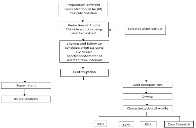
2.5 AuNPs synthesis using raspberry leave extract
In the typical experimental method, a standard gold(III) chloride solution from Sigma-Aldrich, gold atomic spectroscopy standard concentrate, 1.00 g/L Au standard solution was used to prepare 50 mL standard solutions of 25 mg/L, 50mg/L, 75 mg/L and 100 mg/L, which were placed in 250 ml flask. 5 mL raspberry leave extract were added drop by drop to each flask at room temperature while it was shaking at 100 rpm with a shaker (TH 15, Edmund Buhler). Different synthesis time 0 min (at the very beginning of the reaction), 5 min, 10 min, 15 min, 30 min and 60 min were monitored using UV-Visible spectrophotometery (BIOTEK, Synergy spectrometer) by measuring the absorbance between 400-800 nm, the range aims to analyze the surface plasmon resonance (SPR) peak. Different concentrations of gold (III) solution and synthesis times were used to get the optimum condition and other initial condition were selected based on literature review (Bappi Paul, 2015; Chun-Gang Yuan, 2017; Jia Yu, 2016). After synthesis following the mentioned conditions, the particles were separated from the solution by centrifugation at 4000 rpm for 25 minutes using centrifuge (scientific K3 serious) at 4000 rpm for 25 minutes. Then the supernatant was separated from precipitate by manual decantation and the precipitate was dried in a desiccator overnight.The Au-(III) concentration in the initial standard gold (III) solution and in the supernatant was analyzed for Au-(III) concentration using a MP-AES (Agilent 4200 MP-AES, Santa Clara, CA). The pH was measured using a pH meter (GLP 21, Crison).Triplicate analysis was done for each test. The best optimum condition was selected for synthesis of AuNPs.
21
Figure 4: General flow chart for gold (0) synthesis
2.6 Synthesis of multi-metallic composite particles
2.6.1 Collection and characterization of acid mine drainage
Acid mine drainage wastewater was collected from São Domingos at São Domingos mining area. The characterization of the wastewater was done using MP-AES (Agilent 4200 MP-AES, Santa Clara, CA) and flame atomic absorption spectroscopy (FAAS) (novAA 350, analytikjena). Selected main metallic components Fe, Al, Zn, Cu, based on preliminary study, were analyzed. The pH of AMD was measured using pH meter (GLP 21, Crison).
2.6.2 Synthesis of multi-metallic composite particles using raspberry leave extract
The multi-metallic composite particle synthesis process was optimized in terms of wastewater-extract ratio (v/v) and different synthesis time. Wastewater-extract ratios (v/v) of, 75:25, 85:25, 95:5 and different synthesis time of 0.5h, 1h, 2h, 3h, and 6h were investigated. Based on the ratio mentioned volume of acid mine drainage wastewater was placed in 250 ml glass bottle and a certain volume of raspberry leave extract was measured and added drop by drop to each bottles at room temperature while bottles were shaking at 100 rpm on a shaker (TH 15, Edmund Buhler). 5 ml of solution was collected from each bottle at different time intervals: 0.5h, 1h, 2h, 3h, 6h and centrifuged at 14000 rpm
22
for 20 minutes (ROTOFIX 32, hettich zentrifugen,). Then the precipitate and supernatant were separated by manual decantation. The precipitate was dried in a desiccator overnight. The initial concentration Al, Fe, Zn and Cu of AMD and the filtrate were analyze using FAAS (novAA 350, analytikjena) and MP-AES (Agilent 4200 MP-AES, Santa Clara, CA). The pH was measured using pH meter (GLP 21, Crison). Considering the sustainability and feasibility of the process, the conditions in terms of wastewater-extract (v/v) ratio and reduction time that allow obtaining the highest metal reduction percentage was used for production composite particles. Triplicate analysis was done for each experiment. The general synthesis method was shown in fig. 5 below.
Figure 5: General flow chart for synthesis of multi-metallic composite particles
2.7 Characterization of AuNPs and multi-metallic composite particles
The characterization (size, shape, morphology, surface area, crystal structure, elemental composition, surface charge and stability) of AuNPs and multi-metallic composite particles were done using transmission electron microscopy (TEM), energy-dispersive X-ray spectroscopy (EDS/EDX), X-ray diffraction (XRD), and zeta potential. The formation AuNPs were monitored using UV-Vis spectrophotometer in a range between 400-800 nm.
23
2.7.1 UV-Vis spectrophotometer
The formation AuNPs were monitored using UV-Vis spectrophotometer (BIOTEK, Synergy) in a range between 400-800 nm.
2.7.2 Transmission electron microscopy (TEM)
The images of AuNPs and multi-metallic composite particles were recorded with TEM for determination of the shape, and size of the particles and diffraction.
2.7.2 Energy-dispersive X-ray spectroscopy (EDS/EDX)
The elemental composition of AuNPs and multi-metallic composite particles was determined using EDS coupled with TEM.
2.7.3 X-ray diffraction (XRD)
The crystallographic structures of obtained AuNPs and multi-metallic particles were analyzed by X-ray diffraction (XRD) using a PANalytical XPERT-PRO powder diffractometer operating at 45 kV and 30 mA with Cu Ka radiation filtered by Ni. The XRD patterns were recorded using an X'Celerator detector with a step size (2θ) of 0.03o and a time per step of 1000 s. Peak analysis and the identification of crystalline phases were based on comparison using High-Score Plus software with the ICDD PDF-2 database. From the width of the XRD peaks the crystallite size was calculated by using Scherrer's equation below,
(1) Where,
τ is the mean size of the ordered (crystalline) domains, which may be smaller or equal to the grain size;
K is a dimensionless shape factor, with a value close to unity. The shape factor has a typical value of about 0.9, but varies with the actual shape of the crystallite;
λ is the X-ray wavelength;
β is the line broadening at half the maximum intensity (FWHM), after subtracting the instrumental line broadening, in radians. This quantity is also sometimes denoted as Δ(2θ); θ is the Bragg angle (in degrees).
2.7.4 Zeta potential
The Zeta potential measurements of AuNPs and multi-metallic particles were performed using Malvern Nano Zetasizer (Nano-Z590, Nano Series) equipment at 146 mV, by adding the precipitates to aqueous solutions prepared using Milli-Q water with different pH from 1 to 12. Immediately after the particles addition, the solution was submitted during 5 minutes ultrasound (FB15054, Fisherbrand) treatment. The pH of the Milli-Q water was adjusted using 5% NaOH or 5% HNO3 as required.