U
m
inho | 20
1
6
Carla Isabel Mor
eir
a de F
aria
Immobilized antimicrobial agents: po
tential de
velopment of microbial resis
tance
Outubro de 2016
Universidade do Minho
Escola de Engenharia
Carla Isabel Moreira de Faria
Immobilized antimicrobial agents:
Dissertação de Mestrado
Mestrado Integrado em Engenharia Biomédica
Trabalho efetuado sob a orientação da
Professora Doutora Maria Olívia Pereira
Carla Isabel Moreira de Faria
Immobilized antimicrobial agents:
potential development of microbial resistance
Outubro de 2016
Universidade do Minho
Escola de Engenharia
iii
DECLARAÇÃO
Nome: Carla Isabel Moreira Faria
Endereço eletrónico: [email protected] Telefone: 914190258
Cartão do Cidadão: 13913591
Título da dissertação: Immobilized antimicrobial agents: potential development of
microbial resistance
Orientadora:
Professora Doutora Maria Olívia Baptista de Oliveira Pereira
Ano de conclusão: 2016
Mestrado Integrado em Engenharia Biomédica
É AUTORIZADA A REPRODUÇÃO PARCIAL DESTA DISSERTAÇÃO, APENAS PARA EFEITOS DE INVESTIGAÇÃO, MEDIANTE DECLARAÇÃO ESCRITA DO INTERESSADO, QUE A TAL SE COMPROMETE.
Universidade do Minho, _____/_____/_________
v
A
CKNOWLEDGEMENTS
The accomplishment of this work marks the end of an important stage of my life and it was only possible due to the collaboration and contribution, directly or indirectly, of several people. Therefore, I would like to thank all those who contributed decisively to the completion of this work.
In particular, to my supervisor, Prof. Dr. Maria Olívia Pereira, for support, availability, sympathy, comprehension, suggestions and knowledge transmitted throughout this work.
To Diana, for teaching me all the laboratory techniques, for all the knowledge transmitted, for being always available, for the tireless help, for the support and comprehension and also for the answers to all my questions that arose during this work.
To my friends, for the friendship, and especially the MOP group for the sharing knowledge, for the support and the fun moments in the laboratory.
To my parents for always encouraging me to continue to accomplish my goals, for the tremendous help and care that were very important during this process, and rest of the family in general.
Finally, special thanks to Henrique for the constant presence, care, attention, patience and support during this so important academic stage and Tomás for making my days happier.
vii
A
BSTRACT
With an aging society and the increasing use of medical implants and devices, the problem of biomaterial-associated infections (BAI) will increase in coming years. The development of materials able to prevent bacterial colonisation is a promising approach to deal with BAI and several strategies to confer biomaterials with antimicrobial properties are emerging. They have, however, some limitations to be solved, namely the potential development of microbial resistance to the antimicrobial agents immobilized on the functionalized surfaces.
This study aimed to evaluate the potential development of resistance by Staphylococcus
aureus and Staphylococcus epidermidis adhered on polydimethylsiloxane (PDMS)
functionalized with different antimicrobial compounds (vancomycin, rifampicin and benzalkonium chloride, BAC). PDMS functionalization was performed using a mussel-inspired coating strategy.
A preliminary optimization of antimicrobials immobilization to confirm their contact-killing activity was followed by leaching assays. The ability of antimicrobial surfaces to prevent biofilm formation was further assessed by XTT assay. The potential development of resistance towards the immobilized antimicrobials was evaluated by continuously recovering the cells adhered on the surfaces and allowing them to adhere to new surfaces for a total of 10 passages. Results showed that antimicrobial surfaces exhibited contact-killing activity being the best performance achieved for a basic pH (8.5) and an overnight incubation. The leaching assays revealed that the increase of the antimicrobial concentration produced higher inhibition zones, hence more antimicrobial release from the functionalized surfaces. Antimicrobial surfaces were able to impair biofilm establishment, although complete biofilm eradiation was not achieved. The possible development of resistance of these remaining cells was then investigated and results showed that cells recovered from BAC-functionalized surfaces did not express propensity for developing resistance, as they have susceptibility patterns similar to the cells recovered from the unmodified surfaces. Conversely, cells recovered from the surfaces modified with antibiotic exhibited a higher MBC as compared to cells recovered from unmodified PDMS. Surfaces functionalized with rifampicin were not tested for potential development of resistance because its immobilization without leaching could not be achieved. This study highlighted the importance of evaluating the potential development of microbial resistance towards immobilized antimicrobials, namely when antibiotics are used to modify biomedical surfaces. Although it is required to test BAC cytotoxicity after its immobilization, overall results also emphasized its potential to be used in the design of materials able to prevent BAI without fostering bacterial resistance.
ix
R
ESUMO
Com o envelhecimento da sociedade e o aumento do uso de implantes e dispositivos médicos, a problemática das infeções associadas a biomateriais (BAI) deverá aumentar nos próximos anos. A melhor forma de lidar com estas infeções consiste na modificação dos biomateriais conferindo-lhes propriedades antimicrobianas. Existem, contudo, limitações que precisam de ser solucionadas, nomeadamente o potencial desenvolvimento de resistência microbiana aos agentes antimicrobianos após a sua imobilização nas superfícies.
Este trabalho teve como objetivo avaliar o potencial desenvolvimento de resistência de
Staphylococcus aureus e Staphylococcus epidermidis aderidos em superfícies de
polidimetilsiloxano (PDMS) funcionalizadas com diferentes compostos antimicrobianos (vancomicina, ripampicina e cloreto de benzalcónio, BAC). A funcionalização do PDMS foi realizada utilizando uma estratégia de revestimento inspirada na adesão dos mexilhões. Primeiro, procedeu-se à otimização da imobilização dos antimicrobianos para confirmar a sua capacidade de matar por contacto, seguindo-se os ensaios para avaliar a sua libertação da superfície. A capacidade das superfícies antimicrobianas para prevenir a formação de biofilme foi avaliada através de um ensaio de XTT. O potencial desenvolvimento de resistência aos agentes antimicrobianos imobilizados foi em seguida avaliado através da contínua recuperação das células aderidas a estas superfícies antimicrobianas, seguindo-se a sua adesão a novas superfícies, processo repetido num total de 10 passagens. Os resultados demonstraram que as superfícies preparadas apresentaram propriedades antimicrobianas, obtendo-se um melhor desempenho para um pH alcalino (8.5) e um tempo de incubação de 16-18 horas. Os ensaios de libertação revelaram que um aumento da concentração do agente produziu maiores zonas de inibição, logo uma maior libertação. Estas superfícies foram capazes de prejudicar o estabelecimento de biofilme, contudo, não foram capazes de o erradicar completamente. O potencial desenvolvimento de resistência destas células foi depois investigado e os resultados demonstraram que as células recuperadas das superfícies funcionalizadas com BAC não apresentaram tendência para induzir resistência uma vez que estas exibiram o mesmo perfil de suscetibilidade que as células aderidas às superfícies não modificadas. Pelo contrário, as células em contacto com as superfícies funcionalizadas com vancomicina, apresentaram uma MBC superior à das células recuperadas das superfícies não modificadas de PDMS. A imobilização da rifampicina não foi possível de efetuar sem libertação pelo que não foi testada nos ensaios de resistência.
Este estudo salienta a importância de estudar o potencial desenvolvimento de resistência por parte de antimicrobianos imobilizados, sobretudo quando antibióticos são usados para modificar a superfícies de biomateriais. Apesar de ser necessário testar a citotoxicidade do BAC após a sua imobilização, foi ainda salientado o potencial do BAC na produção de materiais para prevenir as BAI sem despoletar resistência bacteriana.
xi
TABLE OF CONTENTS
Acknowledgements ... v
Abstract ... vii
Resumo ... ix
List of Figures ... xiii
List of Tables ...xv
List of Abbreviations ... xvii
Scope and Aims ... XIX 1. Introduction ... 21
1.1 Biomaterial-associated infections ... 21
1.1.1 Routes of infection ... 22
1.1.2 Main causative organisms and infections incidence... 24
1.1.3 Biofilm formation on biomaterial surfaces ... 25
1.2 Strategies to fight BAI ... 28
1.2.1 Main treatment options ... 28
1.2.2 Preventing strategies ... 30
1.2.3 Surface modification to prevent BAI ... 31
1.3 Development of resistance towards immobilized compounds ... 35
1.3.1 The emergence of multi-drug resistant strains ... 35
1.3.2 Resistance mechanisms ... 36
1.3.3 How to evaluate the potential development of bacterial resistance ... 39
2. Materials and Methods ... 41
2.1 Microorganisms and culture conditions ... 41
2.1.1 Bacterial strains ... 41
2.1.2 Media and growth conditions ... 41
2.1.3 Bacteria preservation ... 41
2.2 Antimicrobial compounds ... 42
2.2.1 Benzalkonium chloride ... 42
xii
2.2.3 Rifampicin ... 42
2.2.4 MIC and MBC determination ... 42
2.3 Surface modification ... 43
2.3.1 Polydimethylsiloxane preparation ... 43
2.3.2 Polydopamine coating ... 44
2.3.3 Antimicrobial compounds immobilization ... 45
2.4 Antibacterial performance of modified surfaces ... 45
2.4.1 Contact-killing assay ... 45
2.4.2 Leaching assay ... 46
2.4.3 Biofilm formation on the modified surfaces ... 46
2.5 Evaluation of resistance development ... 47
2.6 Statistical analysis ... 48
3. Results and Discussion ... 49
3.1 Susceptibility of planktonic cultures to antimicrobials ... 49
3.2 Optimization of antimicrobials immobilization ... 50
3.3 Leaching assay ... 53
3.4 Biofilm formation on modified surfaces ... 56
3.5 Evaluation of development of resistance ... 57
4. Conclusions ... 61
5. Future work ... 63
References ... 65
Supplementary material I – Optimization of sonication time... 73
xiii
L
IST OF
F
IGURES
Figure 1. Schematic model representing the distinct developmental stages of microbial biofilms (Monroe, 2007).
I. Conditioning film; II. Reversible attachment; III. Irreversible attachment; IV. Stronger adhesion between the bacteria and the surface; V. Dispersion of single cells from the biofilm matrix... 26
Figure 2. Antibacterial strategies developed to prevent biomaterial-associated infections. Some compounds can
prevent the attachment of bacteria by coating of medical device surfaces. Biofilm development can be prevented by killing the early surface colonizers. Biofilm can be destroyed by agents which penetrate in biofilm matrix and kill biofilm-associated cells (di Luca, Maccari, & Nifosì, 2014). ... 32
Figure 3. Consumption of antibiotics in Europe (European Centre for Disease Prevention and Control (ECDC),
2012). ... 36
Figure 4. Schematic representation of different mechanisms of resistance developed by bacteria Ho does
Ba te ial Resista e happe ? | Da Volte a, .d. ... 38
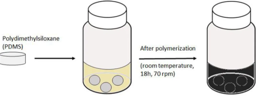
Figure 5. Schematic representation of immobilization strategy of pDA onto PDMS material. The coupons of PDMS
were submerged in bicine buffer during 18 h. ... 45
Figure 6. Absence or presence of an inhibition zone (denoted with a circle) around modified PDMS coupons on
an agar plate, after 72 h incubation with S. aureus, indicating presence or absence of leaching of antimicrobial compounds. ... 53
Figure 7. Absence or presence of an inhibition zone (denoted with a circle) around modified PDMS coupons on
an agar plate, after 72 h incubation with S. epidermidis, indicating absence or leaching of antimicrobial compounds. ... 55
Figure 8. Metabolic activity of biofilm cells adhered to unmodified PDMS, pDA-coated PDMS, pDA-coated BAC
and pDA-coated vancomycin. Significant differences were found for (***) p< 0.001, compared to PDMS control. ... 56
Figure 9. Optimization of sonication times (represented in minutes) to detach S. aureus adhered to PDMS and
PDMS coated with pDA. ... 73
Figure 10. Representative images of contact-killing assay: bacterial growth can be observed on unmodified PDMS
and PDMS-coated surfaces where a drop of a bacterial suspension of S. aureus was added. No growth was visible for surfaces immobilized with BAC (at a concentration of 1 mg/mL) as well as in the negative control (pDA-coated PDMS without bacteria). ... 74
xv
L
IST OF
T
ABLES
Table 1. Incidences and causative organisms of infections associated with commonly used medical devices and
implants (Busscher et al., 2012; Roosjen et al., 2006). ... 24
Table 2. Summary of mechanisms of action and main disadvantages associated to some antibacterials used in
the development of antimicrobial coatings to prevent BAI. ... 33
Table 3. Summary of different mechanisms developed by bacteria to protect themselves from the antimicrobial
agents. ... 38
Table 4. MIC and MBC of antimicrobials BAC and vancomycin against planktonic cultures of S. aureus and S.
epidermidis. MIC and MBC are expressed in µg/mL. ... 49
Table 5. Optimization of BAC and vancomycin immobilization onto PDMS surfaces for S. aureus using
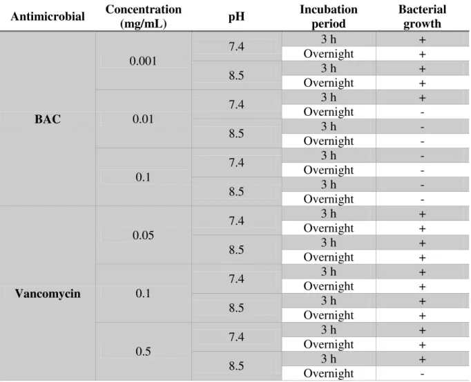
polydopamine as an intermediate. Different concentrations, pH values and incubation periods of time
were tested. Visible growth was used as an indicator of contact-killi g a ti ity a d it as ta ulated as +
fo g o th a d - fo o isi le g o th. ... 50
Table 6. Optimization of immobilization of BAC, vancomycin and rifampicin onto modified PDMS surfaces using
polydopamine as an intermediate against S. epidermidis. Different concentrations were tested. Visible
bacterial growth was used as an indicator of contact-killi g a ti ity a d it as ta ulated as + fo
a te ial g o th a d - fo o isi le g o th. ... 51
Table 7. Antimicrobial susceptibility of planktonic cultures of S. aureus against BAC and vancomycin: MIC on
day 0 and after 10 passages in a sub-inhibitory concentration. MIC and MBC are expressed in µg/mL. ... 57
Table 8. Antimicrobial susceptibility of adhered cells of S. aureus against BAC and vancomycin: MIC and MBC
after 10 passages in contact with unmodified PDMS, pDA-coated PDMS and pDA-coated PDMS surfaces functionalized with antimicrobials. MIC and MBC are expressed in µg/mL. ... 58
xvii
L
IST OF
A
BBREVIATIONS
AFM Atomic force microscopy
AMP Antimicrobial peptide
BAC Benzalkonium chloride
BAI Biomaterial-associated infection(s)
CFU Colony forming units
DDD Defined daily dose
DNA Deoxyribonucleic acid
DOPA Dihydroxyphenylalanine
ECDC European Centre for Disease Prevention and Control
EPS Extracellular polymeric substances
EUCAST European Committee on Antimicrobial Susceptibility Testing
HCl Hydrochloric acid
HPLC High performance liquid chromatography
MAP Mussel adhesive protein(s)
MBC Minimum bactericidal concentration
MHB Mueller Hinton Broth
MIC Minimum Inhibitory Concentration
NaCl Sodium chloride
OD Optical density
PBS Phosphate-buffered saline
pDA Polydopamine
PDMS Polydimethylsiloxane
PEG Polyethylene glycol
PMS Phenazine methosulfate
QAC Quaternary ammonium compounds
SCV Small colony variant
SD Standard deviation
SEM Scanning electron microscopy
xviii
TSB Tryptic Soy Broth
XTT
S
COPE AND
A
IMS
The use of biomaterial implants and medical devices is an increasingly common procedure in modern healthcare. Despite their benefits, the problem of biomaterial-associated infections (BAI) has also been increasing. These infections generally involve microbial colonization and biofilm formation on biomaterials, which results in a higher antimicrobial and host immune system resistance. Several studies have been conducted to prevent the formation of these microbial biofilms. Immobilization of antimicrobials such as quaternary ammonium compounds (QAC) and antibiotics has been proposed as a promising strategy to confer antimicrobial properties to medical devices and, therefore, to reduce the risk of infection. Despite the promising results reported in the literature, most of these strategies fail to proceed into clinical trials and important factors involved in the pathogenesis of these infections are often neglected, namely toxicity issues and the potential development of resistance towards antimicrobials immobilized. So, it is crucial to understand the fate of bacteria that manage to adhere to antimicrobial surfaces to develop effective antimicrobial coatings able to combat these infections.
The main purpose of the present thesis is to assess the potential development of resistance by S. aureus and S. epidermidis against three antimicrobials, currently under investigation for use in medical devices, after their immobilization. To achieve this goal, two antibiotics (vancomycin and rifampicin) and a QAC will be immobilized onto polydimethylsiloxane (PDMS) surfaces using a mussel-inspired coating strategy. Their immobilization will be then optimized so that PDMS exhibits antimicrobial properties but without antimicrobials release. Their ability to impair biofilm establishment will be also performed. Once obtained antimicrobial surfaces that meet these requirements, the potential development of microbial resistance towards immobilized antimicrobials will be finally investigated.
KEYWORDS: BACTERIAL RESISTANCE, ANTIMICROBIAL COATINGS, POLYDOPAMINE,
21
1.
I
NTRODUCTION
1.1
Biomaterial-associated infections
The last decades have been characterized, as never before in human history, by the broadest application of medical devices in all areas of medicine (Campoccia et al., 2013). Every year, millions of patients improve their quality of life through surgical procedures that involve medical devices that are implanted or not (Khan et al., 2014). These devices play an important role in human life to support and restore function after wear, trauma, surgical intervention or even to improve appearance (Domingues et al., 2015; Zimmerli & Sendi, 2011). Devices like joint and vascular prostheses, catheters, lenses, dental implants and others are increasingly being used since the 90s (Moraes et al., 2013). As a result of the increased life expectancy and the increasing demand for medical care from the aging population, the number of age-related diseases also increased. Thus, the need to carry out new treatments arises, which may involve the use of implants and long-term pharmaceutical administration (Campoccia et al., 2013; Khan et al., 2014).
In 2007, it was estimated that, worldwide, the use of medical devices approached half a billion devices per year, with catheters alone acounting for about 400 million pieces (Campoccia et al., 2013). The trend is that medical devices are being used increasingly, continuously and simultaneously (Baio, 2011).
Despite its great benefits, it has been recognized for more than half a century that the presence of a biomaterial implant or device in host tissue strongly predisposes for infection (Zaat et al., 2010). These devices provide foreign surfaces to the human body, to which microorganisms can adhere and start forming biofilms (structured communities of microorganisms that adhere to one another on a living or abiotic surface and produce extracellular polymeric substances which protect them from the external environment) (Desrousseaux et al., 2013). Accordingly, in modern medicine, biomaterial-associated infections (BAI) are the number one cause of failure of biomaterial implants and devices, resulting in high costs to the health care system (Domingues et al., 2015; Moraes et al., 2013; Zaat et al., 2010). For example, in the USA, more than 5 million central venous catheters are implanted annually, of which, more than 80000 lead to catheter-related bacteremia
22 (Desrousseaux et al., 2013). A study conducted in four European countries showed that bloodstream infections related to catheters accounted for over 1000 deaths with associated osts of et ee € a d € illio a ually a d pe ou t y (Desrousseaux et al., 2013). Therefore, the occurrence of BAI is clearly recognized as a world problem.
1.1.1 Routes of infection
As aforementioned, the undesirable complication often associated to the use of implants or medical devices is the occurrence of an infection caused by microorganisms. Microorganisms can be acquired from several sources including the operating room environment, surgical e uip e t, lothi g o y edi al p ofessio als, eside t a te ia o the patie t’s ski , and bacteria already in the body (Hetrick & Schoenfisch, 2006). The best documented route of infection is direct contamination of an implant during surgery (perioperative contamination), such as in orthopedic, cardiovascular, plastic reconstructive, general surgery, and neurosurgery (Domingues, 2013; Zimmerli & Sendi, 2011). These infections occur after invasive procedures in the superficial or deep layers of the incision or in the organ or space that was manipulated or traumatized, and can be diagnosed 30 days after the surgical procedure (Amaral et al., 2013; WHO Guidelines for Safe Surgery, 2009). This way of contamination implies that an implant becomes contaminated with microorganisms before or during implantation into the human body (Domingues, 2013).
Perioperative infection accounts for about 15 % of all healthcare-associated infections and about 37 % of the hospital-acquired infections associated to surgical patients. In Western countries, the frequency of these infections is 15-20 % of all cases and, generally, surgery is responsible for 2-15 %. In general, perioperative infections lead to an average increase in length of hospital stay of 4-7 days. More specifically, patients infected with these kind of infections are twice as likely to die and to spend time in an intensive care unit and five times more likely to be readmitted after discharge (WHO Guidelines for Safe Surgery, 2009).
Despite the preparation performed in the skin before surgery, bacteria are always present. Quantitatively, the risk of acquiring an infection in the surgical site is much greater if it is contaminated by more than 105 microorganisms per gram of tissue. However, in the presence
of a foreign material, such as an implant, the amount of microorganisms to produce a necessary infection is much lower (Domingues, 2013; WHO Guidelines for Safe Surgery, 2009).
23 Although sterilization and the use of aseptic techniques greatly reduces the levels of bacteria found in hospital settings, pathogenic microorganisms are still found at the site of approximately 90 % of all implants, so it is crucial to continue preventing this way of contamination (Hetrick & Schoenfisch, 2006).
The second route of infection is called postoperative because it occurs after the surgery, during the hospitalization period (Domingues, 2013). After biomaterial implantation, there is a 6 h decisive period, which is critical for the long-term success of the implant. Over this period, an implant is particularly susceptible to surface colonization (Hetrick & Schoenfisch, 2006). This way of infection is mainly caused by direct contamination of open wounds or by the use of invasive devices like infusion tubes, catheters, or drains (Domingues, 2013). Postoperative infection can be triggered by different variables, that encompasses procedural variables (including type and length of procedure) and patient variables (such as the general medical and physical condition of patients before surgery) (Peterson, 2006). The most important variable is the type of surgery performed. Some studies show that the duration of the surgery is related to the infection rate. Simple procedures with short operative times and minimal incisions generally result in lower rates of postoperative infection as opposed to the complex procedures with long operative times (Peersman et al., 2006; Peterson, 2006). At last, biomaterials can also be infected by the hematogenous route. Although the risk of developing a BAI is higher for events related to surgery, there is a residual risk for the possibility of late infections caused by microorganisms from local infections elsewhere in the body that are spread through the blood (Campoccia et al., 2013; Domingues, 2013). Most hematogenous infections are caused by infected skin lesions that produce recurrent bacteremia. This is supported by the fact that the majority (56 %) of the infections suspected to be hematogenous are caused by staphylococci. Other examples, such as dental or other surgical interventions, bacteriuria, intestinal surgery and pneumonia, have also been suggested as possible causes of hematogenous spread of microorganisms, which can cause temporal or chronic bacteremia, leading to infections. (Domingues, 2013; Gottenbos et al., 2001).
Another possible mechanism for hematogenous spreading from the intestinal tract is bacterial translocation, when bacteria, mainly Gram-negative strains, escape through the intestinal wall. Thus, BAI due to hematogenous spreading of bacteria to an implant site may occur any time after implantation (Domingues, 2013; Gottenbos et al., 2001).
24
1.1.2 Main causative organisms and infections incidence
The most commonly isolated pathogens from infected biomaterial surfaces include Gram-positive Staphylococcus epidermidis and Staphylococcus aureus, causing up to 60 % of all prosthetic hip implant infections since 1980. Additionally, Gram-negative organisms such as
Escherichia coli and Pseudomonas aeruginosa are also isolated (Bruellhoff et al., 2010;
Domingues, 2013; Hetrick & Schoenfisch, 2006; Joo & Otto, 2012; Subbiahdoss et al., 2011).
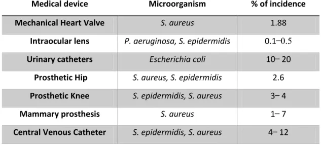
Table 1 presents the percentage of incidence of BAI associated to different biomedical
implants and devices and the main causative organism.
Table 1. Incidences and causative organisms of infections associated with commonly used medical devices and implants. Incidence data refers to the lifetime of the implant or device. (Busscher et al., 2012; Roosjen et al., 2006).
Medical device Microorganism % of incidence
Mechanical Heart Valve S. aureus 1.88
Intraocular lens P. aeruginosa, S. epidermidis 0.1 ̶ 0.5
Urinary catheters Escherichia coli 10 ̶ 20
Prosthetic Hip S. aureus, S. epidermidis 2.6
Prosthetic Knee S. epidermidis, S. aureus 3 ̶ 4
Mammary prosthesis S. aureus 1 ̶ 7
Central Venous Catheter S. epidermidis, S. aureus 4 ̶ 12
S. aureus is both a commensal bacterium and a human pathogen and it is a dangerous and
versatile microorganism that can cause a wide variety of diseases (Nair et al., 2014; Otto, 2014). S. aureus is responsible for approximately 23 % of infections associated with prosthetic joints and is the leading cause of bacteremia and infective endocarditis as well as osteoarticular, skin and soft tissue and respiratory tract infections (Otto, 2014; Subbiahdoss et al., 2011; Tong et al., 2015).
Skin infections caused by S. aureus are commonly community-acquired, whereas respiratory tract infections are predominantly nosocomial infections. Among the range of nosocomial pathogens, S. aureus is the most common and associated with high morbidity and mortality. In hospitalized patients with debilitated conditions, such as in patients suffering from immune
25 deficiencies or viral infections, S. aureus is often responsible for developing pneumonia (Otto, 2014).
S. epidermidis is a microorganism present in the epithelial surfaces of every human being. It
can be widely found on the skin, where is part of the commensal bacterial microflora (Hetrick & “ hoe fis h, ; O’Ga a & Hu ph eys, . In a similar way to S. aureus, S. epidermidis is a major cause of nosocomial infections (Cheung et al., 2010). Almost 50 % of the infections associated with catheters, artificial joints and heart valves are caused by this microorganism (Subbiahdoss et al., 2011).
Although S. aureus infections are characterized by progressing rapidly and are generally more severe than S. epidermidis infections, S. epidermidis has the capacity to breach the epithelial barrier and adhere to the surfaces of indwelling medical devices during device insertion and form biofilms, having been recognized as an important opportunistic pathogen (Cheung et al., 2010; Hetrick & Schoenfisch, 2006; Otto, 2012).
P.aeruginosa is a ubiquitous microorganism that grows in many environmental sites (Lovewell
et al., 2014). This bacterium is frequently associated with hospital acquired infections and is responsible for acute infections commonly associated with burn wounds and invasive instrument procedures (Lanini et al., 2011; Lovewell et al., 2014). It can be encountered in chronic infections typically in patients with persistent lung disease and immunocompromised (Lovewell et al., 2014).
Patient-to-patient transmission through contaminated medical devices is a well-established mechanism of P. aeruginosa spreading in healthcare settings (Lanini et al., 2011). Furthermore, this microorganism, which is able to live in biofilm mode, is resistant to a variety of chemicals, including antibiotics, detergents and hospital disinfectants, which facilitates its long-term persistence in hospital settings and diffusion between patients (Høiby, 2011; Lanini et al., 2011).
1.1.3 Biofilm formation on biomaterial surfaces
The ability to form biofilms is a universal attribute of almost all bacteria. Bacteria are able to grow adhered to almost every surface, forming highly complex communities called biofilms. Biofilms are composed of cells that grow in multicellular aggregates which are embedded in an extracellular matrix produced by the bacteria themselves (López et al., 2010).
26 In human life, biofilms can be found in many contexts, such as natural, medical and industrial environment. The mechanisms used by bacteria to form biofilms differ because they often depend on the environmental conditions and attributes of the strain concerned (López et al., 2010).
Biofilms can be composed of single or multiple species, depending on the device and its duration of use in the patient. For example, urinary catheter biofilms may initially be composed of single species, but longer exposures inevitably leads to biofilms composed by multispecies (Donlan, 2001).
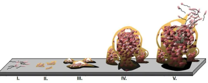
In biofilm composition, cells represent almost 10-25 % of biofilm volume while the matrix represents 75-90 % (Garrett et al., 2008; Moraes et al., 2013). Its formation follows sequential steps starting with bacterial adhesion to the substrate, followed by proliferation and accumulation of extracellular matrix in multiple layers, culminating in a bacterial community that remains in the produced matrix. From this community, some microorganisms will become detached and be transported to nearby areas, spreading over the surface of the biomaterial (Moraes et al., 2013). The different steps involved in biofilm formation are schematized in Figure 1.
Figure 1. Schematic model representing the distinct developmental stages of microbial biofilms (Monroe, 2007). I. Conditioning film; II. Reversible attachment; III. Irreversible attachment; IV. Stronger adhesion between the bacteria and the surface; V. Dispersion of single cells from the biofilm matrix.
Immediately after insertion of a medical device in the body, the conditioning film occurs, in which proteins such as fibrinogen and immunoglobulins are deposited on the surface of the implant, facilitating the adherence of bacteria to the implant (Vickery et al., 2013).
27 The step II involves the weak adhesion of planktonic cells to a surface and the production of EPS. Planktonic bacterial cells can approach surfaces under bacterial motility, or under physical forces, such as Brownian motion, van der Waals attraction forces, gravitational forces, surface electrostatic charge, and hydrophobic interactions (Kim et al., 2012; Kostakioti et al., 2013). Bacteria are attracted or repelled depending on the levels of nutrients, pH, ionic strength and temperature. The properties of the medium, along with the composition of the bacterial cell surface, affect the speed and direction of bacteria towards or away from the surface. After intercepting the surface, bacterial adhesion is mediated by additional extracellular adhesive appendices and secreted adhesins (Kostakioti et al., 2013). The initial attachment is dynamic and reversible, during which bacteria can detach if perturbed by repulsive forces, or in response to nutrient availability (Kostakioti et al., 2013). At this stage, as the adhesion is not final, the development of the biofilm and subsequent infection can be avoided by wash, antibiotics and host defences (Moraes et al., 2013). If conditions are favourable, bacteria reinforce the EPS production, consolidating the bacteria-surface bond (step III) (Garrett et al., 2008).
Step IV of biofilm development can be characterized by stronger adhesion between the bacteria and the foreign material, leading to cellular aggregation and the subsequent growth and maturation processes (Hetrick & Schoenfisch, 2006; Kim et al., 2012). Specific chemical reactions between compounds on the cell and substrate surfaces result in irreversible molecular bridging. Both polysaccharides and adhesin proteins within the bacterial membrane facilitate the attachment to substrate surfaces (Hetrick & Schoenfisch, 2006). Contact with the surface initiates responses that lead to changes in gene expression regulating factors favouring sessility, such as those involved in extracellular matrix formation (Kostakioti et al., 2013). Irreversible attachment is reached by bacteria that can resist the shear forces and maintain a constant grip on the surface and if provided with an appropriate supply of nutrients (Hetrick & Schoenfisch, 2006; Kostakioti et al., 2013).
Step V involves the dispersion of single cells from the biofilm matrix (Kim et al., 2012; Kostakioti et al., 2013). Within the mature biofilm there is a community that can sustain and maintain the biofilm architecture, providing a favourable environment for the resident bacteria. However, there may occur dispersion of cells caused by shear stresses. In addition, bacteria have developed ways to realize if the environment where they are, is favourable and may remain in the biofilm or return to planktonic mode. Biofilm dispersal can be triggered by
28 several cues, such as alterations in nutrient availability, oxygen fluctuations and increase of toxic products, or other stress-inducing conditions (Kostakioti et al., 2013).
Biofilms play an extremely important role in human health, because they protect bacteria from antibiotics and host immune responses. Biofilm formation is critical in the colonization of the implant surface, in the low efficiency of the host immune response, as well as in reducing the effectiveness of the antimicrobial treatment (Moraes et al., 2013).
In biofilm lifestyle, bacteria exhibit extreme resistance to antibiotics. In some cases, it has been found that killing bacteria in a biofilm requires approximately 1000 times the antibiotic dose necessary to achieve the same results in a suspension of planktonic cells (Hetrick & Schoenfisch, 2006).
The number of bacterial infections involving biofilms varies between 65 % and 80 % of all infections (Hetrick & Schoenfisch, 2006; Joo & Otto, 2012). Thus, biofilm development is the primary cause of BAI and because it is difficult to eliminate biofilm, removal of the contaminated device is often the only way to treat these infections (Desrousseaux et al., 2013; Hetrick & Schoenfisch, 2006; Moraes et al., 2013).
In this way, bacterial adhesion is often regarded as the most critical step to act in order to prevent BAI (Hetrick & Schoenfisch, 2006).
1.2
Strategies to fight BAI
1.2.1 Main treatment options
Infections associated with implanted biomaterials are a frequently occurring problem in modern healthcare (Engelsman et al., 2010). Treatment of these infections usually involves both medical and surgical measures, depending upon the cause and timing of the infection, and the condition of the host (Al-Mayahi et al., 2014).
The first treatment option is the administration of antimicrobial agents. Antibiotics are currently the preferred treatment strategy for bacterial infections (Al-Mayahi et al., 2014; Engelsman et al., 2010; Kostakioti et al., 2013). Conventional antibiotics work by preventing bacterial cell division (bacteriostatic) or killing the cell (bactericidal) (Kostakioti et al., 2013).
29 BAI are typically caused by commensal bacteria which adhere to the biomaterial surface and have the ability to form a biofilm on the implant surface. The extracellular matrix produced by cells, which hinders the diffusion of the antibiotic in the biofilm, along with the presence of metabolically inactive cells, contribute to fact that microorganisms become less susceptible to the action of the antimicrobial agent (Al-Mayahi et al., 2014; Engelsman et al., 2010; Kostakioti et al., 2013; Zaat et al., 2010; W. Zimmerli, 2014).
In the biofilm lifestyle, microorganisms are in a stationary phase of growth because oxygen and glucose are limited. Accordingly, successful treatment of BAI should consider this aspect. Some studies showed that most antimicrobial agents have a minimum bactericidal concentration (MBC), which is much higher during the stationary than the logarithmic phase of growth. The high stationary-phase MBC and the lack of efficacy against adherent bacteria are predictive of the failure of antibiotics in BAI (Zimmerli, 2014).
Other studies have showed that bacteria can also be located inside macrophages surrounding a biomaterial implant, where they remain protected against antibiotic treatment. Thus, both the biofilm mode of growth on the surface of a biomaterial implant as well as the bacterial localization in peri-implant tissues offer protection to the bacteria involved in BAI against routine antibiotic treatment, which may compromise the antibiotic efficacy (Engelsman et al., 2010).
BAI are usually treated with vancomycin, often in combination with rifampicin. Vancomycin has the ability to effectively penetrate the biofilm and to substantially reduce the number of viable bacteria (Engelsman et al., 2010). In a previous study, it was observed that treatment with rifampicin and vancomycin eradicated S. epidermidis from implanted biomaterial, but, despite the presence of rifampicin, bacteria in the surrounding tissue could survive. Thus, to eliminate bacteria in peri-implant tissue, alternative antibiotic combinations may be needed (Engelsman et al., 2010; Zaat et al., 2010; W. Zimmerli, 2014).
Although antibiotics have shown to be the best in the elimination of bacterial pathogens, high evidence indicates that they extensively damage the host microflora, create an environment where these pathogens can prevail, and they increase the selective pressure for resistance to antibiotics (Kostakioti et al., 2013; Vergidis & Patel, 2013; Wu et al., 2015).
The use of antibiotics is considered a pillar in the treatment of BAI, but it is often unsuccessful and insufficient to fight the infection because once antibiotics are removed, the biofilm is rapidly repopulated from cells that persisted during the treatment, resulting in recurrent
30 infections and chronic low-grade inflammation (Al-Mayahi et al., 2014; Engelsman et al., 2010; Kostakioti et al., 2013; Vickery et al., 2013).
The second treatment option is surgical therapy, when the infected device is removed but, as in antibiotics therapy, it is not fully effective if performed alone. Assays performed with antibiotic treatment in the presence of biofilm showed that antimicrobial therapy failed in all cases if it was instituted before device removal, whereas implant removal combined with antibiotic therapy was effective in all cases (Al-Mayahi et al., 2014; Campoccia et al., 2013; Engelsman et al., 2010; Kostakioti et al., 2013; Vickery et al., 2013).
Thus, the most effective solution for the treatment of BAI is to combine medical and surgical therapy (Vickery et al., 2013).
The typical procedure involves two stages in which the infected device is removed, the infection site is thoroughly cleaned, and antibiotics are systemically and locally delivered for a prolonged period of time. A new implant is then inserted when the infection is fully controlled and the surrounding tissue is not compromised (Busscher et al., 2012). However, the result of the insertion of a new device after BAI is uncertain, which increases patient pain and suffering, the length of hospital stay and consequent costs (Engelsman et al., 2010; Hetrick & Schoenfisch, 2006).
1.2.2 Preventing strategies
Bacterial biofilm contamination of surfaces in clinical workspaces is likely ubiquitous, and serves as a potential source of infection. The most common source of microorganisms is through the hands and skin. In a hospital setting, the transfer of these microorganisms can cause many infections, depending on the patient's condition. Therefore, the first and simplest strategy to prevent these infections is through the aseptic care (Xin, 2014).
Contamination of the environment surrounding the patient in the hospital is also an important source of bacteria. Typically, biofilms are found in wet areas, but studies have showed that bacteria can also exist on dry surfaces as biofilms which are protected from desiccation and have increased resistance to removal by detergents and the action of disinfectants. For these reasons, the risk of obtaining a nosocomial infection if the previous patient who has occupied the room had an infection caused by multi-resistant organism is increased. The existence of
31 multi-resistant bacteria may explain its permanency in the environment, despite the implementation of cleaning protocols increasingly rigorous (Vickery et al., 2013).
The same happens with the surgical instruments. Due to multi-resistant nature of the bacteria, cleaning and disinfection procedures are often ineffective in the decontamination of heat-sensitive instruments, such as endoscopes. Studies showed that approximately 1.8 % of endoscopes that were used remained infected with bacteria from the previous patient (Vickery et al., 2013).
Regarding biomaterial implants, the technically successful act of placing the device in the body does not guarantee the absence of an infection (Busscher et al., 2012). As aforementioned, an infection can occur by different routes (perioperative, postoperative and hematogenous contamination). Infections originated by the first two routes can be minimized through the disinfection and sterilization protocols, but no surgical site is truly sterile and pathogens are present in most operating rooms. These infections, when they occur, have a higher risk because bacteria adhered to the biomaterial surface can grow into a biofilm, becoming undetected by the immune system. Thus, an effective protection can only be offered by the integration of the biomaterial in host so that it establishes a normal immune response into the implant site (Busscher et al., 2012).
Facing this problem, there was the need to find different solutions that would improve the compatibility and integration of biomaterial implants and medical devices. These strategies include modifying the surface of the biomaterial to avoid the colonization of microorganisms (Busscher et al., 2012; Desrousseaux et al., 2013).
Currently, surface modification is the most promising strategy and with the best results to reduce the incidence of BAI (Busscher et al., 2012; Desrousseaux et al., 2013)
1.2.3 Surface modification to prevent BAI
The colonization of surfaces by bacteria is known to affect the function of several specific interfaces, such as those found in medical devices (Hasan et al., 2013). To combat this problem, investigations have focused on creating various strategies to eliminate or substantially reduce the extent of bacterial attachment and subsequent biofilm formation on these surfaces, such as surface modification (Hasan et al., 2013; Xin, 2014).
32 The properties of the surface can greatly influence the adhesion of bacteria. For instance, smooth surfaces act against the attachment of bacteria, unlike rough surfaces that favour the attachment. In addition, the hydrophilicity decreases the adhesion of bacteria, in contrast to hydrophobic surfaces (Lorenzetti et al., 2015).
Nowadays, surface modification of medical devices has often been reported as the approach with more potential to prevent biofilm formation (Bazaka et al., 2012; Xin, 2014). A summary of the surface modification strategies is illustrated in Figure 2.
Figure 2. Antibacterial strategies developed to prevent biomaterial-associated infections. Some compounds can prevent the attachment of bacteria by coating of medical device surfaces. Biofilm development can be prevented by killing the early surface colonizers. Biofilm can be destroyed by agents which penetrate in biofilm matrix and kill biofilm-associated cells (di Luca et al., 2014).
Surface modification can be explored in two main areas. The first involves the use of biocides (antimicrobial coatings), in the development of coatings that may release these antimicrobial agents or kill microorganisms by contact. The other strategy is the development of anti-adhesive materials that prevent the attachment of bacteria (Campoccia et al., 2013; Chen et al., 2013; Desrousseaux et al., 2013; Sileika et al., 2011). A variety of anti-adhesive and antimicrobial coating strategies are strongly being explored to prevent BAI. Table 2 describes some of the strategies that have been studied and their main disadvantages.
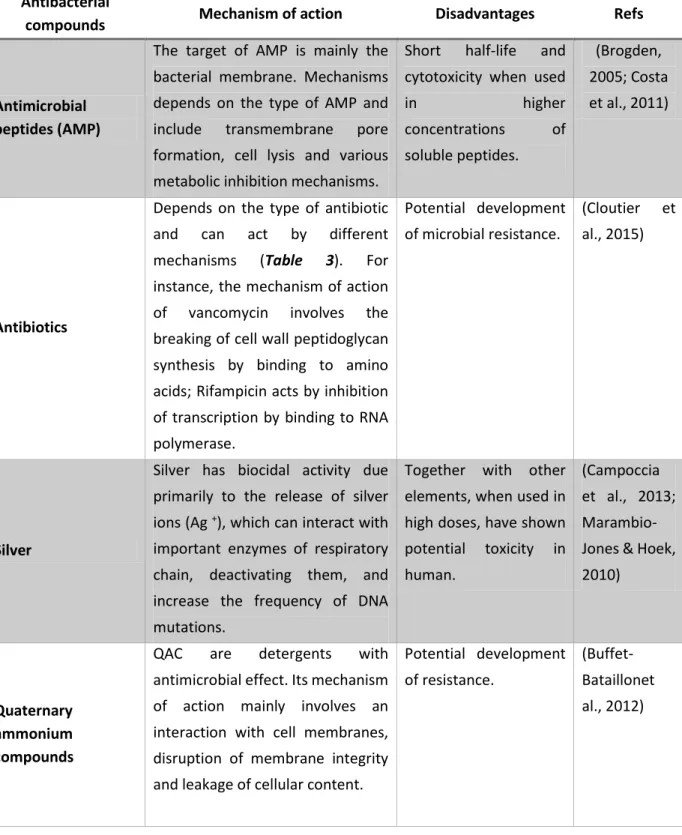
33 Table 2. Summary of mechanisms of action and main disadvantages associated to some antibacterial used in the development of antimicrobial and anti-adhesion coatings to prevent BAI.
Antibacterial
compounds Mechanism of action Disadvantages Refs
Antimicrobial peptides (AMP)
The target of AMP is mainly the bacterial membrane. Mechanisms depends on the type of AMP and include transmembrane pore formation, cell lysis and various metabolic inhibition mechanisms.
Short half-life and cytotoxicity when used
in higher concentrations of soluble peptides. (Brogden, 2005; Costa et al., 2011) Antibiotics
Depends on the type of antibiotic and can act by different mechanisms (Table 3). For instance, the mechanism of action of vancomycin involves the breaking of cell wall peptidoglycan synthesis by binding to amino acids; Rifampicin acts by inhibition of transcription by binding to RNA polymerase. Potential development of microbial resistance. (Cloutier et al., 2015) Silver
Silver has biocidal activity due primarily to the release of silver ions (Ag +), which can interact with important enzymes of respiratory chain, deactivating them, and increase the frequency of DNA mutations.
Together with other elements, when used in high doses, have shown potential toxicity in human.
(Campoccia et al., 2013; Marambio-Jones & Hoek, 2010)
Quaternary ammonium compounds
QAC are detergents with antimicrobial effect. Its mechanism of action mainly involves an interaction with cell membranes, disruption of membrane integrity and leakage of cellular content.
Potential development of resistance.
(Buffet-Bataillonet al., 2012)
34 Table 2. Summary of mechanisms of action and main disadvantages associated to some antibacterial used in the development of antimicrobial and anti-adhesion coatings to prevent BAI (continuation).
Anti-adhesion
compounds Mechanism of action Disadvantages Refs
PEG-based coatings
When bacteria approach the PEG molecules, the compression of the PEG chains results in elastic repulsive force and the removal of water from hydrated PEG chains creates an unfavourable osmotic stress. This combination acts as repulsive forces preventing bacterial attachment.
Surface overwhelming by continuous protein attack and coating degradation (hydrolysis, chain cleavage, surface removal). (Banerjee et al., 2011) Heparin
It is an anticoagulant that possess a strong negative electrical charge (repelling bacteria negatively charged) and presents hydrophilic properties (forming a highly hydrated layer between the bacteria and the surface). These characteristics may prevent bacterial adhesion.
Due to their
biodegradable nature, these coatings have a limited life time.
(Campoccia et al., 2013; Desrousseaux et al., 2013; Sin et al., 2009) Zwitterionic polymers
Zwitterionic polymers are polymers composed of molecules containing both a positive and negative charge, conferring an overall neutral charge balance and makes the polymers ultra-hydrophilic. This combination can prevent not only the adsorption of proteins but also the adhesion of bacteria.
The usage of organic solvents which may affect the integrity of biomaterials.
(Mi & Jiang, 2014; Raynor et al., 2009)
Biosurfactants
Biosurfactants are amphiphilic
compounds produced by
microorganisms with distinct surface and emulsifying activities. The adsorption of biosurfactants to a surface modifies its hydrophobicity, interfering in the microbial adhesion, making them antiadhesive agents against pathogens.
Amounts of produced biosurfactant are very low.
(Gudiña et al., 2010;
Rodrigues et al., 2006)
35
1.3
Development of resistance towards immobilized compounds
1.3.1 The emergence of multi-drug resistant strains
In recent decades, technological advancement along with the development of new drugs resulted in a significant reduction in mortality and increase of life expectancy. However, the overuse of antibiotics has led to the emergence of bacterial resistance, which is a natural phenomenon triggered by mutations in bacteria in order to protect themselves from antibacterial agents (Loureiro et al., 2016; Priyendu et al., 2015).
Bacterial resistance is a public health problem at global level and affects several areas such as medicine, production of animal food and agriculture, so it is very difficult to control and an inevitable event today. In hospital settings, antibiotics used to treat patients may enter the hospital sewer system, becoming a source of resistant organisms which spreads to other areas. Resistant strains can also arise from using antibiotics in sub-therapeutic concentrations (Priyendu et al., 2015). As a consequence, antibiotic resistance causes an elevated mortality and morbidity rate and an increase of treatment costs (Lin et al., 2015).
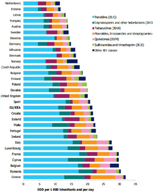
The consumption of antibiotics in Europe is very variable. According to the report from European Centre for Disease Prevention and Control (ECDC) Surveillance Report: Surveillance of antimicrobial o su ptio i Eu ope, the country that has the highest consumption of antibiotics is Greece (31.9), while the Netherlands has the lowest consumption (11.3), expressed as defined daily dose (DDD) per 1000 inhabitants and per day. Figure 3 shows the consumption of antibiotics in Europe.
Portugal reached the maximum consumption level in 2002 (26.5 DDD) reducing gradually until 2012 (22.7 DDD), but this is still higher than in other countries (ECDC, 2012).
It is believed that these significant differences in the consumption of antibiotics in different countries is due to incidence of infections acquired in the community, cultural and social determinants, structure of health care, available resources, knowledge about antibiotics, the pharmaceutical market and the practices of existent legislation (Ferech, 2006).
From the study of the relationship between the use of antimicrobials and antimicrobial resistance in Europe, it was found that the countries with a lower consumption of antibiotics, are also the countries where the level of resistance is lower. The opposite is also true and
36 Portugal is included on this side (Bronzwaer et al., 2002). For this reason the emergence of resistance strains is a matter of concern and deserves the best attention.
Figure 3. Consumption of antibiotics in Europe (ECDC, 2012).
1.3.2 Resistance mechanisms
For decades antibiotics were successfully used to treat patients with microbial infections. Over time, many infectious organisms, such as bacteria, were able to survive and develop resistance to specific antibiotics to which they once were susceptible, causing continuous infections. The rapid growth and evolution of microorganisms, such as bacteria, facilitates the development
37 of resistance to antimicrobials A ti i o ial D ug Resistance | NIH: National Institute of Alle gy a d I fe tious Diseases, .d. .
Nowadays, the concept of resistance is widely accepted and it is well known that bacteria are drastically more resistant to antibiotics, mainly in biofilms, as aforementioned.
This is a growing concern because species resistant to all known antibiotics have arisen and the emergence rate of antimicrobial resistance is unpredictable (Cooper et al., 2010).
Bacterial resistance may arise in different ways. Resistance can be intrinsic to the bacteria because of their genetic content and it is inherited from parents to progeny. Bacterial resistance can also be acquired or adaptive (due to the conditions of the surrounding environment) (Priyendu et al., 2015).
Acquired resistance
Acquired resistance may occur due to acquisition of a resistance gene by horizontal transfer of genes from resistant bacteria or by spontaneous mutations (Priyendu et al., 2015). In chronic infections bacteria are aggregated and very close, enabling the horizontal transfer of encoded genes for antibiotic resistance of a bacterium to another (Bjarnsholt et al., 2013). As this type of resistance is innate, bacteria maintain this state permanently (Priyendu et al., 2015).
Adaptive resistance
Adaptive resistance is developed according to the surrounding environmental conditions, such as the presence of antibiotics. This type of resistance involves a struggle for survival where organisms must adapt to the conditions of the environment faster than other organisms. Bacteria have an excellent ability to adapt to new conditions, thus leading to their survival (Priyendu et al., 2015).
This state of resistance is considered transient because when these conditions are removed it is reached the initial state (Fernández et al., 2011; Priyendu et al., 2015).
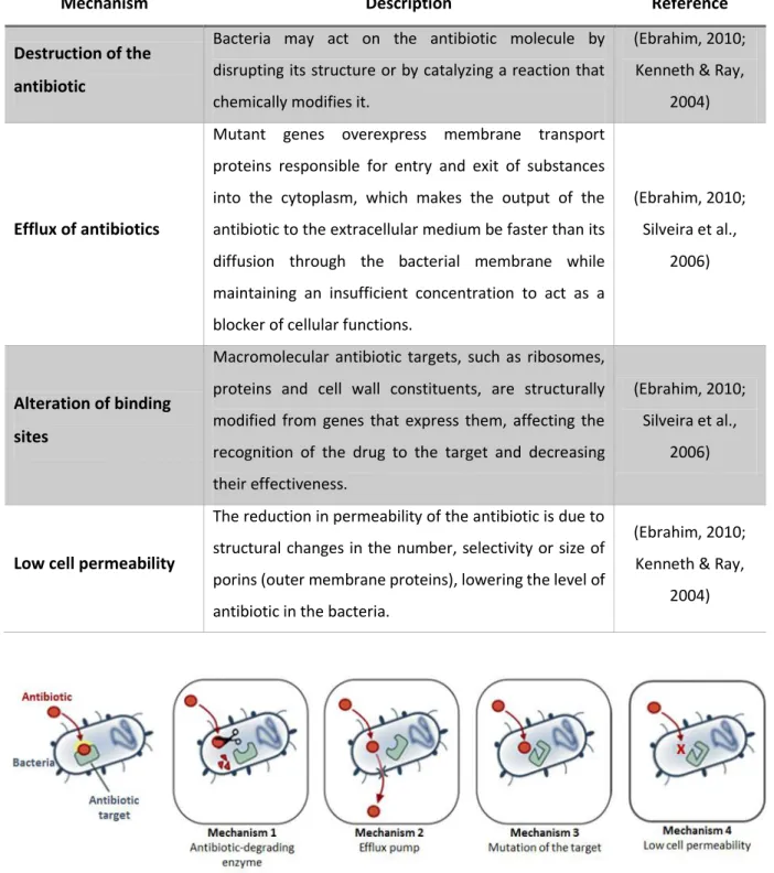
Bacteria can develop one or more mechanisms of resistance simultaneously against antibiotics (Priyendu et al., 2015; Silveira et al., 2006). Among the different resistance mechanisms, the most important are summarized in Table and their illustration are shown in Figure 4.
38 Table 3. Summary of different mechanisms developed by bacteria to protect themselves from the antimicrobial agents.
Mechanism Description Reference
Destruction of the antibiotic
Bacteria may act on the antibiotic molecule by disrupting its structure or by catalyzing a reaction that chemically modifies it.
(Ebrahim, 2010; Kenneth & Ray,
2004)
Efflux of antibiotics
Mutant genes overexpress membrane transport proteins responsible for entry and exit of substances into the cytoplasm, which makes the output of the antibiotic to the extracellular medium be faster than its diffusion through the bacterial membrane while maintaining an insufficient concentration to act as a blocker of cellular functions.
(Ebrahim, 2010; Silveira et al.,
2006)
Alteration of binding sites
Macromolecular antibiotic targets, such as ribosomes, proteins and cell wall constituents, are structurally modified from genes that express them, affecting the recognition of the drug to the target and decreasing their effectiveness.
(Ebrahim, 2010; Silveira et al.,
2006)
Low cell permeability
The reduction in permeability of the antibiotic is due to structural changes in the number, selectivity or size of porins (outer membrane proteins), lowering the level of antibiotic in the bacteria.
(Ebrahim, 2010; Kenneth & Ray,
2004)
Figure 4. Schematic representation of different mechanisms of resistance developed by bacteria Ho does Ba te ial
Resista e happe ? | Da Volte a, .d.
Using the mechanisms aforementioned, bacteria can overcome the action of antibiotics, even the most promising ones (Priyendu et al., 2015).
39 Facing this phenomenon of resistance, it is necessary to create new drugs able to overcome this ability of bacteria. However, there is always the possibility of emerging organisms resistant to these new agents (Cooper et al., 2010).
1.3.3 How to evaluate the potential development of bacterial resistance
In order to determine if bacteria are susceptible or resistant to an antimicrobial agent, it is necessary to evaluate their behaviour in vitro, pharmacological characteristics and later clinical trials (Kenneth & Ray, 2004).
The first step are in vitro tests where bacteria are exposed to the antimicrobial in a wide range of concentrations and the main parameter to be determined is the MIC (Kenneth & Ray, 2004). The results obtained from in vitro tests cannot be considered the only source of data. It should be taken into account pharmacological aspects of the antimicrobial agent and also information about the nature of disease and characteristics of the infection (Kenneth & Ray, 2004).
Currently, both in the development of new antibiotics such as the optimization of old antibiotics, predicting risk of resistance became, as never before, an increasingly important step in the drug development process for both pharmaceutical companies and researchers (Andersson, 2015).
Cooper et al. (2010) studied the behaviour of the bacteria when they are continuously exposed to honey. In ancient times honey was used to treat wounds. As with antibiotics, the extensive use of honey can trigger a selective pressure for the emergence of honey-resistant strains. To test the possible development of resistance two methods were tested. In the first method, initial bacteria were repeatedly cultivated (in fresh medium) in a sub-lethal concentration of the agent for 10 successive days. The MIC was determined on days 0 and 10. In the second method, bacteria were exposed to stepwise increasing concentrations of honey for 10 successive days. MIC of honey were determined before and after this period.
Overall, the results showed that, at the end of the training period, the MIC values remained very close to the initial values of MIC, confirming the potential of honey to treat wounds, without developing resistance.
In another study conducted by Tambe et al. (2001), the risk of development of resistance by
40 evaluated. The used method involved the passage of the culture 10–20 times through sub-inhibitory concentrations of different antimicrobials, alone and in combination. The MIC of each antimicrobial, before and after passages, was determined and compared. The results of this study showed that the resistance develops more easily in the combination of antibiotics than for the antiseptics and more easily to rifampicin than to minocycline. It was also verified that catheters impregnated with antiseptics may have a low risk of colonization by bacteria resistant to antibiotics (Tambe et al., 2001).
Duran et al., (2012) evaluated the association between the antibiotic susceptibility patterns and the antibiotic resistance genes in staphylococcal isolates. Antimicrobial susceptibility was performed in a total of 298 staphylococci clinical isolates. For a rapid diagnosis of antibiotic resistance genes, a molecular method was performed. The genes implicated in resistance to oxacillin, gentamicin, erythromycin, tetracyclin and penicillin were amplified using multiplex PCR method, in which several different DNA sequences were simultaneously amplified, followed by an electrophoresis (Duran et al., 2012). For all antibiotics tested, resistance was found in at least one gene of the several tested and most of staphylococci tested possessed the same gene, the blaZ gene, which confers resistance to beta-lactams. The results showed that this study produced different results, once the phenotypic antibiotic susceptibility patterns were not similar to those obtained by genotyping done by multiplex PCR (Duran et al., 2012).
The presence of bacterial variants called small colony variants (SCV), originated by gene mutations in stress response, is another way to detect the potential development of resistance (Melter & Radojevič, 2010). These variants are not particularly virulent but have the ability to persist viable inside host cells and also exhibited resistance to various antibiotics and even to antiseptics (Kahl, 2014). The most evident feature of these SCV is their small colony size on conventional agar plates, their fastidious growth in pin-point colonies and homogeneous appearance (Kahl, 2014; Proctor et al., 2006).
The methods presented to study the possible development of resistance are simple and easy to apply. Furthermore, the fact that there are always emerging resistant organisms suggests that antimicrobial susceptibility should be monitored continuously.
41
2.
M
ATERIALS AND
M
ETHODS
2.1
Microorganisms and culture conditions
2.1.1 Bacterial strains
In this work, two bacterial species, commonly associated to BAI infections were used:
- Staphylococcus aureus GB 2/1 isolated from explanted voice prostheses at the University
Medical Centre of Groningen (the Netherlands) ;
- Staphylococcus epidermidis GB 9/6 also isolated from explanted voice prostheses at the
University Medical Centre of Groningen (the Netherlands).
2.1.2 Media and growth conditions
During the accomplishment of this work different culture media were used and they were prepared according to the supplier instructions:
- TSB (Tryptic Soy Broth, 30 g/L, Liofilchem);
- TSA (TSB supplemented with Agar, 12,5 g/L, Liofilchem); - MHB (Mueller Hinton Broth, 21 g/L, Liofilchem).
For inoculum preparation, initially, two colonies were inoculated in 20 mL of TSB medium and incubated overnight at 37 °C, 120 rpm. Thereafter, bacteria were collected by centrifugation (9000 g for 5 min at room temperature), washed and suspended in fresh medium to prepare a bacterial suspension. Cellular concentration was determined by measuring optical density (OD) at 640 nm, and adjusted using calibration curves previously prepared.
2.1.3 Bacteria preservation
All strains were stored at -80 °C in liquid medium with glycerol 20 % (v/v). For each experiment, these strains were rehabilitated in TSA plates and placed at 37 °C for 24 h. The plates were then stored at 4 °C up to one week.
42
2.2
Antimicrobial compounds
2.2.1 Benzalkonium chloride
Benzalkonium chloride (BAC), a quaternary ammonium compound, widely used in clinical disinfectant formulations, was purchased from Sigma. A stock solution was prepared and stored at 4 °C. Working solutions were prepared therefrom.
2.2.2 Vancomycin
Vancomycin, a glycopeptide antibiotic exerting a broad spectrum of activity against Gram-positive bacteria was obtained from Sigma (European Pharcopea). A stock solution was prepared and kept at -20 °C. From this solution, several aliquots were also prepared for further work.
2.2.3 Rifampicin
Rifampicin (AppliChem), belonging to rifamycin’s lass of a ti ioti s, is ofte asso iated to vancomycin to treat BAI. A stock solution was prepared and stored at -20 °C, from which, work solutions were then prepared.
2.2.4 MIC and MBC determination
A microdilution test was used to determine the minimal inhibitory (MIC) and bactericidal concentrations (MBC). Antimicrobials solutions were prepared in MHB and added to the wells of 96-well microtiter plate with round bottom (Orange, USA), with several concentrations being tested (ranging from 40 µg/mL to 0.16 µg/mL for BAC and from 64 µg/mL to 0.25 µg/mL for vancomycin). A bacterial inoculum, diluted to reach a final concentration of 5 × CFU/mL, was added to the microtiter plate that was, then, incubated at 37 °C, 120 rpm for 24 h, making a total volume of 200 µL. In this assay, two controls were used, one without bacteria as a negative control and one without the antimicrobial compound as a positive control. MIC was determined by measuring the optical density at 640 nm, where clear wells (OD=negative control) were an indication of bacterial growth inhibition. MBC was determined by adding 10 µL from each well with no visible growth on a TSA plate and defined as the lowest