Journal of the Brazilian Chemical Society
This is an Open Access article distributed under the terms of the Creative Commons
Attribution License, which permits unrestricted use, distribution, and reproduction in any medium,
provided the original work is properly cited. Fonte: http://www.scielo.br/scielo.php?
script=sci_arttext&pid=S0103-50532015001102290&lng=en&nrm=iso. Acesso em: 16 abr. 2018.
REFERÊNCIA
ALMEIDA, Richardson Alves de et al. Cinerascetns, nee e tdes roo H siioas cinerascens:
MALDI LIFT-TOF-MS/MS de novo sequence and ioaging snalHsis. Journal of the Brazilian Chemical
Society, São Paulo, v. 26, n. 11, . 2290-2297, nov. 2015. Dis onível eo:
<htt ://eee.scielo.ir/scielo. h sscri tssci_arttext&
idsS0103--5053-2015001102290&lngsen&nrosiso>. Acesso eo: 16 air. 2018. doi:
htt ://dx.doi.org/10.593-5/0103--5053-.20150219.
Article
J. Braz. Chem. Soc., Vol. 26, No. 11, 2290-2297, 2015. Printed in Brazil - ©2015 Sociedade Brasileira de Química 0103 - 5053 $6.00+0.00
A
http://dx.doi.org/10.5935/0103-5053.20150219*e-mail: [email protected]
Cinerascetins, New Peptides from Hypsiboas cinerascens:
MALDI LIFT-TOF-MS/MS de novo Sequence and Imaging Analysis
Richardson A. Almeida,*,a,b Marcelo Gordo,c Felipe M. A. da Silva,a,d
Rafael C. de Araújo,a Marcelo H. S. Ramada,e Fernando Y. Abrão,f Túlio O. G. Costa,a Hector H. F. Koolen,a,d Afonso D. L. de Souzaa and Carlos Bloch Jr.b
aDepartamento de Química, Universidade Federal do Amazonas, 69077-000 Manaus-AM, Brazil bLaboratório de Espectrometria de Massa, Embrapa-Recursos Genéticos e Biotecnologia,
70700-900 Brasília-DF, Brazil
cDepartamento de Biologia and dGrupo DeMpSter de Espectrometria de Massas, Universidade
Federal do Amazonas, 69077-000 Manaus-AM, Brazil
eDepartamento de Biologia Celular, Universidade de Brasília, 70910-900 Brasília-DF, Brazil fInstituto de Patologia Tropical e Saúde Pública, Universidade Federal de Goiás,
74605-050 Goiânia-GO, Brazil
The continuous search for antimicrobial candidates pushes the pursuit of compounds in the most diverse organisms. Amphibians are known as a prolific source of antibacterial peptides. Based on the rich biodiversity of the Amazon region the unexplored green-tree frog (Hypsiboas cinerascens) was studied for its skin secretion peptide content. Chromatographic separations and established tandem mass spectrometry (MS/MS) methods were used for sequencing the primary structures of the purified compounds. De novo sequencing lead to the identification of five new peptides related to hylaseptin P1, displaying an aminated C-terminal. Sequencing of the complementary deoxyribonucleic acid (cDNA) analysis allowed the disambiguation of isobaric amino-acids for C-01. Matrix assisted laser desorption ionization (MALDI) was carried out, demonstrating the
in situ co-occurrence of the identified peptides in the dorsal skin. The major peptide C-01 was
synthesized and assayed against a selection of microorganisms displaying minimal inhibitory concentrations (MICs) ranging from 4 to 16 µM.
Keywords: anuran, antimicrobial peptide, Hypsiboas cinerascens, skin secretion
Introduction
Peptides are short chains of amino acids connected to
one another in a sequence by peptide bonds.1 This type of
compounds is widespread through all organisms such as
plants, microorganisms and animals.2 In animals, peptides
are involved in the complex coordination of the organism,
mainly attributed to hormonal and neural activities.3
Peptides also are involved in cell growth control that regulate not only cell proliferation but an extraordinary range of cell activities, including matrix protein deposition and resolution, the maintenance of cell viability, cell
differentiation, inflammation, and tissue repair.4
Beyond these activities, a sub-class of peptides named antimicrobial peptides (AMPs) represents a promising source of active compounds to overcome increasing
microbes strains resistant to some antibiotics.5 Most of
these peptides are cationic and hydrophobic possessing
the ability to permeabilize microorganisms membranes.6
The amphipathic nature of AMPs allows the interaction with bacterial membranes through electrostatic bonds, resulting in a disruption process and consequently the cell
death.7 This process can occur by more than one manner
(detergent-like, barrel stave and pore toroidal mechanisms)8
making it difficult for microorganisms to gain resistance
to these peptides.9
Amphibians represent the main source of AMPs, these compounds are synthesized and stored in the granular
Almeida et al. 2291 Vol. 26, No. 11, 2015
bioactive peptides are synthesized as larger proteins with a signal sequence and an acidic pro-piece that are cleaved to release the mature active peptide before or at the time of
secretion from granular.6 The broad spectrum of pathogenic
microorganisms that are inhibited by amphibian AMPs11
attract attention for the discovery of new structures present in the skin secretions of these vertebrates.
Amphibians belonging to the Hylidae are known as a promising source of AMPs. Members of this family are commonly referred to as “tree-frogs” with adaptations suitable for an arboreal lifestyle, including forward-facing eyes providing binocular vision, and adhesive pads on
the fingers and toes.12 In the non-arboreal species, these
features may be greatly reduced, or absent.12 Several
Hylidae genera have been reported as potent AMPs producers, such as Phyllomedusa, Hyla and Hypsiboas,
all being found in the Amazon region.13 Hypsiboas is one
of the less studied genera found in Brazil, where only
H. puntacta had its peptide content previously evaluated.13
The green-tree frog (H. cinerascens) is a nocturnal and arboreal species largely found in the rain forests of Central
and South America14 not previously studied for its skin
secretion’s peptide content.
The discovery of new amphibian AMPs requires the use of sensitive analytical techniques, since the amount of material is limited. In this case, mass spectrometry (MS) plays a central role when coupled to chromatographic techniques allowing the identification of several peptides in a single run, and with a restricted amount of material. Ambiguities such as the identification of isobaric residues (leucine and isoleucine) can be overcome by combination with molecular biology tools. Considering the increasing need for new antibiotics and the fact that the amphibians
comprise a known source of biologically active molecules,11
this work aimed for the identification of AMPs from the skin secretion of the tree frog H. cinerascens. An approach comprising high performance liquid chromatography (HPLC), MS and complementary deoxyribonucleic acid (cDNA) sequencing was applied enabling the identification of new AMPs named cinerascetins. Additionally, antimicrobial assays were performed for the synthetic peptide C-01.
Experimental
Frog skin secretions
Adult specimens of Hypsiboas cinerascens (n = 7) whose sex were not determinated were captured after acquisition of a license provided by the Instituto Brasileiro do Meio Ambiente e dos Recursos Naturais Renováveis
(IBAMA, 22533-1) at Adolpho Ducke Reserve (Manaus, Amazonas State, Brazil) in the rainy season of 2010. All frogs were identified at the Department of Biology from the Federal University of Amazonas. Skin secretions were obtained by a gentle transdermal electrical stimulation
of the dorsal skin (6 V)15 and collected in falcon tubes of
15 mL after washing the cutaneous surface of the specimens with distilled water. Skin secretions of all specimens were combined, frozen and lyophilized yielding 1 mg of crude secretion. After collection, the specimens were released, with the exception of two, that were euthanized for cDNA sequencing and imaging analyses. The secretion was kept at 4 °C prior to being snap-frozen with liquid nitrogen, lyophilized and stored at –80 °C prior to analyses.
Chromatographic analysis and peptide isolation
The total lyophilized skin secretion was dissolved in 0.6 mL of water with 0.1% trifluoroacetic acid and injected into a reverse phase (C18) column (Vydac 218TP
250 × 10 mm2, 5 µm particle size). The chromatographic
system was a LC10 AD-VP (Shimadzu). Peptides were eluted out by performing a gradient of acetonitrile with 0.1% trifluoroacetic acid ranging from 5 up to 95% over
a period of 60 min under flow of 2.5 mL min-1. Peptide
elution was monitored at 216 and 280 nm.16 All solvents
used for chromatographic and MS techniques were HPLC grade purchased from J. T. Baker and the water was purified by a Milli-Q system.
Structural characterization
Chromatographic fractions were dissolved in 10 µL of deionized water and mixed with α-cyano-4-hydroxycinnamic acid matrix solution (5 mg of matrix, 250 µL of deionized water, 50 µL acetonitrile with 0.1% trifluoroacetic acid) in a proportion of 1/3 µL followed by spotting matrix assisted laser desorption ionization
(MALDI) target plate.16 For mass spectra acquisition, a
MALDI-time-of-flight in tandem (MALDI-TOF-TOF) UltraFlex III mass spectrometer (Bruker Daltonics) was operated in the reflector mode for MALDI-TOF MS peptide mass fingerprint at a range of m/z 600-4000 and in the “LIFT™” mode for MALDI-TOF-TOF tandem mass spectrometry (MS/MS) fragmentation experiments, on fully manual mode using FlexControl software v. 2.2. To process
the data obtained and perform manual de novo sequencing,17
Flex Analysis v.3.0 software (Bruker Daltonics) was employed. The ambiguities on the fragmentation for peptides containing isobaric amino acids were approached by the molecular cloning of precursor-encoding cDNA.
Sequence comparison
All obtained sequences were aligned and subjected to similarities search using the FASTA 3 program on the Expasy Molecular Server (www.expasy.ch) and online BLAST analysis (blastp) from the National Center for Biotechnology Information (NCBI). Secondary structure prediction was performed using SOPMA, also at this
server.18
Molecular cloning of precursor-encoding cDNA
One specimen was euthanized by injection of a 2% lidocaine solution directly in the brain. Immediately, the skin was removed, frozen with liquid nitrogen and mechanically pulverized. The ribonucleic acid (RNA) isolation of approximately 10 mg of the skin was performed using trizol reagent (Invitrogen). Spectrophotometric analysis ensured the purity and quantity of RNA. The protocol for gene cloning and cDNA sequencing was carried according
to Brand et al.19 Briefly, the total RNA (1 µg) was used for
the first strand cDNA synthesis using a superscript reverse transcriptase kit (Invitrogen) and an oligo(dT)-anchor primer (5’-GACCACGCGTATCGATGTCGACTTTT
TTTTTTTTTTTTT-3’).20 The cDNA amplification
reactions employed the four degenerated 5’ primers PPS-1 (5’-ATGGCTTTCCTGAARAARTCBCTTTT Y C T T G TA C TAT T C C T T G S - 3 ’ ) , P P S - 1 A ( 5 ’ - AT G G C T T T C C T G A A G A A AT C T C T T T T C C T T G T A C T A T T C C T T G G - 3 ’ ) , P P S - 2 ( 5 ’ - AT G G C T T T C C T G A A R A A R T C B C T T T T Y C T T G TAT TAT T T C T C G G 3 ’ ) a n d P P S -2A (5’-ATGGCTTTCCTGAAGAAATCTCTTTT CCTTGTATTATTTCTCGC-3’) that were designed based on the highly conserved 5’-signal regions of previously
described AMPs cDNAs of anurans of the Hylidae family.21
Imaging analysis
The same euthanasia process was carried for other specimen, and immediately the whole dorsal skin was surgically removed. The dorsal skin was stretched over a glass plate and dried at room temperature. After dryness, the dorsal skin was fixed in a MALDI plate and covered with a thin layer of matrix solution (5 mg of α-cyano-4-hydroxycinnamic acid, 250 µL of deionized water, 50 µL acetonitrile with 0.1% trifluoroacetic acid). The imaging analysis was performed on an Ultraflex III instrument on a reflective positive mode, with laser intensity at 30% and 200 µm of distance from each acquisition point. The data acquisition and post analysis was performed using
Flex-imaging 3.0 software (Bruker Daltonics) which was programmed to map molecular components ranging from 600 to 4000 m/z. The software Biomap was used to perform the ion co-localization analysis. The complete preparation of the dorsal frog skin fragment for MALDI imaging
analysis has been described elsewhere.19
Solid phase peptide synthesis
The peptide C-01 was manually synthesized by the solid phase approach using the 9-fluorenylmethoxycarbonyl (Fmoc)/t-butyl chemistry according to the previously
employed methodology.22 After chemical de-protection
and lyophilization, the peptide was purified by reverse phase HPLC.
Antimicrobial assay
Candida albicans American Type Culture Collection (ATCC) 90028, Cryptococcus neoformans ATCC 28957,
Escherichia coli ATCC 25922 and Staphylococcus aureus ATCC 25923 were used to evaluate the antimicrobial activity of C-01. Dermaseptin-1 (DS01) peptide, a known antimicrobial peptide isolated from frog skin, was used as positive control and pure water was used as negative control. The minimal inhibitory concentrations (MICs) were evaluated by broth microdilution test according to Clinical
and Laboratory Standards Institute (CLSI) protocols.23 For
yeasts, CLSI M27-A3 protocol was used. Briefly, yeast cell suspensions from 48 h old Sabouraud dextrose agar (SDA) were prepared in 0.85% (v/v) sodium chloride and diluted at appropriate densities in Roswell Park Memorial Institute (RPMI) 1640 broth (with L-glutamine, without bicarbonate,
pH 7.0). A total of 1 × 10³ cells were mixed at different
peptides concentration ranging from 256 to 0.5 µM at a final volume of 100 µL. Experimental tests were incubated at 35 ± 2 °C for 48 and 72 h for C. albicans and C. neoformans, respectively. MIC was determined as the concentration that no visible cell growth was observed after incubation period. All tests were performed in triplicate.
For bacteria, M07-A9 protocol was used.24 Prior to
testing, bacteria were transferred to inclined casoy agar and incubated at 35 ± 2 °C for 24 h. Bacteria cells suspension were prepared in 0.85% (v/v) NaCl, diluted and adjusted to
0.5 McFarland (1 × 10⁸ colony-forming unit (CFU) mL-1).25
Muller Hinton broth was used for antibacterial test. The final volume of 100 µL, containing different peptides concentration and 5 µL of bacteria inoculum were incubated at 35 ± 2 °C for 24 h. The MIC was determined as the concentration that no visible cell growth was observed after
Almeida et al. 2293 Vol. 26, No. 11, 2015
Results and Discussion
Peptide identification
The pooled skin secretion obtained from H. cinerascens was purified by reverse phase HPLC (Figure 1). Twenty fractions were manually collected and aliquots of each one containing the major peaks in the chromatogram were subjected to MALDI LIFT-TOF-MS/MS analysis. Chromatographic fractions eluting at 42.41, 34.90, 37.94, 36.64 and 34.90 min (Figure 1) displayed ions whose monoisotopic masses were m/z 2386.43, 2395.25, 2165.30, 2393.43 and 2490.50, respectively. MS/MS experiments allowed de novo sequencing of the major peaks. The manual interpretation of the product spectra displayed similar fragmentation behaviors, consistent with peptides sharing similar sequences named cinerascetins (C-01 to C-05). The chemical similarities among the isolated peptides demanded a carefully inspection of the fragmentation pattern.
The main peak observed at the chromatographic step (C-01) displayed the m/z 2386.4328 (–1.5 ppm) whose primary structure presented a sequence with 25 amino acid residues (GVI/LDAI/LKAI/LAKAAGKAAI/LQAAGEHI/
L-NH2). The monoisotopic mass of C-01 it showed to
be 1 Da below the theoretical value (2387.3696). The observed m/z suggested a post-translational modification. Based on the sequencing and the recorded protonated peptide we proposed the existence of a carboxyamidated
C-terminal residue. C-02 eluted at 34.90 min (m/z 2395.2525,
+1.3 ppm) displaying a similar sequence to C-01, differing only at the residue number 18, where a histidine residue is found instead of a glutamine/lysine. The peptide C-03 (m/z 2165.2939, 3.6 ppm) eluted at 37.94 min and its sequence is composed of 24 amino acid residues (GVSVI/LAI/LAGTI/
LAKAAGKAAI/LEAAI/L-NH2). The remaining peptides
C-04 (m/z 2393.4300, –2.6 ppm) and C-05 (m/z 2490.5047, 3.3 ppm) eluted in 36.64 and 34.50 min, respectively, containing 25 and 26 amino acid residues characterized as
GVI/LDKI/LKAI/LAKAAGKAAI/LKAAGESI/L-NH2 and
GVVI/LDKI/LKAI/LAKAAGKAAI/LQAAGEVV-NH2.
For C-02 to C-05 the same 1 Da shifts on the recorded masses were observed, indicating that all identified peptides include a carboxyamidated C-terminal residue. AMPs with such modifications are largely found in Anuran. For the studied genus, H. pulchellus displayed
peptides with the same post-translational modification.27
Disambiguation of isobaric residues and confirmation of the post-translational modifications were carried by the cDNA analysis.
Figure 1. Chromatographic profile of the total skin secretion obtained from H. cinerascens. The fractions containing cinerascetins-01 to 05 (C-01 to C-05)
Cloning of cinerascetins biosynthetic precursor-encoding cDNAs
In order to establish the isobaric residues such as leucine and isoleucine and residues with near masses (glutamine/lysine) the biosynthetic precursors were cloned. Four different cinerascetin-encoding sequences were consistently cloned from the skin secretion cDNA library. Unfortunately, from the MS-based identification only C-01 was encoded (Figure 2), whereas the remaining ones represent peptides not identified by MS. Those peptide sequences were named C-06, C-07 and C-08, since they displayed similar sequences when compared with the MS-identified ones. The ambiguities were resolved for C-01, where the obtained sequence was
GVLDAIKAIAKAAGKAALQAAGEHI-NH2. All
encoded peptides displayed a glycine at the end of the mature peptide followed by a stop codon, which is a confirmation of C-terminal amination during the peptide cleavage. NCBI-BLAST comparisons were performed with these four encoded peptides revealing that they possess novel sequences with high identities to known AMPs from Hypsiboas punctatus: 93% of identity with
hylapseptin-P1;28 Phyllomedusa distincta: 65% with
dermadistinctin-K;29 and Phyllomedusa sauvagii: 71%
with dermaseptin-S5.30 On the other hand, cinerascetins
displayed different sequences when compared with peptides
reported from H. semilineatus,31 H. albopunctatus,32
H. biobeba,33 H. raniceps,21 and H. pulchellus27
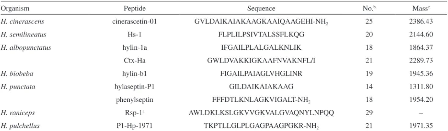
(Table 1).
Imaging analysis
Imaging of biological tissues enables the molecular mapping of ions under almost native conditions, preserving morphological and molecular informations. Aiming to
map the cinerascetins on the skin tissue and to compare their distribution on the skin, the total dorsal tissue of
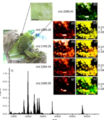
H. cinerascens was submitted to imaging acquisition. To facilitate observation of results, a representative fragment of analyzed skin is demonstrated in Figure 3. Pictorial representation of C-01 (m/z 2386.43), C-02 (m/z 2395.25), C-03 (m/z 2165.29), C-04 (m/z 2393.43) and C-05 (m/z 2490.50) are shown in Figures 3c to 3g, respectively. Co-localization between C-01 (green) and the other related peptides (red) are shown in Figures 3h to 3k, respectively. The areas shared between two ions are shown in yellow. The technique was able to detect all reported cinerascetins in this work. The results indicate that C-01 is present in a great area of the dorsal tissue and that the other related peptides shared the same area. Thus, based on the primary structure and spatial location, the peptides C-02, C-03, C-04 and C-05 probably have similar biological role as C-01. Table 1. General characteristic of antimicrobial peptides derived from Hypsiboas species
Organism Peptide Sequence No.b Massc
H. cinerascens cinerascetin-01 GVLDAIKAIAKAAGKAAIQAAGEHI-NH2 25 2386.43
H. semilineatus Hs-1 FLPLILPSIVTALSSFLKQG 20 2144.60
H. albopunctatus hylin-1a IFGAILPLALGALKNLIK 18 1864.37
Ctx-Ha GWLDVAKKIGKAAFNVAKNFL/I 21 2289.73
H. biobeba hylin-b1 FIGAILPAIAGLVHGLINR 19 1945.36
H. punctata hylaseptin-P1 GILDAIKAIAKAAG 14 1311.80
phenylseptin FFFDTLKNLAGKVIGALT-NH2 18 1954.20
H. raniceps Rsp-1a AWLDKLKSLGKVVGKVALGVAQNYLNPQQ 29 –
H. pulchellus P1-Hp-1971 TKPTLLGLPLGAGPAAGPGKR-NH2 21 1971.35
aDetermined only by cDNA analysis; bNo.: number of residues; cmonoisotopic protonated peptide [M + H]+; dstructure confirmed by cDNA sequencing.
Figure 2. Nucleotide and translated amino acid sequences of cloned skin
secretion-derived cDNA encoding the biosynthetic precursor of C-01. The signal peptide is shown double-underlined, mature peptide in single-line underline and stop codon is indicated with asterisk. Glycine amino acid residue indicates a possible amination.
Almeida et al. 2295 Vol. 26, No. 11, 2015
Solid phase synthesis and antimicrobial assay
Based on the structure of peptides characterized here and knowledge about the amphibian peptides antimicrobial potential, C-01 was selected to be manually synthesized for its antimicrobial evaluation. The peptide was prepared on a solid phase system and the purified peptide assayed against the yeast Cryptococcus neoformans, the fungus Candida
albicans, the gram-positive bacteria Staphylococcus
aureus and the gram-negative bacteria Escherichia coli,
all human pathogens.34 The synthetic peptide was also
evaluated against Xanthomonas axonopodis pv. glycines, a plant pathogenic bacteria. The peptide dermaseptin-01 (DS01) obtained from Phyllomedusa hypochondrialis was used as positive control due to its antimicrobial
properties.15
The peptide C-01 was able to inhibit the growth of all tested microorganisms with MIC values ranging from 4 to 16 µM (Table 2). The uncommon broad spectrum activity of C-01 was shown to be more active than peptides previously described in Hypsiboas such as Rsp-1 (MIC
of 20 µM against S. aureus)21 and hylaseptin-P1 (MIC of
24.2 µM against E. coli).28 Over the last years a wide range
of antimicrobial cationic peptides have been shown to play
a significant role in host defenses.35 The mode of action
of these peptides against gram-positive bacteria is well
known,36 where the ability to form channels in lipid bilayer
membranes seems to be most accepted theory.35 On the
other hand the mechanism against gram-negative bacteria
and fungi still remains unclear.36 The cationic nature of
C-01 must be taken into account to determine the possible mechanism (76% of α-helix according to self-optimized prediction method with alignment (SOPMA) prediction). Similar anuran peptides such as dermaseptin-01 are also cationic, and it has been suggested that the membrane surface in the presence of negatively charged lipids is associated with a “carpet-like” manner, breaking the lipid bonds due the reduction of repulsive electrostatic forces
between positively charged peptides.37 The similarities
among dermaseptin-01 and C-01 may be the key to establish the mechanism of action, however, more studies to confirm this hypothesis are needed. Additionally, the presence of a carboxyamidated C-terminal moiety has been shown as a factor of increasing the antimicrobial activity, possibly by a decrease of degradation rate by
carboxypeptidases.38
Conclusions
Using chromatography and MS it was possible to identify and characterize five new peptides. These compounds displayed sequences with 24 to 26 amino acid residues. Molecular cloning of precursor-encoding cDNA was performed and four sequences were identified, being one previously detected by MS. Imaging analysis with the dorsal skin allowed the localization of the MS identified peptides throughout the whole tissue. These peptides were assigned as new peptides related to hylaseptin-P1, displayed the C-terminal region with amination as post translational modifications. The synthetic peptide C-01 displayed in vitro antimicrobial activity against the tested organisms. The presented results are part of the continuous search for novel substances that can serve as models for new antibiotics. This work constitutes the first report on the peptide constitution of H. cinerascens.
Table 2. Antimicrobial activities of C-01
Pathogen MIC / µM a C-01b DS01c Waterd C. albicans 10.00 7.64 n.d.e C. neoformans 16.00 8.00 n.d.e E. coli 16.00 1.00 n.d.e S. aureus 10.00 8.00 n.d.e X. axonopodis pv. glycines < 4.00 n.d.e n.d.e aMIC (µM): minimal peptide concentrations required for total inhibition
of cell growth in liquid medium; bC-01: synthetic peptide; cDS01: positive
control; dwater: negative control; en.d.: not detected.
Figure 3. Imaging analysis of H. cinerascens skin. (a) Adult specimen
of H. cinerascens; (b) analysis of the skin fragment; (c)-(g) pictorial representation of C-01 to C-05; (h)-(k) colocalization between C-01 and the other peptides; (l) global spectrum of detected ions.
Supplementary Information
Supplementary data (imaging and MALDI LIFT-TOF-MS/MS spectra of C-01, C-02, C-03, C-04 and C-05 and sequences of cDNA of C-06, C-07 and C-08) are available free of charge at http://jbcs.sbq.org.br as PDF file. Acknowledgments
Th e au th ors t ha nk Co ns el ho Na ci on al de Desenvolvimento Científico e Tecnológico (CNPq) and Fundação de Amparo à Pesquisa na Amazônia (FAPEAM). José de Lima Cardozo Filho and Eder Alves Barbosa (Embrapa) are specially acknowledged for the collaborations.
References
1. Nelson, D. L.; Cox, M. M.; Lehninger Principles of Biochemistry, 5th ed.; W. H. Freeman: New York, 2008.
2. Hancock, R. E. W.; Chapple, D. S.; Antimicrob. Agents Chemother. 1999, 43, 1317.
3. Cardoso, M. H.; Cobacho, N. B.; Cherobim, M. D.; Pinto, M. F. S.; Santos, C.; Maximiano, M. R.; Barros, E. G.; Dias, S. C.; Franco, O. L.; Clin. Toxicol. 2014, 4, 1.
4. Deuel, T. F.; Annu. Rev. Cell Biol. 1987, 3, 443.
5. Zasloff, M.; Proc. Natl. Acad. Sci. U. S. A. 1987, 84, 5449. 6. Rinaldi, A. C.; Curr. Opin. Chem. Biol. 2002, 6, 799. 7. Brogden, K. A.; Nat. Rev. Microbiol. 2005, 3, 238. 8. Zasloff, M.; Nature 2002, 415, 383.
9. Nicolas, P.; Vanhoye, D.; Amiche, M.; Peptides 2003, 24, 1669.
10. Mor, N. P.; Annu. Rev. Microbiol. 1995, 49, 277.
11. Conlon, J. M.; Kolodziejek, J.; Nowotny, N.; Biochim. Biophys. Acta 2004, 1696, 1.
12. Gerhardt, H. C.; Science 1978, 199, 992.
13. Mignogna, G.; Severini, C.; Simmaco, M.; Negri, L.; Erspamer, G. F.; Kreil, G.; Barra, D.; FEBS Lett. 1992, 302, 151. 14. Bernarde, P. S.; Machado, R. A.; Turci, L. C. B.; Biota Neotrop.
2011, 11, 117.
15. Brand, D. G.; Leite, J. R. S. A.; Silva, L. P.; Albuquerque, S.; Prates, M. V.; Azevedo, R. B.; Carregaro, V.; Silva, J. S.; Sá, V. C. L.; Brandão, R. A.; Junior, C. B.; J. Biol. Chem. 2002, 277, 49332.
16. Costa, T. O. G.; Almeida, R. A.; Melo, J. T.; Koolen, H. H. F.; Silva, F. M. A.; Leite, J. R. S. A.; Prates, M. V.; Bloch Jr., C.; Pinto, A. C.; J. Braz. Chem. Soc. 2012, 12, 2133.
17. Biemann, K.; Annu. Rev. Biochem. 1992, 61, 977.
18. Garnier, J.; Osguthorpe, D.; Robson, B.; J. Mol. Biol. 1978, 120, 97.
19. Brand, D. G.; Leite, J. R. S. A.; Mandel, S. M. S.; Mesquita,
D. A.; Silva, L. P.; Prates, M. V.; Barbosa, E. A.; Vinecky, F.; Galasso, M. J. H.; Kuckelhaus, S. A.; Sampaio, R. N.; Junior, F. J. R.; Andrade, A. C.; Bloch Jr., C.; Biochem. Biophys. Res. Commun. 2006, 347, 739.
20. Magalhães, M. T. Q.; Barbosa, E. A.; Prates, M. V.; Verly, R. M.; Munhoz, V. H. O.; Araújo, I. E.; Bloch Jr, C.; PLoS One 2013, 8, e59255.
21. Magalhães, B. S.; Melo, J. A.; Leite, J. R.; Silva, L. P.; Prates, M. V.; Vinecky, F.; Barbosa, E. A.; Verly, R. M.; Mehta, A.; Nicoli, J. R.; Bemquerer, M. P.; Andrade, A. C.; Bloch Jr., C.; Biochem. Biophys. Res. Commun. 2008, 377, 1057.
22. Chan, W. C.; White, P. D.; Fmoc Solid Phase Peptide Synthesis: A Practical Approach, 3rd ed.; Oxford University Press: Oxford,
2000.
23. Clinical Laboratory Standards Institute (CLSI); Reference Method for Broth Dilution Antifungal Susceptibility Testing of Yeasts: CLSI document M27-A3, 3rd ed.; Clinical Laboratory
Standards Institute: Wayne, PA, 2008.
24. Clinical Laboratory Standards Institute (CLSI); Methods for Dilution Antimicrobial Susceptibility Testing for Bacteria that Grow Aerobically: CLSI document M07-A9, 9th ed.; Clinical
Laboratory Standards Institute: Wayne, PA, 2012.
25. Koolen, H. H. F.; Soares, E. R.; Silva, F. M. A.; Almeida, R. A.; Souza, A. D. L.; Quim. Nova 2012, 35, 771.
26. Koolen, H. H. F.; Soares, E. R.; Silva, F. M. A.; Oliveira, A. A.; Souza, A. Q. L.; Medeiros, L. S.; Filho, E. R.; Cavalcanti, B. C.; Pessoa, C. O.; Moraes, M. O.; Salvador, M. J.; Souza, A. D. L.; Nat. Prod. Res. 2013, 27, 2118.
27. Siano, A.; Húmpola, M. V.; de Oliveira, E.; Alberício, F.; Simonetta, A. C.; Lajmanovich, R.; Tonarelli, G. G.; J. Nat. Prod. 2014, 77, 831.
28. Prates, M. V.; Força, M. L. S.; Regis, W. C. B.; Leite, J. R. S. A.; Silva, L. P.; Pertinhez, T. A.; Araújo, A. L. T.; Azevedo, R. B.; Spisni, A.; Bloch Jr., C.; J. Biol. Chem. 2004, 13, 13018. 29. Verly, R. M.; Moraes, C. M.; Resende, J. M.; Bemquerer, M. P.;
Piló-Veloso, D.; Valente, A. P.; Almeida, F. C.; Bechinger, B.; Biophys. J. 2009, 18, 2194.
30. Mor, A.; Nicolas, P.; Eur. J. Biochem. 1994, 219, 145. 31. Nacif-Marçal, L.; Pereira, G. R.; Abranches, M. V.; Costa,
N. C. S.; Cardoso, S. A.; Honda, E. R.; de Paula, S. O.; Feio, R. N.; Oliveira, L. L.; Toxicon 2015, 99, 16.
32. Castro, M. S.; Ferreira, T. C. G.; Cilli, E. M.; Crusca Jr., E.; Mendes-Giannini, M. J. S.; Sebben, A.; Ricart, C. A. O.; Souza, M. V.; Fontes, W.; Peptides 2009, 30, 291.
33. Castro, M. S.; Matsushita, R. H.; Sebben, A.; Souza, M. V.; Fontes, W.; Protein Pept. Lett. 2005, 12, 89.
34. Bataglion, G. A.; Silva, F. M. A.; Santos, J. M.; Santos, F. N.; Barcia, M. T.; Lourenço, C. C.; Salvador, M. J.; Godoy, H. T.; Eberlin, M. N.; Koolen, H. H. F.; Food Res. Int. 2014, 64, 472. 35. Bechinger, B.; Lohner, K.; Biochim. Biophys. Acta 2006, 1758,
Almeida et al. 2297 Vol. 26, No. 11, 2015
36. Epand, R. M.; Vogel, H. J.; Biochim. Biophys. Acta 1999, 1462, 11.
37. Bechinger, B.; Zasloff, M.; Opella, S. J.; Protein Sci. 1993, 2, 2077.
38. Nascimento, A. C. C.; Fontes, W.; Sebben, A.; Castro, M. S.; Protein Pept. Lett. 2003, 10, 227.
Submitted: July 1, 2015 Published online: August 28, 2015