Diana Filipa Duarte Lobo Licenciada em Bioquímica
Cerebrovascular and Blood-Brain Barrier
Impairments in Machado-Joseph Disease
Dissertação para obtenção do Grau de Mestre em Genética
Molecular e Biomedicina
Orientadores:
Rui Jorge Gonçalves Pereira Nobre, PhD, CNC UC
Catarina Sofia Oliveira Miranda, PhD, CNC UC
I
Diana Filipa Duarte LoboLicenciada em Bioquímica
Cerebrovascular and Blood-Brain Barrier
Impairments in Machado-Joseph Disease
Dissertação para obtenção do Grau de Mestre em Genética
Molecular e Biomedicina
Orientadores:
Rui Jorge Gonçalves Pereira Nobre, PhD, CNC UC
Catarina Sofia Oliveira Miranda, PhD, CNC UC
III
Copyright © 2017 Diana Filipa Duarte Lobo, FCT/UNL and UNL. Faculty of Sciences and Technology, and the New University of Lisbon have the perpetual right without geographic limits of the publication and storage of this dissertation through printed exemplars, in digital format or through any other know means that exist or may be invented, it is also entitle to the divulgation through scientific repositories and admitting the copy and distribution of the dissertation for educational and research proposes without commercial intent as long as it is given credit to the author and editor.V
O trabalho aqui apresentado foi realizado no grupo de investigação do Prof. Doutor Luís Pereira de Almeida, Grupo de Vectores e Terapia Génica do Centro de Neurociências e Biologia Celular, Universidade de Coimbra, Portugal. Este trabalho foi financiado pelo FEDER através do Programa Operacional Regional Centro 2020, do Programa Operacional de Factores de Competitividade (COMPETE 2020) e por Fundos Nacionais através da FCT (Fundação para a Ciência e a Tecnologia) – projectos BrainHealth2020 (CENTRO-01-0145-FEDER-000008), ViraVector (CENTRO-01-0145- FEDER- 022095), CortaCAGs (POCI-01-0145-FEDER-016719) e POCI-01-0145-FEDER-007440, bem como pelos projectos EXPL/NEU-NMC/0331/2012 (FCT) e pelos SynSpread, ESMI e ModelPolyQ no âmbito do EU Joint Programme - Neurodegenerative Disease Research (JPND), os dois últimos co-financiados pelo programa H2020 da União Europeia, GA No. 643417; ainda pela “National Ataxia Foundation” (USA), pela AFM-Telethon (Proj. nº 21163), pelo American Portuguese Biomedical Research Fund (APBRF) e pelo Richard Chin and Lily Lock Machado Joseph Disease Research Fund.The work presented here was carried out in the research group of Prof. Dr. Luís Pereira de Almeida, Group of Vectors and Gene Therapy of the Center for Neurosciences and Cell Biology, University of Coimbra, Portugal. This work was funded by the ERDF through the Regional Operational Program Center 2020, Competitiveness Factors Operational Program (COMPETE 2020) and National Funds through FCT (Foundation for Science and Technology) - BrainHealth2020 projects (CENTRO-01- 0145-FEDER-000008), ViraVector (CENTRO-01-0145-FEDER-022095), CortaCAGs (POCI-01-0145- FEDER-016719) and POCI-01-0145-FEDER-007440, as well as the EXPL/NMC/0331/2012 (FCT) and the SynSpread, ESMI and ModelPolyQ under the EU Joint Program - Neurodegenerative Disease Research (JPND), the last two co-funded by the European Union H2020 program, GA No.643417; by National Ataxia Foundation (USA), AFM-Telethon (Proj. nº 21163), the American Portuguese Biomedical Research Fund (APBRF) and the Richard Chin and Lily Lock Machado-Joseph Disease Research Fund.
VII
Acknowledgments
No geral, quero demonstrar a minha mais sincera gratidão a todos os que de uma maneira ou outra contribuíram para que este trabalho fosse possível, tanto a nível profissional como pessoal.
Em primeiro lugar, agradeço ao Professor Doutor Luís Pereira de Almeida por me ter dado a oportunidade de realizar este trabalho no grupo de Vectores e Terapia Génica do Centro de Neurociências da Universidade de Coimbra. Queria também agradecer toda a disponibilidade, simpatia e conhecimento partilhado ao longo deste ano.
Aos meus orientadores, Doutora Catarina Miranda e Doutor Rui Nobre, por me terem aceitado como sua aluna, mas também por toda a atenção, disponibilidade e paciência. Em particular, agradeço todo o conhecimento científico transmitido. A eles devo também parte do trabalho aqui apresentado, nomeadamente tudo o que envolve manipulação de animais.
Gostaria de agradecer também aos Doutores José Sereno, João Castelhano e Miguel Castelo-Branco do Instituto de Ciências Nucleares Aplicadas à Saúde (ICNAS) da Universidade de Coimbra pela disponibilidade em terem realizado as experiências de DCE-MRI apresentadas nesta dissertação. Ao Centro Microscopia de Coimbra (MICC), em particular à Doutora Luísa Cortes e à Doutora Margarida Caldeiras pela disponibilidade e pelos ensinamentos de microscopia. Agradeço também a todos os meus professores que me acompanharam até aqui.
Como não poderia deixar de ser, também gostaria de deixar um agradecimento muito especial a todos os membros do grupo de Vetores e Terapia Génica pelo auxílio mas também pelo bom ambiente de trabalho. Quero deixar um agradecimento especial à Ana Cristina, à Dina, à Inês, à Patrícia e à Sara por me terem ensinado muito e pela amizade que ofereceram à sua “caçula”.
À minha família agradeço por tudo, em especial por apesar das muitas dificuldades ter tornado possível este meu percurso académico. Por fim, mas não menos importante, agradeço ao meu “respectivo” pela paciência, pela amizade e motivação, e também deixo um grande obrigado a todos os meus amigos, incluindo uns “Bandalhos” e o Designer Pedro Silva.
IX
Table of Contents
Abstract ... XI Resumo ... XIII Abbreviations List ... XV Figures Index ... XVII Table index ... XVII
1. Introduction ... 1
1.1 Central Nervous System (CNS)-barriers ... 1
1.2 The blood-brain barrier (BBB)... 1
1.2.1 Physiological functions of BBB ... 1
1.2.2 Anatomical features: Neurovascular Unit ... 2
1.2.3 Junctional complexes in the BBB ... 5
1.2.4 Routes of transport across BBB ... 8
1.3 The Blood-Cerebrospinal Fluid Barrier (BCSFB)... 9
1.4 BBB dysfunction in neurodegenerative disorders: ... 10
1.4.1 Multiple sclerosis ... 11
1.4.2 Parkinson’s disease ... 12
1.4.3 Alzheimer’s disease ... 12
1.4.4 PolyQ diseases: the particular case of Huntington’s disease ... 13
1.5 Machado-Joseph disease (MJD): ... 14
1.5.1 Genetics and protein physiology ... 14
1.5.2 Clinical features ... 15
1.5.3 Neuropathology and pathogenesis ... 15
1.5.4 Mouse models of MJD ... 17
1.5.5 Can BBB be impaired in MJD? ... 18
1.6 Objectives ... 18
2. Materials and Methods ... 19
2.1 Animals ... 19
2.2 EXPERIMENT 1: ... 19
2.2.1 Evans blue (EB) injection, Sacrifice and Tissue collection ... 19
2.2.2 EB quantification by spectrophotometry ... 19
2.2.3 EB detection by fluorescence microscopy ... 20
2.3 EXPERIMENT 2 ... 20
2.3.1 Sacrifice and Cerebellum dissection ... 20
2.3.2 Immunofluorescence ... 20
2.3.3 Immunofluorescence quantitative analysis ... 20
2.3.4 Protein Extraction and Western blotting ... 21
2.3.5 Dynamic Contrast-Enhanced-Magnetic Resonance Imaging (DCE-MRI) ... 22
2.4 Statistical Analysis ... 24
3. Results ... 25
X
3.2. Unraveling the mechanisms of BBB disruption in the cerebellum of a MJD transgenic
mouse model ... 27
3.2.1 Localization of ataxin-3 aggregates within cerebellar blood vessels in MJD transgenic mice. ... 27
3.2.2 Global alterations in the cerebellar vasculature of MJD mice ... 28
3.2.3 Fibrin extravasation in the cerebellum of MJD transgenic mice ... 30
3.2.4 Altered expression of TJ-associated proteins in the cerebellum of MJD mice ... 31
3.2.5 In vivo evidence of CNS-barriers disruption using DCE-MRI ... 33
4. Discussion ... 35
XI
Abstract
Central Nervous System (CNS)-barriers are essential to maintain brain homeostasis, protection and nutrition. Blood-brain barrier (BBB) is mainly constituted by brain endothelial cells, pericytes and astrocytes that restrict the communication between blood and the brain parenchyma. Blood-cerebrospinal fluid barrier (BCSFB) controls molecular exchange between blood and the cerebrospinal fluid in the epithelial cells of choroid plexus. Both barriers express tight junction (TJ) proteins that limit the paracellular permeability between adjacent cells.
In several neurodegenerative diseases, BBB dysfunction has been associated with neuroinflammation and TJs disruption with consequent enhancement of pathogenesis. Machado-Joseph Disease (MJD), also a neurodegenerative disorder, is caused by an expansion in CAG repeats in MJD1 gene that codifies for mutant ataxin-3 protein and causes neurodegeneration and neuroinflammation.
The aim of this work was to evaluate the cerebrovascular and CNS-barriers integrity in MJD. To accomplish that, we first assessed BBB permeability by quantifying the Evans blue (EB) extravasation in the brain of a transgenic mouse model of MJD. In a second experiment, we aimed at investigating which mechanisms were involved in BBB disruption, by analyzing: the presence of mutant ataxin-3 in cerebellar blood vessels, fibrin extravasation across BBB, and the expression of TJ-associated proteins. Finally, perfusion and vascular permeability were evaluated by Dynamic Contrast Enhanced-Magnetic Resonance Imaging (DCE-MRI).
The results of this work showed that BBB is disrupted in this MJD mouse model, which was demonstrated by Evans blue and fibrin extravasation. Both barriers showed alterations in TJs expression. Occludin was cleaved in both barriers, claudin-5 was upregulated in BBB, whereas ZO-1 showed a tendency to be decreased in BCSFB. Furthermore, it was demonstrated the presence of ataxin-3 aggregates in cerebellar blood vessels. Finally, DCE-MRI confirmed an increased blood volume and higher vascular permeability in MJD mice.
In conclusion, this work demonstrated that cerebrovasculature and CNS-barriers are impaired in MJD.
Keywords: Machado-Joseph Disease (MJD), Neurodegenerative disease, Blood-brain barrier (BBB), Blood-Cerebrospinal Fluid Barrier (BCSFB), Tight Junction (TJ), Dynamic Contrast Enhanced-Magnetic Resonance Imaging (DCE-MRI).
XIII
Resumo
As barreiras do sistema nervoso central são essenciais na homeostasia, proteção e nutrição neuronal. A barreira hematoencefálica (BHE) é constituída principalmente por células endoteliais, pericitos e astrócitos, que restringem a comunicação entre o sangue e o parênquima cerebral. A barreira sangue-líquido cefalorraquidiano (BLCR) está localizada nas células epiteliais do plexo coroide e controla a troca de substâncias entre o sangue e o líquido cefalorraquidiano. Ambas as barreiras expressam proteínas tight junctions (TJs) que limitam a permeabilidade entre células adjacentes.
Em várias doenças neurodegenerativas, as disfunções na barreira hematoencefálica têm sido associadas a neuroinflamação e alterações das TJs que, consequentemente promovem a progressão da doença. A doença de Machado-Joseph (DMJ) é, também, uma doença neurodegenerativa causada por uma expansão de CAG no gene MJD1/ATXN3 que codifica a ataxina-3 mutante e leva à neurodegenerescência e neuroinflamação.
O objetivo principal deste trabalho foi avaliar a vasculatura e a integridade das barreiras do sistema nervoso central na DMJ. Para o efeito, num modelo animal da doença, avaliámos a permeabilidade da BHE ao Evans blue e, posteriormente, investigámos os mecanismos envolvidos na disrupção da barreira, analisando: a presença de ataxina-3 mutante em vasos sanguíneos do cerebelo, a extravasão da fibrina e a expressão das TJs. Por fim, a permeabilidade vascular foi ainda analisada por ressonância magnética.
Os resultados deste trabalho demonstraram a rutura da BHE neste modelo animal, que foi evidenciada pela extravasão de Evans blue e fibrina. Ambas as barreiras mostraram alterações na expressão das TJs. A ocludina foi clivada em ambas as barreiras, a claudina-5 está aumentada na BHE e a ZO-1 mostrou-se tendencialmente reduzida na BLCR. Para além disso, a ataxina-3 mutante foi detectada em vasos sanguíneos do cerebelo. A análise por ressonância magnética mostrou ainda um aumento no volume sanguíneo e confirmou o aumento da permeabilidade vascular no cerebelo de animais transgénicos.
Em conclusão, este trabalho demonstrou que existem alterações na vasculatura e nas barreiras do sistema nervoso central na DMJ.
Palavras-chave: Doença de Machado-Joseph (DMJ), Doenças Neurodegenerativas, Barreira Hematoencefálica (BHE), Barreira Sangue-Líquido Cefalorraquidiano (BLCR), Proteínas tight junctions (TJ), Ressonância Magnética.
XV
Abbreviation List
AJ Adherens Junction a.u. Arbitrary Units
AUC Area Under the Curve BBB Blood-brain Barrier
BCSFB Blood-Cerebrospinal Fluid Barrier BSA Bovine Serum Albumin
CAG Cytosine, Adenine, Guanine CD31 Cluster of Differentiation 31 CNS Central Nervous System CoIV Collagen IV
CSF Cerebrospinal Fluid
DAPI 4',6-diamidino-2-phenylindole
DCE-MRI Dynamic Contrast Enhanced-Magnetic Resonance Imaging DCN Deep Cerebellar Nuclei
EB Evans blue
HA Hemagglutinin
JAM Junctional Adhesion Molecule MJD Machado-Joseph Disease MMP Metalloproteinase
PBS Phosphate Buffered Saline PolyQ Polyglutamine
ROI Region of Interest SCA Spinocerebellar Ataxia SEM Standard Error of Mean
Tg Transgenic
TJ Tight Junction
VEGF Vascular Endothelial Growth Factor
WT Wild Type
XVII
Figures Index
Figure 1.1 Neurovascular unit 2
Figure 1.2 Junctional complexes 5
Figure 1.3 Different routes of transport across the BBB 9
Figure 1.4 Structure of BCSFB and junctional complexes from epithelial cells of the choroid plexus
10 Figure 1.5 The mechanism underlying mutant ataxin-3 cellular toxicity in MJD 17
Figure 2.1 Representative example of quantification of extravascular fibrin 21
Figure 2.2 Coronal T1-weighted anatomical MRI sequence of the defined ROI in the cerebellum of mice
23 Figure 2.3 Schematic interpretation of tissue enhancement curve produced by
DCE-MRI technique with the injection of contrast agent
24
Figure 3.1 EB extravasation in the cerebellum of MJD transgenic mice suggests BBB disruption
26
Figure 3.2 Localization of mutant ataxin-3 aggregates within cerebellar blood vessels of MJD transgenic mice
28
Figure 3.3 Global alterations in the cerebellar vasculature of MJD transgenic mice 29
Figure 3.4 Fibrin extravascular deposition in the cerebellum of MJD mice 30
Figure 3.5 Expression levels of tight junction (TJ)-associated proteins in the cerebellum of MJD mice differ from wild-type controls
32
Figure 3.6 in vivo evidence of alterations in the perfusion and vascular permeability in the cerebellum of MJD transgenic mice using DCE-MRI
33
Figure 4.1 Schematic representation of the neurovascular unit impairments in MJD, according to the theory postulated in the present study
38
Table index
1
1. Introduction
1.1 Central Nervous System (CNS)-barriers
The Central Nervous System (CNS) homeostasis is essential for the reliable transmission of chemical and electrical signals among neurons, allowing the regulation of defined neurotransmitters and ionic concentrations around synapses and axons. This homeostasis is mainly accomplished by the barrier systems and extracellular fluids that surround the CNS, which simultaneously protects it from circulating toxins and participates in immune surveillance, reviewed in (Abbott et al 2010).
Thereby, CNS cells have controlled access to peripheral fluids through three major barriers: 1) the Blood-brain barrier (BBB); 2) the blood-cerebrospinal fluid barrier (BCSFB); 3) and the arachnoid epithelium of the meninges. BBB is an interface between blood and the brain parenchyma interstitial fluid, constituted mainly by brain endothelial cells which form brain capillaries. In order to limit contact between blood and the cerebrospinal fluid (CSF), choroid plexus epithelial cells enclose the fenestrated capillaries constituting the BCSFB. Another distinct barrier is formed by the avascular arachnoid epithelium of the meninges, sealing the subarachnoid space filled with CSF from fenestrated capillaries of dura mater. CNS barriers are reviewed in (Saunders et al 2008).
The CSF is predominantly secreted by the choroid plexus of lateral ventricles, circulating through all the four ventricles and through the subarachnoid space, being then absorbed into the venous outflow system (Sakka et al 2011). The contribution of the CSF to CNS homeostasis relies on the communication with brain parenchyma interstitial fluid, which regulates electrolyte balance, circulation of active molecules and promotes elimination of waste products.
Circumventricular organs (area postrema and median eminence, neuro-hypophysis, pineal gland, sub-fornical organ and lamina terminalis) constitute sites of physiological communication with periphery, being responsible for food intake and body temperature regulation. However, they do not represent a blood-CNS barrier (Weiss et al 2009).
1.2 The blood-brain barrier (BBB)
BBB concept was firstly introduced by Lena Stern to describe a complex structure located in the blood vessels of the CNS (Stern & Gautier 1921). Despite the fact that brain endothelial cells are the central element of BBB, they are integrated in a functional concept of the brain designated as “neurovascular unit”, which is essential for BBB development and function. Although being referred as a barrier between systemic circulation and brain parenchyma, it also functions as a selective carrier of blood components required for CNS physiology.
1.2.1 Physiological functions of BBB
BBB constitutes a barrier at several levels: paracellular, transcellular and through efflux transporters. At paracellular level, intercellular junctional complexes restrict molecular and cellular transit between adjacent brain endothelial cells, assuring a physical barrier between blood and the brain interstitial fluid. In addition, brain endothelial cells in BBB limit transcellular movement of molecules in and out of brain tissue through specific transporters and restricted vesicular transport. Besides control of paracellular and transcellular movement, some molecules that are capable to cross BBB are removed back to the blood by efflux transporters expressed in the BBB. These transporters allow ion and neurotransmitter regulation, independently of oscillations in the blood concentration (Abbott et al 2010).
2
BBB also controls protein content of the brain tissue, which is much lower than plasma, avoiding infiltration of plasma proteins such as albumin, fibrin and pro-thrombin that can damage neural tissue through induction of neuroinflammation. For instance, thrombin and plasmin in the brain can lead to glial activation and albumin induces astrocyte proliferation, both resulting in glial scar formation (Gingrich & Traynelis 2000, Nadal et al 1995).
Moreover, since communication between the periphery and CNS is essential for body homeostasis, namely to provide brain nutrition, BBB acts as a carrier for the exchange of some molecules from blood, through passive diffusion or selective transporters in brain endothelial cells (Abbott et al 2010).
In conclusion, BBB is a complex and dynamic structure adapting its functions in response to the needs of CNS in healthy and disease conditions.
1.2.2 Anatomical features: Neurovascular Unit
The concept of neurovascular unit has been defined to integrate the components that are essential to all functional roles of the CNS, including BBB dynamism (Zlokovic 2011). The neurovascular unit is constituted mainly by vascular cells (endothelial cells, pericytes and vascular smooth muscle cells), glia (astrocytes, microglia, and oligodendrocytes), neurons and the basement membrane. Brain endothelial cells are covered by endothelial basement membrane, which in part is bordered by pericytes. The parenchymal basement membrane encloses the perivascular space along with the astrocytic end-feet. Astrocytes communicate with both neurons and brain endothelial cells, enabling the communication among the different neurovascular unit
components (Lecuyer et al 2016) (see Figure 1.1).
Figure 1.1. Neurovascular unit. Brain endothelial cells of capillaries are enrolled in the endothelial basement membrane and partially covered by pericytes. Surrounded by the parenchyma basement membrane is the perivascular space. Astrocytes contact with brain endothelial cells through their terminals called end-feet. Other intervenients in the neurovascular unit organization are microglia and neurons.
Brain endothelial cells
Brain endothelial cells constitute the larger interface between blood and the brain. These specific cells are distinct from other endothelial cells in what concerns their morphology and function. The principal morphologic difference is the absence of fenestrations due to junctional
3
complexes, namely tight junctions (TJs) and adherens junctions (AJs), which lead to low paracellular diffusion (Feletou 2011). However, other phenotypic variations differentiate brain endothelium from the remaining endothelial cell types, such as the larger size and the higher amount of cytosolic mitochondria, low levels of pinocytotic vesicles and absence of lysosomal fusion with endocytic vacuoles (Bernacki et al 2008). Another phenotypic feature is the expression of drug and neurotransmitter metabolizing enzymes, such as acetylcholinesterase, alkaline phosphatase, gamma-glutamyl transpeptidase and monoamine oxidases. This enzymatic activity provides protection against high levels of neurotransmitters in the brain, which can damage neuronal tissue, so as other damaging molecules (Wilhelm et al 2011).Moreover, brain endothelial cells membrane receptors and transporters show distinct expression in luminal (blood) and abluminal (CNS) sides, which enable an unidirectional route of transport across BBB for specific molecules (Banks 2016). The luminal membrane is also distinct from the abluminal one due to a negative charge that is conferred by a glycocalyx coverage, a mesh of proteoglycans, glycosaminoglycans, glycoproteins and glycolipids (van den Berg et al 2006). Glycocalyx maintains BBB integrity by preventing the movement of macromolecules (based on their charge) to the endothelial surface, influencing flow resistance, protecting brain endothelial cells from oxidative stress and preventing the contact between endothelium and red blood cells, platelets and leukocytes. In normal conditions, leukocytes cannot interact with their receptors in brain endothelial cells, thus avoiding their infiltration into the CNS (Kolarova et al 2014). In addition, inflammatory responses are also modulated by glycocalyx, since it can bind to cytokines and, consequently, restrict them to interact with endothelial cell receptors (Reitsma et al 2007). On the other hand, glycocalyx acts as a sensor of biochemical and mechanical forces of blood, transducing it into conformational changes and, consequently, into endothelial cellular responses (Reitsma et al 2007).
Therefore, junctional complexes, glycocalyx and the different distribution of abluminal and luminal receptors give rise to brain endothelial cell polarization, a phenotypic characteristic translated into unique endothelial functions, such as unidirectional transport.
Astrocytes
Astrocytes have multiple functions in the CNS that are essential to many metabolic processes, including: the modulation of synaptic transmission, neurotransmitter uptake and promotion of neurovascular coupling as reviewed in (Cabezas et al 2014, Newman 2003). In CNS, two main types of astrocytes have been described: the protoplasmic astrocytes, which are present in grey matter, around neuronal bodies and synapses, and the fibrous astrocytes present in the white matter associated with Ranvier nodes and oligodendroglial cells (Oberheim et al 2012).
Protoplasmic astrocytes have cytoplasmic appendices, whose endings, called end-feets, interact with both neurons and brain endothelial cells, forming another layer of the neurovascular unit (see Figure 1.1). Although brain endothelium is separated from astrocytes by pericytes and basement membrane, connexins allow for the communication between these two cell types (Winkler et al 2011). The contact with brain endothelial cells during neurodevelopment is responsible for promoting barrier properties, namely polarization of these cells by maintaining TJ and cytoskeleton-associated proteins integrity (Michele & Campbell 2003).
Moreover, as a part of the neurovascular unit, astrocytes synchronize neuronal metabolic demands with local cerebral blood flow. They control exchanges between the nervous system and the vasculature, participate in the formation of parenchymal basement membrane and modulate junctional complexes expression by brain endothelium (Anderson & Nedergaard 2003, Janzer & Raff 1987). Paracrine regulation of BBB by astrocytes is a result of signaling molecules release, such as vascular endothelial growth factor (VEGF), a pro-angiogenic factor that influences brain endothelium growth and glial-derived neurotrophic factor, which specifically induces the expression of the TJ protein claudin-5 in brain endothelial cells (Almutairi et al 2016, Igarashi et al 1999, Shibuya 2008).
4
Astrocytes also secrete key proteins of the parenchymal basement membrane, as described below (Proctor et al 2005, Sixt et al 2001). Another product released by astrocytes is transforming growth factor-β that has been described to have a dual role in BBB. On one hand, it stabilizes the barrier through the modulation of the expression of P-glycoprotein efflux transporter, which eliminates xenobiotics from CNS (Dohgu et al 2004). On the other hand, transforming growth factor-β is associated with neuroinflammation in diseases such as Alzheimer’s and multiple sclerosis (Lecuyer et al 2016). Furthermore, when activated, astrocytes can stimulate the ubiquitin-proteasome system, leading to degradation of TJ proteins (Chang et al 2015, Filous & Silver 2016).
Pericytes
Pericytes are found enwrapped in pre-capillary arterioles, capillaries, and post-capillary venules (Winkler et al 2011). As part of the neurovascular unit, these cells are located between brain endothelial cells and astrocytes, covering part of capillary outer surfaces (Sims 1991) (see Figure 1.1). Contact spots of pericytes with brain endothelium are provided by gap junctions and AJs, where there is no basement membrane (Cuevas et al 1984, Sa-Pereira et al 2012, Winkler et al 2011). During development, pericytes promote angiogenesis by secretion of angiogenic factors. Simultaneously, they respond to platelet-derived growth factor B, which is secreted by brain endothelial cells as a recruitment signal to pericytes (Darland et al 2003, Winkler et al 2011). Besides, pericyte attachment to brain endothelium is promoted by notch signaling, by upregulation of an adhesion protein, N-cadherin (Li et al 2011). Like astrocytes, pericytes maintain barrier properties and influence the expression of TJ-associated proteins (Bonkowski et al 2011, Haseloff et al 2005). However, these effects may occur indirectly, since it has been demonstrated that pericytes act on brain endothelium through the induction of astrocyte polarity and also inhibition of transcellular vesicular trafficking, which is important to control BBB permeability (Armulik et al 2010, Daneman et al 2010).
In addition to their vital role during embryogenesis, pericytes remain crucial in adulthood, namely due to brain endothelium stabilization and maturation, establishment of barrier properties and basement membrane maintenance (Allinson et al 2012, Jeansson et al 2011). In fact, pericytes contribute to basement membrane integrity through the regulation of laminin production and fibronectin secretion (Stratman et al 2009).
Moreover, pericytes are mediators of neurovascular coupling, since they are responsive to vasoactive substances, expressing contractile proteins and vasoactive agents. These substances allow for modulation of capillary blood flow in response to CNS needs (Hamilton et al 2010).
Pericyte phagocytic properties were also suggested to be important in BBB disruption models, due to the participation in the clearance of neurotoxic circulating plasma proteins, including immunoglobulins, fibrin and albumin (Winkler et al 2014, Yates et al 2017).
Basement membrane
BBB integrity is also maintained by both parenchymal and endothelial basement membranes, which provide cell adhesion, structural support and a signaling interface between brain endothelial cells and the surrounding environment (see Figure 1.1). Endothelial basement membrane surrounds pericytes and brain endothelial cells, whereas parenchymal basement membrane delineates perivascular space filled with interstitial fluid.
The main basement membranes constituents are collagen IV (CoIV), fibronectin, laminins, agrin, nidogen-1 and 2 and heparan sulfate proteoglycans (Morris et al 2014). CoIV, produced by brain endothelial cells, pericytes and astrocytes, confer mechanical resistance to basement membrane and stabilize it, retaining laminins (Poschl et al 2004). Laminins are essential to pericytes function, which in turn regulate the expression of these basement membrane glycoproteins (Armulik et al 2010). Another basement membrane glycoprotein, fibronectin, is also secreted by astrocytes, pericytes and brain endothelial cells and its insoluble
5
form acts as a linker between basement membrane and plasma membrane of both pericytes and brain endothelial cells (Almutairi et al 2016, Cuevas et al 1984, Tilling et al 1998). Additionally, heparin sulfate proteoglycan agrin, is crucial to maintain BBB integrity through stabilization of AJs and TJs proteins (Steiner et al 2014). Integrins and growth factors provide communication between basement membrane components and cells attachment, forming a dynamic basement membrane (Yurchenco 2011). The dynamism is also accomplished by MMPs (metalloproteinases) and its inhibitors that degrade or limit basement membrane degradation, respectively, both in physiological and inflammatory conditions (Weiss et al 2009, Yong 2005).Neurons
Despite being the basic building block of the CNS, neurons’ influence in the BBB activity is still poorly understood. However, it is known that these cells modulate blood vessel function (Cardoso et al 2010). In fact, the contact of neuronal projections with astrocytes and brain endothelial cells induces the regulation of cerebral blood flow by neuronal mediators, which is mainly accomplished by noradrenergic, serotonergic and cholinergic neurons, among others (Hawkins & Davis 2005, Weiss et al 2009). However, a lot of knowledge on this subject is still lacking and needs further investigation.
Microglia
Microglia represents another component in the barrier established between endothelium and neurons. Its high proximity with BBB starts during development in which microglia release VEGF that stimulates angiogenesis, which is reviewed in (Arnold & Betsholtz 2013). In an adult brain, microglia function as part of the immune system, alternating between the inactive form and the activated one. However, in pathological conditions, activated microglia promotes disease progression and BBB breakdown (Dudvarski Stankovic et al 2016). This fact contradicts the idea of an immune-privileged CNS, since microglia is capable of phagocyting microorganisms, whole neurons and even parts of blood vessels (Brown & Neher 2014, Jolivel et al 2015). Accordingly, microglia act as a defense line in the perivascular space, protecting brain from harmful molecules that can cross BBB.
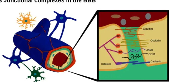
1.2.3 Junctional complexes in the BBB
Figure 1.2. Junctional complexes. TJs between adjacent brain endothelial cells are constituted by transmembrane proteins (claudins, occludin and junction adhesion molecules), which are attached to actin cytoskeleton by cytoplasmic accessory proteins, such as zonula occludens-1 (ZO-1). AJs are mainly formed by the transmembrane proteins cadherins and catenins that are bound to the cytoskeleton. JAM= Junctional Adhesion Molecule; CD31= Cluster of Differentiation 31.
6
The endothelium of brain capillaries has barrier features mainly due to the formation of junctional complexes, including TJs and AJs (Figure 1.2). These intercellular protein complexes lead to low paracellular permeability between adjacent brain endothelial cells and participate in the polarization of these cells. Additionally, gap junctions have also been identified at the BBB, allowing for a tight communication between brain endothelium cells and pericytes through proteins of the connexin family (Nagasawa et al 2006, Rodrigues & Granger 2015). Gap junction plaques provide neighboring brain endothelial cells with ions and small molecules, contributing for the transmission of intercellular signals between adjacent cells (Stamatovic et al 2016). It was shown that in Drosophila, gap junctions are essential for the coordinated signaling established in BBB glia (Speder & Brand 2014).
1.2.3.1 Tight junction (TJ)
In the BBB, TJs confer a high electrical resistance of approximately 1800Ωcm2 (Bazzoni & Dejana 2004). Structurally, TJs are constituted by a group of transmembrane adhesion proteins (occludin, claudins and junctional adhesion molecules) and accessory cytoplasmic proteins like zonula occludens (ZO) and cingulin, among others. Accessory cytoplasmic proteins are responsible for linking transmembrane proteins to the actin cytoskeleton, providing TJs with intracellular signaling and physical support (Stamatovic et al 2016) (Figure 1.2).
Multiple cells of the neurovascular unit modulate the formation and maintenance of TJ, namely astrocytes, pericytes and neurons (Abbott et al 2010). Alterations in intracellular and extracellular calcium concentration affect TJs assembly and, consequently, BBB permeability. Extracellular calcium signaling cascade involves phosphokinase-C and heterotrimeric G protein; when extracellular calcium is removed, BBB permeability increases (Stevenson & Begg 1994). In turn, intracellular calcium regulates the proximity of adjacent brain endothelial cells by affecting cytoplasmic TJ proteins location, such as ZO-1. In the absence of intracellular calcium, ZO-1 is not localized in the membrane, where it exerts its function (Stuart et al 1994). In addition, phosphorylation of TJ proteins constitutes to signaling mechanisms of its regulation that finally contribute to BBB dynamism (Huber et al 2001).
Claudins
Claudins are the major component of TJs. They are phosphoproteins with four transmembrane domains and a molecular weight of around 22 kDa. These proteins attach adjacent brain endothelial cells through claudin-claudin hemophilic interactions, while the carboxylic end is connected to cytoplasmic ZO proteins (Bernacki et al 2008) (Figure 1.2). Despite the existence of 26 isoforms of claudins, the most abundant and relevant one in BBB is claudin-5, whose absence leads to disruption of this barrier in mice, despite the presence of other isoforms (Gunzel & Yu 2013). Claudins form size and charge-selective pores and each claudin controls the diffusion of certain molecules’ size (Morita et al 1999). For example, claudin-5 allows the diffusion of molecules with <800 Da (Nitta et al 2003). Together, claudins and occludins form heteropolymers and transcellular tracts containing channels for the selective transport of ions and hydrophilic molecules (Matter & Balda 2003). Claudin phosphorylation is one of the regulation forms of TJs’ function and consequently regulates BBB permeability. Phosphorylation results in TJs assembly or disassembly, depending on the claudin phosphorylated and the phosphokinase responsible for it. For instance, phosphorylation mediated by phosphokinase-A leads to assembly of claudin-16 but, inversely, leads to claudin-3 disassembly (Findley & Koval 2009). On the other hand, myosin light chain kinase-mediated phosphorylation has been associated with an increase of BBB permeability, in inflammation conditions, due to claudins disassembly (Haorah et al 2005). This reflects the extraordinary complexity of these structures as well as the tight physiological regulation that they require.
7
OccludinOccludin is a phosphoprotein with 63 kDa and four transmembrane domains, which regulates paracellular transport (Hirase et al 1997). Together with claudin, the two extracellular loops of occludin constitute the TJ paracellular component, while the cytoplasmic C-terminal domain interacts with cytoplasmic ZO proteins (Zlokovic 2008) (Figure 1.2). Furthermore, occludin is a regulatory protein and its localization in the cell modulates BBB integrity (Huber et al 2001). The function of this TJ-associated protein is also regulated through phosphorylation by several kinases. It can be phosphorylated on serine, threonine and tyrosine residues (Bauer et al 2014). For instance, tyrosine phosphorylation prevents occludin binding to ZO cytoplasmic proteins, leading to an increased paracellular permeability since the transmembrane proteins will no longer be linked to actin cytoskeleton (Kale et al 2003). Small G proteins such as Rho also regulate TJs assembly through occludin phosphorylation, promoting an increase in BBB permeability when occludin is phosphorylated (Hirase et al 2001).
Junctional adhesion molecules (JAMs)
Junctional adhesion molecule (JAM)-A, B and C are immunoglobulins with a single transmembrane domain and an extracellular fragment with two immunoglobulin-like loops (Petty & Lo 2002). In the intercellular cleft, they form homotypic cell-cell contacts between adjacent brain endothelial cells, enhancing its adhesion (Bazzoni et al 2000a). In addition, JAM proteins interact with cytoplasmic TJ proteins, like ZO proteins, in order to anchor to the actin cytoskeleton (Bazzoni et al 2000b). They are also involved in regulation of the immune status within the CNS, since they modulate leukocyte migration through BBB due to its close association with platelet endothelial cellular adhesion molecule 1 (also denominated as cluster of differentiation 31 (CD31)) (Martin-Padura et al 1998). Furthermore, a protein with similar structure to JAM, endothelial cell-selective adhesion molecule is also a transmembrane protein associated with extravasation of neutrophils during inflammation (Stamatovic et al 2016, Wegmann et al 2004).
Cytoplasmic proteins involved in TJ formation
In addition to the previous proteins that belong to the intercellular cleft, there are also cytoplasmic proteins that are essential to TJ formation and brain endothelial cells regulation. Cytoplasmic TJ proteins provide not only structural support to BBB and BCSFB, but also integrate several signaling pathways that regulate activity of brain endothelial cells and epithelial cells from choroid plexus and TJs assembly. Some of these are ZO proteins, cingulin, AG-6, 7H6 and others. ZO-1, 2 and 3 are membrane-associated guanyl kinase-like proteins, which connect claudins, occludin and JAM proteins to the actin cytoskeleton, allowing effectiveness of TJs’ regulation (Vorbrodt & Dobrogowska 2004). In fact, ZO proteins may even translocate to the nucleus regulating proliferation by interacting with transcription factors (Bauer et al 2014, Betanzos et al 2004). The absence of ZO-1 has demonstrated its relevance at the establishment of junctional proteins placement and angiogenesis, since its knock out leads to lethal phenotype during embryogenesis in mice (Katsuno et al 2008).
ZO-1 has two isoforms: ZO-1 α- and ZO-1 α+. Isoform α- is restricted to endothelium, thus present in BBB, and some atypical epithelial cells, such as renal glomerular podocytes and between Sertoli cells; while isoform α+ is found in most epithelial-cell junctions (Willott et al 1992). With similar functions, the myosin-like phosphoprotein named cingulin, stands between transmembrane proteins and the cytoskeleton, binding to ZO proteins and myosin (Cordenonsi et al 1999). The 7H6 phosphoprotein is associated with the impermeability of the BBB to ions and large molecules (Satoh et al 1996). In conditions of low ATP levels, 7H6 protein dissociates from the other TJ proteins, which leads to an increase of paracellular permeability, demonstrating the dynamic properties of junctional complexes in response to environmental circumstances (Huber et al 2001).
Other cytoplasmic proteins described in the BBB are Ca2+-dependent serine protein
8
kinase with inverted orientation of protein-protein interaction domains, small guanosine triphosohateases, G-protein signaling 5, and ZO-1-associated nucleic acid-binding protein. Together, these proteins are responsible for ensuring the integrity of TJs and structural support of the brain endothelium and its regulation (Bernacki et al 2008).
1.2.3.2 Adherens junction (AJ)
In order to allow TJs assembly, it is not only necessary the presence of other junctional complexes, AJs, but also the crosstalk between these two complexes to maintain a dynamic BBB (Tietz & Engelhardt 2015). Consequently, AJs are essential to BBB integrity and its absence leads to barrier disruption (Wolburg & Lippoldt 2002). These junctional complexes are located at the basal region of lateral plasma membrane and mediate paracellular permeability between brain endothelial cells through the stabilization of cell-cell interactions. AJ main proteins are the transmembrane glycoproteins, cadherins, which interact homotipically to enhance adhesion between brain endothelial cells (Takeichi 1995). To anchor cadherins to the actin cytoskeleton, cytoplasmic proteins β and ϒ-catenins mediate cadherin connection with α-catenin, which in turn interact to actin (Nieset et al 1997). Catenin and vascular endothelial-cadherin have been identified in the human cortex (Vorbrodt & Dobrogowska 2004). CD31 is also present in human AJs, interacting with β-catenin (Matsumura et al 1997).
Regarding the cross-talk between the two types of junctional complexes, interactions of AJs with TJs start during development when AJs initiate cell-cell contacts and promote TJ maturation, maintenance and plasticity (Tietz & Engelhardt 2015). Vascular endothelial-cadherin promotes upregulation of claudin-5 expression. This has been demonstrated due to increased paracellular permeability as a consequence of claudin-5 downregulation, in the absence of vascular endothelial-cadherin (Taddei et al 2008). However, TJ proteins also have a modulation role in AJs, namely ZO-1 essential role in vascular endothelial-cadherin integrity (Tornavaca et al 2015). Furthermore, AJs are also regulated by phosphorylation, namely by tyrosine phosphorylation of β-catenin which results in disruption of this junctional complex (Roura et al 1999).
1.2.4 Routes of transport across BBB
Intact and functional BBB restricts the entry of compounds according with certain factors: high polar surface area, tendency to form more than 6 hydrogen bonds, presence of rotatable bonds in the molecule, a molecular weight higher than 450 Da and a high affinity of binding to plasma proteins. Besides that, molecules with cationic nature have advantage over acids when penetrating BBB, due to the negatively charged glycocalyx of brain endothelium, as reviewed in (Abbott et al 2010).
Nevertheless, one of the main roles of BBB is the regulation of the transport of nutrients, cells and other molecules into and out of the brain. Through paracellular pathway, by crossing TJs, some water-soluble agents can penetrate BBB into the CNS. On the other hand, lipid-soluble molecules can enter the brain passively through transcellular route (Liu et al 2004). Blood gases, oxygen and carbon dioxide diffuse through lipid membranes of brain endothelial cells, depending on their concentration gradient (Abbott et al 2010). Furthermore, there are specific transport systems on the luminal and abluminal membranes, such as solute carriers and ATP-binding cassette transporters. Solute carriers control the transcellular traffic of small hydrophilic molecules, such as glucose, amino acids, nucleosides and choline. ATP-binding cassette transporters transport, with consume of ATP, lipid-soluble substances out of the brain. The most significant of these transporters are Multidrug Resistance-Associated Proteins and Breast Cancer Resistance Proteins (Begley 2004). P-glycoprotein is a Multidrug Resistance-Associated Protein, and it was the first identified in humans in several organs such as the intestine, kidney, pancreas, peripheral immune cells and in BBB (Loscher & Potschka 2005). Its wide activity in the BBB is
9
demonstrated by its expression in many cells, specifically brain endothelial cells, astrocytes, microglia and pericytes (Beaulieu et al 1997, Lee & Bendayan 2004).Additionally, specific receptor-mediated transcytosis allows the entry of large molecules, such as insulin and transferrin, using the vesicular trafficking machinery of brain endothelial cells promoted by receptor-ligand interactions (Abbott et al 2006). Conversely, absorptive-mediated transcytosis allow the entrance of molecules into the brain with less specificity than receptor-mediated transcytosis, based only on charge interactions (Jones & Shusta 2007).
Regarding cell movement across BBB, it is known that perivascular space functions as a niche for immune cells, namely mononuclear cells that in the absence of pathology can enter the brain through diapedesis, without rearranging TJ (Bechmann et al 2001). In pathological scenarios, leukocytes, monocytes and macrophages can penetrate BBB by promoting TJs rearrangements, keeping up with microglia (Davoust et al 2008).
Figure 1.3. Different routes of transport across the BBB. a) Passive diffusion of lipid soluble molecules; b) ATP-binding cassette (ABC) transporter efflux xenobiotics and lipid soluble metabolites; c) Solute carriers (SLC) mediate transport of polar substances; d) Receptor (RMT) and adsorptive- mediated transcytosis (AMT) involves vesicular transport; e) Cell movement may occur through transcellular or paracellular route involving TJ disarrangement. Pgp= P-glycoprotein; BCRP= Breast Cancer Resistance Protein; MRP= Multidrug Resistance Protein. Reviewed in (Abbott et al 2010).
1.3 The Blood-Cerebrospinal Fluid Barrier (BCSFB)
BCSFB is localized in the choroid plexus of cerebral ventricles, being an interface between blood and CSF. Main functions of BCSFB are: to prevent the entry of toxic substances to the CSF, such as toxins; to provide glucose, oxygen, ions and vitamins necessary to CSF production; to absorb and eliminate the waste products from this fluid; to maintain Ca2+
homeostasis and hormones transport, as reviewed in (Engelhardt & Sorokin 2009).
Although BBB and BCSFB have a similar function, their structure are quite different. BCSFB is constituted by epithelial cells of the choroid plexus, which are wrapped in the subepithelial basement membrane (see Figure 1.4). The intermediate interstitium separates the basement membrane and the voluminous vasculature, which is formed by fenestrated capillaries. The high content of blood vessels in the choroid plexus produce the high blood flow necessary to CSF production. In addition, these capillaries are highly permeable, in contrast with brain endothelial cells from BBB (Tietz & Engelhardt 2015).
10
Figure 1.4. Structure of BCSFB and junctional complexes from epithelial cells of the choroid plexus. Distance between adjacent epithelial cells is reduced by the existence of tight junctions (TJ) and adherens junctions (AJ). Claudin- 1, 2, 3 and 11 among others together with occludin and junctional adhesion molecule-A (JAM-A) form the transmembrane component of TJs, whereas zonula occludens-1 (ZO-1) constitutes the cytoplasmic link to the cytoskeleton. AJs are formed by cadherin and catenins. Adapted from (Tietz & Engelhardt 2015).
TJs in BCSFB are expressed in epithelial cells, restricting the paracellular permeability between adjacent cells. However, these junctional complexes in BCSFB are leakier than the ones formed in BBB. In fact, TJ-associated proteins involved in BCSFB are different from BBB. Although there are some common components, such as occludin, they express different claudins and different ZO-1 isoforms. In BCSFB, claudins 1, 2, 3, 9, 11, 19 and 22 are predominantly expressed, so as ZO-1 isoform α+ which is associated with epithelial cells (Liddelow et al 2013); whereas claudins 3, 5 and 12 and ZO-1 isoform α- are associated with the endothelium of BBB (Hawkins & Davis 2005, Willott et al 1992). The epithelial cells of choroid plexus also form AJs between them and express efflux transporters, such as P-glycoprotein (Mealey et al 2008). Besides paracellular flux of molecules, another route of transport of BCSFB is through transcytosis, specially macromolecules, such as transferrin (Xiao & Gan 2013).
1.4 BBB dysfunction in neurodegenerative disorders:
BBB dysfunction is a natural event during aging and occurs mainly due to chronically elevated levels of pro-inflammatory cytokines, such as tumour necrosis factor-α, interleukin-6 and interleukin-1β, which regulate the expression of TJ-associated proteins. In particular, high levels of tumour necrosis factor-α in ageing are associated with TJs loss (Elahy et al 2015). Nevertheless, in some neurodegenerative diseases, the BBB is even more dysfunctional than in normal ageing process. In fact, many of these disorders have a late onset, which in part may be correlated with BBB alterations. Implications and causes of BBB dysfunction in Multiple Sclerosis, Parkinson’s, Alzheimer’s and Huntington’s diseases are discussed below.
11
Table 1.1. BBB alterations in neurodegenerative diseases. Multiple sclerosis, Parkinson’s, Alzheimer’s and Huntington’s diseases and the respective abnormalities in neurovascular unit, TJs, BBB transporters and in extravasation of some substances from this barrier.Neurodegenerative Disease
BBB alteration Reference
Multiple sclerosis Astrocyte activation (Lopes Pinheiro et al 2016)
Infiltration of activated leukocytes (Lassmann et al 2007) (Larochelle et al 2011) Occludin dephosphorilation and TJ
disruption
(Morgan et al 2007) Decreased P-glycoprotein levels (Kooij et al 2010) Fibrin extravasation (Yates et al 2017)
Parkinson’s disease Decreased ZO-1 and occludin
expression
(Chen et al 2008) Reduced contact between neurons
and brain endothelial cells
(Farkas et al 2000) Decreased levels of P-glycoprotein (Kortekaas et al 2005)
Alzheimer’s disease Downregulation of ZO-1 expression (Kook et al 2013)
Reduction in pericytes (Halliday et al 2016) Altered amyloid-β clearance (Deane et al 2004) Reduced P-glycoprotein activity (Deo et al 2014)
Huntington’s disease Increased brain blood vessels
density
(Drouin-Ouellet et al 2015) Impaired vascular reactivity (Hsiao et al 2015)
Decreased levels of occludin and claudin-5
(Drouin-Ouellet et al 2015) Redistribution of claudin-5 (Lim et al 2017)
1.4.1 Multiple sclerosis
In multiple sclerosis, neurodegeneration is preceded by demyelination and axonal loss, mainly due to inflammatory and immune system reactions. This may lead to a variety of symptoms, such as muscle weakness, affected vision and coordination, balance impairment and memory loss.
As regulators of BBB function and mediators of inflammation, activated astrocytes have a dual effect in BBB in multiple sclerosis, where their protective role is represented by the production of retinoic acid, which has an antioxidant effect in brain endothelial cells (Mizee et al 2014). Conversely, Pinheiro et al. described BBB dysfunction as an initial event in this disease that further results in astrocyte activation due to the infiltration of toxic molecules in the CNS (Lopes Pinheiro et al 2016). Activated astrocytes are known to release pro-inflammatory cytokines that affect BBB integrity (Lee et al 2012, Lopes Pinheiro et al 2016). In addition, activated astrocytes release monocyte chemoattractant protein-1, a chemokine found to induce TJ degradation (Stamatovic et al 2009). In fact, alterations in junctional complexes were found in the experimental autoimmune encephalomyelitis model for multiple sclerosis. Occludin dephosphorylation, associated with TJ disruption, was demonstrated to be associated with inflammation and to occur prior to BBB disruption, suggesting that it might be a target for signaling processes in this disease model (Morgan et al 2007, Sakakibara et al 1997). Moreover, microvascular P-glycoprotein levels are decreased in multiple sclerosis, possibly affecting the efflux of toxic molecules, such as fibrin that is a neurotoxic blood-born protein affecting multiple sclerosis progression (Kooij et al 2010, Yates et al 2017).
12
All previously described events lead to BBB disruption documented in this disease. Consequently, it was also demonstrated that activated leukocytes, plasma proteins and inflammatory agents from periphery are able to infiltrate the brain parenchyma through brain capillaries (Bruck et al 1997, Larochelle et al 2011, Lassmann et al 2007). Migration of leukocytes leads to demyelination and boosts BBB permeability, increasing subsequent leukocyte infiltration (Biernacki et al 2004, Seguin et al 2003). Some authors suggested that BBB remains persistently dysfunctional in multiple sclerosis, while others found evidences that BBB dysfunction is transient and dynamic, as a focal reaction in the site of a newly developing inflammatory lesion (Claudio et al 1995, Eisele et al 2016, LeVine 2016).
Furthermore, BCSFB have also been implicated in this disease, since TJ-associated proteins such occludin, ZO-1 and claudins were found downregulated in this disease (Kooij et al 2014).
1.4.2 Parkinson’s disease
Parkinson’s disease is characterized by motor symptoms and neuropsychiatric disturbances caused by neurodegeneration especially in dopaminergic neurons of substantia nigra. Other pathological hallmark is neuroinflammatory course and immune response that lead to progressive neurodegeneration (Yan et al 2014). At the molecular level, intranuclear aggregates of α-synuclein (Lewy bodies) are formed within neurons and other CNS cells affecting their function. An example of this picture is the accumulation of α-synuclein in astrocytes that disturbs its activity (Song et al 2009).
In Parkinson’s patients, dopaminergic neurons of substantia nigra extend their axons to the striatum, the site where post-mortem studies have found BBB dysfunction in these patients (Gray & Woulfe 2015). Besides, in the striatum of the 1-methyl-4-phenyl-1,2,3,6-tetrahydropyridine mouse model of Parkinson’s disease there was a decreased expression of TJ proteins ZO-1 and occludin associated with enhanced paracellular permeability in brain endothelial cells (Chen et al 2008). This may be explained by microglia activation and pro-inflammatory cytokine expression caused α-synuclein aggregation (Gu et al 2010). Consequently, inflammatory molecules lead to BBB disruption due to the attraction of lymphocytes to the damaged site (Blum-Degen et al 1995, Gonzalez-Scarano & Baltuch 1999). Moreover, in Parkinson’s disease there is reduced contact between neurons and brain endothelial cells, which disturb neurovascular coupling essential to BBB function (Farkas et al 2000, Thiollier et al 2016). In the brain endothelial cells, there is evidence that P-glycoprotein, a BBB protein responsible for the removal of toxic compounds out of the brain, has its activity reduced in the midbrain, which can be associated with α-synuclein accumulation (Bartels 2011, Kortekaas et al 2005).
Consequently, BBB disruption can enhance Parkinson’s disease progression by inducing apoptotic death of nigral dopaminergic neurons (Rite et al 2007). Loss of integrity of these neurons can be explained by thrombin leakage through BBB into the brain, enhancing an intense inflammatory reaction (Carreno-Muller et al 2003). Another consequence of BBB increased permeability is the impairment in α-synuclein transport across BBB. This transport occurs bidirectionally and is assured by the lipoprotein receptor-related protein-1, which in conditions of systemic inflammation, can lead to the increased uptake of α-synuclein (Sui et al 2014).
Regarding the sequence of events, it is suggested that BBB disruption is an early event, which enhances α-synuclein aggregation due to the leakage of neurotoxic agents (Gray & Woulfe 2015).
1.4.3 Alzheimer’s disease
Cognitive decline and loss of memory in Alzheimer’s disease result from several events: amyloid-β aggregates forming plaques, tau protein in neurofibrillary tangles, inflammation, neurovascular dysfunction and neuronal loss (Zlokovic 2011). Amyloid-β activates microglia and
13
astrocytes through Toll-like receptors, leading to the production of reactive oxygen species and cytokines, thus triggering an intense inflammatory response in CNS (Caldeira et al 2017, Guerriero et al 2016). This leads to another Alzheimer’s disease hallmark, BBB breakdown with TJ disruption, which has been demonstrated in Alzheimer’s disease patients and mouse models of the disease (Kook et al 2013, van de Haar et al 2016).BBB dysfunction in Alzheimer’s disease is, in part, explained by dysregulation of vasoconstrictors and vasodilators, along with oxidative stress in brain endothelial cells, impair BBB structure and function (Aliev et al 2009, Palmer et al 2012, Sole et al 2015). Pericyte reduction leads to short coverage of brain endothelial cells, enhancing vascular dysfunction. Ultimately, neurovascular unit impairment results in BBB disruption and consequent extravasation of immunoglobulin G and fibrin, plasma proteins that are toxic to neurons (Halliday et al 2016). Additionally, Alzheimer’s disease has been linked to changes in the expression of TJ proteins. For example, there is disruption of ZO-1 expression, caused by high intracellular Ca+ and
metalloproteinases levels induced by amyloid-β42 (Kook et al 2013).
On the other hand, an intact BBB may regulate amyloid-β accumulation in the CNS, through receptor-mediated transport mechanisms which control both the efflux and influx of this protein into the brain (Mackic et al 2002). Amyloid-β is exported from the brain across BBB through low-density lipoprotein receptor-related protein 1 and 2 transporter and P-glycoprotein, whereas receptor for advanced glycation end products is responsible for the import to the brain (Bell et al 2007, Lam et al 2001, Storck et al 2016). In Alzheimer’s patients, inflammation and oxidative stress disturb amyloid-β transport across BBB (Jaeger et al 2009). Low density lipoprotein receptor-related protein 1 transporter is impaired, resulting in a decreased efflux of amyloid-β out of the brain (Deane et al 2004). In addition, P-glycoprotein is decreased in this disease leading to an impaired amyloid-β clearance (Deo et al 2014). Simultaneously, higher levels of receptor for advanced glycation end products activity promote increased amyloid-β transcytosis into the brain, causing its accumulation in brain parenchyma. Besides, the interaction of these receptors with amyloid-β triggers the intracellular Ca2+ signaling cascade and MMPs
secretion resulting in TJ disruption (Kook et al 2013, Stuart et al 1996). Consequently, amyloid-β accumulation leads to further paracellular permeability, as explained above. Therefore, in Alzheimer’s, an accumulation of other circulating proteins, like immunoglobulins, albumin, fibrinogen and thrombin, was also demonstrated (Halliday et al 2016).
1.4.4 PolyQ diseases: the particular case of Huntington’s disease
Polyglutamine (polyQ) diseases are neurodegenerative disorders characterized by the expansion of the cytosine-adenine-guanine (CAG) repeat in genes encoding a long polyglutamine tract in the respective protein, which causes alterations in its function. Huntington’s disease, dentatorubral-pallidoluysian atrophy, spinal and bulbar muscular atrophy and spinocerebellar ataxia types (SCA) 1, 2, 3 6, 7 and 17 are the nine members of polyQ diseases (Gatchel & Zoghbi 2005, Matos et al 2011, Shao & Diamond 2007).Huntington’s disease is an autosomal dominant disorder with a CAG repeat expansion in the exon 1 of the huntingtin gene, which leads to a mutant huntingtin protein. This mutated protein forms oligomers, and, ultimately, insoluble aggregates (Scherzinger et al 1997). In both YAC128 and R6/2 transgenic mouse models and human patients, there have been evidences of increased density and decreased size of blood vessels, a fact that is independent from the neuronal loss in Huntington’s disease (Drouin-Ouellet et al 2015, Franciosi et al 2012). On the other hand, in both Huntington’s animal models and human patients, BBB has shown structural and functional changes resulting in increased permeability (Drouin-Ouellet et al 2015, Franciosi et al 2012).
Aggregates of mutant huntingtin have been found in many cells of the neurovascular unit, including brain endothelial cells and astrocytes (Bradford et al 2009, Waldvogel et al 2015). In the presence of inflammation, astrocytes express this mutant protein and VEGF-A, which causes neurovascular changes that include proliferation of primary brain endothelium, leading to
14
increased vessel density. Impaired astrocytes may also be responsible for low pericyte coverage as observed by Hsaio et al. (Hsiao et al 2015). Furthermore, it has been demonstrated that mutant huntingtin can be transmitted by a non-cell autonomous mechanism, and that this protein is expressed peripherally by circulating monocytes and leukocytes. Thus, peripheral cells can transport it from blood to the CNS and increase its toxicity in neurovascular unit cells (Drouin-Ouellet et al 2015, Weiss et al 2012). In addition, Huntington’s paracellular permeability at BBB is increased due to reduction in TJ-associated protein expression, namely occludin and claudin-5 (Drouin-Ouellet et al 2015). More recently, a study performed in an in vitro model of Huntington’s disease demonstrated similar levels of claudin-5 expression comparing to control, but showed a redistribution of this protein in the brain endothelial cell. In this case, instead of a typical transmembrane location, claudin-5 was detected in the cytoplasm (Lim et al 2017).
Regarding the sequence of events, it has been suggested that BBB dysfunction in Huntington’s disease is a primary event of neurodegeneration, possibly due to the susceptibility of brain endothelial cells to oxidative stress caused by high levels of mutant huntingtin, which may influence cellular energy metabolism (Browne et al 1999, Drouin-Ouellet et al 2015). All of these vascular alterations and BBB damage enhance neurodegeneration and, consequently, Huntington’s disease progression, as well as alter brain perfusion and reduce toxin clearance of CNS (Waldvogel et al 2015).
1.5 Machado-Joseph disease (MJD):
1.5.1 Genetics and protein physiology
Also called spinocerebellar ataxia type 3 (SCA3), Machado-Joseph disease (MJD) is the most common autosomal dominant spinocerebellar ataxia worldwide. In this polyQ disorder, the mutated protein is ataxin-3, encoded in MJD1 gene on chromosome 14q32.1 (Takiyama et al 1993). The exon 10 of this gene contains CAG repeats encoding for a polyglutamine fragment at the C-terminus, interrupted by a single lysine. While in healthy individuals, the MJD1 gene contains 10 to 51 CAG repeats, in MJD patients the number of CAG repeats reaches 55 to 87 repeats (Kawaguchi et al 1994, Lima et al 2005). When the number of CAG triplets is between these two intervals, the disease shows incomplete penetrance (Ichikawa et al 2001). Although it is not completely understood, polyQ expansion within mutated ataxin-3 possibly leads to its toxic gain of function (Nóbrega 2012, Schmidt et al 1998). MJD usually has a late onset, with the first symptoms arising between the age of 20 and 50 years. Some authors suggest that age onset is negatively correlated with the number of CAG repeats (Jardim et al 2001). Due to the instability of mutant alleles, the number of repeats may increase between generations leading to an anticipated disease onset and the possibility of mosaicism related to the number of glutamines (Igarashi et al 1996, Maciel et al 1997, Sequeiros & Coutinho 1993). The extremely rare cases of homozygosity of this mutation show considerable more severity, suggesting a gene dosage effect (Carvalho et al 2008).
Ataxin-3 expression has been described in many different cell types, throughout peripheral and CNS tissues, being present in the cytoplasm, nucleus and mitochondria (Paulson et al 1997, Trottier et al 1998). Structurally, ataxin-3 contains a catalytic Josephin domain located in the N-terminus of the protein with ubiquitin interacting motifs, whereas a flexible C-terminus contains the polyQ fragment (Mao et al 2005, Masino et al 2003). This protein can undergo post-transcriptional modifications, being mono- or oligo-ubiquitinated, enhancing its deubiquitinating activity (Todi et al 2009). Ataxin-3 can also experience proteolysis by caspases and calpains, for example, during cell apoptosis, in which a polyQ fragment is released (Berke et al 2004).
The wild-type form (non-expanded) of ataxin-3 has at least 20 different isoforms, the lengthiest form having 42 kDa. Though its functions are still poorly understood, it is known that ataxin-3 is a deubiquitinating enzyme involved in protein quality control, especially within the proteasome system, regulating the ubiquitinated status of many proteins; furthermore, it is also