Sara Catarina Fernandes Pereira
Novel Biomarkers for metabolic inheritance
of obesity through male gametes
setembro de 2019
No
ve
l Bi
oma
rke
rs
fo
r me
tabo
lic
in
he
ritance
o
f o
bes
ity
thr
ough
ma
le
gam
etes
Sa ra C at arin a F er nan des Pe re ira UMin ho | 2019Sara Catarina Fernandes Pereira
Novel Biomarkers for metabolic inheritance
of obesity through male gametes
setembro de 2019
Dissertação de Mestrado
Bioquímica Aplicada
Especialização em Biomedicina
Trabalho efetuado sob a orientação de:
Doutor Marco G. Alves
ii
DIREITOS DE AUTOR E CONDIÇÕES DE UTILIZAÇÃO DO TRABALHO POR
TERCEIROS
Este é um trabalho académico que pode ser utilizado por terceiros desde que respeitadas as regras e boas práticas internacionalmente aceites, no que concerne aos direitos de autor e direitos conexos. Assim, o presente trabalho pode ser utilizado nos termos previstos na licença indicada.
Caso o utilizador necessite de permissão para poder fazer um uso do trabalho em condições não previstas no licenciamento indicado, deverá contactar o autor, através do RepositóriUM da Universidade do Minho.
Atribuição-NãoComercial-SemDerivações CC BY-NC-ND
iii
AGRADECIMENTOS
Em primeiro lugar, gostava de agradecer ao professor Marco Alves pela oportunidade de participar neste projeto de investigação, por toda a paciência, apoio e motivação. Agradeço também toda a confiança que depositou em mim e por acreditar que eu era capaz de atingir as metas que me eram propostas. Ao professor Mário Sousa, agradeço por me ter aceitado no seu grupo e por todo a seu apoio, sem o qual não seria possível a realização deste projeto. Agradeço também ao professor Pedro Oliveira por todo o seu apoio na orientação deste projeto, pelas suas dicas valiosas e por toda a sua disponibilidade. A sua ajuda foi essencial para a realização deste trabalho. À professora Mariana Monteiro e à professora Carolina Lemos, agradeço a sua disponibilidade e ajuda no tratamento dos dados deste trabalho. Agradeço também à minha orientadora na Universidade do Minho, professora Sandra Paiva, pelo carinho com que me acolheu na Escola de Ciências, pela sua boa disposição, disponibilidade e motivação que foram essenciais para que este projeto fosse realizado.
Agradeço também as instituições que financiaram este projeto, nomeadamente à Fundação para a Ciência e a Tecnologia, ao Fundo Europeu de Desenvolvimento Regional e à Sociedade Portuguesa de Diabetologia.
A toda a equipa do laboratório de Biologia Celular do ICBAS, agradeço do fundo do coração todo o vosso apoio e carinho com que me acolheram. Agradeço os incontáveis cafés e a boa disposição que vos caracteriza. Um agradecimento especial à Raquel Bernardino e à Ana Martins que me acompanharam nos primeiros passos que dei no laboratório e por todo o seu apoio, tanto na bancada, como no tratamento dos resultados e escrita desta tese. À Ana Martins e ao Bruno Moreira, tenho também a agradecer o trabalho que realizaram com as células de Sertoli, uma componente essencial deste trabalho. Deixo também um agradecimento especial à Ana Maria Silva, não só pela sua disponibilidade e apoio no laboratório, mas também por todos os livros que me emprestou ao longo do ano, obrigada por todas as sugestões de leitura.
Agradeço aos meus colegas, Anette Veiga, Cassandra Santos, João Ribeiro e Patrícia Braga, por todo o seu apoio e amizade. Foi um prazer trabalhar em equipa com vocês. A vós, que também terminam mais uma fase na vossa formação académica, desejo toda a felicidade e maior sucesso (vocês merecem!). Agradeço as minhas amigas de sempre, Sara Eusébio e Sara Vila Cova, por estarem presentes em todas as etapas importantes de minha vida, pelo vosso apoio e motivação. Ver-vos lutar pelos vossos sonhos inspira-me a também lutar pelos meus.
iv
À minha “família bioquímica”, Guilherme Mello, Marcelo Fonseca, Pedro Corte-Real, Rita Araújo e Sandra Gandarela, obrigada por todo o vosso apoio e carinho. Disseram-me que os amigos que se fazem na faculdade são para sempre e eu tive a sorte de encontrar os melhores do mundo.
Ao Álvaro Martins, agradeço todo o apoio e carinho que me deu durante todo o meu mestrado. Sem dúvida, o meu maior cheerleader.
Finalmente, mas não menos importante, agradeço a toda a minha família. Tudo aquilo que sou, devo-o a vocês. Aos meus avós, obrigada por terem acreditado em mim e nos meus sonhos e por todo o vosso incentivo. Aos meus pais, que abdicaram dos seus sonhos para que eu e os meus irmãos tivéssemos mais chances de atingir os nossos, obrigada por todo o vosso sacrifício. Não há nada no mundo que possa retribuir tudo que fazem por nós. Aos meus irmãos, que testam o limite da minha paciência todos os dias, obrigada por fazerem de mim uma pessoa melhor.
v
DECLARAÇÃO DE INTEGRIDADE
Declaro ter atuado com integridade na elaboração do presente trabalho académico e confirmo que não recorri à prática de plágio nem a qualquer forma de utilização indevida ou falsificação de informações ou resultados em nenhuma das etapas conducente à sua elaboração.
vi
NOVOS BIOMARCADORES ASSOCIADOS À HERANÇA METABÓLICA DA
OBESIDADE ATRAVÉS DOS GAMETAS MASCULINOS
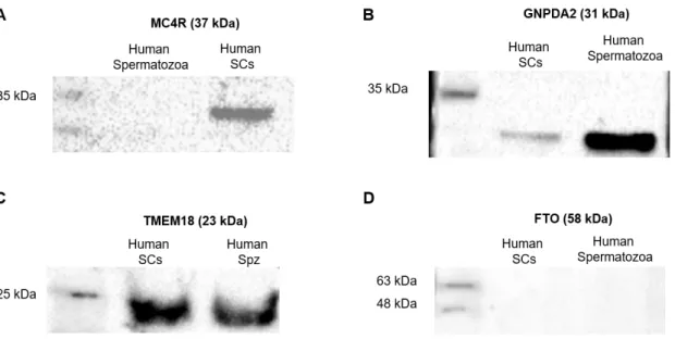
Os genes relacionados à obesidade (ORG) têm sido apontados como causas de um fenótipo de sobrepeso e/ou obesidade, sugerindo que crianças nascidas de pais obesos possuem uma maior predisposição para desenvolver distúrbios metabólicos. Este projeto investigou mecanismos moleculares relacionados com a transmissão de um fenótipo de sobrepeso e/ou obesidade. Foi colocada a hipótese de que os ORG, recetor de melanocortina-4 (MC4R), gene de obesidade e de massa de gordura associada (FTO), glucosamina-6-fosfato deaminase 2 (GNPDA2) e proteína transmembranar 18 (TMEM18) são fatores importantes nas células de Sertoli (SCs) e espermatozoides. Propusemos também que a expressão destes ORG poderia estar relacionada com o desenvolvimento embrionário e taxa de gravidez de casais em tratamentos de reprodução medicamente assistida. Neste trabalho, identificamos a expressão de FTO, MC4R, TMEM18 e GNPDA2 e respetivas proteínas em SCs humanas. Identificamos ainda a expressão de FTO, MC4R, GNPDA2 e respetivas proteínas em espermatozoides humanos. De seguida, avaliamos se a expressão de ORG nas SCs respondia a hormonas associadas à obesidade, tratando as células com doses crescentes de leptina, grelina e glucagon-like protein (GLP-1), mimetizando níveis hormonais relatados em indivíduos desnutridos, normais, obesos e gravemente obesos. A expressão de GNPDA2 e TMEM18 aumentou após tratamento das SCs com as concentrações mais elevadas de leptina e grelina, respetivamente. De seguida, investigamos se a expressão de ORG poderia estar relacionada com a qualidade espermática e o desenvolvimento do embrião. Verificamos que a idade paterna e o índice de massa corporal (BMI) não se correlacionam com a expressão de ORG em espermatozoides. No entanto, a expressão de MC4R e FTO está correlacionada com a viabilidade espermática (r= -0.3111) e contagem total de espermatozoides (r=0.5042), respetivamente. A expressão de FTO está também correlacionada com a qualidade do embrião, particularmente com a taxa de fertilização (r= 0.4751), a taxa de clivagem do embrião (r= 0,6530) e a taxa de embriões de alta qualidade (r= 0.6544). A expressão de MC4R nos espermatozoides também está correlacionada com a taxa de gravidez bioquímica (r= 0.4502), um parâmetro associado ao início do processo de implantação pelo embrião. Em suma, a abundância de ORG no espermatozoide mostrou estar associada a parâmetros relevantes da qualidade do esperma e associada ao sucesso clínico das técnicas de reprodução medicamente assistida. No entanto, mais estudos são necessários para desvendar o papel desses ORG nos espermatozoides e SCs. Palavras-chave: Fertilidade, Metabolismo, genes relacionados à obesidade, Célula de Sertoli, qualidade espermática.
vii
NOVEL BIOMARKERS FOR METABOLIC INHERITANCE OF OBESITY
THROUGH MALE GAMETES
Obesity-related genes (ORG) have been pointed out as causes for an overweight/obese phenotype, suggesting that children born from obese parents could have a genetic predisposition to develop metabolic disorders. This project aimed to unveil molecular mechanisms related with the inheritance of an overweight/obese phenotype through sperm. We hypothesized that ORG, Melanocortin-4 receptor (MC4R), Fat mass and obesity (FTO), Glucosamine-6-phosphate deaminase 2 (GNPDA2), and Transmembrane protein 18 (TMEM18) were present and could have a role in Sertoli cells (SCs) and sperm physiology. In addition, we also proposed that ORG expression was important for embryo development, being correlated with pregnancy rates in couples seeking paternity through medical-assisted reproduction treatments.
We identified the expression of MC4R, TMEM18, GNPDA2, FTO, and respective proteins, in human SCs. We further identified the expression of MC4R, GNPDA2, FTO, and respective proteins, in human spermatozoa. First, we evaluated if ORG expression in SCs responded to hormonal dysregulation associated with obesity. SCs were treated with increasing doses of obesity-related hormones, leptin, ghrelin, and glucagon-like protein 1 (GLP-1), mimicking the hormonal levels reported in undernourished, normal, obese and severely obese individuals. The expression of GNPDA2 and TMEM18 was increased after exposure to the highest concentration of leptin and ghrelin, respectively. Afterward, we investigated if the abundance of ORG could be associated with the overweight/obesity effects in sperm quality and embryo development. Paternal age and body mass index were not correlated with the abundance of ORG in spermatozoa. However, the expression MC4R and FTO was correlated with sperm quality through sperm viability (r=-0.3111) and total sperm count (r=0.5042), respectively. Furthermore, the expression of FTO was also correlated with embryo quality, particularly with the fertilization rate (r= 0.4751), embryo cleavage rate (r= 0.6530), and high-quality embryo rate (r= 0.6544). The expression of MC4R in spermatozoa was also correlated with the biochemical pregnancy rate (r= 0.4502), a parameter that is associated with the initiation of the implantation process by the embryo. In sum, the abundance of ORG in spermatozoa was associated with relevant parameters of sperm quality and with the clinical success of assisted reproduction techniques. Further studies are necessary to unveil the role of these ORG in spermatozoa and Sertoli cells.
viii
TABLE OF CONTENTS
DIREITOS DE AUTOR E CONDIÇÕES DE UTILIZAÇÃO DO TRABALHO POR TERCEIROS ... I AGRADECIMENTOS ... III DECLARAÇÃO DE INTEGRIDADE ...V NOVOS BIOMARCADORES ASSOCIADOS À HERANÇA METABÓLICA DA OBESIDADE ATRAVÉS DOS GAMETAS MASCULINOS ... VI NOVEL BIOMARKERS FOR METABOLIC INHERITANCE OF OBESITY THROUGH MALE GAMETES ... VII LIST OF ABBREVIATIONS ...XI LIST OF FIGURES ... XIII LIST OF TABLES ... XIV
1. INTRODUCTION ... 2
1.1. THE DECLINE OF FERTILITY IN MODERN SOCIETIES ... 2
1.2. SPERMATOGENESIS AND SPERMIOGENESIS AT BRIEF ... 4
1.3. SERTOLI CELLS, THE SENTINELS OF SPERMATOGENESIS ... 6
1.4. HORMONAL MODULATORS OF THE MALE REPRODUCTIVE SYSTEM ... 8
1.4.1. Leptin ... 11
1.4.2. Ghrelin ... 12
1.4.3. Glucagon-like protein 1 ... 12
1.5. OBESITY AND MALE INFERTILITY:IS THERE A LINK? ... 14
1.5.1. Hormonal dysregulation ... 16
1.5.2. Fat accumulation on the scrotum ... 17
1.5.3. Accumulation of environmental toxic substances ... 18
1.5.4. Inflammation and Oxidative stress ... 18
1.6. EPIGENETICS OF OBESITY ... 19
1.7. MONOGENIC OBESITY ... 22
1.7.1. Melanocortin-4 receptor gene ... 23
1.7.2. Fat mass and obesity-associated gene ... 24
ix
1.7.4. Transmembrane protein 18 gene ... 27
1.8. THE HEALTH OF FUTURE GENERATIONS ... 29
2. OBJECTIVES ... 30
3. MATERIALS AND METHODS... 33
3.1. HUMAN SERTOLI CELL CULTURE AND EXPERIMENTAL DESIGN ... 33
3.1.1. Experimental Design ... 33
3.2. PATIENT’S CHARACTERIZATION AND STUDY DESIGN FOR SPERMATOZOA ... 34
3.2.1. Body Mass index determination ... 35
3.2.2. Assisted-Reproduction Techniques data analysis... 35
3.3. RNA ISOLATION FROM HUMAN SERTOLI CELLS AND HUMAN SPERM ... 36
3.4. COMPLEMENTARY DNA(CDNA) SYNTHESIS ... 36
3.5. POLYMERASE CHAIN REACTION (PCR) ... 37
3.6. REAL-TIME POLYMERASE CHAIN REACTION (QPCR) ... 37
3.7. WESTERN-BLOT (WB) ... 39
3.8. IMMUNOFLUORESCENCE STAINING (IF) ... 40
3.9. STATISTICAL ANALYSIS ... 41
4. RESULTS ... 43
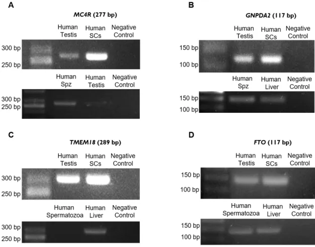
4.1. IDENTIFICATION OF ORG IN HUMAN SERTOLI CELLS AND HUMAN SPERMATOZOA ... 43
4.2. EXPRESSION OF ORG IN HUMAN SERTOLI CELLS AFTER STIMULATION WITH OBESITY-RELATED HORMONES 46 4.2.1. GNPDA2 expression is increased in human SCs after treatment with leptin concentration found in obese men ... 47
4.2.2. TMEM18 expression is increased in human SCs after treatment with ghrelin concentration found in normal men ... 48
4.2.3. Treatment of human SCs with different GLP-1 concentrations has no effect on the expression of the selected ORG ... 49
4.3. TMEM18 IS NOT EXPRESSED IN HUMAN SPERMATOZOA AND THERE IS NO CORRELATION BETWEEN THE EXPRESSION OF ORG BY HUMAN SPERMATOZOA AND BODY MASS INDEX ... 51
4.4. THERE IS NO CORRELATION BETWEEN THE EXPRESSION OF ORG IN HUMAN SPERMATOZOA AND PATERNAL AGE 52 4.5. CORRELATION BETWEEN SPERM QUALITY AND BMI, AGE AND ORG EXPRESSION ... 53
x
4.5.2. Correlation between GNPDA2 mRNA abundance and sperm parameters ... 55
4.5.3. Correlation between FTO mRNA abundance and sperm parameters ... 57
4.5.4. ORG expression and spermogram classification ... 58
4.6. CORRELATION BETWEEN ORG EXPRESSION AND EMBRYO DEVELOPMENT ... 59
4.6.1. Correlation between MC4R mRNA abundance and embryo quality ... 60
4.6.2. Correlation between GNPDA2 mRNA abundance and embryo quality ... 61
4.6.3. Correlation between FTO mRNA abundance and embryo quality ... 62
4.7. CORRELATION BETWEEN ORG EXPRESSION AND PREGNANCY ... 63
5. DISCUSSION ... 67
6. CONCLUSION ... 75
7. REFERENCES ... 78
xi
LIST OF ABBREVIATIONS
5α-Dihydrotestosterone DHT 17β-Estradiol E2 α-Melanocyte-stimulating hormone α-MSH β2-microglobulin β2-MGB Agouti-related protein AgRP Asthenozoospermia AT AT-rich interactive domain-containing protein 5B ARID5B Biochemical pregnancy rate BP Body mass index BMI Blood-testis barrier BTB c-Jun N-terminal kinase JNK Cyclic Adenosine 3’,5’-monophosphate cAMP Fat mass and obesity gene FTO Follicle-stimulating hormone FSH Glyceraldehyde-3-phosphate dehydrogenase GAPDH Glucagon-like protein GLP-1 Glucosamine-6-phosphate deaminase 2 GNPDA2 Gonadotropin-releasing hormone GnRH Hypothalamic-pituitary-gonadal HPG Immunofluorescence staining IF Intracytoplasmic sperm injection ICSI In vitro fertilization IVF Iroquois homeobox protein 3 IRX3 Iroquois homeobox protein 5 IRX5xii
Lactate dehydrogenase LDH Luteinizing hormone LH Melanocortin-4 receptor MC4R Membrane glucose transporters GLUTs N6-methyladenosine m6A
Normozoospermia NZ Obesity-related gene ORG Obstructive azoospermia A Oligoasthenoteratozoospermia OAT Oligospermia O Oligoteratozoospermia OT Peroxisome proliferator-activated receptor gamma PPARG Phosphate-buffed saline PBS Phosphatidylinositol 3-kinase PI3-K Polymerase chain reaction PCR Protein kinase A PKA Proton/Monocarboxylate transporters MCTs Quantitative polymerase chain reaction qPCR Reactive oxygen species ROS Sertoli cells SCs Sex hormone-binding globulin SHBG Teratozoospermia T
Transmembrane protein 18 TMEM18 Western-blot WB World Health Organization WHO
xiii
LIST OF FIGURES
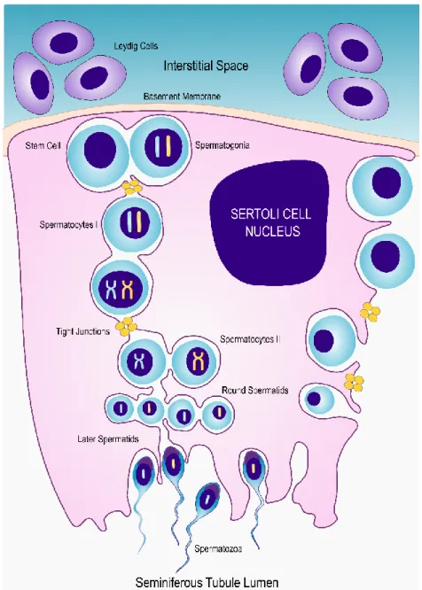
Figure 1: Schematic representation of the interaction between Sertoli cells and germ cells, during
spermatogenesis. ... 5
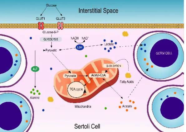
Figure 2: Schematic representation of Sertoli Cell metabolism and its main metabolic pathways. ... 7
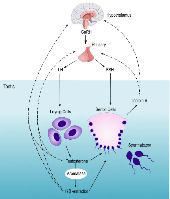
Figure 3: Schematic representation of the male Hypothalamus-Pituitary-Gonadal axis. ... 9
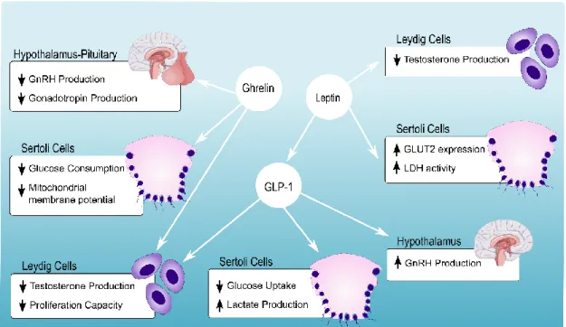
Figure 4: Schematic resume of obesity-related hormones effects on the male reproductive tract... 13
Figure 5: Identification of ORG in human Sertoli cells and spermatozoa by conventional PCR. ... 43
Figure 6: Identification of ORG protein in human Sertoli cells and spermatozoa by Western-Blot. ... 44
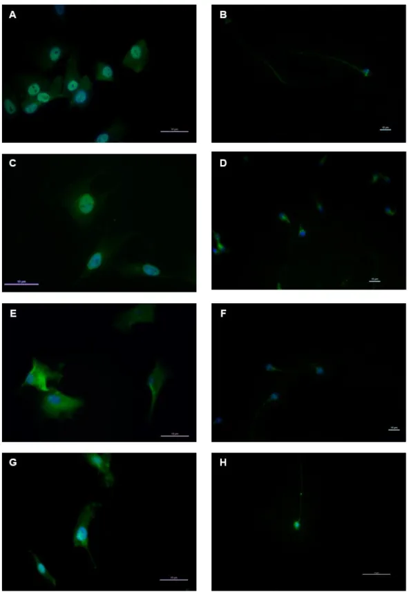
Figure 7: The results of immunofluorescence staining for the studied ORG in human Sertoli Cells and Spermatozoa. ... 45
Figure 8: Effect of leptin on the expression of ORG in humans Sertoli Cells. ... 47
Figure 9: Effect of ghrelin on the expression of ORG in humans Sertoli Cells ... 49
Figure 10: Effect of GLP-1 on the expression of ORG in humans Sertoli Cells.. ... 50
Figure 11: Pearson correlation between ORG expression in human spermatozoa and paternal body mass index (BMI). ... 52
Figure 12: Pearson correlation between the expression of selected ORG in human spermatozoa and paternal age. ... 53
Figure 13: Pearson correlation between MC4R mRNA abundance and sperm parameters.. ... 55
Figure 14: Pearson correlation between GNPDA2 mRNA abundance and sperm parameters. ... 56
Figure 15: Pearson correlation between FTO mRNA abundance and sperm parameters.. ... 57
Figure 16: Effect of ORG mRNA abundance in human spermatozoa on spermogram classification. .... 59
Figure 17: Pearson correlation between MC4R mRNA abundance in spermatozoa and embryo quality ... ... 61
Figure 18: Pearson correlation between GNPDA2 mRNA abundance in spermatozoa and embryo quality. ... 62
Figure 19: Pearson correlation between FTO mRNA abundance in spermatozoa and embryo quality. . 63
Figure 20: Pearson correlation between ORG expression in human spermatozoa and biochemical pregnancy. ... 64
Figure 21: Effects of ORG expression in human spermatozoa and ongoing pregnancy. ... 65
xiv
LIST OF TABLES
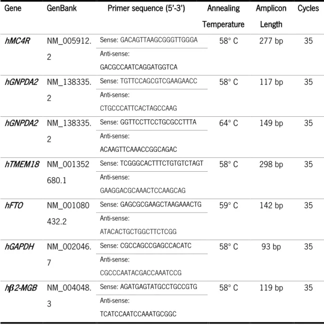
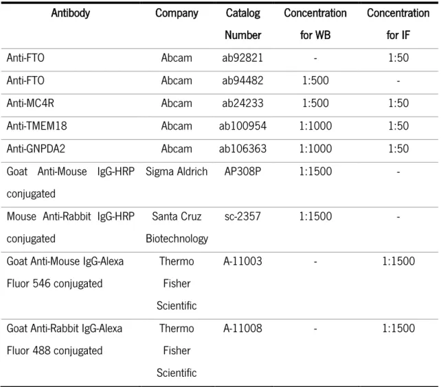
Table 1: Summary of studies investigating the effects of obesity on classic sperm parameters. ... 15 Table 2: Primer sequences and PCR conditions used to assess gene expression and mRNA abundance in human SCs and spermatozoa. GAPDH and β2-microglobulin were used as housekeeping controls. ... 38 Table 3: List of antibodies, and respective concentrations, used for identification of proteins associated with ORG expression. ... 41 Table 4: Pearson correlation coefficients between classical sperm parameters and paternal BMI and age.
1
Chapter I
2
1. INTRODUCTION
1.1. The decline of fertility in modern societies
A large diversity of living things cohabitate on earth and all sustain their evolutive success through reproduction. It is not only relevant to enhance the chances of survival of the species but also to ensure continuity and propagation. However, it is well-known that fertility rates are decreasing worldwide. Several authors had discussed this phenomenon, but the reasons behind it are complex and difficult to unveil and thus, a consensus has not been reached yet. The anthropologist Marvin Harris proposed, in 1989, that people adjust their fertility to the economic value of children’s labor, explaining why fertility rates were higher in developing countries at the time (1). Several recent works highlight that the increased incidence of metabolic diseases, known to induce alterations in the male and female reproductive tracts, is associated with the reported decrease in birth rates worldwide. This subject has been overlooked for years but is now clear that deserves special attention from researchers, stakeholders, media, and politicians.
Infertility, by definition, describes the inability to conceive children after 12 or more months of regular unprotected sex and it has been recognized by the World Health Organization (WHO) as a major health problem that affects individuals on both, modern and in developing societies. In a more accurate medical context, infertility is a disease of the reproductive system that prevents a sexually active couple to achieve a successful pregnancy in a period of 12 months of unprotected sex (2). Nowadays, it is assumed that infertility affects over 72 million people. However, due to the difficulty of evaluating infertility rates in developing countries, the number of affected people is must certain underestimated (3). In addition, the contribution of the male factor alone or in combination with the female factor is expected to account for almost 2/3 of the infertility cases (4).
Although medical care has substantially increased in modern society, the incidence of metabolic diseases keeps rising worldwide at a worrying level. Like other noncommunicable diseases, metabolic disorders are largely caused by lifestyle, mainly through poor dietary choices and lack of physical activity. Several authors also consider the rise in metabolic diseases as a consequence of the technologic, economic and social advances accomplished in the last decades. As human populations became more urban, carbohydrate and fiber-rich diets were replaced by sugar and fat-rich diets, while a sedentary lifestyle is promoted. In addition, most of these disorders have a strong
3
genetic component and are usually associated with inborn errors that could be inherited by future generations (5).
One of the most common metabolic disorders is obesity that is commonly defined by an excess of fat accumulation, resulting from a positive balance between caloric intake and energy expenditure. Even though the consumption of high-fat diets and sedentary lifestyle are major factors for the accumulation of excess weight, recent studies provided clear evidence that the development of obesity is also strongly influenced by genetics (6). Nowadays, the obesity epidemic has reached alarming proportions. According to the WHO, in 2016, more than 1.9 billion adults and 41 million children were overweight or obese (7) and these statistics can become even more serious in the years to come. It is well known that an increased body mass index (BMI) has detrimental effects in all systems of the body and is associated with the development of cardiovascular diseases, cancer, musculoskeletal disorders, diabetes mellitus, and other health complications, such as infertility (8). The growing rates of obesity incidence, along with comorbidities associated are concurrent with the serious concerns on the reproductive health of the individuals in modern societies. Nowadays, at least 1 in 7 couples will be affected by infertility (9) and the fertility rates are expected to keep falling as the number of obese individuals at reproductive age rise. Indeed, the metabolic abnormalities caused by obesity are associated with polycystic ovary syndrome and anovulatory infertility in women (9). Unsurprisingly, 50% of the women diagnosed with polycystic ovary syndrome are overweight or obese, supporting that obesity plays a pathogenic role in the development of the syndrome (10). Moreover, obese women are known to develop more pregnancy complications and to be more prone to suffer more miscarriages in the first trimester of pregnancy that normal-weight pregnant women (11). Thus, there are several clinical, epidemiologic and molecular studies showing a clear relationship between overweight/obesity and less fertility in females and a lower probability to conceive (12). Nevertheless, only recently has emerged the concept of subfertility or infertility caused by overweight or obesity in the male. Indeed, the increased incidence of male obesity is being coincident with the increase in the cases of subfertility and/or infertility in males from westernized countries. Moreover, recent studies showed that obesity in the male partner may equally impair embryo development as when occurring in the female partner (13, 14). Of note, overweight men are usually associated with lower reproductive potential and more likely to be oligospermic (a condition where semen has a very low concentration of sperm), or azoospermic (a condition where semen has no sperm) when compared to males with normal BMI values (15). These individuals are also more likely to suffer from mechanical
4
dysfunctions in the reproductive tract, including erectile dysfunction (16). Although many studies have associated the infertility epidemic of the couple with the increasing number of overweight and obese men, the molecular mechanisms by which spermatogenesis or sperm quality are impaired are only starting to be unveiled.
1.2. Spermatogenesis and spermiogenesis at brief
Spermatogenesis is the process where male germ cells are formed. It occurs in the testis and can be divided into three crucial phenomena: differentiation of spermatogonial stem cells into spermatocytes via mitosis, production of haploid spermatids from primary spermatocytes via meiosis and spermiogenesis (17). Sertoli cells (SCs) are the main mediators of this process and are responsible for limiting the expansion of the spermatogonial population, providing critical factors necessary for the success of spermatogenesis (18). These cells are also responsible for the formation of the blood-testis barrier (BTB), which is a structured biological barrier formed by tightly adjacent SCs that control the intratubular environment while also providing protection against the host immune system (19). The maintenance of the seminiferous tubule is achieved by a balance between germ cell apoptosis and regeneration. These processes are controlled by a complex network of endocrine and paracrine signals. SCs have receptors to follicle-stimulating hormone (FSH) and testosterone, the main hormonal regulators of spermatogenesis, both acting as germ cell survival factors (18). These hormones regulate glucose uptake and lactate production by SCs (20). Sertoli and germ cells communicate by both paracrine signals and direct membrane contact, for example, through the c-kit receptor, expressed on the surface of spermatogonia, and stem cell factor, produce by SCs (18).
Spermatogonia and SCs adhere to the basement membrane of the seminiferous tubule. In humans, it is possible to distinguish three types of spermatogonium: type A (dark) are reserve spermatogonia cells and do not undergo mitosis and type A (pale) undergo through mitotic division to give rise to type B (19). Under the correct conditions, each type B spermatogonia undergoes through one mitotic division, producing primary spermatocytes. After having completed meiosis I, primary spermatocytes give rise to secondary spermatocytes. When meiosis is completed, haploid spermatids are formed (17, 21) (Figure 1).
5
Figure 1: Schematic representation of the interaction between Sertoli cells and germ cells, during spermatogenesis. The SCs are located at the base of seminiferous tubule and provide support for the developing spermatogonia. SCs are responsible for the limitation of the expansion of the spermatogonial population providing critical factors necessary for the success of spermatogenesis. Under the right conditions, diploid spermatogonia undergo through meiotic division to form haploid spermatids. Finally, spermatids undergo cytodifferentiation to form spermatozoa. [Adapted from: “Blood-Testis Barrier: How Does the Seminiferous Epithelium Feed the Developing Germ Cells?” (23)]
Spermiogenesis can be described as a series of cytodifferentiation that spermatids undergo to form spermatozoa capable of motility (22). At the beginning of this process, spermatids have a round shape and central nucleus. Spermiogenesis starts with nuclear condensation and migration of the
6
nucleus to the periphery of the cell. Meanwhile, the acrosome is assembled, which is a Golgi-derived organelle that can be described as a specialized lysosome, essential for the interaction with the egg during fertilization, and microtube-based axoneme, essential for mobility (19, 22). The latter will become the central structure of the flagellum, which is assembled soon after meiosis is completed. Following these events, spermatids shed a large part of its cytoplasm and the elongated spermatids are released from the seminiferous epithelium. This last event is called spermiation. Yet, spermatozoa released into the seminiferous tubules lumen are still not able to move. The seminiferous tubules converge into the epididymis, tubular structures behind the testis where sperm maturation occurs. However, the exact molecular process behind sperm maturation is largely unknown (24). In humans, the process of formation and maturation of spermatozoa takes around 74 days (22).
1.3. Sertoli Cells, the sentinels of spermatogenesis
SCs, also known as nurse cells, are responsible for fulfilling the physical and nutritional needs of the developing germ cells. Each SCs has the ability to support around 30 to 50 developing germ cells. These cells are characterized by a very complex cup-structure. This three-dimensional structure is constantly changing, allowing SCs to interact with the developing germ cells and the epithelium cells of the seminiferous tubules (25). Mature SCs produce extracellular matrix components, such as collagen and laminin, to form specialized junctions and guarantee the maintenance of the seminiferous epithelium (25). Concurrently, SCs secret a great panoply of specific products, which are essential for the developing germ cells. The transference of these products can only be accomplished due to the close relationship between germ cells and SCs. Moreover, adjacent SCs form tight junctions, creating the BTB (26). These SCs characteristics allow them to control the intratubular environment. Since developing germ cells are unable to use glucose as an energy substrate, SCs convert most of the glucose into lactate, which can then be used by germ cells as an energy substrate. The uptake of glucose from the interstitial space by the SCs occurs via specific integral membrane glucose transporters (GLUTs) being that GLUT1 and GLUT3 appear to have the most relevant role regarding SCs metabolism (27), though GLUT2 appears to be highly expressed (28). The conversion of glucose into lactate is divided into two fundamental steps. In the first step, glucose is metabolized into pyruvate. Pyruvate is then converted into lactate by lactate dehydrogenase (LDH), synchronously with the oxidation/reduction of NADH to NAD+. Finally, lactate is exported into the intratubular fluid of the seminiferous tubules
7
by specific proton/monocarboxylate transporter 4 (MCT4). Herein, lactate becomes available to the developing germ cells, where it is captured via specific monocarboxylate transporter 1 and 2 (MCT1 and MCT2) (29). Alternatively, pyruvate can also be converted into alanine via alanine transaminase or be transported to the mitochondria matrix, where it is converted in acetyl-CoA and directed to the tricarboxylic acid cycle. Furthermore, acetate is also produced at high rates by SCs. Although its role remains to be further elucidated, acetate is an intermediate for the synthesis of fatty acids and appears to be related to the high demand of lipids needed by the developing germ cells (Figure 2).
Figure 2: Schematic representation of Sertoli Cell metabolism and its main metabolic pathways. Glucose is converted into pyruvate by glycolysis. Pyruvate can participate in several metabolic pathways. In these cells, the majority of pyruvate is converted into lactate by LDH, which is then exported into the intratubular fluid and captured by germ cells through proton/monocarboxylate transporters. Alanine transaminase (ALT) promotes the convention of pyruvate into alanine which, acts as an energy substrate reservoir. Alanine can again be converted into pyruvate through alanine transamination. On the mitochondria, acetyl-CoA fuels the tricarboxylic acid (TCA) cycle, responsible for energy production. Finally, acetyl-CoA can also be converted into acetate which appears to be an important intermediate of fatty acids synthesis. The transport of metabolites is represented by the dashed lines.
8
The high metabolic plasticity of SCs allows them to metabolize other substrates in addition to glucose such as ketone bodies, fatty acids, and glycogen. These characteristics allow SCs to ensure metabolites production even in glucose privation situations (30).
1.4. Hormonal modulators of the male reproductive system
Due to the importance of glucose to the maintenance of spermatogenesis, the energy state of the organism can induce several alterations in the reproductive system. The glucose metabolism in SCs and the reproductive events are mainly controlled by the endocrine system, predominantly by FSH, sex steroid hormones, and insulin. The hypothalamic-pituitary-gonadal (HPG) axis is responsible for the coordination of reproductive events (Figure 3). The hormonal messengers associated with this axis can interact not only with the developing germ cells but, also with their caretakers. The gonadotropin-releasing hormone (GnRH) is produced by the hypothalamus and stimulates the production of the pituitary hormones, FSH and luteinizing hormone (LH) (31). In the seminiferous tubules, the SCs are the only cells that possess FSH and testosterone receptors. Nowadays, it is described that FSH can activate at least 5 signaling pathways on SCs. The crosstalk between them determines the final cellular consequence. Among the 5 known signaling pathways, thecyclic adenosine 3′,5′-monophosphate (cAMP)-dependent protein kinase (PKA) pathway, the mitogen-activated protein kinase (MAPK) pathway, and the phosphatidylinositol 3-kinase (PI3-K) pathway are the most well studied (31). The cAMP-PKA pathway was the first to be identified. Herein, the activation of the FSH receptor culminates with the expression of various isoforms of cAMP-responsive element modulator, which are required for spermatocytes and spermatids survival (32). The stimulation of the MAPK pathway by FSH is responsible for the proliferation of the SCs and occurs in a stipulated period of 15 days postpartum, in humans. The number of SCs remains relatively constant throughout adulthood, approximately 3,700 million cells per testis. The activation of the PI3-K pathways by FSH was first described in granulosa cells (33). The increase of cAMP levels mediated by FSH-receptor activation promotes the activation of PI3-K, which is a key enzyme involved in several biological responses, such as mitogenesis and glucose uptake (34). On SCs, PI3-K promotes the uptake of glucose and promotes transferrin production, both essential for the maintenance of spermatogenesis. Moreover, androgen aromatization is stimulated by SCs and germ cells by the phospholipase A2 pathway (35).
9
Figure 3: Schematic representation of the male Hypothalamus-Pituitary-Gonadal axis. GnRH stimuli on the pituitary promote the synthesis of gonadotropin hormones LH and FSH. While LH will promote steroidogenesis in LCs, FSH promotes spermatogenesis through the stimulation of SCs. As a result, LCs produce testosterone, a crucial regulator of spermatogenesis, while SCs produce inhibin B. A small part of testosterone suffers aromatization by testicular aromatase, producing 17β-Estradiol. These hormones promote a negative feedback loop between the testis, hypothalamus, and pituitary (represented by the dashed lines).
Testosterone produced by Leydig cells (LCs), after LH stimulation, can interact with SCs by the androgen receptor. Although FSH and testosterone display redundant functions on SCs, there are some distinct differences between the action of these hormones. First, testosterone is able to
10
activate the MAPK, CREB phosphorylation and CREB-mediated transport of SCs without the up-regulation of cAMP (36). This may explain how testosterone, but not FSH, can maintain spermatogenesis independently, suggesting the cAMP is not essential for spermatogenesis. Moreover, the expression of some genes is exclusively regulated by the interaction between DNA and the androgen receptor, where testosterone stimulation is required (37). The action of testosterone through the non-classical pathway promotes the attachment of germ cells to SCs and promotes the reformation of the BTB (38). In its turn, the SCs produce inhibin B, which, along with testosterone and 17β-estradiol (E2), provides a negative loop that reduces the expression of GnRH by the hypothalamus (39).
The sex steroid hormones dihydrotestosterone (DHT), mainly produced on the prostate by 5α-reductase, and E2 were reported to modulate glucose consumption and lactate production in SCs. DHT increase glucose consumption without increasing lactate production, suggesting it stimulates the Krebs cycle. This hypothesis proposes that DHT can metabolically modulate SCs to a more efficient energy status (40). Alternatively, E2 does not seem to interfere with lactate production in SCs (40). However, in vitro human SCs treated with E2 demonstrated increased production of acetate (41). This metabolite appears to play a key role in the progression of spermatogenesis, particularly as a precursor of cellular constituents’ synthesis (41). Furthermore, both DHT and E2 are able to downregulate the transcript levels of glucose transporters, GLUT1 and GLUT2 after a 50h treatment on in vitro human SCs. These results appear to describe a metabolic shift from an exponential phase of glucose consumption to a stationary phase (42).
Insulin is a hormone produced by the islets of Langerhans of the pancreas and a powerful modulator of SCs metabolism (43). The insulin receptor was identified in rat SCs by OonK RB and colleagues suggesting that insulin had specific functions in the maturation and activity of these cells (44). But many studies showed new functions for this hormone in these cells. For instance, insulin-deprived SCs also presented decreased transcript levels of genes associated with lactate metabolism (LDH and MCT4) (45). In addition, those insulin-deprived cells present decreased glucose consumption and acetate production (41). Although all these in vitro results need further clarifications, insulin appears to be an important regulator of SCs metabolism. These results suggest that insulin dysregulation could be one of the main causes of infertility in diabetic men (46). More recently, other hormones associated with the energy homeostasis and appetite regulation were reported as metabolic modulators of SCs.
11
1.4.1. Leptin
Leptin is a peptide hormone produced mainly by white adipocytes, which is related to adipose tissue mass and decrease of food intake (47). The action of this hormone is modulated by the membrane-spanning leptin (or obesity) receptor (48). The hypothalamus is suggested as the primary target of this hormone. Leptin, by itself, is not capable to lead to the termination of a meal but it can interact with other hypothalamic hormone pathways associated with appetite-regulation (47, 49). In nutrient deprivation situations, the decreased plasma leptin levels induce neuroendocrine responses to food restrictions. The hypothalamic neurons express neuropeptide Y and agouti-related protein (AgRP) (47). The first acts on Y1 and Y5 receptors and stimulates food intake by increasing motivation to eat, delaying satiety and augmenting meal size (50). The AgRP is an antagonist to the melanocortin-4 receptor (MC4R) and increases food intake by antagonizing the effect of the α-melanocyte-stimulating hormone (α-MSH) (51). Under the stimuli of leptin, pro-opiomelanocortin neurons express cocaine and amphetamine-related peptide and α-MSH. These peptides induce the decrease of food intake by promoting short-term satiety (49) and interacting with other appetite-regulation pathways, including the endocannabinoid system (52).
The actions of leptin in the hypothalamus were suggested to act as modulators of the reproductive axis (53). Leptin is able to increase LH and FSH secretion by stimulation of growth and differentiation of pituitary cells, regardless of the presence or absence of GnRH (54). Nonetheless, studies have suggested that leptin mediates the HPG axis by regulating kisspeptins produced by the Kiss1 gene (55). However, the mechanisms by which leptin interacts with the HPG axis is complex and combines stimulatory and inhibitory effects. Leptin can also interact directly with the testis (56). In fact, leptin can also induce a decrease in testosterone expression by inhibiting expression of the cAMP-stimulated steroidogenic acute regulatory protein and interfering with cAMP signaling (57). Martins AD and other members from our group were the first to clearly demonstrate the presence of leptin receptor in human SCs and that leptin could directly act on these cells (28). The authors reported that leptin concentration found in lean men induce an increase of GLUT2 protein level and LDH activity, in SCs. These results suggested that leptin is a hormonal regulator of the nutritional support of spermatogenesis since all concentrations of leptin tested decrease human SCs acetate production (28) (Figure 4).
12
1.4.2. Ghrelin
Ghrelin, known as the hunger hormone, is another peptide involved in a large group of physiological functions, including food intake, sleep, body weight, inflammation and others (58). It is known that ghrelin promotes food intake both in rodents and in humans and reduces insulin secretion (59). In what concerns the reproductive system, ghrelin appears to be an important integrator of energy homeostasis control. It was proposed that ghrelin can inhibit the proliferative activity of immature germ cells and LCs (60), avoiding excess build-up of germ cells which is crucial for spermatogonia survival (61). Furthermore, recent in vitro studies have reported that ghrelin is able to inhibit testicular secretion of testosterone (62) while suppressing LH and follicle-stimulating hormone FSH secretion by the pituitary (63), events that could also promote the apoptosis of germ cells. The presence of the growth hormone secretagogue receptor on human SCs was confirmed by immunohistochemistry technique (64). Martins AD and other members from our group demonstrated that ghrelin could modulate human SCs metabolic phenotype, decreasing the glucose consumption and mitochondrial membrane potential. Curiously, LDH activity and lactate production remain unaltered. These results suggest that ghrelin can act as an energy sensor for human SCs in a dose-dependent manner (65) (Figure 4).
1.4.3. Glucagon-like protein 1
Glucagon-like protein 1 (GLP-1) is a hormonal derived from proglucagon gene, mainly secreted by endocrine L-cells from intestines (66). Along with leptin and ghrelin, GLP-1 is one of the prominent players in glucose homeostasis, gastrointestinal motility, and appetite. The release of GLP-1 is stimulated by the presence of nutrients (predominantly sugars and fats) in the stomach and proximal intestine or through direct contact between nutrients and L-cells. GLP-1 diffuses through the capillaries and lymph. Herein, it mediates the activation of the GLP-1 receptor, present throughout the periphery (pancreas, stomach, adipose tissue, heart, and others) (67) and the central nervous system (68). The endogenous role of GLP-1 in the regulation of appetite is a concept well accepted. This hormone can decrease gastric emptying and intestinal motility. Meanwhile, the absorption and digestion of nutrients are optimized. In the pancreas, it stimulates the secretion of insulin, proliferation of islet, coupled with inhibition of glucagon secretion. The stimulation of the hypothalamus by GLP-1 induces satiety and decreases in caloric intake (69). Due to these proprieties, GLP-1 analogs are prescribed for the treatment of diabetes, a condition characterized, in a simple sum, by hyperglycemia and insulin resistance (70).
13
The expression of GLP-1 receptor was first identified in isolated human SCs by Martins AD and colleagues (71), which reported that exposure to increasing doses of GLP-1 did not alter the expression of this receptor. The authors reported that exposure to GLP-1 decreased glucose consumption while increasing lactate production in human SCs (Figure 4). Meanwhile, no effects were detected in mitochondria functionality in human SCs exposed to the GLP-1 levels found in healthy individuals.
Figure 4: Schematic resume of obesity-related hormones effects on the male reproductive tract. In the pituitary, Ghrelin suppresses LH and FSH secretion leading to the inhibition of testosterone secretion. Ghrelin levels are related to decreased glucose consumption and decreased mitochondrial membrane potential of SCs, in a dose-dependent manner. Androgens are an important regulator of leptin secretion. LCs are known to express leptin receptors, which promote the reduction of testosterone secretion, upon high circulating leptin levels. Leptin also decreases SCs acetate production suggesting that it has a direct action on SCs mitochondrial function. Furthermore, increased levels of GLUT2 expression and LDH activity are also associated with leptin. It can also stimulate GLP-1 secretion, in a dose-dependent manner. In its turn, GLP-1 can interact with the HPG axis, promoting the secretion of GnRH by the hypothalamus. In LCs, it can decrease the secretion of testosterone. While on SCs, it promotes lactate production, even though glucose consumption is decreased. Down arrows stand for downregulation and up arrows stand for upregulation.
14
The stimuli of GLP-1 seem to be vital for eliciting the production of lactate by SCs while, consuming lower amounts of glucose. These results illustrate the metabolic plasticity that characterizes SCs. Furthermore, the authors proposed that that GLP-1 modulates the glucose metabolism of SCs and, it may have an anti-apoptotic effect in the developing germ cells (71). Additionally, this hormone is able to decrease testosterone secretion (72), suggesting that it may affect the HPG axis. Further, this hormone seems to increase GnRH secretion, which suggests that it may have an important role in puberty development (73).
1.5. Obesity and Male Infertility: Is there a link?
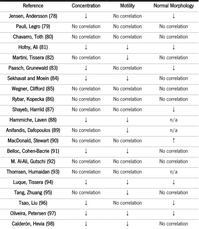
Obesity results from a disturbance between energy intake and expenditure. From this point of view, it is easy to associate excess of weight and obesity as the result of lack of exercise, a sedentary lifestyle and the consumption of diets rich in fat. However, the origin of obesity is multifactorial and complex. Several of those factors are very difficult to unveil, and for instance, the development of obesity has an evident environmental contribution. Indeed, some environmental compounds are classified as obesogens, a class of chemical compounds that can enhance adipogenesis and promote lipid accumulation. The exposure to these compounds, usually through diet, is known to impair energy metabolic pathways and to disrupt signaling pathways that control food intake (74). Obesogens are reported to have great stability and be structurally similar to androgens. Further, a large portion of these compounds is lipophilic, which means that they can disrupt the endocrine system. These effects will be reflected in the reproductive systems of both males and females (75-77). However, the impact of obesity on the fertility potential of males has been disregarded for several decades. In addition, previous studies focused on obesity and classic sperm parameters (namely, concentration, motility, and morphology) have reported mixed results (summarized in Table 1).
While several studies showed that excess weight is related to low semen volume and sperm motility as well as aberrant sperm morphology (99-101), other studies found no significant correlation (79, 80, 102). The observed discrepancies in the literature are likely caused by the several limitations associated with studies performed in humans. In addition, sperm function can be altered by innumerable lifestyle factors, such as smoking, alcohol consumption, and recreational drugs, which are confounding factors difficult to control in human studies.
15
Table 1: Summary of studies investigating the effects of obesity on classic sperm parameters. Reference Concentration Motility Normal Morphology Jensen, Andersson (78) ↓ No correlation ↓
Pauli, Legro (79) No correlation No correlation No correlation Chavarro, Toth (80) No correlation No correlation No correlation
Hofny, Ali (81) ↓ ↓ ↓ Martini, Tissera (82) No correlation ↓ No correlation Paasch, Grunewald (83) ↓ No correlation ↓ Sekhavat and Moein (84) ↓ ↓ No correlation
Wegner, Clifford (85) No correlation No correlation No correlation Rybar, Kopecka (86) No correlation No correlation No correlation Shayeb, Harrild (87) No correlation No correlation ↓ Hammiche, Laven (88) ↓ ↓ n/a Anifandis, Dafopoulos (89) No correlation ↓ n/a MacDonald, Stewart (90) No correlation No correlation ↑ Belloc, Cohen-Bacrie (91) ↓ ↓ No correlation
M. Al-Ali, Gutschi (92) No correlation No correlation No correlation Thomsen, Humaidan (93) No correlation No correlation n/a
Luque, Tissera (94) ↓ ↓ ↓ Tang, Zhuang (95) No correlation ↓ No correlation
Tsao, Liu (96) ↓ No correlation ↓ Oliveira, Petersen (97) ↓ ↓ ↓
Calderón, Hevia (98) ↓ ↓ No correlation Furthermore, the limitations of the BMI evaluation as an adiposity calculator can also be questioned (103). Obesity is also associated with the development of other complications, such as diabetes and oxidative stress, which can also impair sperm parameters (104, 105), promote sperm DNA fragmentation (80, 106, 107) and induce aberrant sperm mitochondrial function (106, 108). Beyond weight gain and adiposity, there are other relevant factors associated with obesity impact on sperm quality, including hormonal dysfunction, fat accumulation in the male reproductive tract,
16
accumulation of environmental toxic substances or even inflammation and oxidative stress. Below we briefly discuss each of these factors.
1.5.1. Hormonal dysregulation
Obesity causes whole body strong hormonal dysregulation. Due to the essential role of hormones in the maintenance of glucose homeostasis, the hormonal disruption affects not only the signaling pathways regarding food intake control but also other functions intimately associated with energy homeostasis, such as reproduction (109). In humans, leptin and sex hormones blood concentrations appear to be strongly related (110). Usually, androgens suppress leptin production (110), supporting that testosterone is an important regulator of leptin. Concurrently, leptin has been found to exert important effects on gonadal organs through leptin receptors that were reported in testicular cells, including in the surface of LCs (110, 111) and SCs (28). Obesity is associated with leptin resistance (112, 113), resulting in increased leptin levels as a response to increased adiposity. Besides, leptin resistance is associated with a lack of responsiveness to leptin’s appetite-suppressing effects (114). Fortunately, leptin resistance is reportedly reversible since the reduction of food intake and body weight allow peripherical leptin to more easily access its hypothalamic sites (115). The excess of circulating leptin, found in obese men, appears to contribute to reducing testosterone concentrations due to impairment of LCs (116). Further, high levels of leptin are known to modulate SCs metabolism, decreasing lactate dehydrogenase activity (28) and both events have the potential to compromise male fertility. Concordantly, higher serum leptin levels found in obese infertile men were correlated with abnormal sperm morphology (81).
Contrarily to leptin, ghrelin resistance has not been associated with obesity (117). It was reported a significantly lower basal level of ghrelin in obese subjects in comparison to lean subjects. Even though the mechanism(s) underlying the decreased ghrelin levels associated with obesity remain unclear, it was hypothesized that ghrelin acts as a counter-regulatory hormone that limits energy intake in obese people (118). On the male reproductive tract, Martins AD and colleagues suggested that ghrelin can act as an energy sensor for human SCs in a dose-dependent manner, modulating the nutritional support of spermatogenesis (65). This hypothesis also supports that ghrelin balance can contribute to the metabolic dynamics within the male reproductive axis in situations of energy deficit (63, 109). Consequently, ghrelin balance disruption can induce severe complications upon the metabolism of the testis.
17
The incretin effect, a process that describes the increase of insulin plasma levels along with the decrease of glucagon levels, after a meal, is also impaired in obesity (119). This evidence suggests that incretin hormones, such as GLP-1, may be altered in obese subjects. However, the findings regarding GLP-1 levels in obesity were inconsistent and its variations have not been conclusively determined (119, 120). Meanwhile, some evidence suggested that leptin may exert a regulatory effect on GLP-1 secretion and that impairment of leptin regulation associated with obesity may also be associated with impairment of GLP-1 regulation (121). Although the effects of GLP-1 in male reproduction remain overlooked, its contribution to glucose homeostasis suggests that it may be important for the regulation of spermatogenesis (71).
Hyperglycemia, which is usually found in obese men, seems to cause a decrease in sex hormone-binding globulin (SHBG) production by the liver (122). SHBG is responsible for the transport of sex hormones through the blood to the target tissue. Naturally, with a decrease of SHBG concentration, testosterone availability will also decrease. Hyperglycemia per se has long been associated with compromised male infertility. The frequency and duration of hyper- and hypoglycemic events, the degree of glucose control, age and other factors are important to evaluate the occurrence of infertility (123). However, it has been reported that poor glucose control, such as in diabetic men, is associated with impairment of sperm motility (124), sperm DNA fragmentation (125) and hormonal dysregulation (126). Additionally, gonadotropins secretion tends to progressively decrease in obese men (111). Overweight is also associated with increased androgen aromatization by the adipose tissue. This may result in a decrease of total and free testosterone in serum, a condition known as hypogonadism (127).
1.5.2. Fat accumulation on the scrotum
Obese individuals have an excess and abnormally distributed fat in the testis, a condition known as scrotal lipomatosis, also associated with infertility (128). The abnormal distribution of scrotal fat can form a diffuse sheet of fat with variable thickness and diffuse fat covers the cord veins and the spermatic cord. Additionally, a lobular pattern of fat distribution can be found in the internal spermatic fascial tube (128). One of the main reasons why lipomatosis induces infertility is the impairment of thermoregulation in the testis of those individuals. Ideally, the scrotal sac is composed of thin skin with minimal subcutaneous fat, dense sweat glands, and scant hair distribution. These characteristics are essential for testicular aeration and heat radiation dispersion. Furthermore, vasodilation of the scrotal vessels and activation of the sweat glands are crucial for
18
the maintenance of the testicular temperature 2-4⁰ C lower than the body temperature (129). Excess scrotal fat increases insulation, rising scrotal temperature and promoting testicular germinal atrophy and spermatogenic arrest. The reason behind germ cells' vulnerability to heat stress lies in their higher mitotic activity. Specifically, hyperthermic testis induces germ cell apoptosis (130), autophagy (131), DNA damage and generation of reactive oxygen species (ROS) (132). The excess of fat can compress the cord veins of the testis, being the cause of testicular ischemia, a condition characterized by the increased tension within the testis. Furthermore, the compromise blood pumping results in a venous status of the testis and testicular congestion (128) which, translates in metabolic impairment of the gonads.
1.5.3. Accumulation of environmental toxic substances
Some environmental toxins, usually agriculture pesticides or industrial compounds, act as endocrine disruptors (133). Indeed, some evidence suggests that obese individuals are at higher risk to suffer from the action of these disruptors since a large portion of them are liposoluble (134) and tend to accumulate in fat. However, the interaction between adipose tissue, environmental toxins, and male fertility remains to be elucidated. Still, the presence of organochlorines, a class of toxins usually found in pesticides (135), has already been reported in the seminal fluid of men. Some studies have already evaluated the impact of these compounds on sperm quality parameters. The exposure to some organochlorines is associated with decreased sperm counts (136), while aromatic hydrocarbons, another class of toxins, are reportedly described as major contributors for the dysfunction of sperm parameters (137). Although the presence of liposoluble toxins in the testis is likely one of the ways by which obesity induces infertility in males, this topic has been overlooked over the years.
1.5.4. Inflammation and Oxidative stress
The balance between death/regeneration of developing germ cells is essential for the maintenance of spermatogenesis. ROS are important modulators of several apoptotic signaling pathways, including the p38 MAPK pathway. Damaged germ cells produce ROS, which can then activate the p38 MAPK pathway thus starting the apoptotic process (138). This process highlights the importance of ROS in the regulation of testicular germ cell population under stress conditions. ROS produced by testicular macrophages also mediates steroidogenesis. The close physical association between the LCs and the leucocytes suggests that they are functionally related. Indeed, the
19
presence of ROS inhibits the mobilization of cholesterol to the mitochondria, which is a crucial step for steroidogenesis. Concurrently, the degradation of the steroidogenic machinery is also promoted by ROS and other inflammatory cytokines (139). On the other hand, sperm cells are highly susceptible to oxidative stress due to the limited amount of antioxidant machinery and cytoplasm present in mature spermatozoa (140). Obese individuals have a high metabolic rate, to restore the energetic homeostasis of the body. As a result, oxygen consumption rates increase, leading to ROS overproduction through the mitochondrial respiratory chain (141). This situation is aggravated by the permanent state of inflammation caused by the rupture of adipocytes due to triglyceride saturation (142). Consequently, macrophages invade the tissues, where pro-inflammatory cytokines are released (143). In the testicular environment, ROS is a major contributor to sperm cell dysfunction, inducing DNA damage and compromising cell membrane integrity in spermatozoa (144). Moreover, paternal obesity is associated with altered DNA methylation, meaning that methylation changes in sperm may alter the embryo development and phenotype of the offspring (145). This close relationship between inflammation and oxidative stress in the testis deserves special merit, particularly to understand the subfertility and infertility associated with overweight and obesity.
1.6. Epigenetics of obesity
Family studies involving twins and adopted children have demonstrated that adiposity is highly heritable and that the development of obesity has a strong genetic contribution (6, 146). These reports suggested that children born from obese parents could have a genetic predispose to develop metabolic disorders. Furthermore, obesity was found to be strongly related to genetics and epigenetics causes (147).
Epigenetics can be described as an ensemble of heritable changes, which can regulate gene expression without altering the DNA sequence. Genomic imprinting is an epigenetic process where genes are expressed in a similar way to the parental cell, however, to this day, the maintenance of epigenetic marks through the generation is not completely understood (148). Epigenetic marks include DNA methylation and histone modifications and many imprinted genes have been associated with growth and metabolism. When failures in imprinting occur, the expression of growth and differentiation factors is altered and metabolic disturbances, such as obesity, can be developed (147).
20
DNA methylation is an epigenetic regulatory mark that is found on the 5 carbon of cytosine residues. It is obtained through the addition of a methyl group to a cytosine positioned next to guanine. It actives or represses gene transcription at specific sites based on the methylations levels of the promoter region (hypermethylation leads to transcription inactivation and hypomethylation facilitates gene activation) (149, 150). Histone modifications are another type of epigenetic marks that regulate DNA transcription (151). The nucleosome is a histone octamer (a tetramer of H3 and H4 histones, flanked by two H2A-H2B dimmers) that was designed to pack DNA in a manner that allows efficient storage but, it does not allow transcriptional enzymes to access the DNA promoter. Post-translational modification of the histones can lead to promotion or repression of certain genes. For example, H3 and H4 post-translation hyperacetylation and methylation of lysine 4 and 36 of H3 histone open the DNA structure and allow gene translation. On the other way, methylation of Lysine 9, 20 27 on H3 and ubiquitination of H2A result in a closed configuration and to the repression of gene expression (151).
Recent studies have demonstrated that obese children often have obese parents (152, 153). The confirmation of this fact led to the hypothesis that early environmental influences can change epigenetic variation and therefore, affect the metabolism and increase the risk for the development of chronical diseases. Therefore, it has been a matter of great concern about how the dietary choices of the mother can have long-lasting effects on the future health of the newborns. Indeed, it is known that infants born from obese mothers are born larger for their gestational age, suggesting that infants are born with increased adiposity (154, 155).
Pregnancy is considered a natural inflammatory state due to the activation of maternal leucocytes and increased cytokines. Once combined, obesity and pregnancy induce a state of exacerbated inflammation and, consequently, promotes intra-placental inflammatory cascades and damage of placental trophoblast barrier (156, 157). Inflammation is known for inducing epigenetic modifications, especially in cancer (158). Nevertheless, little is known about how these epigenetic modifications contribute to the propagation of metabolic diseases.
Epigenetic modifications are crucial in the coordination of organogenesis and embryonic development (159). Alterations in the intrauterine environment may cause powerful epigenetic consequences. Little is known about how fetal overnutrition influences DNA methylation or other epigenetic modifications. Nonetheless, it was already reported that fetal hepatic expression of insulin-like growth factor was altered in the fetal livers of high-fat-fed mouse pups due to DNA methylation and microRNA regulation (160). Also, increased fetal lipids may favor the formation of
21
adipocytes over myocytes in early organogenesis (161). These studies, along with others, suggest that maternal obesity and high-fat diet consumption may lead to alterations in neurological pathways associated with appetitive, which profoundly alter offspring feeding behavior (162-164). Regarding the paternal genetic information, it was suggested that, for the safe delivery of paternal DNA to the oocyte, it was required a replacement of canonical histones with specific sperm protamine proteins. This process of protamination would remove epigenetic modification, explaining why sperm could not transmit epigenetic changes to the embryo. Because of this, the genetic inheritance of predisposition to metabolic diseases has been attributed to the mother (165). This concept has recently been refuted. In fact, about 5-15% of the chromatin remains nucleosome-bound (165). It was postulated that retained histones may contain modifications that play an epigenetic role in embryonic regulation (166). Moreover, gene ontology analysis revealed that genes carrying histones were associated with metabolic and development processes (167). Also, the position of the histone is described as unaltered in spermatozoa from lean or obese man (167). DNA methylation may also be an important sperm epigenetic regulator. Paternal obesity is associated with altered DNA methylation, meaning that methylation changes in sperm may alter the development of the embryo and phenotype of the offspring (168).
Recent studies have suggested that male obesity may not only decrease reproductive capacity, but it can also negatively affect the offspring’s health. Even though it was speculated for a long time that sperm carried potential epigenetic factors that might mediate offspring health, only in recent years this hypothesis has been taken into consideration. In mice, it was reported that males fed with a fat-diet could totally or partially transmit the obese phenotype to the offspring through subsequent generations. Later, it was verified that F0 sperm microRNA content was altered and global DNA methylation was decreased. These paramutations were inherited by the next generations (169). Another study reported that male mice fed with a high fat-diet presented increased body weight and adiposity; and impaired glucose tolerance and insulin sensitivity. The female offspring presented the same phenotype with the exception of normal adiposity, although the underlying epigenetic modifications transmitted to the offspring were not elucidated (170). One possible epigenetic factor that could induce these phenotypes could be transfer-RNA derived small RNA (tsRNA), which is a small class of non-coding RNA. A study carried out by Chen Q and colleagues revealed that sperm tsRNA from obese male mice once introduced into early healthy embryos could affect the expression of genes related to the metabolic regulation pathway. These effects were still present in the adulthood of the offspring (171).
22
Furthermore, significantly different sperm DNA methylation percentages were found between obese and normal-weight men at specific methylated regions (172). Genes that are involved in neurological diseases and metabolic regulation are known for been epigenetic hotspots in gametes (173). Additionally, various genes associated with appetite regulation, such as the fat mass and obesity (FTO) gene and the melanocortin-4 receptor (MC4R), were found to be methylated in a different way between sperm collected from obese and lean men (173). These results suggest that male overweight and obesity are traceable in sperm epigenome and could the transmitted to the descendants.
1.7. Monogenic obesity
According to the WHO, monogenic disorders result from a single defective gene on the autosomes, presented in all cells of the body. The mutations can be spontaneous, with no previous family history background. However, it can be inherited by future generations, usually according to Mendel’s Laws. Although rare, it is hypothesized that over 10 000 human diseases have a monogenic cause (174). Even though recent genetic technology has allowed huge advances in molecular medicine, monogenic disorders are hard to study, due to its variability. Epigenetic changes, cis and trans position of the mutation are some of the variants that explain the genetic heterogeneity of these disorders. Moreover, phenotypes can be mask by other influencing factors, such as the environment and ethnicity backgrounds (175).
Monogenic obesity is caused by mutations in genes that are associated with the endocrine system. Usually, these mutations occur in the gene of the leptin/melanocortin axis, which is responsible for food intake regulation (176). Examples of single-gene disorders that also include syndromic obesity, and are associated with classic pleiotropic syndromes are the Prader-Willi (177) and Bardet-Biedl (178). These syndromes are also associated with mental retardation, dysmorphic features and organ-specific development abnormalities (176). In 1997, it was described the first human single genetic defect that leads to severe obesity without the absence of developmental delay: a mutation in prohormone convertase 1 gene causes a frameshift and a creation of a premature stop codon causing a defective prohormone processing, which leads to obesity in rodents and humans (179). In the same year, it was also reported that the deletion of a single guanine nucleotide in the leptin gene was related to severe obesity, enhancing the role of leptin as a regulator of energy balance (180). Nowadays, more than 20 single-gene disorders have been