UNIVERSIDADE DA BEIRA INTERIOR
Ciências da Saúde
Effects of Wi-Fi Radiation on Metabolism of Rat
Immature Seminiferous Tubules Ex Vivo
Patrícia Feliciano Pinto
Dissertação para obtenção do Grau de Mestre em
Ciências Biomédicas
(2º ciclo de estudos)
Orientador: Prof. Doutor José Eduardo Brites Cavaco
Co-orientador: Prof. Doutor António Eduardo Vitória do Espírito Santo
O conteúdo do presente trabalho é da exclusiva responsabilidade do autor:
Acknowledgments
I would like to thank to my supervisor Professor José Eduardo Cavaco for the opportunity, the motivation, enthusiasm and support given through this year.
I also have to be grateful to professor Espírito Santo for the help given in the early stages of this ambitious project in the construction of the set up.
I would like to express my gratitude towards Professora Sílvia for all the help and for triggering in me the interest for the area of reproductive biology.
In addition to that, I have to thank Sara Correia, Henrique, Marilia, Ana Manuela and Ana Margarida for all of their willingness to help and share their knowledge.
I also have to acknowledge my friends from CICS, specially Joana, Mariana, Rita and Sara for their friendship and companionship throughout this past year.
Last but not least, I have to thank from the bottom of my heart to my family, to my parents and sister for all the support, words of motivation and for always believing in me.
A special acknowledgment to Fábio, who was my companion, confident and support and always encouraged me to go further.
Resumo
Nas últimas décadas, vários estudos evidenciaram uma diminuição da fertilidade masculina. À medida que as tecnologias sem fios, Wi-Fi, e o tempo gasto com a sua utilização estão a aumentar, a relação entre estes dois fatos tem sido um tema de investigação.
Existem vários artigos que demonstram as consequências negativas da radiação eletromagnética de dispositivos sem fios na fertilidade masculina, afetando os parâmetros espermáticos nomeadamente reduzindo a motilidade e viabilidade, aumentando a percentagem de espermatozóides com uma morfologia anormal e diminuindo a concentração espermática. Existem inclusivamente estudos que revelam que a radiação eletromagnética destes aparelhos potencia o stress oxidativo e aumenta as espécies reativas de oxigénio, sendo também capaz de provocar alterações histopatológicas nos órgãos reprodutores masculinos e até mesmo alterar a produção de hormonas importantes para a fertilidade masculina, como a testosterona. No entanto, todos os estudos mostram falhas na concepção de um modelo realista de exposição à radiação e não existem estudos sobre os efeitos da radiação eletromagnética no metabolismo testicular.
Uma função reprodutiva normal é afectada pelo processo de espermatogénese, que por sua vez está dependente do metabolismo testicular, mais especificamente das células de Sertoli que fornecem suporte nutricional às células germinativas em desenvolvimento. As células de Sertoli, localizadas no interior dos túbulos seminíferos, em situações normais, metabolizam a maioria da glucose a lactato, sendo o lactato o substrato preferido para as células germinativas em desenvolvimento obterem energia. Contudo, alguns estudos já demonstraram que em circunstâncias específicas, as células de Sertoli podem usar outros substratos em vez da glucose para obter energia.
Para realizar o estudo, foi construído um set up de exposição constituído por diferentes componentes eletrónicos comercialmente disponiveis para expor os túbulos seminíferos em cultura, de uma forma realística à radiação eletromagnética. Para a validação do modelo de exposição construído, espermatozóides de ratos adultos foram expostos durante 1 hora à radiação eletromagnética do set up desenvolvido. Desta experiência os resultados revelaram uma diminuição significativa na motilidade dos espermatozóides do grupo exposto em relação aos do grupo controlo, validando o modelo.
Do estudo de metabolismo realizado, expondo túbulos seminíferos de ratos de 20 dias ao equipamento construído, os nossos resultados mostraram que a radiação eletromagnética diminuiu significativamente o consumo de glucose, no entanto contraditoriamente, a produção de lactato aumentou significativamente. A actividade da enzima lactato
desidrogenase foi avaliada e embora não significativo, a radiação electromagnética causou um aumento da mesma. Os resultados obtidos contraditórios entre o consumo de glicose e a produção de lactato sugerem que, quando expostos a radiação eletromagnética de dispositivos Wi-Fi, as células responsáveis pelo metabolismo testicular, nomeadamente as células de Sertoli nos túbulos seminiferos, podem usar vias metabólicas alternativas para a produção de lactato e consequentemente obter energia.
Em conclusão, este estudo demonstrou que o modelo de exposição à radiação eletromagnética de aparelhos Wi-Fi foi criado e validado com sucesso e que a radiação eletromagnética proveniente destes equipamentos, para além de causar alterações negativas nos parâmetros espermáticos das células germinativas, promove alterações no metabolismo glicolítico normal, sugerindo a utilização de uma via alternativa de obtenção de energia que pode ter efeitos na espermatogénese e afectar a fertilidade masculina.
Palavras-chave
Wi-Fi; Radiação Eletromagnética; Fertilidade Masculina; Túbulos Seminíferos; Metabolismo
Resumo Alargado
Actualmente, a infertilidade, definida pela impossibilidade de alcançar uma gravidez desejada após um ano de relações sem o uso de qualquer método contracetivo, é um problema de saúde que incide cada vez mais em casais que pretendem ter filhos, sendo afectada pelos mais diversos factores. Sendo que cerca de 30% dos casos de infertilidade são atribuídos a factores masculinos, é de extrema importância o desenvolvimento de um trabalho contíguo entre a investigação e a medicina de maneira a perceber as perspectivas clinicas e os tratamentos a adoptar para minimizar o problema da infertilidade masculina. Vários factores sendo eles psicológicos, bioquímicos ou ambientais têm vindo a ser apontados por terem um papel no aumento do número de casos de infertilidade. De facto, existem evidências de que problemas de infertilidade masculina são mais comuns em países desenvolvidos, o que nos leva a supor que o estilo de vida nestes países possa contribuir para o número crescente de casos de homens inférteis. Sendo os países desenvolvidos também caracterizados por um maior avanço tecnológico, é plausível questionar se as novas tecnologias podem também ser um factor capaz de influenciar a função reprodutiva masculina. De facto, a exposição a radiação eletromagnética tem sido associada a vários desfechos adversos à saúde. Alguns desses resultados relatados são tumores cerebrais, aumento do risco de cancro de mama, função imunológica alterada, dano de células nervosas, doenças cardiovasculares, abortos espontâneos, problemas de sono e até efeitos de curto prazo na cognição e no comportamento. Desta maneira e uma vez que a internet e os aparelhos wireless que utilizam redes Wi-Fi são cada vez mais um instrumento de lazer e trabalho presente no nosso dia-a-dia, têm surgido estudos acerca dos efeitos da radiação emitida por este tipo de aparelhos no aparelho reprodutor masculino. Vários estudos demostraram que a radiação eletromagnética proveniente de equipamentos wireless têm efeitos negativos na fertilidade masculina, sendo capaz de afectar os parâmetros espermáticos reduzindo a motilidade e viabilidade, aumentando a percentagem de espermatozóides com uma morfologia anormal e diminuindo a concentração espermática. Existem outros estudos que revelam que este tipo de radiação é também capaz de provocar alterações histopatológicas nos órgãos reprodutores masculinos e alterar a produção de hormonas importantes como a testosterona, essencial para uma função reprodutiva normal. No entanto, os estudos acerca deste tema apresentam falhas na concepção de um modelo realista de exposição à radiação e os efeitos desta radiação no metabolismo testicular ainda não foram objecto de estudo. Assim, para colmatar a principal falha encontrada foi criado um
set up de exposição à radiação eleromagnética de 1.4GHz, passível de ser utilizado noutras
experiências onde se pretenda fazer uma exposição à radiação electromagnética mimetizando uma situação real de utilização de internet através da transmissão de pacotes de informação. Para a validação do set up construído, espermatozóides de ratos adultos foram expostos
durante 1 hora à radiação eletromagnética do aparelho. Desta experiência os resultados revelaram uma diminuição significativa na motilidade dos espermatozóides do grupo exposto em relação aos do grupo controlo.
Tendo em conta que neste trabalho utilizámos cultura de túbulos seminíferos, verificou-se através de análise histológica se o tempo de 72h em cultura não provocava alterações histológicas nos túbulos, concluindo-se que não houveram alterações. Além disso, uma vez que também era pretendido criar um novo modelo para o estudo do metabolismo em ratos, de acordo com os dados encontrados na literatura, procurou-se determinar a melhor idade para estudar o metabolismo, sendo analisadas secções histológicas de ratos de 19, 20, 21 e 22 dias de maneira a confirmar qual a idade em que o epitélio dos túbulos seminíferos tinha uma população de células de Sertoli bem estabelecida, de extrema importância sendo estas as responsáveis pelo metabolismo testicular, e células germinativas apenas numa fase inicial da espermatogénese. Concluiu-se que ratos com 20 dias possuem um epitélio dos túbulos seminíferos que reúne as condições pré-determinadas, constituindo a idade ideal para estudar o metabolismo testicular em ratos.
O metabolismo testicular, onde as células de Sertoli têm um papel fundamental, é de extrema importância para a espermatogénese, processo fisiológico no qual se produzem os espermatozóides a partir de células germinativas. Estas células, localizadas no interior dos túbulos seminíferos, em situações normais, metabolizam a maioria da glucose a lactato, sendo o lactato o substrato preferido para as células germinativas em desenvolvimento obterem energia. Sendo através do metabolismo glicolítico testicular que as células germinativas obtêm suporte nutricional, um metabolismo testicular alterado tem consequências no processo de espermatogénese e consequentemente na função reprodutiva do homem. Não obstante, alguns estudos já demonstraram sob circunstâncias específicas, as células de Sertoli podem usar outros substratos em vez da glicose para obter energia. Efectivamente, existem estudos que sugerem que que alguns mecanismos metabólicos como o efeito de Warburg podem ocorrer não só em situações de cancro como também nas células de Sertoli. O ciclo de Krebs e a glutaminólise podem ser uma alternativa sendo a glutamina, a leucina e a alanina apontadas como possíveis substratos para obtenção de energia
Para o estudo do metabolismo testicular, túbulos seminíferos de ratos de 20 dias em cultura foram expostos ao equipamento construído. Os nossos resultados revelaram uma diminuição significativa da concentração de glucose extracelular, no entanto contraditoriamente existiu um aumento significativamente da produção de lactato. A actividade da enzima lactato desidrogenase, responsável pela conversão de piruvato em lactato, foi avaliada e embora não significativo, a radiação electromagnética causou um aumento da mesma. Tendo em conta que em situações normais a glucose é transformada em piruvato e posteriormente em lactato pela enzima lactato desidrogenase, seria de esperar que o aumento de lactato fosse acompanhado por um aumento do consumo de glucose, o que não se verificou. Os resultados
obtidos contraditórios entre o consumo de glucose e a produção de lactato sugerem que, quando expostas a radiação eletromagnética proveniente de dispositivos Wi-Fi, as células responsáveis pelo metabolismo testicular, nomeadamente as células de Sertoli nos túbulos seminíferos podem adoptar outra via metabólica para obtenção de energia como as mencionadas anteriormente.
Em conclusão, o nosso estudo clarificou que as radiações eletromagnéticas provenientes de equipamentos Wi-Fi têm efectivamente efeitos negativos nos parâmetros espermáticos, como a diminuição da motilidade e são efetivamente capazes de promover alterações no metabolismo glicolítico normal, que pode ter efeitos na espermatogénese e afectar a fertilidade masculina.
Abstract
In recent decades, several studies have shown a decline in male fertility. As wireless technologies, which use Wi-Fi, and the time spent with them are increasing, the relationship between these two facts has been a subject of investigation. There are several articles that demonstrate the negative consequences of electromagnetic radiation of wireless devices on male fertility, affecting sperm parameters: reducing motility and viability, increasing the percentage of spermatozoa with an abnormal morphology and decreasing sperm concentration. There are also studies that show that the electromagnetic radiation of these devices enhances oxidative stress and increases reactive oxygen species, and is also capable of causing histopathological changes in the male reproductive organs and even alter the production of hormones important for male fertility, such as testosterone. However, all studies show flaws in the design of a realistic model of radiation exposure and there are no studies concerning the effects of electromagnetic radiation on testicular metabolism.
A normal reproductive function is affected by the process of spermatogenesis, which in turn is dependent on the testicular metabolism, more specifically the Sertoli cells that provide nutritional support. These cells, located within the seminiferous tubules, in normal situations, metabolize the majority of glucose into lactate, with lactate being the preferred substrate for developing germ cells to obtain energy. However, some studies have already demonstrated in specific circumstances, Sertoli cells may use other substrates instead of glucose to obtain energy.
To carry out the study, an exposure set up was developed with different electronic components commercially available to expose the seminiferous tubules in culture, in a realistic way to the electromagnetic radiation. For the validation of the developed set up, spermatozoa of adult mice were exposed for 1 hour to the electromagnetic radiation of the apparatus. From this experiment the results showed a significant decrease in sperm motility of the exposed group comparing to the control group, validating the model.
From the metabolism study performed, exposing seminiferous tubules from 20-day-old rats to the built equipment, our results showed that electromagnetic radiation significantly decreased glucose consumption, however, contradictly lactate production increased significantly. The lactate dehydrogenase activity was evaluated and although not significant, the electromagnetic radiation caused an increase. Contradictory results between glucose consumption and lactate production suggest that, when exposed to electromagnetic radiation from Wi-Fi devices, cells responsible for testicular metabolism, namely Sertoli cells in the seminiferous tubules, may use alternative metabolic pathways to produce lactate and consequently obtain energy.
In conclusion, this study showed that the exposure model was successfully created and validated and that electromagnetic radiations from Wi-Fi equipment, in addition to causing negative changes in the sperm parameters of the cells, promote changes in normal glycolytic metabolism, suggesting the use of an alternative way of obtaining energy which may have effects on spermatogenesis and affect male fertility.
Keywords
Table of contents
1. Introduction ... 1
1.1 Anatomy and physiology of the male reproductive system ... 3
1.2 Spermatogenesis ... 5
1.3 Hormonal control of testicular function ... 6
1.4 Testicular metabolism ... 7
1.5 Insights on wireless technologies ... 10
1.6 Physiological effects of EMR from wireless devices on male reproductive system .. 10
2. Aims of the project ... 17
3. Materials and methods ... 21
3.1 Chemicals ... 23
3.2 Instruments ... 23
3.3 Animals ... 26
3.4 Development of a new model to study the effects of Wi-Fi on seminiferous tubules metabolism ex vivo ... 26
3.4.1 Determining the ideal age for the development of the model ... 26
3.4.2 Assessing the possible effects of 72h in culture medium ... 26
3.4.3 Validating the model ... 26
3.5 Effects of Wi-Fi on rat testicular metabolism ex vivo ... 27
3.6 Ex vivo culture of immature rat SeT ... 28
3.7 Histological analysis ... 28
3.8 Sperm extraction ... 29
3.9 Evaluation of sperm parameters... 29
3.9.1 Motility... 29
3.9.2 Viability ... 29
3.9.3 Morphology ... 30
3.10 Total protein extraction ... 30
3.11 Quantification of glucose and lactate ... 30
3.13 Statistical analysis ... 31
4. Results ... 33
4.1 Development of a new model to study the effects of Wi-Fi on seminiferous tubules metabolism ex vivo ... 35
4.1.1 20 day-old is the ideal age for the development of the model ... 35
4.1.2 72 hours in culture medium have no effects on seminiferous tubules ... 36
4.1.3 Validating the model ... 37
4.2 EMR from Wi-Fi altered SeT glycolytic metabolism ... 40
4.2.1 EMR from Wi-Fi device inhibited glucose consumption ... 40
4.2.2 EMR from Wi-Fi device increases lactate production ... 41
4.2.3 EMR from Wi-Fi device increases LDH activity... 41
5. Discussion ... 43
6. Conclusions... 49
List of Figures
Figure 1: Organization of the male reproductive organs. ... 3
Figure 2: Diagram of the testis. ... 4
Figure 3: Diagram of spermatogenesis. ... 5
Figure 4: Diagram of the hypothalamic–pituitary–gonadal axis ... 6
Figure 5: Schematic representation of glycolisys. ... 8
Figure 6: Schematic illustration of the glucose metabolism of Sertoli cells (SCs ... 9
Figure 7: Testes section of a rat after exposure to 2.4 GHz electromagnetic radiation .... 11
Figure 8: The wifly module. It is a standalone device that enables wireless access to LAN 24 Figure 9: mbed board and its connection to wifly terminals ... 25
Figure 10: The internet router TP-LINK model number TL-WR740. ... 25
Figure 11: Disposition of the petri dish containg sperm cells for the exposure to the Wi-Fi network. ... 27
Figure 12: Set up disposition. ... 28
Figure 13: Schematic representation of the orientation of the fields used for the viability analysis ... 29
Figure 14: Histological sections from rat seminiferous tubules stained with hematoxylin and eosin. ... 35
Figure 15: Nuclear morphology of the major cell types found within the human seminiferous epithelium, showing the progress of spermatogenesis. ... 36
Figure 16: Histological sections from 22 day-old rat seminiferous tubules stained with hematoxylin and eosin. ... 37
Figure 18: Random fields observed during viability analysis. Sperm cells stained with
eosin/nigrosin. ... 38
Figure 19: Sperm cells found during sperm morfology analysis from the group exposed 1 hour to EMR. ... 39
Figure 20: Effect of 1hour EMR exposure on epididymmal spermatozoa. ... 40
Figure 21: Wi-Fi effects on glucose consumption. ... 41
Figure 22: Wi-Fi effects on lactate production. ... 41
Figure 23: Wi-Fi effects on LDH activity. ... 42
List of Tabels
Table 1: Recent studies of the negative effects of EMR radiation from wireless devices.
Abbreviations
ADP Adenosine Diphosphate ATP Adenosin Triphosphate BTB Blood-Testis Barrier
CAT Catalase
DHT 5α-Dihydrotestosterone DIP Dual In-Line Package DNA Deoxyribonucleic Acid
DSSS Direct Sequence Spread Spectrum
E2 17-β-Estradiol
EMR Electromagnetic Radiation
FHSS Frequency Hopping Spread Spectrum FSH Follicle-Stimulating Hormone
GLUT1 Glucose Transporter 1 GLUT2 Glucose Transporter 2 GLUT3 Glucose Transporter 3 GLUT8 Glucose Transporter 8 GLUTs Glucose Transporters
GnRH Gonadotropin-Releasing Hormone GPx Glutathione Peroxidase
HPG-axis Hypothalamic-Pituitary-Gonadal-axis IDE Integrated Development Environment
IEEE Institute Of Electrical And Electronics Engineers ISC Industrial-Scientific-Medical
LAN Local Area Network
LCs Leydig Cells
LDH Lactate Dehydrogenase LED Light-Emitting Diode
LH Luteinizing Hormone
MCT4 Monocarboxylate Transporter 4 NAD+ Nicotinamide Adenine Dinucleotide
OFDM Orthogonal Frequency Division Multiplexing PBS Phosphate-Buffered Saline
PCB Printed Circuit Board
RAM Random Access Memory
RF Radiofrequency
RIPA Radioimmunoprecipitation Assay Buffer ROS Reactive Oxygen Species
SCs Sertoli Cells
SeT Seminiferous Tubules SOD Superoxide Dismutase SPI Serial Peripheral Interface
T Testosterone
TCP/IP Transmission Control Protocol/Internet Protocol UART Universal Asynchronous Receiver/Transmitter
USB Universal Serial Bus Wi-Fi Wireless Networking
1.1 Anatomy and physiology of the male reproductive system
The male reproductive system is formed by the testis, a system of spermatic channels – vasaefferentia, epididymis, vas deferens, ejaculatory duct and part of the male urethra, seminal
vesicles, bulbourethral glands, prostate gland and penis 1. The general location of these
structures is shown in figure 1.
Figure 1: Organization of the male reproductive organs. Sagital section of pelvis showing placement of male reproductive organs. Adapted from 2.
The testis are responsible for the production, nurturing and storage of the male sex cells or gametes called spermatozoa, and the production of androgens, the male sexual hormones 3.
The testicle is a oval shaped organ located outside the body cavity suspended by the body wall by a spermatic cord which contains the vas deferens, a testicular nerve and three coiled blood vessels- the testicular artery and two testicular veins. Each testicle is covered by the
tunica vaginalis and below this covering is located the tunica albuginea. The testicle is
divided into approximately 250 testicular lobules wedge shaped separated by septa of connective tissue, each containing one to three seminiferous tubules (SeT; figure 2A), where the sperm is produced in a process called spermatogenesis 4.
The SeT have a lumen lined by a dense seminiferous epithelium containing two types of cells, Sertoli cells (SC¸figure 2B) and male germ cells and are covered with a collagenous basement membrane with contractile myoid cells that promote the movement of mature sperm and testicular fluids through the tubules 4.
A
B
Figure 2: Diagram of the testis. A) The diagram shows a partially sagittal section of the testis. B) A
seminiferous tubule cross section shows spermatogonia (SG) near the periphery, nuclei of Sertoli cells (Sc), primary spermatocytes (PS), and late spermatids (LS) near the lumen, with intersticial cells (IC) also called Leydig cells in the surrounding connective tissue. The seminiferous tubules are covered with a collagenous basement membrane with contractile myoid cells (M) X400. H&E. Adapted from 5.
SCs are pyramid shaped cells with irregular nucleus whose base lines against the basement membrane of each SeT and the tip is orientated towards the middle of the tube 6. This cells,
also called nurse cells have the function of nurturing, supporting the sperm cells during their differentiation, secretion of testicular fluid into the tubular cavity, proteins, like androgen binding protein and hormones like inhibin and Mullerian-inhibiting substances, production of enzymes that convert testosterone (T) to estrogen and 5α-dihydrotestosterone (DHT) and phagocyte degenerated sperm cells 7. The adjacent SCs have tight junctions that provide a
blood-testis barrier (BTB) which controls the chemical composition of testicular fluid in the SeT and protects the spermatocytes from an attack from the immune system as these are haploid cells and the immune system would recognize them as foreign 8 9. The number of SCs
is determined at puberty (except in some cases discussed later) and the number of sperm production is related with the number of SCs 10.
The interstitial space in the testicle contains small arteries capillaries and veins and in this space is where products like oxygen and glucose diffuse to the SeT from the blood 11. This is
the only way for the sperm cells to get glucose and oxygen because there are no blood vessels inside the SeT 12. Hormones also pass from the interstitial space to the SeT through the
basement membrane and waste products produced in the SeT move from the SeT to the interstitial space and leave this space by small veins 13. Also in the interstitial space are
Leydig cells (LC; Figure 2B). These cells are round and polygonal and have vesicular round nucleus with prominent nuclear membranes and one or two nucleolus. Their function is to synthesize and secrete androgenic steroid hormones 12
1.2 Spermatogenesis
Normal male fertility is based on a normal spermatogenesis and this process represented in figure 3, has an extremely importance to men’s fertility 13. Spermatogenesis is a process
regulated by the hypothalamic-pituitary-gonadal axis (HPG-axis) that happens in the SeT and is highly dependent on SCs, by which a diploid spermatogonium transforms into four haploid spermatids and is characterized by continuous cellular differentiations 14. Immature germ
cells, called spermatogonial stem cells, lay on the basement membrane where they replicate mitotically to guarantee the germ cell line. In this process, two diploid daughter cells are derived from a diploid parent cell, so each spermatogonium has 46 chromosomes. While the spermatogonial cells continue to proliferate, some of them begin another cell division process, meiosis, and become primary spermatocytes. Subsequently, these cells undergo the first division of meiosis and form the haploid secondary spermatocytes with 23 chromosomes. The secondary spermatocytes then undergo a second meiotic division which differentiates one secondary spermatocyte into four haploid equalized round spermatids 15.
Figure 3: Diagram of spermatogenesis. The initial cells in this pathway are called spermatogonia,
which yield primary spermatocytes by mitosis. The primary spermatocyte divides meiotically (Meiosis I) into two secondary spermatocytes and each secondary spermatocyte divides into two spermatids by Meiosis II. These develop into mature spermatozoa, also known as sperm cells. Adapted from 16
Thereafter, spermiogenesis starts to transform these round spermatids into elongated spermatids, which through a process called spermiation are released into the lumen of the tubule as immature spermatozoa. In this way, in the SeT, the initial stages of spermatogenesis are present in the basal side of the tubule and mature spermatids are nearest to the lumen of the SeT 17.
1.3 Hormonal control of testicular function
The hormonal control of testicular function, represented in figure 4, is in charge of the hypothalamus, which produces pulses of gonadotropin-releasing hormone (GnRH) that leads to the secretion of the gonadotropins from the anterior pituitary gland: the follicle-stimulating hormone (FSH) and the luteinizing hormone (LH). LH stimulates LC to secrete T which diffuses into the SeT and enters the SCs which convert it to DHT 12. Then, T and DHT leave
the SCs and enter the testicular fluid to be in contact with the germ cells and contribute to spermatogenesis 18. FSH acts on SCs, stimulating the synthesis of 17-β-estradiol (E2) from T in
the testis. Then, FSH along with T stimulate spermatogenesis 20.
Figure 4: Diagram of the hypothalamic–pituitary–gonadal axis (HPG axis) which includes the
hypothalamus, pituitary gland, and gonadal glands. The hypothalamus releases the gonatrophin releasing hormone (GnRH) (1) Which stimulates the anterior pituitary to release luteinizing hormone (LH) and follicle stimulating hormone (FSH) (2). FSH acts on Sertoli cells (SCs) stimulating the synthesis of estrogen from testosterone (T) and spermatogenesis (3) and LH stimulates Leydig cells to produce T (4). T is essential for spermatogenesis (5) and it also has somatic and psychological effects at other body sites (6). A negative feedback mechanism is induced when testosterone reaches high concentrations in the blood, reducing or inhibiting the release of GnRH by the hypothalamus (7), which in turn will decrease LH and FSH production in the pituitary. Inhibin is secreted by SCs and also exhibits a negative feedback effect (inhibitory response) on the production of FSH by the pituitary (8). Adapted from 19
Estradiol also has stimulating effects on spermatogenesis and it is discussed that the male germ cells are specially stimulated by estradiol and not testosterone as there are not androgen receptors in the germ cells. A negative feedback mechanism is induced when T reaches high concentrations in the blood, reducing or inhibiting the release of GnRH by the hypothalamus, which in turn will decrease LH and FSH production in the pituitary. Inhibin is secreted by SCs and also exhibits a negative feedback effect (inhibitory response) on the production of FSH by the pituitary 21.
1.4 Testicular metabolism
A normal spermatogenesis and fertility capacity of sperm depends on a correct testicular glucose metabolism. The process by which glucose is transformed into energy is called glycolysis, represented in figure 5. The first step of glycolysis is irreversible and consists of phosphorylation of glucose into glucose-6-phosphate, in the presence of adenosine triphosphate (ATP) and the enzyme hexokinase acting with Mg2+ ion as cofactor. Then
6-phosphate is isomerized into fructose-6-phosphate, assisted by the enzyme glucose-phosphate isomerase. In the third step of the glycolysis, a second phosphorylation reaction is observed in which the fructose-6-phosphate is transformed into fructose-1,6-bisphosphate with the intervention of the phosphofructokinase enzyme, which has, as cofactor, the Mg2+
ion. Then, due to the action of an aldolase, fructose-1,6-diphosphate is cleaved into two isomeric trioses: phosphoglyceraldehyde and phosphodihydroxyacetone. Next, the only oxidation occurs during the glycolysis process by converting 3-phosphoglyceraldehyde to 1,3-diphosphoglyceric acid. This oxidation takes place in the presence of inorganic phosphate and is catalyzed by a dehydrogenase which has the nicotinamide adenine dinucleotide (NAD+) as
cofactor. During the step, the energy released by the oxidation is transferred to the formation of a new phosphate bond. As the phosphoglyceraldehyde is oxidized, the phosphodihydroxyacetone will become phosphoglyceraldehyde and oxidized in turn. For each of the glucose molecules that "enters" the glycolysis process, oxidation of two phosphoglyceraldehyde molecules to diphosphoglyceride acid will occur. In the next step hydrolysis of the diphosphoglyceride occurs and the energy released by the hydrolysis is transferred to the synthesis of ATP from adenosine diphosphate (ADP) and inorganic phosphate. Finally, 3-phosphoglyceric acid is the subject of several reactions and is transformed into pyruvic acid with the phosphorylation of one more ADP in ATP 22. Thus, in
the course of glycolysis, for each glucose molecule, two molecules of pyruvic acid are produced. At the beginning of the process, energy was invested (2 ATP were consumed). At the end of the process energy was recovered in the form of 4 ATP. The balance is therefore 2ATP per glucose molecule 22. Pyruvate can then follow three pathways: be converted to
alanine by the action of alanine aminotransferase, enter the tricarboxylic acid cycle or, especially under low-oxygen conditions, be converted to lactic acid by lactate dehydrogenase (LDH) action 23.
In the testis, glucose metabolism is carried out by SCs once they support spermatogenesis not only spatially and energetically, but is also required for hyperactivated motility of fully developed germ cells (figure 6) 24 25. Although SCs have the ability to metabolize various
substrates, they preferentially use glucose 26 23.
Lactate is one of the most important products secreted by SCs for maintenance of germinative cells during spermatogenesis because it’s their preferred subtract to obtain energy, so SCs convert the most part of glucose into lactate 28. The control on the production
rate of lactate is on charge of specific glucose transporters (GLUTs) that control the membrane passage of glucose from the extracellular space to SCs 29. So far, there have been
identified four GLUTs- GLUT1, GLUT2, GLUT3 and GLUT8 in SCs. GLUT1, GLUT2 and GLUT3 have been identified in the plasmatic membrane of SCs but not GLUT8, so GLUT1, GLUT2 and GLUT3 are assumed to be the primary responsible for import glucose into SCs 31 32 33 34 35. After
being transported to the intracellular compartment of the cell, glucose suffers glycolysis 35.
Since LDH is responsible for the conversion of pyruvate into lactate it has an enhanced importance once lactate is the preferred subtract of germ cells to produce ATP. After produced, lactate is exported from SCs through the active membrane monocarboxylate transporter isoform 4 (MCT4) 23. Alanine can also be a substrate, because it can be converted
to pyruvate and then used by SCs 36.
Figure 6: Schematic illustration of the glucose metabolism of Sertoli cells (SCs). In SCs, glucose from
interstitial space enters through high-affinity glucose transporters (GLUTs), present in the plasmatic membrane: GLUT1, GLUT2 and GLUT3. Glucose is converted to pyruvate which can follow three distinct paths. It can be converted to alanine by the action of alanine aminotransferase (represented as ALT); it can be converted into acetyl-CoA by the action of pyruvate dehydrogenase; or it can be converted to lactate by the action of lactate dehydrogenase (LDH). Acetyl-CoA enters the mitochondria to be used in the tricarboxylic acid (TCA) cycle, and/or can be converted into acetate. Acetate and lactate are exported to the interstitial space by monocarboxylate transporter isoform 4 (MCT4).
It has been shown that SC glucose metabolism is predominantly regulated by the endocrine system especially by sex steroid hormones, follicle-stimulating hormone and insulin 23 38 39.
Moreover, glucose may itself regulate its own metabolism and transport. Some studies with cultured mammalian cells showed increased rates of glucose uptake in response to glucose deprivation, suggesting glucose can regulate its metabolism 39.
Since glucose metabolism has such an important role in spermatogenesis, dysfunctions in SC glucose metabolism and transport may lead to male subfertility or infertility.
1.5 Insights on wireless technologies
Wireless devices like mobile phones and computers are increasingly present in our lives and progressively wireless networking (Wi-Fi) interfaces are being incorporated in household devices like audio equipment, bathroom scales, games, running shoes and are present in devices that we use in our daily life like computers or advanced mobile phones 40. Wi-Fi
communication is based on pulse radiofrequency (RF) signals with no signal between the bursts 41. The first version of communication standards was IEEE (Institute of Electrical and
Electronics Engineers) 802.11 family and appeared in 1997. Since then, many other versions of IEEE have emerged such as IEEE 802.11a, b, g and n 40. Wi-Fi may have different frequency
ranges and modulations. The 2.400-2.4835GHz range which belongs to the industrial-scientific-medical (ISC) band (2.4-2.5GHz) is where most of the Wi-Fi devices operate (IEEE 802.11, b, g and n). This band is used for digital communication devices such as cordless phones, wireless interfaces as ZigBee or Bluetooth and for medical purposes. Other Wi-Fi devices operate near 5GHZ, using IEEE 802.11 a or n. The physiological difference of these frequencies is the penetration depth in the body as the higher the frequency, the shorter the penetration 42. The bandwidth of the channel is 20MHz for all IEEE 802.11 version except IEEE
802.11n which has two 20MHz channels, allowing a higher rate of data transmission. According to the version of IEEE 802.11 that is used, there are different modulations techniques for the pulse such as Direct Sequence Spread Spectrum (DSSS), Frequency Hopping Spread Spectrum (FHSS) and Orthogonal Frequency Division Multiplexing (OFDM). Other parameter that varies according the IEEE 802.11 version is the data rate per stream ranging from 1-2 Mbits-1 to 600 Mbit-1 for the original and 808.11 n versions respectively. The data
rate influences the RF energy necessary, the higher the data rate, the more energy it requires and consequently more pulses 43.
1.6 Physiological effects of EMR from wireless devices on male
reproductive system
Electromagnetic radiation (EMR) has been prove to have various physiological effects on male reproductive system (see table 1), and having negative consequences on male fertility.
Several studies have found an association between EMR and oxidative stress showing that Wi-Fi radiation can decrease significantly the activity of reactive oxygen species (ROS) scavenging enzymes: superoxide dismutase (SOD), catalase (CAT) and glutathione peroxidase (GPx) 44 45. There are also evidences that EMR from Wi-Fi devices contribute to
deoxyribonucleic acid (DNA) damage 46 47 48. EMR can also alter the production of hormones
crucial for the proper functioning of the male reproductive system like T 45 49 50. Radiation
from wireless devices may even be harmful to male fertility by causing histopathological and ultrastructural changes in the testes such as increase in membrana propria thickness and collagen fibers as well as an irregular basal membrane, irregularities in SCs more specifically a nucleus with irregular contour, high number of immature cells in the lumen, tubules with reduced spermatogenic cell lines and also tubules without lumen 51 52. Dasdag and his
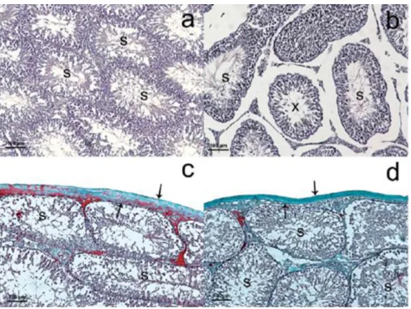
colleagues found observable differences in the tubules diameter and tunica albuginea thickness after long term exposure to 2.4GHz radiation (Figure 7) 53.
Figure 7: Testes section of a rat after exposure to 2.4 GHz electromagnetic radiation. A and C are
from control and B and D are from exposed group. Seminiferous tubules diameter (S) and tunica albugínea thickness (arrows) decreased in exposed group. Also a tubule with disorganized view due to loss of germinal epithelium (b) is seen in the figure (arrowhead). H&E (a, b), Masson Trichrome (c, d). 53
It has also been reported degenerative changes in spermatogenic cells, sharp edge craters, shrinkage on the surface of degenerating cells in seminiferous epithelium, visible debris of degenerating cells and residual cytoplasm and ruptured sperm head and distorted tail 54. In
group but not the 1-hour group showed a significant decrease in the number of germ cell layers. The exposure for 1 and 7 hours also caused a decline in seminal vesicles weight. As in Shokri et al., 55 other studies also found not only a significant decrease in seminal vesicles
weight but also in epididymis. The EMR from wireless devices has been shown to have a negative effect in sperm parameters such as decrease in viability, sperm count and motility
45. Yan et al., 56 exposed rats to 1.9GHz EMR in a cycle of 3 hours of exposure followed by a
30 minute period without expose and again 3 more hours of exposing for 18 weeks and the results revealed a majority of sperm cells without motion, dead or with straight rigid tails in the exposed rats.
There are also evidences of negative effects of prenatal exposure to EMR radiation on the sperm quality of the descendants. Odaci et al., 57 exposed pregnant rats to 900MHz EMR for
1h/day during days 13-21 of pregnancy and analyzed the epididymis of the rats with 60 days who were born from those exposed female rats. They found a lower sperm motility and viability and also histopathological changes in the epididymis and alterations on spermatogenesis.
However there are also studies that found no effects of EMR on the male reproductive system. Dasdag and his colleagues who exposed rats to long term 2.4GHz EMR found no significant differences in sperm concentration, sperm motility and total morphological defects 53.
Besides, in other studies, Dasdag observed no differences of short (20min/day for 1 month) and long (2h/7days for 10 months) periods of EMR radiation on the apoptotic cell number in the testes 59 60. Also Saygin et al. 59 did not observe any significant effect of Wi-Fi (2.45GHz)
on the diameter of seminiferous tubules.
Although there are many studies about the effects of Wi-Fi radiation on several parameters related to male fertility, there are no studies regarding the effects of EMR from wireless devices on testicular metabolism.
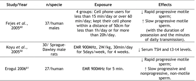
Table 1: Recent studies of the negative effects of EMR radiation from wireless devices. Supportive.
Study/Year n/specie Exposure Effects
Fejes et al.,
200560 37/human males
4 groups: Cell phone users for less than 15 min/day or over 60
min/day; kept their cell phone within a distance of 50cm for
less than 1h/day or for more than 20h/day.
↓ Rapid progressive motile sperm;
↑ Slow progressive motile sperm.
(with the duration of possession and the minutes
of daily transmission). Koyu et al., 200550 30/ Sprague– Dawley male rats EMR 900MHz, 2W/kg, 30min/day
for 5days/week, for 4 weeks. ↓ Serum TSH and t3-t4 levels. Erogul 200661 27/human EMR 900MHz for 5 min.
↓ Rapid progressive motile sperm;
↑ Slow progressive and nonprogressive, non-motile
Yan et al., 200756
16/Sprague-Dawley male
rats
EMR 1.9Hz, distance of 1 cm for 6h/day for 18 weeks.
↓ Sperm motility; ↑ Sperm cells dead; Clumps of sperm cells.
Wdowiak et al.,
200762 304/human males
3 groups: No use of mobile phones, sporadic use for the period of 1-2 years or regular use for more than 2 years.
↓ Sperm motility and vitality; ↑ Abnormal morphology of
sperm cells; (with the increase of mobile
phone use.) Agarwal et al.,
200863 361/human males
4 groups according to daily active cell phone use: group B <2 h/day, group C 2–4 h/day and
group D >4 h/day and a group A as control.
↓Sperm count, viability and normal morphology (with the
increase of cell phone use).
Agarwal et al., 200964
32/human males
EMR 850MHz, 1.46W/kg, at a distance of 2.5cm for 60 min.
↓ Sperm motility and viability; ↑ ROS level; No significant differences in
DNA integrity. De Iuliis et al.,
200946 22/human males EMR 1.8GHz, 0,4W/kg-27.5W/kg SAR, incubated for 16h. ↓ Motility and viability. Kesari et al.,
201065 12/Wistar male rats EMR 0,9W/kg SAR, 2h/day, for 35 days. ↓ Protein kinase C; ↑ Apoptosis.
Chalabi & Al-Wattar 201166
300/human males
4 groups with different hours of active mobile phone use: 4h/day, 3h/day, 2h/day, no
active use;
2 groups according the duration of use in years: 1-3 years or 4-6
years;
3 groups according the position of storage: trouser pocket, waist
pouch or in the shirt pocket
↓ Sperm count, motility and normal morphology with the increase of active mobile phone use, duration of use in
years and proximity of storage position to the
testes.
Gutschi et al., 201167
2110/human males
2 groups: men that use cell phones and men that don’t use
cell phones. ↑ % Of abnormal morphology and teratozoospermia; ↓ Proportion of progressive motile sperm; ↑ Testosterone and luteinizing hormone levels.
(in the cell phone users group)
Meo et al.,
201149 40/male rats
Mobile phone EMR placed inside the cage and a call was given for 30min/day or 60min/day for 3
months.
↑ Proportion of hypospermatogenesis and
maturation arrest; ↓ Serum testosterone level.
(in the group exposed to 60min/da). Esmekaya et al., 201168 30/ Wistar albino male rats EMR 900MHz, 1.20W/kg, 20min/day for 3 weeks.
Induced oxidative injury in testes by ↑ nitric oxide levels
and ↓ antioxidant defense mechanisms. Kesari et al., 201169 12/Wistar male rats EMR 10GHz, 0.014W/kg, power density of 0.21mW/cm2, 2 h/day
for 45 days. ↑ ROS levels and apoptosis. Falzone et al.,
201170 12/human males EMR 900-MHz SAR of 2.0 W⁄kg, 1 h.
↓ Morphometric parameters, such as the analysis of major
and minor axis, area, perimeter and acrosome;
↓ Sperm binding to the hemizona. Oni et al.,
201171
10/human
males Wi-Fi EMR 2.45GHz for 1 hour.
No effects on sperm concentration.
Avendaño et al.,
201247 24/human males Wi-Fi EMR 2.4GHz for 4 hours. ↓ Sperm progressive motility. Lee et al., 201272 50/Sprague– Dawley male rats EMR simultaneously 848,5MHz and 1950MHz, 45 min/day, 5 days/week with a total of 12
weeks
Did not observed adverse effects on rat spermatogenesis Çelik et al., 201251 30/Wistar-Kyoto male rats
2 cell phones, SAR values of 1,58 for 3 months
↑ Membrana propria thickness and collagen fibers;
Irregular basal membrane; Nucleus with irregular
contour in Sc. Atasoy et al.,
201248
10/Wistar albino rats
2.437GHz from a Wi-Fi device, 24h/day for 20 weeks.
↓ Testicular biopsy score; ↑ Level of
8-hydroxy-2’-deoxyguanosine; ↓ CAT and GPX activity. Nisbet el al.,
201273 albino rats 33/Wistar 1800 and 900MHz, 2h/day/90days
↑ Plasma testosterone level ↑ Epididymal sperm motility and normal morphology Khavanin et al.,
201374
28/Wistar male rats
3 exposure groups: EMR 915 MHz,8h/day for 14 and 21 days (group 2 and 3 respectively) and
950MHZ for 8h/day/14days (group 4).
↓ Sperm viability (was more notable in experimental group 3) ↓Sperm motility Shahin et al., 201445 40/male mice EMR 2.45GHz, 0.018W/kg, 0.029812mW/cm2 power density, 2h/day for 30 days.
↓ Sperm count, sperm viability, seminiferous tubule
diameter and serum testosterone level. ↑ROS production, total
nitrite and nitrate concentration and in mda; ↓ SOD, CAT and GPX activity.
Karaman et al., 201452
21/Wistar albino male
rats
2 exposure groups: EMR SAR of 1,52W/Kg, group 1 talk mode for
8 hours and standby for 8 hours for 20 days, group 2 same exposure but after those 20 days
the rats were exposed to stand by mode for more 20 days.
↑ Immature cells in the lumen;
↓ Spermatogenic cell lines; Tubules without lumen.
Kumar & Shukla,
201454 24/Swiss male rats
EMR from cell phone during 3h followed by 30 minutes of rest,
followed by another 3h exposure/day/5months.
Degenerative changes in spermatogenic cells, sharp edge craters, shrinkage on the surface of degenerating
cells in seminiferous epithelium, visible debris of
degenerating cells and residual cytoplasm and ruptured sperm head and
distorted tail. Dasdag et al., 201453 16/Wistar albino rats 2.4 GHz radiation for 24h/day/12months. ↑ Percentage of head defects; ↓ Seminal vesicles and
epididymis weight; ↓ Seminiferous tubules
diameter and tunica albuginea thickness. Gorpinchenko et
al., 201475
31/human males
EMR, frequency range of 900-1800MHz for 5 hours in combined
standby/talk mode.
↓Spermatozoa with progressive movement.
Bahaodini et al.,
201576 14/male rats
EMR 1 mT,50 Hz low frequency, for 85 days 24 h/day.
↓ Lumen diameter and area of the seminiferous tubules; ↓ Total diameter and cross
sectional area; ↑ Number of seminiferous
tubules per unit area of testis
Shokri et al.,
201555 27/Wistar rats
2 exposure groups: 2.45GHz Wi-Fi radiation, 1 hour/day/2
months and another and 7hours/day/2 months.
↓ Percentage of motile sperm, concentration and
proportion of normal to abnormal sperm; ↓ Seminal vesicles weight;
↓ Germ cell layers and ↑apoptotic cells (only in the
7h/day group) EMR- Electromagnetic radiation; SAR- Specific absorvation rate; ROS- Reactive oxygen species ; CAT- Catalase; GPX- Glutathione Peroxidase; SOD- Superoxide Dismutase; DNA- Deoxyribonucleic acid. ↑- Increased significantly; ↓- Decreased significantly.
the male reproductive organs and even alter the production of important hormones such as T. However, the possible effects of Wi-Fi networks on testicular metabolism has never been studied which lead us to the development of this project.
A normal spermatogenesis and fertility capacity of sperm depends on a correct testicular metabolism, which in turn depends on the correct functioning of several metabolic pathways. The processes involved on testicular metabolism may be affected by several factors such as environmental factors, a sedentary lifestyle and even the use of devices with Wi-Fi connection. This last factor has been receiving increasing attention from researchers since more and more devices using Wi-Fi connections, such as mobile phones and computers, are present in our daily life. Thus, we are almost constantly emerged in EMR from Wi-Fi networks. Recent advances have highlighted that the exposure to EMR from Wi-Fi networks is an important contributor to the decline of male reproductive health. In fact, there are several papers that evidence the negative consequences of EMR from wireless devices on male fertility affecting the sperm parameters, increasing the oxidative stress and ROS, contributing to histopathological chances in the male reproductive organs and even alter the production of important hormones such as T. However, the possible effects of Wi-Fi networks on testicular metabolism has never been studied which lead us to the development of this project.
The general aim of the research described in this work was to disclose the association between EMR from Wi-Fi devices and male infertility, dissecting its possible effects on testicular metabolism, particularly glucose metabolism, and the subsequent consequences for male reproductive health. It is also intended to develop a new model for the study of the effects of EMR on testicular metabolism using 20-day-old rats.
To achieve this, we first aimed to perform histological analysis to observe the cell populations present in SeT at 19, 20, 21 and 22 days in order to understand what is the most appropriate age for the development of the new model. Secondly we intended to perform an histological analysis to SeT after 72 hours in culture medium to assess if there were any observable histological changes in the tissue after the time of incubation. Then, we wanted to built an Wi-Fi exposure set up, to expose our SeT cultures to EMR on a realistic way. As there are studies showing that EMR has effects on sperm parameters, we decided to confirm these results by exposing sperm cells from an adult rat to EMR from our Wi-Fi network and assess their mobility, viability and morphology after exposure, validating our set up. Lastly, to disclose the effects of Wi-Fi on testicular metabolism, the metabolite production of lactate and glucose consumption were measured as well as LDH activity.
3.1 Chemicals
Dulbecco’s Modified Eagle Medium Ham’s Nutrient Mixture F12 (DMEM:F12), Bradford reagent, 3-Isobutyl-1-methylxanthine and phenylmethanesulfonyl fluoride were obtained from Sigma-aldrich (St. Louis, USA); gentamicin was obtained from Alfagene (Carcavelos, Portugal); Protease Inhibitor Cocktail 5 MammCell/Tissue was obtained from PVL (Famões, Portugal); Hematoxilin and aqueous eosin 1% were obtained from Leica Microsystems (Wetzlar, Germany); inclusion agent for histology Histosec was obtained from Merk (Darmstadt, Germany).
3.2 Instruments
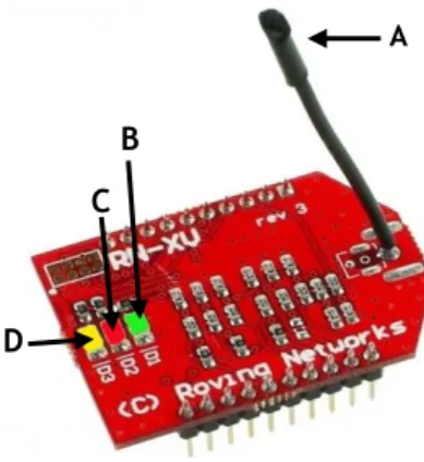
The system used to create an internet network was constituted by three components: The WiFly module, the mbed and the internet router.
The wifly module, represented on figure 8, is a standalone device that enables wireless access to a local area network (LAN). It is certified to operate on 2.4GHz IEEE802.11b/g networks and it has a flash memory of 8Mbit and a random access memory (RAM) of 128KB. It also has a slave interface universal asynchronous receiver/transmitter (UART) which is a computer hardware device for asynchronous serial communication in which the data format and transmission speeds are configurable and the electric signaling levels and methods (such as differential signaling, etc.) are handled by a driver circuit external to the UART, and a serial peripheral interface (SPI) which is a synchronous serial communication interface specification used for short distance communication, primarily in embedded systems. The module has internally implemented the transmission control protocol/internet protocol (TCP/IP) stack that is the suite of communications protocols used to connect hosts on the Internet. Once properly set up, the radio trough the wire antenna automatically establishes a connection to the Wi-Fi network. The firmware allows establishing a communication channel between the radio channel and the UART. There were used two wifly modules, one had the function of sending data packets and the other had the function of receiving the data packets. Every time this communication was made the information (data packets) had to always first pass through the internet router. The wifly module also has three light-emitting diodes (LED’s) a green one, a red one and a yellow one, each one providing information about the status: If the red LED is blinking rapidly it indicates that the module is not connected to the wireless network. Contrarywise, if the LED is off, the module is connected to the wireless network; If the yellow LED is blinking, each blink means it is either sending or receiving data; if the green LED is on and solid, it indicates the module is connected over TCP, if it is blinking rapidly, it means that no IP address is assigned, if it is blinking slowly, it means that the IP address is assigned but still not conected to TCP. The minimum operating temperature is -40°C and the maximum operating temperature is 85°C. The humidity range operating values are bellow 90% 77.
A
B C
D
Figure 8: The wifly module. It is a standalone device that enables wireless access to LAN (local area
network). The module is constitued by a flash memory, a RAM (Random Access Memory), an interface UART (Universal Asynchronous Receiver/Transmitter), a hardware device for asynchronous serial communication in which the data format and transmission speeds are configurable and the electric signaling levels and methods are handled by an external driver circuit; a SPI (Serial Peripheral Interface), a synchronous serial communication interface specification used for short distance communication; a TCP/IP (Transmission Control Protocol/Internet Protocol) stack, a suite of communications protocols used to connect hosts on the Internet and a wire antenna (A) that establishes a connection to the Wi-Fi network. The module also has three LED’s that provide information about the status of the module. If the green LED (B) is on and solid, the module is connected over TCP, if it is blinking rapidly, no IP address is assigned, if it is blinking slowly, the IP address is correctly assigned; if the red LED (C) is blinking rapidly, the module is not connected to the wireless network, if it is off, the module is correctly connected; If the yellow LED (D) is blinking, it is either sending or receiving data. Adapted from77
Mbed, (represented in figure 9, panel A) is a platform and operating system for internet-connected devices based on 32-bit RAM Cortex-M microcontrollers. Such devices are also known as Internet of Things devices. The application for the mbed platform was developed using the mbed online integrated development environment (IDE), which is an online code editor and compiler in which the code was written and compiled within a web browser, and compiled on the cloudusing the ARMCC C/C++ compiler. It was used the mbed Microcontroller Board- mbed NXP LPC1768- a demo-board based on an NXP microcontroller, which has an ARM Cortex M3 core, running at 96 MHz, with 512 KB flash, 64 KB RAM. It is packaged as a small dual in-line package (DIP) form-factor which is an electronic component package with a rectangular housing and two parallel rows of electrical connecting pins for prototyping with through-hole printed circuit boards (PCBs), stripboard and breadboard, and includes a built-in universal serial bus (USB) flash programmer. There is also a USB port through which it supplies power to the system and a reboot bottom to restart running the program. There were used 2 mbed boards, each one connected to a wifly module. The Wifly terminals are identified in figure 9, panel B and the RN-XV-171 was connected to the mbed according to the figure. The minimum operating temperature is -65°C and the maximum operating temperature is 150°C. The humidity range operating values are not specified 78.
A B a
b
Figure 9: mbed board and its connection to wifly terminals. A) mbed board. The mbed board is a
demo-board based on an NXP microcontroller, which has an ARM Cortex M3 core, RAM, a small DIP (dual in-line package) form-factor which is an electronic component package with a rectangular housing and two parallel rows of electrical connecting pins for prototyping with through-hole PCBs (printed circuit boards), a stripboard and breadboard, and a built-in USB (Universal Serial Bus) flash programmer. It has also a USB port (a) through which it supplies power to the system a reboot bottom to restart running the program (b). B) The mbed and Wifly terminals are represented in the figure, through this scheme it is possible to understand how to connect the wifly module to the mbed board. Adapted from78
The internet router used was TP-LINK model number TL-WR740N, represented in figure 10, a 150MBps wireless router with 9V power and a frequency band of 2.4 GHz. The minimum operating temperature is 0°C and the maximum operating temperature is 40°C. The humidity range operating is 10 to 90% of humidity 79.
b a
Figure 10: The internet router TP-LINK model number TL-WR740. A 150MBps wireless router with 9V
power and a frequency band of 2.4 GHz. a- Wi-Fi horn used to transmit and receive signals; b- Wi-Fi hardware. Adapted from 79
3.3 Animals
One wistar male rat (Rattus norvegicus) 19-day old, 21-day old, 22-day-old and 3-months old as well as twenty-five wistar male rats (Rattus norvegicus) 20-days old, were obtained and housed at CICS-UBI animal facilities under a 12-hour light/dark cycle, with food and water available ad libitum. Animals were handled in compliance with the guidelines established by the “Guide for the Care and Use of Laboratory Animals” published by the U.S. National Institutes of Health (NIH Publication No. 85‐23, revised 1996) and the European Union rules for the care and handling of laboratory animals (Directive 2010/63/EU). In accordance with the Portuguese law (Ordinance no. 1005/92 of 23 October), the research team requested a permission to perform this animal experimentation study to the Portuguese “Direção Geral de Veterinária” (Portuguese Veterinarian and Food Department). All rats were euthanized with CO2.
3.4 Development of a new model to study the effects of Wi-Fi
on seminiferous tubules metabolism ex vivo
3.4.1 Determining the ideal age for the development of the model
There are studies that indicate that mitotic division of SCs ceases after the 15 day of post-natal development in rats, preceding the formation of the hematopoietic barrier created by the inter-sertoli tight junctions between days 16 and 19 80. Another study indicates that the
mitotic division of the SCs ceases at 18 days 81. In view of the number of published studies on
the metabolism of SCs 82 83, a model using a rat with an epithelium of seminiferous tubules
containing a well established SC population and germ cells exclusively in the early stages of spermatogenesis would constitute an excellent model for the study of metabolism in rats. In order to assess the best age to implement this model, histological sections from SeT from 19 days-old, 20 days-old, 21 days-old and 22 days-old rats were analysed to assess which type of cells were present and to observe the SCs population.
3.4.2 Assessing the possible effects of 72h in culture medium
As the possible effects of the culture medium in SeT with rats at this age have never been assessed, histological analysis was performed comparing t=0 and t=72 of SeT of a 22-day-old rat in culture medium.
3.4.3 Validating the model
Several articles show that radiation from Wi-Fi devices has effects on sperm 60. In this way, to
validate the set up and built Wi-Fi network, spermatozoa from six 3 months-old adult rat were extracted, placed at a Petri dish and exposed 1 hour to the Wi-Fi network in the incubator at 33ºC as represented in figure 11. The sperm parameters were analyzed and compared to the control, constituted by spermatozoa extracted and place at a petri dish in
the incubator for 1 hour at 33ºC but not exposed to the Wi-Fi network. For each rat, one epididymis was used for exposure and the other one as control.
A
B
C
D
Figure 11: Disposition of the petri dish containg sperm cells for the exposure to the Wi-Fi network. A-
Wifly; B- mbed; C- Wi-Fi router; D- The petri dishes are placed between the receptor and sender.
3.5 Effects of Wi-Fi on rat testicular metabolism ex vivo
Twenty-four 20-day-old rats were used to study the effects of Wi-Fi on testicular metabolism. The SeT were extracted and cultured, the exposed group (12 rats) received the EMR from the created Wi-Fi network during 72h. The 72 hours were chosen as the exposure time once daily most people are exposed to EMR from Wi-Fi networks 24 hours a day, day after day. Thus, the choice of this exposure time is intended to be as close as possible to the actual situation. The control group (12 rats) was not exposed to the Wi-Fi network.
Every 12 hours it was verified the correct functioning of the system by accessing the network created through the router IP and checking if there was a correct transfer of data packets which indicated that there was continuous communication. Also visually it was possible to check the correct functioning of the system by observing on the mbed a constantly lit green LED which indicates that the mbed has connected correctly to the router and an yellow LED flashing rapidly, indicating that the mbed boards are receiving and sending data packets. A new data packet was transferred every 0.5 seconds. The set up is represented in figure 12.
A B
C
D D
Figure 12: Set up disposition. A- Wifly; B- mbed; C- Wi-Fi router; D- The culture plates are placed
between the receptor and sender. At each time 8 plates were expose.
3.6 Ex vivo culture of immature rat SeT
Testicles from twenty-four 20-day-old rats and one 22 day-old rat were removed, trimmed free of fat, washed in cold phosphate-buffered saline (PBS) and placed in Dulbecco's modified Eagle's medium/Ham's F12 culture medium (Sigma‐Aldrich, St. Louis, USA) supplemented with 20 mg/L gentamicin sulfate, 0.1 mM 3‐isobutyl‐1‐methylxanthine, and 1 μg/L of bovine serum albumin (BSA) 10% at 33°C. Tunica albuginea was cut and peeled back to expose tubules. As the immature SeT are very tangled, it is difficult to pick individual fragments, thus, the SeT from one testicle was reparted to the 12 wells of a culture plate (Nunclon D 12 well multidishes; Nunc, Roskilde, Denmark), each well containing 5 ml of pre‐warmed culture medium. In this way, 48 plates were used, each one corresponding to a testicle.
3.7 Histological analysis
Testicles from 19 day-old, 20 day-old and 21 day-old animals were extracted and the entire testicles were included side to side in paraffin in order to obtain sections with many cross sections of the SeT which alow us to observe the SeT epithelium. The paraffin sections (5 µm) of SeT were deparaffinized in xylene for 10 minutes and rehydrated in graded alcohols (1 minute in ethanol 100%, 1 minute in ethanol 70% and 1 minute in running water). Then the paraffin sections were stained and differentiated first 7 minutes in hematoxylin, then 1 second in hydrochloric ethanol (differentiator), 4 minutes in running water, 1 minute in eosin 1% and 10 seconds in running water. Subsequently the sections were dehydrated 50 second in ethanol 95%, 1 minute and 30 seconds in ethanol 100% and 2 minutes in ethanol 100%. For the