einstein. 2016;14(3):439-42
MEDICAL DEVELOPMENTS
This content is licensed under a Creative Commons Attribution 4.0 International License.
A pictorial presentation of 3.0 Chicago Classification for
esophageal motility disorders
Apresentação em imagens da Classificação de Chicago versão 3.0
das doenças da motilidade esofagiana
Fernando Augusto Herbella1, Priscila Rodrigues Armijo1, Marco Giuseppe Patti2
ABSTRACT
High resolution manometry changed several esophageal motility paradigms. The 3.0 Chicago Classification defined manometric criteria for named esophageal motility disorders. We present a pictorial atlas of motility disorders. Achalasia types, esophagogastric junction obstruction, absent contractility, distal esophageal spasm, hypercontractile esophagus (jackhammer), ineffective esophageal motility, and fragmented peristalsis are depicted with high-resolution manometry plots.
Keywords: Manometry/methods; Esophageal motility disorders; Esophageal achalasia/classification
RESUMO
A manometria de alta resolução mudou vários paradigmas da motilidade digestiva. A Classificação de Chicago, na versão 3.0, definiu critérios manométricos para as doenças da motilidade esofagiana. O presente artigo é um atlas das dismotilidades descritas. Tipos de acalásia, obstrução ao nível da junção esofagogástrica, contrações ausentes, espasmo esofagiano distal, esôfago hipercontrátil, motilidade esofagiana ineficaz e peristalse fragmentada são mostradas em traçados de manometria de alta resolução.
Descritores: Manometria/métodos; Transtornos da motilidade esofágica; Acalásia esofágica/classificação
INTRODUCTION
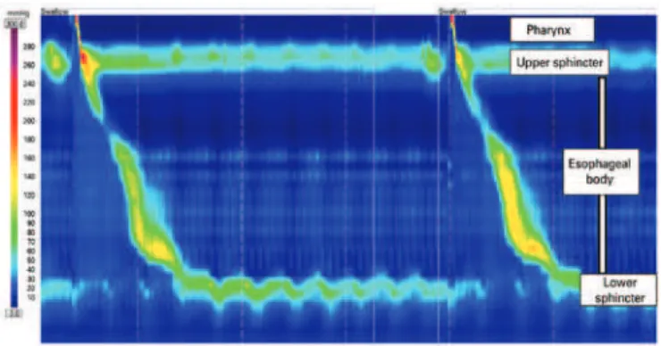
High resolution manometry (Figure 1) has clear and inherent advantages over conventional manometry, despite its higher cost.(1) High resolution manometry
detailed analysis of esophageal peristalsis changed several esophageal motility paradigms, including new manometric parameters and different classification for named “motility
disorders based on pressure topography”, the Chicago classification,(2) which was recently revised.(3)
We present a pictorial atlas of the motility disorders according to the 3.0 Chicago Classification with high-resolution plots.
Achalasia
Chicago Classification divided achalasia into three subtypes according to esophageal pressurization(4)
(Figure 2). Type I is characterized by 100% failed contractions and no esophageal pressurization; type II has panesophageal pressurization in at least 20% of swallows; and type III is defined by the presence of preserved fragments of distal peristalsis or premature contractions for at least 20% of the swallows.(3) This
classification may be applied to Chagas’ disease esophagopathy as well, although type III is rarely, if ever, seem.(5)
Figure 1. Normal high resolution manometry
1 Escola Paulista de Medicina, Universidade Federal de São Paulo, São Paulo, SP, Brazil. 2 University of Chicago, Chicago, Illinois, Estados Unidos.
Corresponding author: Fernando Augusto Herbella – Rua Diogo de Faria, 1.087, room 301 – Vila Clementino – Zip code: 04037-003 – São Paulo, SP, Brazil – Phone: (55 11) 3926-7610 E-mail: [email protected]
einstein. 2016;14(3):439-42 440 Herbella FA, Armijo PR, Patti MG
Esophagogastric junction obstruction
Esophagogastric junction obstruction (Figure 3) is characterized by an elevated residual pressure of the lower esophageal sphincter (LES) measured by a new and more sophisticated tool, the integrated relaxation pressure(6) in the absence of criteria for achalasia
(absence of peristalsis).(3) This parameter measures the
mean pressure of the 4 seconds of maximal deglutitive relaxation in the 10-second window beginning at the beginning of the swallow (upper sphincter relaxation). It is a rare finding usually present in patients with dysphagia after operations at the esophagogastric junction.(7)
be noticed in patients with connective tissue diseases, end-stage gastroesophageal reflux disease etc.
Figure 2. Achalasia types
Figure 3. Esophagogastric junction obstruction in a patient after Nissen operation
Absent contractility
Absent contractility is characterized by aperistalsis in the setting of normal LES relaxation and absence of esophageal pressurization(3) (Figure 4). This finding may
Figure 4. Absent contractility in a patient with scleroderma
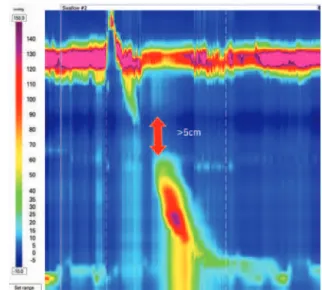
Distal esophageal spasm
Distal esophageal spasm is defined by over 20% of premature contractions as measured by a new parameter, the distal latency (DL) <4.5 seconds(3) (Figure 5). The DL
is the time interval between the beginning of the upper sphincter relaxation and the contractile deceleration point the manometric representation of the transition from the esophageal body to the epiphrenic ampulla regarded as an inflection of the peristaltic axis within 3cm of the proximal margin of the LES.(8)
441
A pictorial presentation of 3.0 Chicago Classification for esophageal motility disorders
einstein. 2016;14(3):439-42
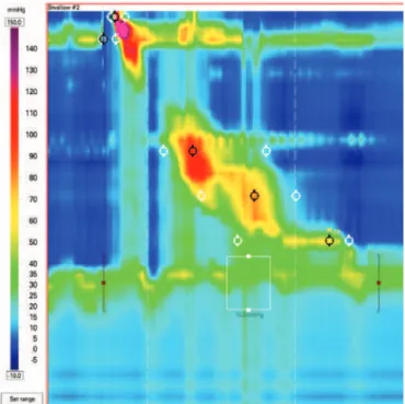
Hypercontractile esophagus
Hypercontractile esophagus (Jackhammer esophagus) is characterized by at least two swallows with hypercontractility as measured by the distal contractile integral (DCI)(3)
(Figure 6). The DCI measures the contractile vigor combining the amplitude versus duration versus length of the distal esophageal contraction exceeding 20mmHg from the transition zone to the proximal margin of the LES.(9) Hypercontractility is defined by DCI
>8,000mmHg.s.cm.(3) It may occur with esophagogastric
junction obstruction, gastroesophageal reflux disease and eosinophilic esophagitis.(10)
Figure 6. Hypercontractile esophagus
Ineffective esophageal motility
Ineffective esophageal motility is defined by
≥50% ineffective swallows (failed or weak – DCI
<450mmHg.s.cm)(3) (Figure 7).
Figure 7. Ineffective esophageal motility in a patient with gastroesophageal reflux disease
Fragmented peristalsis
Fragmented peristalsis ≥50% fragmented contractions
with DCI >450mmHg.s.cm(3) (Figure 8). Although
patients with fragmented peristalsis are more prone to have dysphagia,(11) its clinical significance is still
elusive.
Figure 8. Fragmented peristalsis
DISCUSSION
Motility patterns to define named disorders have been motive of controversy since the era of conventional manometry. Different definitions exist although the classification by Richter was the most used by experts.(12)
High resolution manometry seems to bring a more intuitive and reproducible interpretation compared with conventional manometry,(13) and more sophisticated tools
to define old and new manometric parameters. Despite all improvements, and similarity with conventional manometry, some cases are still unclassified, and the real clinical significance of some Chicago Classification disorders is still under investigation.
REFERENCES
1. Herbella FA, Del Grande JC. [New ambulatory techniques for assessment of esophageal motility and their applicability on achalasia study]. Rev Col Bras Cir. 2008;35(3):199-202. Portuguese.
2. Kahrilas PJ, Ghosh SK, Pandolfino JE. Esophageal motility disorders in terms of pressure topography: the Chicago Classification. J Clin Gastroenterol. 2008;42(5):627-35. Review.
3. Kahrilas PJ, Bredenoord AJ, Fox M, Gyawali CP, Roman S, Smout AJ, Pandolfino JE; International High Resolution Manometry Working Group. The Chicago Classification of esophageal motility disorders, v3.0. Neurogastroenterol Motil. 2015;27(2):160-74.
einstein. 2016;14(3):439-42 442 Herbella FA, Armijo PR, Patti MG
5. Vicentine FP, Herbella FA, Allaix ME, Silva LC, Patti MG. High-resolution manometry classifications for idiopathic achalasia in patients with Chagas’ disease esophagopathy. J Gastrointest Surg. 2014;18(2):221-4; discussion 224-5. 6. Lin Z, Kahrilas PJ, Roman S, Boris L, Carlson D, Pandolfino JE. Refining the
criterion for an abnormal Integrated Relaxation Pressure in esophageal pressure topography based on the pattern of esophageal contractility using a classification and regression tree model. Neurogastroenterol Motil. 2012;24(8):e356-63.
7. Scherer JR, Kwiatek MA, Soper NJ, Pandolfino JE, Kahrilas PJ. Functional esophagogastric junction obstruction with intact peristalsis: a heterogeneous syndrome sometimes akin to achalasia. J Gastrointest Surg. 2009;13(12):2219-25. 8. Pandolfino JE, Roman S, Carlson D, Luger D, Bidari K, Boris L, et al. Distal esophageal spasm in high-resolution esophageal pressure topography: defining clinical phenotypes. Gastroenterology. 2011;141(2):469-75.
9. Roman S, Pandolfino JE, Chen J, Boris L, Luger D, Kahrilas PJ. Phenotypes and clinical context of hypercontractility in high-resolution esophageal pressure topography (EPT). Am J Gastroenterol. 2012;107(1):37-45.
10. Martín-Domínguez V, Pérez-Fernández MT, Marinero A, Jusué-Irurita V, Caldas M, Santander C. Hypercontractile esophagus: Clinical context and motors findings in high resolution manometry. Rev Esp Enferm Dig. 2015;107(5):274-9. 11. Roman S, Lin Z, Kwiatek MA, Pandolfino JE, Kahrilas PJ. Weak peristalsis in
esophageal pressure topography: classification and association with Dysphagia. Am J Gastroenterol. 2011;106(2):349-56.
12. Richter JE. Oesophageal motility disorders. Lancet. 2001;358(9284):823-8. Review.