RESUMO
JUSTIFICATIVA E OBJETIVOS: Determinar o grau de
concordância e correlação entre amostras arteriais e
as obtidas através de um cateter venoso umbilical,
com relação ao pH, bicarbonato, excesso de base (BE)
e lactato, em recém-nascidos prematuros e de termo, criticamente doentes.
MÉTODO: Foram obtidas amostras para gasometria
(0,5 – 1 mL), por punção de artéria radial, e, dentro
do limite de 5 minutos, do cateter venoso umbilical. O método de Bland-Altman foi utilizado para demons-trar a concordância entre as medidas. Os limites de
concordância foram deinidos como a diferença média ± 2 DP. Para as correlações foi utilizado o método de
Pearson.
RESULTADOS: Cento e seis amostras (53 pares) de 53
Agreement and Correlation of pH, Bicarbonate,
Base Excess and Lactate Measurements in Venous
and Arterial Blood of Premature and Term Infants*
Concordância e Correlação das Medidas de pH, Bicarbonato,
Excesso de Base e Lactato no Sangue Venoso e Arterial
de Recém-Nascidos de Termo e Prematuros
Orlei R. Araujo1, Ana Regina Diegues2, Dafne C. Borguignon da Silva2, Andréa de Cássia S. Albertoni3,
Maria Eduarda R. Louzada3, Eloíza A. F. Cabral3, Ronaldo Arkader2, Marta R. Afonso3.
1. Pediatra Intensivista, Coordenador do Serviço de Pediatria do
Hospital Santa Marina
2. Pediatra Intensivista, Unidade de Terapia Intensiva Pediátrica do Hospital Santa Marina
3. Pediatra Neonatologista, Unidade de Terapia Intensiva Neonatal do Hospital Santa Marina
*Recebido do Hospital Santa Marina, São Paulo, SP.
Apresentado em 03 de abril de 2007
Aceito para publicação 24 de agosto de 2007 Endereço para correspondência:
Dr. Orlei R. Araujo
Av. Santa Catarina, 2785 - Vila Santa Catarina 04378-500 São Paulo, SP
Fones: (11) 5013-1263 – 5563-6331 E-mail: [email protected]
©Associação de Medicina Intensiva Brasileira, 2007
pacientes foram analisadas para bicarbonato, pH e BE. Foi dosado lactato em 49 pares de amostras. Houve
concordância em 94,3% dos pares de amostras para
o pH, e este mesmo percentual foi observado para o bicarbonato. Para o excesso de base, a concordância
foi de 96,2%, e de 91,8% para o lactato. As diferen
-ças médias foram 0,03 unidade para o pH, -1,2 mmol/L
para o bicarbonato, -0,24 mmol/L para o excesso de base e 0,33 mmol/L para o lactato. Os coeicientes de
correlação de Pearson (r) foram 0,87 para o pH, 0,76 para o bicarbonato, 0,86 para o excesso de base e 0,95 para o lactato.
CONCLUSÕES: Os valores venosos isolados não
po-dem ser usados como equivalentes aos arteriais para a
avaliação do estado ácido-básico em recém-nascidos.
As amostras venosas poderiam ser usadas de forma serial, para monitorizar tendências ao longo do tempo. Unitermos: artérias, gasometria, recém-nascido, veias
SUMMARY
BACKGROUND AND OBJECTIVES: Determine the
extent of agreement and correlation between arterial samples and venous (obtained from a venous umbili-cal catheter), with respect to measurements of pH, bi-carbonate, base excess and lactate, in critically ill term and premature newborns.
RESULTS: A hundred and six samples (53 pairs) were taken from 53 patients for analysis of bicarbonate, pH and base excess. Lactate was analyzed in 49 pairs of samples. Differences were within the limits of
agree-ment in 94.3% of pairs of samples for pH, and the
same percentage was observed for bicarbonate. There
was agreement in 96.2% of pairs for base excess, and in 91.8% for lactate. Mean differences were 0.03 units
for pH, -1.2 mmol/L for bicarbonate, -0.24 mmol/L for base excess and 0.33 mmol/L for lactate. Pearson’s correlation coeficients (r) were 0.87 for pH, 0.76 for bi-carbonate, 0.86 for base excess and 0.95 for lactate. CONCLUSIONS: Although single venous values can-not be used as equivalent to arterial for assessing acid base status in newborns, venous blood samples can be used serially for monitoring trends over time.
Key Words: arteries, blood gas analysis, newborn, veins
INTRODUCTION
Arterial blood sampling is usually the standard method for measurements of bicarbonate, pH, BE and lac-tate. In newborns, arterial lines are frequently placed in umbilical vessels for blood sampling, since arterial punctures may be dificult, especially in low-weight premature infants. Additionally, radial artery puncture is painful, and can lead to complications like local he-matomas, infection and occlusion or embolization of the artery, with distal ischemic injury1.These risks
in-crease with repeated punctures2.The use of umbilical
artery catheters also poses a risk, especially for neo-natal thrombosis, which carries a signiicant mortality rate3.It has been experimentally demonstrated that the
presence of arterial catheter is enough to start the lo-cal ibrin formation4. Blood sampling from the umbilical
arterial catheter induces a decrease in cerebral blood volume and cerebral oxygenation, depending on the volume e sampling velocity5,6.
Central venous catheters are interesting alternative sources of blood samples. Venous umbilical catheters are the easiest venous line for premature newborns, and are a common procedure in neonatal intensive care units. As these catheters have usually diameters larger than 3-French, blood sampling is feasible, since proto-cols for manipulation with aseptic technique are adopt-ed. One study have demonstrated that central venous pH, bicarbonate, BE and lactate show a high level of agreement with respective arterial values in adult pa-tients7. The purpose of our study was investigating the
extent of agreement between arterial and venous sam-ples, when obtained from a venous umbilical catheter, with respect to acid-basis status, in critically ill term and premature newborns.
METHODS
The study was undertaken in the 17-bed Neonatal In-tensive Care Unit (NICU) within a tertiary hospital in São Paulo, Brazil. Premature and term infants born from March to September, 2005, were the potential subjects. Inclusion criteria were the need for blood gas analy-sis in a critically ill newborn, the presence of a venous umbilical catheter with the tip placed at inferior vena cava below the diaphragm (with placement conirmed by chest and abdominal radiograph), and an informed consent form signed by one or both of the parents. The local ethics committee approved the study. Infants with congenital heart diseases, with signiicant intracardiac shunt, were excluded.
Arterial blood samples (0.5-1 mL) were obtained by ra-dial artery puncture with 25G (Gauge) or 27G needles, in 3 mL heparinised syringes. As simultaneously as possible (up to 5 minutes), samples were obtained from venous umbilical catheters, by the “push-pull” method: after lushing the dead space of the catheter with sa-line, 1 mL of blood was aspirated into a 10 mL non-heparinised syringe. The blood was reinfused, and this procedure was repeated at least 3 times8,9. The syringe
was removed, and a new 3-mL heparinised syringe was attached, and 0.5-1 mL of blood was sampled for gas analysis and lactate dosage. The measurements were performed within 15 minutes, using a Rapid Lab 865 analyzer (Bayer, USA). Multiple samples from the same patient were not allowed.
Data were analyzed with the statistical package SPSS 10.0 (SPSS, USA). The Bland-Altman plots were used to depict agreement between arterial and venous mea-surements. Limits of agreement were deined as the mean of differences ± 2 SD10. Pearson’s correlation
co-eficients were also determined.
RESULTS
neonatal sepsis (11%). Table 1 shows characteristics of
the samples and table 2 the difference means, limits of agreement and correlation coeficients.
Figure 1 depicts Bland-Altman plot for arterial and ve-nous pH. Differences were within the limits of
agree-ment in 94.3% of pairs of samples. The same percent -age was observed for bicarbonate (Figure 2). There was
agreement in 96.2% of pairs for base excess (Figure 3), and in 91.8% for lactate (Figure 4).
2.21 kg (range 0.88–3.5 kg). 14 patients (26.6%) were
premature infants with birth weight lower than 1.5 kg. The mean age at the moment of the sampling was 31.3 hours (range 1-240 hours). All the patients were under
respiratory assistance, either invasive (73%) or nonin
-vasive (27%), and 22 (41.5%) of them were receiving
vasoactive drugs (dobutamine or dopamine, or both). The most frequent diagnoses were respiratory distress
syndrome (54.7%), pulmonary hypertension (17%) and
0,2
0,1
0,0
-0,1
6,9 7,0 7,1 7,2 7,3 7,4 7,5 7,6
MD - 2SD MD
MD + 2SD MD + 2SD
MD
MD - 2SD 6
4
2
0
-2
-4
-6
-20 -10 0 10
Figure 1 – Bland-Altman Plot of the Means of Arterial and Venous pH Measurements (X) and their Differences (Y).
There was agreement in 50/53 pairs of samples (94.3%). MD:
mean difference SD: Standard Deviation
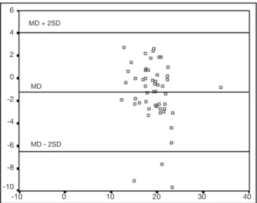
Figure 2 – Plot of the Means of Arterial and Venous Base Excess Measurements (X) and their Differences (Y).
There was agreement in 51/53 pairs of samples (96.2%).
Table 1 – Characteristics of Samples
Variables Number of Samples Mean (SD) Range
Arterial pH 53 7.33 (0.11) 6.96 to 7.53
Venous pH 53 7.29 (0.10) 6.99 to 7.54
Arterial bicarbonate 53 18.54 (3.45) 10.5 to 33.4 Venous bicarbonate 53 19.75 (3.96) 11.4 to 34.2 Arterial base excess 53 -6.60 (3.74) -17.5 to 4.3 Venous base excess 53 -6.35 (3.82) -18.4 to 4.1 Arterial lactate 49 3.24 (1.88) 1.18 to 8.69 Venous lactate 49 2.91 (1.47) 0.76 to 6.73
Table 2 – Agreement and Correlation between Arterial and Venous Samples
Variables N MD (95% CI) Limits of Agreement (MD ± 2 SD) r (Signiicance) pH (units) 53 0.03 (0.018 to 0.048) -0.07 to 0.14 0.87 (p < 0.001) Bicarbonate (mmol/L) 53 -1.21 (-1.92 to -0.50) -6.49 to 4.06 0.76 (p < 0.001) Base excess (mmol/L) 53 -0.24 (-0.84 to 0.35) -4.68 to 4.19 0.83 (p < 0.001) Lactate (mmol/L) 49 0.33 (-0.14 to 0.52) -1.00 to 1.67 0.95 (p < 0.001)
DISCUSSION
The assessment of acid-base status is essential in the management of critically ill newborns. Measurements of pH and base-excess are not only important in diag-nosis of acidosis, but are also useful to monitor the pro-gression of disease2. Lactate is a marker of impaired
tissue oxygenation, as resultant of hypoxia or hypovo-lemia, and has been demonstrated to be a predictor of poor outcome in children with shock or sepsis11,12.
Umbilical catheters also can lead to serious complica-tions to premature and term newborns, like thrombosis or sepsis, but, if the association of an umbilical arterial line could be avoided, the overall risks would be poten-tially diminished. We can assume that collecting blood from a venous umbilical catheter is not more hazardous then collecting from arterial ones. Some studies have suggested that venous pH 1 or pH, bicarbonate, base
excess and lactate7,13 show good statistical agreement
when compared in arterial and venous samples. The limits of agreement used in these studies were in ac-cordance with the Bland and Altman statement, that the interval between 2 SD for the mean difference is not clinically signiicant10. In spite of the general
ac-ceptance of this statement by the authors, clinicians may not have the same opinion. In the study of Rang et al., 45 emergency physicians were asked about the acceptable level of difference between arterial and ve-nous values for blood gases. The limits stated by the clinicians were quite narrower than the 2 SD around the mean difference14. This limited survey suggests
physi-cians responsible for patient care would not be com-fortable with the level of disagreement between arterial and venous samples.
Our study shows mean differences greater than previ-ously reported in adult patients1,7. The sample was
rea-sonably small, limited by the dificulties of simultaneous blood collecting in low birth weight infants, precluding analysis of subgroups. Despite of this, the conidence intervals were narrow for the mean difference (Table 2), indicating that this sample is in fact representative. There were also a high percentage of patients with hemodynamic compromise, as demonstrated by the number of patients with inotropic support. The study of Adrogue et al. has demonstrated that when the hemo-dynamic compromise is critical, agreement between arterial and central venous pH is poor. The proposed mechanism is that the disproportionate decrement in cardiac output leads to an increased ventilation-to-per-fusion ratio with arterial hypocapnia. The venous hy-percapnia results from a greater addition of CO2 per unit of blood transversing the capillaries of the hypo-perfused peripheral tissues15,16. This inluence of CO
2
can explain the lower correlation of arterial and venous bicarbonate that we observed (r = 0.76), as bicarbonate is a calculated value, based on CO2 level. According with our data, for using arterial and venous measure-ments of pH, base excess, bicarbonate and lactate in-terchangeably, clinicians would have to accept a range of disagreement up to ± 0.1 unit for pH, ±5.2 mmol/L MD + 2SD
MD
MD - 2SD 6
4
2
0
-2
-4
-6
-8
-10
-10 0 10 20 30 40
MD + 2SD
MD
MD - 2SD 2,0
1,5
1,0
0,5
0,0
-0,5
-1,0
-1,5
0 1 2 3 4 5 6 7 8
Figure 3 – Plot of the Means of Arterial and Venous Bicarbonate Measurements (X) and their Differences (Y).
There was agreement in 50/53 pairs of samples (94.3%).
Figure 4 – Plot of the Means of Arterial and Venous Lactate Mea-surements (X) and their Differences (Y).
for bicarbonate, ±4.4 mmol/L for base excess and ±1.3 mmol/L for lactate. We think that with these data, single venous blood values cannot be used as equivalents to arterial ones, but venous blood samples from umbilical catheters can be useful in serial collects for identifying trends over time. This recommendation is supported by the high correlation observed between arterial and venous measurements for pH, base excess and espe-cially for lactate.
REFERENCES
01. Kelly AM, McAlpine R, Kyle E - Venous pH can safely replace arterial pH in the initial evaluation of patients in the emergency department. Emerg Med J, 2001;18:340-342.
02. Yildizdas D, Yapicioglu H, Yilmaz HL et al - Correlation of simultaneously obtained capillary, venous, and arterial blood gases of patients in a pae-diatric intensive care unit. Arch Dis Child, 2004;89:176-180.
03. Coleman MM, Spear ML, Finkelstein M et al - Short-term use of umbilical artery catheters may not be associated with increased risk for thrombo-sis. Pediatrics, 2004;113:770-774.
04. Chidi CC, King DR, Boles ET - An ultrastructural study of the intimal injury induced by an indwelling umbilical artery catheter. J Pediatr Surg, 1983;18:109-115.
05. Roll C, Huning B, Kaunicke M et al - Umbilical artery catheter blood
sam-pling volume and velocity: impact on cerebral blood volume and oxyge-nation in very-low-birthweight infants. Acta Paediatr, 2006;95:68-73. 06. Schulz G, Keller E, Haensse D et al - Slow blood sampling from an
um-bilical artery catheter prevents a decrease in cerebral oxygenation in the preterm newborn. Pediatrics, 2003;111:E73-E76.
07. Middleton P, Kelly AM, Brown J et al - Agreement between arterial and central venous values for pH, bicarbonate, base excess, and lactate. Emerg Med J, 2006;23:622-624.
08. Moureau NL - Drawing blood through a central venous catheter. Nursing, 2004;34:28.
09. Barton SJ, Chase T, Latham B et al -Comparing two methods to obtain blood specimens from pediatric central venous catheters. J Pediatr On-col Nurs, 2004;21:320-326.
10. Bland JM, Altman DG - Statistical methods for assessing agreement be-tween two methods of clinical measurement. Lancet, 1986;1:307-310. 11. Hatherill M, Waggie Z, Purves L et al -Mortality and the nature of
metabo-lic acidosis in children with shock. Intensive Care Med, 2003;29:286-291. 12. Duke TD, Butt W, South M - Predictors of mortality and multiple organ
failure in children with sepsis. Intensive Care Med, 1997;23:684-692. 13. Brandenburg MA, Dire DJ - Comparison of arterial and venous blood gas
values in the initial emergency department evaluation of patients with diabetic ketoacidosis. Ann Emerg Med, 1998;31:459-465.
14. Rang LC, Murray HE, Wells GA et al -Can peripheral venous blood ga-ses replace arterial blood gaga-ses in emergency department patients? CJEM, 2002;4:7-15
15. Adrogue HJ, Rashad MN, Gorin AB et al -Assessing acid-base status in circulatory failure. Differences between arterial and central venous blood. N Engl J Med, 1989;320:1312-1316.