w w w . r b o . o r g . b r
Original
article
Analysis
on
the
acromial
curvature
and
its
relationships
with
the
subacromial
space
and
types
of
acromion
夽
,
夽夽
José
Aderval
Aragão
a,b,∗,
Leonardo
Passos
Silva
c,
Francisco
Prado
Reis
b,
Camilla
Sá
dos
Santos
Menezes
a aDepartmentofMorphology,UniversidadeFederaldeSergipe(UFS),Aracaju,SE,BrazilbMedicalSchool,UniversidadeTiradentes(UNIT),Aracaju,SE,Brazil
cOrthopedicsandTraumatologyService,HospitalSantaCasadeBeloHorizonte,BeloHorizonte,MG,Brazil
a
r
t
i
c
l
e
i
n
f
o
Articlehistory:
Received13September2013 Accepted24October2013 Availableonline31October2014
Keywords:
Acromion/anatomy&histology Shouldercollisionsyndrome Rotatorcuff
a
b
s
t
r
a
c
t
Objective:Tocorrelatetheacromialcurvature,usingtheanglesproposed,withthe subacro-mialspaceandtypesofacromion.
Methods:Ninetyscapulaswerestudied.TheacromiawereclassifiedastypesI,IIorIII.The acromialcurvaturewasanalyzedbymeansofthealpha,betaandthetaangles.Wealso measuredthedistancebetweentheanteroinferiorextremityoftheacromionandthe supra-glenoidtubercle(DA).Thescapulasweregroupedinrelationtosexandage.Theangles proposedwereanalyzedinrelationtoeachtypeofacromionandalsoinrelationtothe measurementsofthedistanceDA.
Results:Outofthetotalnumberofacromia,39(43.3%)weretypeI,43(47.7%)typeIIand eight(9%)typeIII.Themeanagesforeachtypeofacromion(I–III)were45.6,55.2and51.1 years,respectively.Theproportionsofthedifferenttypesofacromionvariedinrelationto sexandage.Theevaluationsonthemeanbetaangle(p=0.008)andthetaangle(p=0.028), withcomparisonsinrelationtoeachtypeofacromionandmeasurementsofthedistance DA(p=0.037),wereshowntobestatisticallysignificant.
Conclusion:Theanglesproposedinourstudycanbeusedformorphometricanalysisonthe acromion,especiallyregardingitscurvature,andcancontributetowardsstudiesondiseases oftheshoulderandaidinsurgicalplanningandanalysisoftheacromialslope,bymeans ofradiographyormagneticresonance.
©2014SociedadeBrasileiradeOrtopediaeTraumatologia.PublishedbyElsevierEditora Ltda.Allrightsreserved.
夽
Pleasecitethisarticleas:AragãoJA,SilvaLP,ReisFP,dosSantosMenezesCS.Análisedacurvaturaacromialesuarelac¸ãocomoespac¸o subacromialeostiposdeacrômio.RevBrasOrtop.2014;49:636–641.
夽夽
StudywasdevelopedatDepartmentsofHumanAnatomyoftheFederalUniversityofSergipe(UFS)andTiradentesUniversity(UNIT), Aracaju,SE,Brazil.
∗ Correspondingauthor.
E-mail:[email protected](J.A.Aragão).
http://dx.doi.org/10.1016/j.rboe.2013.10.005
Análise
da
curvatura
acromial
e
sua
relac¸ão
com
o
espac¸o
subacromial
e
os
tipos
de
acrômio
Palavras-chave:
Acrômio/anatomia&histologia Síndromedecolisãodoombro Bainharotadora
r
e
s
u
m
o
Objetivo:Correlacionaracurvaturaacromial,pormeiodosângulospropostos,comoespac¸o subacromialeostiposdeacrômio.
Métodos: Foramestudadas90escápulas.OsacrômiosforamclassificadosemtiposI,II ou III.Acurvaturaacromialfoianalisadapormeiodosângulosalfa,betaeteta.Mensuramos tambémadistânciaentreoextremoanteroinferiordoacrômioeotubérculosupraglenoidal (DA).Asescápulasforamagrupadasemrelac¸ãoaosexoeàidade.Osângulospropostos foramanalisadosemrelac¸ãoacadatipodeacrômioetambémemrelac¸ãoàmedidada distânciaDA.
Resultados: Dototaldeacrômios,39 (43,3%)foramdotipoI,43(47,7%)dotipoII eoito (9%)dotipoIII.AmédiadeidadeparacadatipodeacrômioI-III foide45,6anos,55,2e51,1, respectivamente.Aproporc¸ãodosdiferentestiposdeacrômiovariouemrelac¸ãoaosexoeà idade.Aavaliac¸ãodasmédiasdosângulos(p=0,008)e(p=0,028),comparadasemrelac¸ão acadatipodeacrômioeàsmedidasdadistânciaDA(p=0,037),mostrou-seestatisticamente significativa.
Conclusão: Osângulospropostosnonossotrabalhopodemserusadosparaanálise mor-fométricadoacrômio,emespecialdesuacurvatura,contribuirparaosestudosdasdoenc¸as doombroeauxiliarnaprogramac¸ãocirúrgicaenaanálisedainclinac¸ãoacromialpormeio deradiografiaouressonânciamagnética.
©2014SociedadeBrasileiradeOrtopediaeTraumatologia.PublicadoporElsevier EditoraLtda.Todososdireitosreservados.
Introduction
Studies on the morphology of the acromion have gained importancesinceNeer’swork.Basedonanalysisonhuman cadaversandclinicalobservations,Neerdescribedtheimpact syndrome(IS),anassociationbetweenacromialmorphology and a clinical entity characterized by repeated mechani-cal shocks caused by the rotator cuff in the subacromial compartment.1
Subsequently, other authors confirmed the association betweentheshapeoftheacromionandrotatorcuffinjuries (RCIs).2–4Thereisagreatvarietyofshapesoftheacromionin thepopulation.Biglianietal.5 proposedaclassification sys-temfor theacromion froma study on140 shouldersfrom humancadavers. They identified three typesof acromion: straight(typeI),curved(typeII)andhooked(typeIII).Themore curvedtheacromionis,thegreaterthelikelihoodof dimin-ishedsubacromialspace,withconsequentdevelopmentofIS andRCIs.5
TheclassificationsystemproposedbyBiglianietal.5 has beengreatlyusedtoanalyzetheprevalenceofeach typeof acromioninthepopulationanditsrelationshipwithage.6–8
Althoughgreatlyused,theclassificationoftheacromion intostraight,curvedandhookedtypesisasubjectiveconcept, especiallywithregardtodistinguishingacromiontypesIIand III,andthereisscopeforwideinterobservervariability.9,10
For this reason, some researchers have proposed using angles to understand the variation of the acromial curvature.4,11However,thetraditionalclassificationofBigliani etal.5isstillgreatlyusedbecauseitiseasytorememberand toreproducegraphically,despiteitssubjectivity.
Inthe lightofthissituation,wecreated threeanglesin thepresentstudy,forcorrelationwiththetypesofacromion. Thesewouldalsoprovideanobjectiveideaofthesubacromial space.Wemeasuredthedistancebetweenthesupraglenoid tubercle and the anteroinferior extremity of the acromion (DA),inordertounderstandtheextenttowhichtheacromial curvatureiscapableofinterferingwiththisspaceand thus causingpinchingofthestructuresincludedinthis.
Materials
and
methods
ThisresearchprojectwasapprovedbytheEthicsCommittee forResearchInvolvingHumanBeingsoftheFederal Univer-sityofSergipe,underprotocolno.CAAE0041.0.107.000-08.No freeand informed consentstatement was appliedbecause thiswasastudyoncadavers.Thescapulaswereobtainedin accordancewithLaw8501,ofNovember30,1992,whichmakes provisionsregardingtheuseofunreclaimedcadaversforthe purposesofscientificstudiesorresearch.
Ninety scapulas from dry adult human skeletons were studied.ThesecadaversbelongtotheHumanAnatomy Lab-oratoriesofTiradentesUniversityandtheFederalUniversity ofSergipeandarecataloguedandidentifiedinrelationtosex andage.Amongthese90scapulas,54werefrommalesand 36werefromfemales.Themeanagewas51.9years,ranging from14to81.
Thescapulasweregroupedinrelationtosexandage(≤49
yearsand≥50years).
ThescapulasweredigitizedusinganHPscanner(Deskjet F4180® model). The images were analyzed using the
Figure1–Layoutofstraightlinesandanglesforanalyzing thecurvatureoftheacromion.
Branch,National InstituteofMental Health,Bethesda, MD, USA), which is available for download on the website
http://rsbweb.nih.gov.Thissoftwareenablesprecise measure-mentofdistancesonimages,usingacalibrationparameter.A rulemarkedinmillimeterswasusedforthecalibration.
Thecurvatureoftheacromionwasmeasuredbymeansof threeangles:alpha,betaandtheta(Fig.1).Theangleswere definedbetweenfivestraightlines:A–betweenthe anteroin-ferior extremity ofthe acromion and the midpoint of the supraglenoidtubercle;D–betweentheanteroinferior extrem-ityandangleoftheacromion;F–betweentheangleofthe acromionandthemidpoint ofthe supraglenoidtubercle;G –betweentheanteroinferiorextremityoftheacromionand thecoracoidprocess;andH–betweenthecoracoidprocess andthemidpointofthesupraglenoidtubercle.Theangle˛
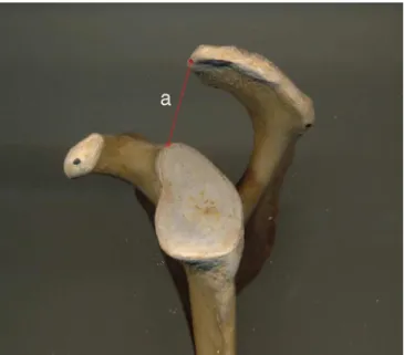
wasbetweenthestraightlinesGandH;theangleˇbetweenF andD;andtheanglebetweenAandD.Theseanglesprovide detailednotionsofthesubacromialspace.Thevariationsin theirmeasurementsprovideanideaofthebehavior ofthe anglingoftheacromionandconsequentlyofthenarrowingor wideningofthesubacromialspace.Thus,wealsomeasured thedistanceDA(Fig.2),whichevaluatedthedistancebetween thebonestructures(acromionandsupraglenoidtubercle).
Figure2–Distancefromtheanteroinferiorextremityofthe acromiontothesupraglenoidtubercle.
Theacromiawerealsoclassifiedinaccordancewiththe modelproposedbyBiglianietal.5(Fig.3).
Thedataobtainedweresubjectedtostatisticalanalysisby meansoftheTukeytest,aftertheFtesthadshownsignificance fortheanalysisofvariance,withasignificancelevelof5%.The distributionofthetypesofacromioninrelationtosexandage wasanalyzeddescriptively.
Results
Ninetydryscapulasfromhumancadavers(90acromia)were studied.Outofthistotal,39(43.3%)wereoftypeI,43(47.7%) oftypeIIandeight(9%) oftypeIII.Themean agesforthe threetypesofacromion(I-III)were45.6,55.2and51.1years, respectively.
Inrelationtosex,therewasvariationinthepercentages relatingtodifferenttypesofacromion(Table1).
Table2showsthedistributionofthetypesofacromionin relationtoage(≤49and≥50years).
To analyze the curvatureofthe acromion,we used the angles˛,ˇandthatweproposed.Weobservedthattheangles
ˇ and presenteda decreasingpattern ofmeans, in com-parisonwiththeacromiontypesI,IIandIII,respectively.On
Table1–Distributionofthetypesofacromionaccording tosex.
Sex TypeI TypeII TypeIII
Female(n=36) 20(55.5%) 10(27.7%) 6(16.8%)
Right 10 5 3
Left 10 5 3
Male(n=54) 19(35.1%) 33(61.1%) 2(3.8%) Right 11 15 1
Left 8 18 1
Total(n=90) 39(43.3%) 43(47.7%) 8(9%)
Table2–Distributionofthetypesofacromionin relationtoageandsex.
Age TypeI TypeII TypeIII
≤49years(n=40) 20(50%) 15(37.5%) 5(12.5%)
Male 9 10 1
Female 11 5 4
≥50years(n=50) 19(38%) 28(56%) 3(6%)
Male 10 23 1
Female 9 5 2
theotherhand,fortheangle˛anditsrelationshipswiththe acromiontypes,wedidnotobserveanyuniformityofbehavior amongitsmeans.
InanalyzingthebehaviorofthedistanceDA,wenoticed thatincreasingcurvatureoftheacromion(fromtypeItotype III)reducedthespacebetweentheanteroinferiorextremityof theacromionandthesupraglenoidtubercle.
Anglesˇ,and˛andthedistanceDAwerecorrelatedwith thetypesofacromion.Thisdescriptiveanalysisispresented inTable3.
Theevaluationofthemeans foreach oftheanglesand forthedistanceDA,inrelationtoeachofthethreeacromion types(Table4)showedthattherewerestatisticallysignificant differencesinthemeansfortheangleˇ(p=0.008),theangle
(p=0.028)andthedistanceDA(p=0.037).Ontheotherhand, fortheangle˛,thedifferenceswerenotstatisticallysignificant (p=0.810).
AccordingtotheTukeytest,thereweredifferencesinthe meansfortheanglesˇand,betweenacromiontypesIandIII, andalsobetweenthemeansforthedistanceDAwithregard totypeIIIandtheothertypesofacromion(IandII).
Table4–MeansoftheanglesandthedistanceDAin relationtoeachtypeofacromion.
Mean Standard deviation
F p-Value
ˇ
TypeI 41.9 4.8 5.50 0.008 TypeII 39.3 4.1
TypeIII 33.6 6.9
TypeI 77.5 8.9 3.88 0.028 TypeII 73.8 8.7
TypeIII 63.7 13.9
˛
TypeI 40.3 8.6 0.21 0.810 TypeII 41.7 8.2
TypeIII 39.3 7.6
Distance
TypeI 29.3 4.5 3.56 0.037 TypeII 28.7 3.8
TypeIII 23.1 6.1
DA,distancefromtheanteroinferiorextremityoftheacromionto thesupraglenoidtubercle.
Discussion
Thesubacromialimpactsyndromeisdirectlyrelatedtothe degreeofinclinationoftheacromion.Thus,variationsinits curvaturechangethedimensionsofthespacebelowthe cora-coacromial arch and may cause lesions of the anatomical structurescontainedinthisregion,especiallythetendonsof therotatorcuff.3
Inthisregard,Biglianietal.5presentedaschemefor clas-sifyingtheacromioninaccordancewiththecurvatureofits lower surface. The hooked type was seen to have a close relationshipwithISandRCIs.5Subsequentstudieshave con-firmedthiscorrelation.4,8,12–16
Otherauthorshaverecognizedtheimportanceofthis clas-sificationandhaveusedittoestablishthefrequencyofeach type of acromion in different populations. In the original description,Biglianietal.5foundthefollowingproportionsfor eachtypeofacromion:straight,17%;curved,43%;andhooked, 39%.
Amongtheother authorswho haveusedthis classifica-tion,theproportionsofeachtypeofacromioninthedifferent
Table3–DescriptiveanalysisontheanglesandthedistanceDAinrelationtoacromiontypesI,IIandIII.
Type TypeI TypeII TypeIII
ˇ ˛ DA ˇ ˛ DA ˇ ˛ DA
Mean 41.9 77.5 40.3 29.3 39.3 73.8 41.7 28.7 33.6 63.7 39.3 23.1 Median 40.4 74.7 38.9 29.0 40.0 75.3 40.9 27.5 35.1 64.5 37.8 23.7 Variance 23.0 79.5 73.6 19.9 17.0 75.5 67.8 14.6 47.0 193.0 57.9 37.8 SD 4.8 8.9 8.6 4.5 4.1 8.7 8.2 3.8 6.9 13.9 7.6 6.1 Minimum 33.4 59.5 22.8 21.0 30.4 51.6 28.8 22.6 24.1 49.8 32.2 16.3 Maximum 51.8 92.7 63.4 36.0 47.3 84.5 57.5 37.7 40.2 76.2 49.3 28.6
studieshavevariedwidely:5.4–67.7%fortypeI;24.2–83%for typeII;and0–42.4%fortypeIII.Theredataareinthereview oftheliteraturebyNatsisetal.10Inourstudy,thefrequency ofeachtypeofacromionusingtheclassificationofBigliani etal.5was43.3%fortypeI;47.7%fortypeII;and9%fortypeIII. Thislargevariabilitybetweenthedifferent authorsmay reflectthesubjectivenatureoftheclassificationmethod,the typeofsample(dryscapulas,scapulasfromcadaversor scapu-lasfrom livingindividuals), thepopulationstudiedandthe methodusedforanalyzingthe acromion(direct inspection, radiographyormagneticresonance).10
Inanalyzingthefrequenciesofthetypesofacromionin relationtoage,wesawthatacromiontypesIIandIIItogether weremorefrequentamongindividualsaged50yearsandover than amongthose aged49 yearsand under: 62%and 50%, respectively.Individualsaged≤49yearsaccountedfor50%of
theacromiaoftypeIand≥50years,only38%.Vähäkarietal.7
studiedradiographsfromasymptomaticindividualsaged21 to71yearsanddidnotperceiveanystatisticallysignificant differencesinthetypesofacromionbetweentheagegroups. Inanalyzingthefrequenciesoftheacromiontypesin rela-tiontosex,wesawthattheproportionoftypeIIIwashigher amongfemales(16.8%versus3.8%),whileParaskevasetal.17 foundagreaterpercentageoftypeIIIinmen(56.2%versus 43.7%)andoftypeIinwomen(56.5%versus43.4%),justlike inourstudy(55.5%versus35.1%).
TheclassificationsystemofBiglianietal.5isveryuseful andwidelyused,butitispurelyvisual,giventhatthe interpre-tationisguidedonlybythemeaningsofthewordsflat,curved andhooked.Thus,this systempresents greatinterobserver variability.
Hence,severalauthorshaveindependentlyformulated dif-ferentmodificationstotheoriginalschemeandhaveproposed analysesofgreateraccuracyfortheacromialcurvature.4,11,13
Epsteinetal.13proposed,alsoonthebasisofvisual clas-sification,thattheacromionshouldbeclassifiedastypeIIif thecurvatureoccurredinthemiddlethirdandastypeIIIif itoccurredintheanteriorthird.Inamoreobjectivemanner thantheseauthors,Toivonenetal.4conducteda retrospec-tivereviewofradiographsinordertodetermineanobjective methodforclassifyingtheshapeoftheacromion.They com-paredthe“objectiveangle”(namedtheacromialangle)with theacromialtypesoftheclassificationofBiglianietal.5and concludedthatacromiontypeIhadanacromialangleof0◦to
12◦;typeII,13◦to27◦;andtypeIII,greaterthan27◦.
Other parametershave been proposed byother authors inordertoanalyze theinfluenceofthemorphologyofthe acromionasanetiologicalfactorforRCIs.Amongthese,the influenceofthethicknessoftheanteriorthirdoftheacromion hasbeencitedasanetiologicalfactorrelatingtosubacromial pathologicalconditions.18
Another morphometricparameter isthe acromial index (AI),whichisobtainedastheratiobetweenthedistancefrom theglenoidcavitytothelateralborderandthedistancefrom theglenoidcavitytothelateralborderofthehumerus.19–21In astudyonaBrazilianpopulation,Miyazakietal.21concluded that RCIs may be associated with greater AI, i.e. greater lateral projection of the acromion. In a subsequent study, Miyazakietal.22comparedtheAIintwodifferentpopulations (BrazilianandJapanese)andconcludedthatthisindexwasa
predictivefactorforRCIsintheBrazilianpopulationbutnot intheJapanesepopulation.
EdelsonandTaitz11proposedaninclinationangleforthe acromionandstatedthatitwasassociatedwithdegenerative alterationssuchthatthemorehorizontaltheacromionwas, thegreaterthedegenerationwouldbe.
Differingfromthatstudy,thepresentstudydidnotanalyze theassociationbetweenacromialcurvatureanddegenerative alterations.Inourview,themainparameteristhecomparison betweentheacromialinclinationandthealterationstothe sizeofthesubacromialspace.
EdelsonandTaitz11alsoanalyzedtheheightoftheheight ofthecoracoacromialarchandperceivedthatthiswasalso associated with degenerative alterations to the acromion. Theseauthorsdidnotfinddegenerationinarchesthatwere morethan15mmabovethesupraglenoidtubercle,but75%of theirsamplewithdegenerativealterationshadanarchheight oflessthan12mm.
LikeinthestudybyEdelsonandTaitz,11weproposedto analyzetheheightofthecoracoacromialarch,butbyusingthe anglesthatwecreated,inordertounderstandtheextentto whichvariationinacromialcurvatureiscapableofalteringthe spacebetweentheanteroinferiorextremityoftheacromion andthesupraglenoidtubercle.Ourevaluationonthemeans for the distance DA between the three different types of acromionshowedstatisticalsignificance(p=0.037).Inrelation toeachofthethreetypesofacromion,weshowedthatthere weredifferencesinthemeansfortheanglesˇ(p=0.008)and
(p=0.028).Inotherwords,amongtheanglesproposedinour study,ˇandmaintainedastrictrelationshipwiththelength ofthesubacromialspace.
One important factor is that measurements similar to those on samples ofdry bone can be obtained by means of radiographs in lateral view of the scapula, i.e. “outlet view”.11,23,24Likewise,theanglesandheightofthe coracoacro-mialarchcanbecalculated.
Conclusion
The beta and theta angles proposed in our study can be usedformorphometricanalysisontheacromion,especially regardingitscurvature,therebycontributingtowardsstudies ondiseasesoftheshoulderandaidinginsurgicalplanningand inanalysisonacromialinclinationbymeansofradiography andmagneticresonance.
Conflicts
of
interest
Theauthorsdeclarenoconflictsofinterest.
r
e
f
e
r
e
n
c
e
s
2. BalkeM,SchmidtC,DedyN,BanerjeeM,BouillonB,LiemD. Correlationofacromialmorphologywithimpingement syndromeandrotatorcufftears.ActaOrthop.
2013;84(2):178–83.
3. MusilD,Sadovsk ´yP,RostM,StehlíkJ,FilipL.Relationshipof acromialmorphologyandrotatorcufftears.ActaChirOrthop TraumatolCech.2012;79(3):238–42.
4. ToivonenDA,TuiteMJ,OrwinJF.Acromialstructureandtears oftherotatorcuff.JShoulderElbowSurg.1995;4(5):376–83.
5. BiglianiLU,MorrisonDS,AprilEW.Themorphologyofthe acromionanditsrelationshiptorotatorcufftears.Orthop Trans.1986;10:228.
6. IkemotoRY,BezerraAD,MonteFA,TellesRB,FujikiEN,Porto LCK.Acrômioemformadegancho:umavariac¸ãoanatômica ouumprocessodegenerativo?RevBrasOrtop.
1995;40(8):454–63.
7. VähäkariM,LeppilahtiJ,HyvönenP,RistiniemiJ,Päivänsalo M,JalovaaraP.Acromialshapeinasymptomaticsubjects:a studyof305shouldersindifferentagegroups.ActaRadiol. 2010;51(2):202–6.
8. SpencerEEJr,DunnWR,WrightRW,WolfBR,SpindlerKP, McCartyE,etal.Interobserveragreementintheclassification ofrotatorcufftearsusingmagneticresonanceimaging.AmJ SportsMed.2008;36(1):99–103.
9. KarasV,ColeBJ,WangVM.Roleofbiomechanicsinrotator cuffpathology:North-Americanperspective.MedSportSci. 2012;57:18–26.
10.NatsisK,TsikarasP,TotlisT,GigisI,SkandalakisP,AppellHJ, etal.Correlationbetweenthefourtypesofacromionandthe existenceofenthesophytes:astudyon423driedscapulas andreviewoftheliterature.ClinAnat.2007;20(3): 267–72.
11.EdelsonJG,TaitzC.Anatomyofthecoraco-acromialarch: relationtodegenerationoftheacromion.JBoneJointSurgBr. 1992;74(4):589–94.
12.OhJH,KimJY,LeeHK,ChoiJA.Classificationandclinical significanceofacromialspurinrotatorcufftear:heel-type spurandrotatorcufftear.ClinOrthopRelatRes.
2010;468(6):1542–50.
13.EpsteinRE,SchweitzerME,FniemanBG,FenlinJN,Mitchell DG.Hookedacromion:prevalenceonMRimagesofpainful shoulders.Radiology.1993;187(2):479–81.
14.JosephCMG,SundeepA,SandipB.Rotatorcufftears: associationwithacromionangulationonMRI.ClinImaging. 2012;36(6):791–6.
15.DiMarioM,FraracciL.MRstudyoftheintrinsicacromial anglein74symptomaticpatients.RadiolMed.
2005;110(3):273–9.
16.NikolaosTR,SoheilM,SuketuV,EdwardE,TheofilosSK,John MI.Theinfluenceoftheacromioclavicularjointdegeneration onsupraspinatusoutletimpingementandtheacromion shape.JOrthopSurg.2009;17(3):331–4.
17.ParaskevasG,TzaveasA,PapaziogasB,KitsoulisP,NatsisK, SpanidouS.Morphologicalparametersoftheacromion.Folia Morphol(Warsz).2008;67(4):255–60.
18.SnyderSJ,KarzelRP,DelPizzoW,FerkelRD,FriedmanMJ. SLAPlesionsoftheshoulder.Arthroscopy.1990;6(4):274–9.
19.NyffelerRW,WernerCM,SukthankarA,SchmidMR,GerberC. Associationofalargelateralextensionoftheacromionwith rotatorcufftears.JBoneJointSurgAm.2006;88(4):800–5.
20.TorrensC,LópezJM,PuenteI,CáceresE.Theinfluenceofthe acromialcoverageindexinrotatorcufftears.JShoulderElbow Surg.2007;16(3):347–51.
21.MiyazakiAN,FregonezeM,SantosPD,SilvaLA,MartelEM, DebomLG,etal.Estudoradiográficodoíndiceacromialesua relac¸ãocomaslesõesdomanguitorotador.RevBrasOrtop. 2010;45(2):151–4.
22.MiyazakiAN,ItoiE,SanoH,FregonezeM,SantosPD,SilvaLA, etal.Comparisonbetweentheacromionindexandrotator cufftearsintheBrazilianandJapanesepopulations.J ShoulderElbowSurg.2011;20(7):1082–6.
23.HyvönenP,PäivänsaloM,LehtiniemiH,LeppilahtiJ,Jalovaara P.SupraspinatusoutletviewinthediagnosisofstagesIIand IIIimpingementsyndrome.ActaRadiol.2001;42(5):441–6.