Urkude Vikas
Mishra Amit
Yadav Mahavir
Tiwari Archana
Analyzing
inhibition
of
BCL11A
gene
expression in K562 cells by RNAi
Authors’ address:
School of Biotechnology, Rajiv Gandhi Proudyogiki Vishwavidyalaya, Bhopal (M.P), India.
Correspondence: Vikas Urkude,
Rajiv Gandhi Proudyogiki
Vishwavidyalaya (State Technological University of Madhya Pradesh), Airport Bypass Road, Bhopal-462033,India. Tel.: +91 755 2742006
E-mail: [email protected]
Article info:
Received: 15 April 2013
Accepted: 21 June 2013
ABSTRACT
RNA interference (RNAi), an effective approach to sequence-specific gene knockdown is widely used for the investigation of regulation of gene expression in various cells. BCL11A (B cell lymphoma 11A) plays a vital role in the evolutionarily different globin gene switches of mammals. In the current study, siRNA complementary to BCL11A was used to inhibit the BCL11A gene expression in erythroleukemic K562 cells and the expression was evaluated through real-time quantitative reverse transcription polymerase chain reaction (qRT-PCR) and western blot analysis. On day 7 of cell culture, 1x10⁶ K562 cells were transfected with lipofectamine containing BCL11A specific siRNA. GAPDH (Glyceraldehyde-3-phosphate dehydrogenase) was used as the reference gene to confirm the relative expression level of BCL11A gene mRNA and BCL11A protein. After 48 h of transfection, BCL11A specific siRNA produced significantly reduction of BCL11A mRNA level in a dose-dependent manner. It also affects the level of BCL11A protein. BCL11A siRNAs were equally effective at reducing the expression level of BCL11A mRNA and protein.
Key words: BCL11A, K562, Thalassemia, RNAi, siRNA
Introduction
RNA interference (RNAi) has recently emerged as a powerful tool for gene knockdown. RNAi has been a recently discovered phenomenon for the analysis of regulation of gene expression in a variety of cells, by which target messenger RNA (mRNA) is cleaved by small interfering complementary RNA (siRNA) (Sarakul et al., 2008). In RNA interference dsRNA facilities the homologous mRNA degradation, in that way causes the diminishing or abolishing gene expression (Fire et al., 1988; Nakayashiki & Nguyen, 2008). The dsRNAs get introduced into the cell and then in the cytoplasm they are processed by an enzyme of the Dicer family into small interfering RNAs (siRNAs). Next, siRNA is assembled into an RNA-induced silencing complex (RISC). Finally, antisense siRNA strands guide the RISCs to complementary RNA molecules, where they cleave and destroy the cognate RNA. This process leaves the genomic DNA intact but suppresses gene expression by RNA degradation.
Beta-thalassemia is an autosomal recessive disorder that affects the body ability to produce protein called hemoglobin. Thalassemia become the most common genetic disorders worldwide; around 4.8γ percent of the world’s population carries globin variants, including 1.67 percent of the population who are heterozygous for α-thalassemia and -thalassemia (Rund & Rachmilewitz, 2005). In India, children born with thalassemia disease reach to 10,000/year. Thalassemia arises due to a quantitative defect in the globin chain production (Chandrashekar & Soni, 2011). Augmented levels of fetal hemoglobin (HbF) can revolutionize the severity of the -hemoglobin disorders, like -thalassemia. Beta-thalassemia is characterized by diverse group of inherited mutations causing odd expression of globin genes, lead to total absence or quantitative reduction in the synthesis of -globin chains.
BCL11A gene encodes a C2H2 type zinc-finger protein, which is similar to the mouse Bcl11a/Evi9 protein. BCL11A on chromosome 2p16 was shown to be a major quantitative trait locus for HbF level and F-cell number in several populations with or without -hemoglobinopathy (Chen et al., 2009). Moreover, BCL11A plays a central role in the evolutionarily different globin gene switches of mammals. As an aspect crucial for gamma-globin gene silencing, BCL11A can supposed to be consider as a beneficial target to increase HbF in a directed manner in beta-thalassemia patients (Sankaran et al., 2010).
K562 is an erythroleukemic cells derived from a chronic myeloid leukemia patient in blast crisis (Ghosh et al., 2013). Towards achieving our goal, in this study we have examined the ability of a siRNA, to inhibit the BCL11A gene expression in vitro in human erythromyeloblastoid leukemia K562 cells and also measure the protein expression level in transfected K562 cells by Western blotting.
Materials and Methods
K562 cells
The human chronic myelogenic leukemia K562 cell line was procured from the National Centre for Cell Science, Pune. The cells were grown in RPMI 1640 (Himedia, AL199S) supplemented with 10% fetal bovine serum, 100 U/mL penicillin, and 1 mg/mL streptomycin. Cultures were incubated at γ7°C and 5% CO2.
siRNA-transfection
BCL11A gene specific siRNA were procured from Santa Cruz Biotechnology, Inc. (sc-43578) along with control siRNA (sc-37007). Control siRNA is recommended as a negative control for evaluating RNAi off-target effects, and in order to verify the accuracy of gene specific siRNA dependent RNAi. siRNA was transfected into cells by lipofectamineTM 2000 (Invitrogen, USA) solution (Dalby et al. 2004). In brief, at 12-24 h before transfection, cultured K562 cells were collected and incubated in 500 μL of cell culture medium without antibiotics and placed into each well of 24-well plate. siRNA was diluted in 50 μL of Opti-MEM I Reduced Serum Medium to the assigned amount (0.2, 0.5, and 1.0 μg). Suitable negative control siRNA were also prepared and diluted in Opti-MEM I Reduced Serum Medium in required amounts. Lipofectamine 2000 was also diluted in 50 μL of Opti-MEM I Reduced Serum Medium and incubated for 5 min at room temperature. The diluted
siRNA and lipofectamine 2000 were mixed and incubated for 20 min at room temperature. Then siRNA-lipofectamine 2000 complexes were added drop by drop to each of the wells containing cells and medium and gently mixed with rocking the plate back and forth. The cells were incubated at γ7°C in the presence of 5% CO2 for 24-96 h at CO2 incubator. At last, the cells were harvested for determination of mRNA level.
Total RNA isolation from the K562 cells
Total RNA was extracted from the K562 cells by using TRIZOL reagent according to manufacturer’s instructions (Invitrogen, USA). TRIZOL reagent is a ready-to-use reagent for the isolation of total RNA from cells and tissues. In RNA isolation the cells were first homogenized, in which the cell pellet was obtained by centrifugation and lysed in TRIZOL reagent by repetitive pipetting. Following homogenization 0.2 mL of chloroform was added per 1 mL of TRIZOL reagent and centrifuged at 12,000 g for 15 minutes at 2 to 8°C. The mixture separates into a lower red, phenol-chloroform phase, an interphase, and a colorless upper aqueous phase. RNA’s were remains exclusively in the aqueous phase. After phase separation RNA precipitation was done by mixing with isopropyl alcohol and incubated at 15 to γ0°C for 10 minutes and then centrifuged at 12,000 g for 10 minutes at β to 8°C. After centrifugation, the RNA pellet was obtained at the side and bottom of the tube. Then the RNA pellet was washed with 75% ethanol, mixed by vortexing and centrifuged at 7,500 g for 5 minutes at β to 8°C. At the end RNA pellet was air dried for 5 to 10 minutes and dissolved in RNase-free water stored at -70°C.
Determination of mRNA level using real time quantitative PCR
acquire) and 72°C for 10 s. Specificity of the PCR products was confirmed by analysis of the dissociation curve. The melting curve analysis program consisted of temperatures 55°C for 15 s with a heating rate of 0.1°C/s and a continuous fluorescence measurement. Target quantities were normalized against standard curve and plotting the threshold cycle of standard against the DNA concentration.
Protein extract preparation and Western blot analysis
Cells were washed with phosphate- buffered saline (PBS) (10 mM, pH 7.4), incubated in 200 ml of RIPA lysis buffer [150 mM NaCl/1% (wt/vol) Nonidet P-40/0.5% Na deoxycholate/0.1% SDS/50 mM Tris·Cl, pH 7.5] on ice for 30 min, and centrifuged at 13,000 g for 45 min at 4°C. The concentration of protein was determined by using absorbance spectroscopy. Whole cell extracts equivalent to 100 μg of total protein were separated by 8% sodium dodecyl sulfate (SDS) polyacrylamide gel electrophoresis and electrotransferred to nitrocellulose membranes. The blot was placed in blocking buffer (10% non‑fat dry milk, 1% Tween‑20; in 20 mM Tris-buffered saline, pH 7.5) for 1 h at room temperature and incubated with appropriate anti-human primary antibody (Santa Cruz Biotechnology; mouse anti-human BCL11A/IgG, 1:1,000; mouse anti‑human GAPDH/IgG, 1:2000) overnight at 4°C. Blots were incubated with anti‑mouse horseradish peroxidase-conjugated secondary antibody (1:3,000) for 1 h and detected by chemiluminescence using ECL Hyperfilm.
Results
Effect of BCL11A siRNAs on the BCL11A expression at the mRNA level
To determine the efficiency of BCL11A inhibition following siRNA treatment, BCL11A expression was analyzed at the mRNA level. After transfecting the K562 cells, the mRNA was extracted and quantitated after 48 h of transfection. Extracted mRNA was reversely transcribed and levels of cDNA were determined by quantitative PCR. The qRT‑PCR data shown include at least three independent experiments. According to the relative qRT‑PCR formula 2‑ΔΔC(t) x100% (Livak & Schmittgen, 2001; Schmittgen & Livak, 2008), the relative expression levels of BCL11A mRNA in K562 cells treated with BCL11A siRNA were significantly lower than cells treated with the control siRNA, at 48 h after transient transfection. By contrast, the control
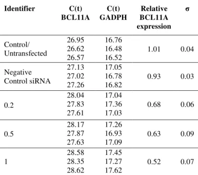
siRNA had a slightly less effect on the expression of BCL11A mRNA. As shown in Table 1, BCL11A siRNA could specifically suppress BCL11A mRNA level in a dose-dependent manner (p<0.05 for all siRNA concentrations compared to both controls), while the level of BCL11A mRNA in negative control was slightly affected. When the mRNA levels were normalized with respect to untransfected control, mRNA level decreased inversely with increasing amounts of used BCL11A siRNA-lipofectamine complex (Figure 1). The knockdown efficiency of each of the BCL11A siRNA are shown in Figure 1 and Table 1 for the three construct: 68%, 63% and 52% respectively.
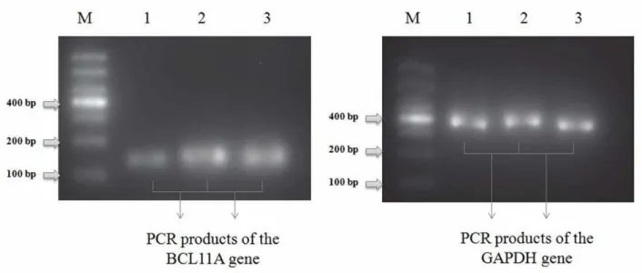
The PCR amplified cDNA was also analyzed in 2% agarose gel electrophoresis, the results revealed that the PCR product of BCL11A gene and GADPH reference gene were specifically amplified and having a size of around 160 and 380 bp, respectively (Figure 2).
BCL11A siRNAs suppressed BCL11A expression at the protein levels in K562 cells
BCL11A protein levels in transfected K562 cells and control K562 cells were assayed by Western blot analysis. Western blot analysis confirmed the results of the qRT‑PCR analysis (Figure 3).
Table 1. Data obtained from qRT-PCR to quantify BCL11A expression in K562 cells following siRNA transfections.
Identifier C(t) BCL11A C(t) GADPH Relative BCL11A expression σ Control/ Untransfected 26.95 26.62 26.57 16.76 16.48 16.52
1.01 0.04
Negative Control siRNA 27.13 27.02 27.26 17.05 16.78 16.82
0.93 0.03
0.2 28.04 27.83 27.61 17.04 17.36 17.03
0.68 0.06
0.5 28.17 27.87 27.63 17.26 16.93 17.09
0.63 0.09
1 28.58 28.35 28.62 17.45 17.27 17.62
Figure 1.
Determination of the levels of BCL11A gene mRNA. The levels of BCL11A mRNA relative to untransfected cell
control were measured. Various amounts of BCL11A siRNA (0.2, 0.5, and 1.0 µg) were transfected into the K562 cells. The
mRNA levels were determined after 48 h incubation. The numbers above of the bars represent the BCL11A/GADPH mRNA ratio. Data are means ± SD of three independent experiments.
Figure 3. Determination of the levels of BCL11A protein in transfected K562 cells by Western blot analysis.
Expression of BCL11A protein significantly decreased in the K562 cell lines treated with BCL11A siRNA as compared with either the negative control siRNA group or untreated cells group. No difference in BCL11A levels among the control siRNA group and non-transfected cells was found. It was found that BCL11A siRNAs were equally effective at reducing the expression level of BCL11A mRNA and protein.
Discussion
Thalassemia is a genetic disorder which occurs due to mutation on alpha and beta globin gene. As a result of this mutation, reduced synthesis of alpha and beta globin chains in the body occurs (Quek & Thein, 2007). Although many of the trials controlling the action of the -globin locus are known, the facts of those regulating normal human hemoglobin switching and reactivation of HbF in adult hematopoietic cells remain to be illuminated. If the molecular trials in hemoglobin switching or -globin gene reactivation were better appreciated and HbF could be totally reactivated in adult cells, the approaching might show the way to cure for these disorders (Sankaran, 2011). The main intention of this study was to observe the knockdown expression of BCL11A gene in K562 cell line by the process of RNA interference. The present investigation firstly advocates that BCL11A plays a very vital role in the evolutionarily different globin gene switches of mammals (Buratowski, 2008). Secondly, RNA interference (RNAi) has been a recently discovered
phenomenon for the analysis of regulation of gene expression in a variety of cells, by which target messenger RNA (mRNA) is cleaved by small interfering complementary RNA (siRNA) (Vikas et al., 2012). The technique is now established in in-vitro systems, and much work is focused on adapting RNAi for in vivo application.
thalassemic cells in vitro, and may possibly be applied to increase the fetal Hb levels in severe forms of -thalassemia in a clinical setting.
This study has accentuated several factors regarding RNAi-based knockdown experiments. Firstly, accessibility of the mRNA target site to RNAi activity is essential. Secondly, off-target knockdown effects, as well as cytotoxicity arising from the RNAi process, the transfection conditions or target knockdown must be carefully considered. Thirdly, while qRT-PCR is an immensely sensitive and powerful technique, it can give misleading results if reliable reference genes and controls are not used. Further investigations can be done for the results obtained in this study on in vivo approach which can give us more significant conclusions.
Acknowledgement
The author is grateful to Mr. Amit Mishra, Dr. Mahavir Yadav and Dr. Archana Tiwari for given me the opportunity to study the interesting work and also provided generous support in the preparation of this manuscript. The authors are also grateful to the Rajiv Gandhi Proudyogiki Vishwavidyalaya for providing the necessary facilities to carry out the study. We thank to all our colleagues for the constructive suggestions for the current research.
References
Buratowski S. 2008. Transcription. Gene expression - where to start?. Science, 322(5909): 1804-1805.
Cao A, Moi P, Galanello R. 2011. Recent advances in beta-thalassemias. Pediatr. Rep., 3(2): e17.
Chandrashekar V, Soni M. 2011. Hemoglobin disorders in South India. ISRN Hematol., 2011:748939.
Chen Z, Luo HY, Steinberg MH, Chui DH. 2009. BCL11A represses HBG transcription in K562 cells. Blood Cells Mol. Dis., 42(2): 144-149.
Dalby B, Cates S, Harris A, Ohki EC, Tilkins ML, Price PJ, Ciccarone VC. 2004. Advanced transfection with lipofectamine 2000 reagent: primary neurons, siRNA, and high-throughput applications. Methods, 33(2): 95-103.
Fire A, Xu S, Montgomery MK, Kostas SA, Driver SE, Mello CC. 1998. Potent and specific genetic interference by double-stranded RNA in Caenorhabditis elegans. Nature, 391(6669): 806-811.
Ghosh D, Saha C, Hossain M, Dey SK, Kumar GS. 2013. Biophysical studies of mutated K562 DNA (erythroleukemic cells) binding to adriamycin and daunomycin reveal that mutations induce structural changes influencing binding behavior. J. Biomol. Struct. Dyn., 31(3): 331-341.
Livak KJ, Schmittgen TD. 2001. Analysis of relative gene expression data using real-time quantitative PCR and the 2(-Delta 2(-Delta C(T)) Method. Methods, 25(4): 402-408.
Nakayashiki H, Nguyen QB. 2008. RNA interference: roles in fungal biology. Current Opinion in Microbiology, 11(6): 494-502.
Quek L, Thein SL. 2007. Molecular therapies in beta-thalassaemia. Br. J. Haematol., 136(3): 353-365.
Rund D, Rachmilewitz E. 2005. Beta-thalassemia. N. Engl. J. Med., 353(11): 1135-1146.
Sankaran VG, Xu J, Orkin SH. 2010. Transcriptional silencing of fetal hemoglobin by BCL11A. Ann. N. Y. Acad. Sci., 1202: 64-68.
Sankaran VG. 2011. Targeted therapeutic strategies for fetal hemoglobin induction. Hematology Am. Soc. Hematol. Educ. Program, 2011: 459-465.
Sarakul O, Vattanaviboon P, Wilairat P, Fucharoen S, Abe Y, Muta, K. 2008. Inhibition of alpha-globin gene expression by RNAi. Biochem. Biophys. Res. Commun., 369(3): 935-938.
Schmittgen TD, Livak KJ. 2008. Analyzing real-time PCR data by the comparative C(T) method. Nat. Protoc., 3(6): 1101-1108. Vikas U, Amit M, Mahavir Y, Archana T. 2012. RNA Silencing: An