J. bras. pneumol. vol.34 número4 en v34n4a10
Texto
Imagem
Documentos relacionados
The probability of attending school four our group of interest in this region increased by 6.5 percentage points after the expansion of the Bolsa Família program in 2007 and
In B, axial contrast-enhanced CT of the chest scan showing a heterogeneous hypervascular mass (asterisk) iniltrating the right serratus anterior and pectoralis muscles, as well as
At that point, the patient was submitted to a computed tomography scan of the chest, which revealed that the areas of consolidation, although smaller in size, persisted in both
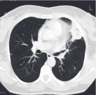
A computed tomography scan of the chest revealed a 4-cm mass with heterogeneous content and pleural extension to the level of the lingula, as well as two micronodules in
At that point, the patient was submitted to a computed tomography scan of the chest, which revealed that the areas of consolidation, although smaller in size, persisted in both
A computed tomography scan of the chest revealed atelectasis of the right upper lobe (caused by occlusion of the upper lobe bronchus) that extended up to the juxtacarinal portion of
The working diagnosis was superior vena cava syndrome, and the patient was submitted to computed tomography of the chest, which revealed a mass in the anterior mediastinum, with
Computed tomography of the chest revealed consolidation with interposed cavitation in the right upper lobe.. Fiberoptic bronchoscopy revealed purulent fluid within
