Role of nuclear factor kappa B and
reactive oxygen species in the
tumor necrosis factor-
α
α
α
α
α
-induced
epithelial-mesenchymal transition
of MCF-7 cells
Department of General Surgery, Tangdu Hospital,
the Fourth Military Medical University, Xi’an, Shaanxi Province, China R. Dong, Q. Wang,
X.L. He, Y.K. Chu, J.G. Lu and Q.J. Ma
Abstract
The microenvironment of the tumor plays an important role in facili-tating cancer progression and activating dormant cancer cells. Most tumors are infiltrated with inflammatory cells which secrete cytokines such as tumor necrosis factor-α (TNF-α). To evaluate the role of TNF-α in the development of cancer we studied its effects on cell migration with a migration assay. The migrating cell number in TNF-α-treated group is about 2-fold of that of the control group. Accordingly, the expression of E-cadherin was decreased and the expression of vimen-tin was increased upon TNF-α treatment. These results showed that TNF-α can promote epithelial-mesenchymal transition (EMT) of MCF-7 cells. Further, we found that the expression of Snail, an important transcription factor in EMT, was increased in this process, which is inhibited by the nuclear factor kappa B (NFκB) inhibitor aspirin while not affected by the reactive oxygen species (ROS) scavenger N-acetyl cysteine. Consistently, specific inhibition of NFκB by the mutant IκBα also blocked the TNF-α-induced upregulation of Snail promoter activity. Thus, the activation of NFκB, which causes an increase in the expression of the transcription factor Snail is essential in the TNF-α-induced EMT. ROS caused by TNF-α seemed to play a minor role in the TNF-α-induced EMT of MCF-7 cells, though ROS per se can promote EMT. These findings suggest that different mechanisms might be responsible for TNF-α- and ROS-induced EMT, indicating the need for different strategies for the prevention of tumor metastasis induced by different stimuli.
Correspondence
J.G. Lu and Q.J. Ma Department of General Surgery Tangdu Hospital
Fourth Military Medical University Xi’an 710038, Shaanxi Province China
E-mail: [email protected]
Received January 24, 2007 Accepted May 21, 2007
Key words
•Tumor necrosis factor-α
•Nuclear factor kappa B •Reactive oxygen species •Snail
•E-cadherin
•Epithelial-mesenchymal
transition
Introduction
The microenvironment of the tumor plays an important role in facilitating cancer pro-gression and activating dormant cancer cells (1). One feature of many solid tumors is the
focal macrophage infiltration has been linked to increased angiogenesis in human breast and colorectal cancer (2,3). Yet, the func-tional significance of cytokines produced in situ by inflammatory and tumor cells is still unclear. Another feature of many tumors is the hypoxia of the tumor cells and the result-ing production of reactive oxygen species (ROS) (4,5). We reasoned that some of the cytokines released from these cells and hy-poxia might serve to enhance the invasive step in breast carcinogenesis through de-fined signaling pathways. Many epithelial tumors undergo an epithelial-mesenchymal transition (EMT) that facilitates their inva-sion. It has been reported that either ROS or nuclear factor kappa B (NFκB) could facili-tate the EMT in certain cell types (6-10) and tumor necrosis factor-α (TNF-α), one of the major factors released from the inflamma-tory cells, could cause NFκB activation and ROS production (11). It has been reported that TNF-α can promote EMT in certain cell types, but the precise mechanism is still unclear (12). Based on the importance of the EMT in carcinoma progression and the preva-lence of TNF-α and hypoxia in the presence of tumors, the present study was designed to determine whether TNF-α and ROS are ca-pable of facilitating EMT and to understand the mechanisms involved. We report that MCF-7 cells undergo an EMT conversion from a relatively benign cancer to a migra-tory phenotype in response to TNF-α. Fur-thermore, our data suggest that this EMT transition is mainly due to the activation of NFκB rather than ROS stimulated by TNF-α. Overall, our finding has important
impli-cations for our understanding of how TNF-α contributes to tumor development.
Material and Methods
Cell culture and material
MCF-7 cells, originally obtained from the American Type CultureCollection, were
maintained in culture in a 37ºC incubator with 5% CO2 in RPMI 1640 medium
supple-mented with 10% fetal bovine serum (FBS). All antibodies (anti-vimentin, anti-E-cad-herin, anti-actin) used were purchased from Santa Cruz Biotechnology, Inc., Santa Cruz,
CA, USA. TNF-α and the ROS-quenching
agent N-acetyl cysteine (NAC) were pur-chased from Sigma (St. Louis, MO, USA).
Cell migration assay
The cell migration assay was done as previously described (13). MCF-7 cells were grown to 80% confluence, serum-starved overnight in RPMI 1640 medium and tryp-sinized and washed in serum-free RPMI 1640 medium before plating onto a 12-well Trans-well plate (12 mm in diameter, 8-µm pore size; Corning Incorporated Costar, Lindfield, NSW, Australia). Twelve-well transwell chambers were incubated with RPMI 1640 medium containing 0.01% bovine serum al-bumin and 0.01% FBS overnight. Cells (5 x 10⁴) were added to the upper well, which
was placed inside a lower well containing RPMI 1640 medium, 10% FBS, and various agents indicated in the Results section. After incubation for 24 h, the filter was removed and cells remaining on the upper membrane surface were scraped off. MCF-7 cells that had migrated to the lower side of the filter were fixed in methanol at 4ºC for 15 min, stained with Toluidine blue and counted un-der a microscope for quantitation of MCF-7 migration.
RT-PCR
RT-PCR was performed as previously described (14). At the beginning of each experiment, cells growing in the log phase were plated onto a 6-well dish at a density of 3 x 10⁵ cells/well and then treated with 10
for extraction of RNA, and 2 µg total RNA was used to prepare cDNA in a 25-µL sys-tem (TRizol, Invitrogen, Carlsbad, CA, USA; MLV Reverse transcriptase, Promega, Madi-son, WI, USA). PCR was then performed on 1 µL cDNA with the following primers: Snail forward, GGGCAGGTATGGAGA GGAAGA; Snail reverse, TTCTTCTGCGC TACTGCTGCG; E-cadherin forward, CAG CACGTACACAGCCCTAA; E-cadherin reverse, GCTGGCTCAAGTCAAAGTCC; GAPDH forward, CCTGGCCAAGGTCAT CCATGAC; GAPDH reverse, CATGTAGG CCATGAGGTCCACCAC.
Construction of the Snail promoter reporter and luciferase assay
A fragment of about 800 bp upstream to the translational start site of Snail was ampli-fied by PCR using the following set of prim-ers: forward CCAGATCTCAAAGCACACT TCCCTTTGCATTG; downward GGGCCA TGGTGGTCGAGGCACTGGGGTC. PCR products were confirmed by sequencing and digested with the indicated restriction nu-cleases before cloned into the promoterless and enhancerless pGL3 Basic vector (Pro-mega). Cells were plated at a density of 104/
well onto a 24-well dish and transiently trans-fected with 300 ng 3X κB luc (which con-tains multiple NFκB sites upstream to the minimal promoter-luciferase gene) or Snail reporter or the combination of Snail reporter and the mutant IκB expression vector in the indicated groups. Fifty nanograms of pBIND vector (Promega) was co-transfected as an internal control. Ten nanograms/mL TNF-α, 50 µM H2O2, 5 mM aspirin, or 5 mM NAC, or different combinations and the con-trol agents were added to the indicated wells 4 h post-transfection, and cells were har-vested 24 h after the transfection for the luciferase activity test. Experiments for each treatment were performed in triplicate. Lu-ciferase activity was assessedusing the Pro-mega luciferase assay system. The luciferase
activity of eachlysis was measured and then normalized to the activity of Renilla driven by the constitutive expression promoter in the vector of pBIND. Fold induction was calculated relativeto the activity observed with the basic pGL3 vector alone.
Western blot analysis
After stimulation, whole cells were washed with cold phosphate-buffered saline and lysed in 10 mM Tris-HCl, pH 7.4, 50 mM NaCl, 5 mM ethylenediamine tetraacetic acid, 1% Nonidet P-40, and 10 µg/mL phen-ylmethylsulfonyl fluoride. Cell extracts were transferred to microcentrifuge tubes, mixed, and left on ice for 10 min. After one freeze/ thaw cycle, they were centrifuged at 12,000
g for 5 min at 4oC. Supernatant samples were
subjected to SDS-PAGE and then transferred to nitrocellulose by electrophoresis. Blots were incubated with primary antibodies (anti-vimentin, anti-E-cadherin, anti-actin) in Tris-buffered saline-Tween-20 plus 2.5% skim milk overnight. After serial washes with Tris-buffered saline-Tween-20, membranes were incubated with the secondary antibody. Im-munoreactive bands were visualized using a peroxidase-conjugated secondary horserad-ish antibody and subsequent ECL detection (Amersham Pharmacia Biotech, Bucks, UK).
Statistical analysis
Data are reported as mean ± SD and the Student t-test was applied for statistical anal-ysis, with the level of significance set at P < 0.05.
Results
TNF-ααααα promotes cell migration
Figure 1A, the number of migrating cells doubled under treatment with TNF-α com-pared to control. In addition, we tested the level of vimentin expression in MCF-7 cells. In the control group, vimentin could rarely be detected while in the TNF-α-treated group it was significantly enhanced (Figure 1B). Consistent with this, the results of the wound-ing assay revealed that a migration distance about 100 µm longer occurred after MCF-7 cell exposure to TNF-α for 24 h (data not shown).
TNF-ααααα increased the transcription of Snail in
an NFκκκκκB-dependent way
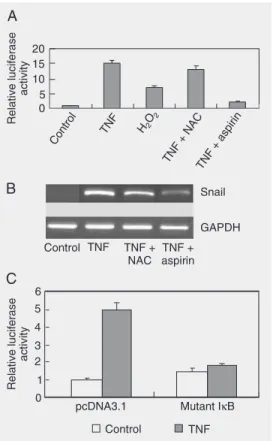
In light of the important roles of NFκB and ROS in EMT, we raised the possibility that NFκB or ROS function importantly in the migration of MCF-7 cells induced by TNF-α. It has also been reported that a re-duced level of E-cadherin expression has a pivotal role in EMT. Thus, we proposed that NFκB or ROS may bridge TNF-α and EMT through E-cadherin. To examine this possi-bility, we tested the expression level of Snail, the transcription repressor of E-cadherin, in the different treatments. We treated cells with the combination of TNF-α plus NAC or aspirin, which were supposed to inhibit the production of ROS and the activation of NFκB, respectively. First, the effect of NAC and aspirin was confirmed by the 3X κB luc reporter assay. As shown in Figure 2A, NAC had little effect on NFκB activity while aspi-rin almost totally blocked the activation of NFκB induced by TNF-α. The results in Figure 2B reveal that the transcription of Snail was enhanced by TNF-α and can be inhibited by the NFκB inhibitor aspirin while it cannot be inhibited by NAC, pointing to the possibility that NFκB was the main me-diator. In contrast, H2O2 induced
up-regula-tion of Snail which was only mildly inhib-ited by aspirin (data not shown). Since aspi-rin has other functions in addition to being an inhibitor of activated NFκB, we also
car-Figure 1. Tumor necrosis fac-tor-α (TNF-α)-induced
epitheli-al-mesenchymal transition in MCF-7 cells. A, In Boyden chamber assays, cells were treated with TNF-α (10 ng/mL)
or the control PBS and incu-bated for 24 h. Cells migrating to the lower side of the filter were counted. The number of migrating cells in the TNF-α
-treated group was significantly larger than control (P < 0.05, Student t-test). Experiments for each treatment were performed in triplicate. Data are reported as means ± SD. B, MCF-7 cells were treated with TNF-α or the
control. Protein was isolated 24
h later, and the levels of vimentin were determined by Western blot. TNF-α significantly
increased the expression of vimentin in MCF-7 cells. Results are representative of two independent experiments.
Figure 2. Tumor necrosis
factor-α (TNF-α) increased the
expres-sion of Snail in a nuclear factor kappa B (NFκB)-dependent way.
A, MCF-7 cells were transfected with 300 ng 3X κB luc and 50 ng
pBIND. Ten nanograms/mL TNF-α, 50 µM H2O2, 5 mM
aspi-rin, 5 mM N-acetyl cysteine (NAC) or different combinations and the control agents were added to the indicated wells 4 h post-transfection, and cells were harvested 24 h after transfection for the luciferase activity test. Aspirin (5 mM) potently blocked the TNF-α- and H2O2-induced
activation of NFκB (P < 0.05).
NAC had no obvious effect on the activation of NFκB induced
by TNF-α. Experiments for each
treatment were performed in triplicate. Results are reported as means ± SD. B, MCF-7 cells were pretreated with aspirin or NAC for 2 h or control. Cells were then incubated with TNF-α
or control. RNA was isolated 24 h later, and the levels of Snail and glyceraldehyde-3-phos-phate dehydrogenase (GAPDH)
were determined by RT-PCR. Similar results were obtained in three independent experi-ments. C, MCF-7 cells were co-transfected with Snail promoter reporter, the internal control vector, and the IκB mutant or the control vector pcDNA3.1 and treated with TNF-α 6 h after
transfection. Results showed that the IκB mutant nearly blocked the induction of Snail
ried out the following experiment. Snail pro-moter was constructed and transfected into MCF-7 cells. As shown in Figure 2C, TNF-α significantly enhanced the promoter
activ-ity of Snail which can be blocked by co-transfection with the mutant IκB expression vector. These data are consistent with previ-ous reports showing that the Snail promoter is regulated by NFκB (15). Finally, we ob-served the expression of E-cadherin both at the mRNA level and at the protein level. In agreement with the fluctuating expression of Snail, a reverse trend to expression of E-cadherin was observed both at the mRNA and protein level (Figure 3A and B). In addition, H2O2 induced down-regulation of
E-cadherin which was only mildly inhibited by the NFκB inhibitor. Notably, the present results do not exclude a direct effect of acti-vated NFκB on E-cadherin itself, which may occur through loss of Snail function.
Inhibition of NFκκκκκB blocked the TNF-ααααα
-induced migrating ability of MCF-7 cells
On the basis of the above data, we further tested the effects of aspirin and NAC on the
TNF-α-induced EMT in MCF-7 cells and
observed that it was aspirin rather than NAC that blocked the enhanced migrating ability of MCF-7 cells induced by TNF-α. In con-trast, the increased migrating ability induced by H2O2 was only mildly inhibited by the
NFκB inhibitor aspirin (Figure 4).
Discussion
The initial process in the metastatic spread of breast carcinomas involves invasion by malignant cells through the extracellular matrix of a basement membrane, followed by their migration into lymphatic or vascular channels (16). The acquisition of migratory properties and weakening of cell-cell adhe-sion are imperative for tumor cell metasta-sis.
The continued expression and functional
Figure 3. Tumor necrosis
factor-α (TNF-α) decreased the
ex-pression of E-cadherin in a nu-clear factor kappa B (NFκ
B)-de-pendent way. MCF-7 cells were pretreated with aspirin, N-acetyl cysteine (NAC) or the control agent for 2 h. Cells were then incubated with TNF-α, H2O2 or
control. RNA and protein were isolated 24 h later, and the lev-els of E-cadherin and glyceral-dehyde-3-phosphate dehydro-genase (GAPDH) were deter-mined by RT-PCR (A) and West-ern blot (B). Similar results were obtained in three independent experiments.
Figure 4. Inhibition of nuclear factor kappa B (NFκB) but not
of reactive oxygen species pre-vents tumor necrosis factor-α
(TNF-α)-induced migration. In
Boyden chamber assays, MCF-7 cells were pretreated with as-pirin, N-acetyl cysteine (NAC) or control agent for 2 h in the volume indicated in Material and Methods. Cells were then incu-bated with TNF-α, H2O2 or
con-trol for an additional 24 h. Cells migrating to the lower side of the filter were counted. TNF-α
-induced MCF-7 migration was inhibited by the NFκB inhibitor
aspirin (P < 0.05, Student t-test). Inhibition of reactive oxygen species production by NAC did not alter TNF-α-induced migration (P > 0.05, Student t-test). H2O2-induced migration
was only moderately inhibited by the inhibition of NFκB (P < 0.05, Student t-test).
Experi-ments for each treatment were performed in triplicate. Data are reported as means ± SD. activity of E-cadherin are required for cells
to remain tightly associated in the epithe-lium. In the absence of E-cadherin, many other cell adhesion and cell junction proteins expressed in epithelial cells are unable to support intercellular adhesion. The central role of E-cadherin in epithelia is demon-strated by the fact that loss of either its expression or function results in the dissolu-tion of the epithelial architecture and the acquisition of a mesenchymal phenotype. This process, referred to as the EMT, occurs within the context of development and
tu-E-cadherin
GAPDH
Control TNF H2O2 TNF
+ NAC
H2O2
+ aspirin
TNF + aspirin
Control TNF H2O2 TNF
+ NAC
H2O2
+ aspirin
TNF + aspirin
A
E-cadherin
Actin
mor progression (17).
Although MCF-7 breast cancer cells are poorly invasive (18), we show that exposure of these cells to TNF-α leads to increased motility. NFκB plays a central role in the EMT. It has been reported that NFκB can facilitate EMT through transcriptional regu-lation of Snail, a transcriptional repressor of E-cadherin (12). It has also been reported that ROS could also promote EMT by acti-vating Rac and the downstream cascade (19).
ROS and NFκB can cross-talk with each
other (20). Cytotoxic ROS signaling appears to be mediated in part by activation of the c-Jun-N-terminal kinase mitogen-activated protein kinase cascade, while in some sys-tems ROS lead to activation of NFκB. Re-markably, new evidence has unveiled the existence of a reciprocal negative control that NFκB exerts on ROS and the c-Jun-N-terminal activities ROS and NFκB have a complicated interaction in the TNF-α sig-naling. On the one hand, NFκB can inhibit ROS production and on the other ROS can function both as an inhibitor and as an acti-vator of NFκB depending on the context, complicating the roles of ROS and NFκB in
the TNF-α-induced EMT. In the present
study, we observed that NFκB plays a vital role in this process through its transcrip-tional regulation of the expression of Snail and then of E-cadherin. We cannot rule out the possibility that other factors function
importantly in the TNF-α-induced EMT,
such as cyclooxygenase-2 (COX-2), an in-ducible enzyme involved in prostaglandin (including prostaglandin E2) biosynthesis, which is overexpressed in several epithelial malignancies including breast cancer and up-regulated under the NFκB signal, while COX-2 plays an important role in metastasis (21-23). It is also possible that other proteins affected by NFκB are vital for EMT and that other factors such as protein kinase or other signal molecules function downstream from, or synergistic or parallel with, the NFκB pathway. All of these hypotheses still need
to be tested. The ROS caused by TNF-α
seemed to play a minor role in the TNF-α -induced EMT of MCF-7 cells, though ROS can promote EMT by themselves, suggest-ing a complicated role of ROS in EMT. On the one hand, this can be explained by the fact that the ROS produced by TNF-α are different in quantity, time and compartment from those produced by treatment with ex-ogenous H2O2 or under hypoxia or other
The present study also pointed out that H2O2 alone can promote EMT in a way
dif-ferent from TNF-α-induced EMT, in which NFκB only plays a minor role. Since EMT can be affected by many signal pathways and kinds of transcription factors (17), an-other transcription factor(s) or signal pathway(s) may be the leading factor(s) of EMT induced by ROS. A study on how ROS promote EMT is currently underway. This result also sheds light on the mechanism of tumor metastasis since hypoxia is another characteristic of tumors due to
overprolifer-ation and nutrient insufficiency, which in turn produce ROS. Thus, antioxidants may also be useful for the prevention of cancer.
Our results imply a critical role for TNF-α in stimulating EMT, in which NFκB
func-tions essentially via the transcriptional up-regulation of Snail and thus the inhibition of E-cadherin, while in ROS-induced EMT NFκB does not seem to be so essential. These data indicate the importance of deter-mining different drug strategies for the pre-vention of cancer metastasis induced by dif-ferent stimuli.
References
1. Bissell MJ, Radisky D. Putting tumours in context. Nat Rev Cancer
2001; 1: 46-54.
2. Leek RD, Lewis CE, Whitehouse R, Greenall M, Clarke J, Harris AL. Association of macrophage infiltration with angiogenesis and prog-nosis in invasive breast carcinoma. Cancer Res 1996; 56: 4625-4629.
3. Etoh T, Shibuta K, Barnard GF, Kitano S, Mori M. Angiogenin expression in human colorectal cancer: the role of focal macro-phage infiltration. Clin Cancer Res 2000; 6: 3545-3551.
4. Moncada S, Palmer RM, Higgs EA. Nitric oxide: physiology, patho-physiology, and pharmacology. Pharmacol Rev 1991; 43: 109-142. 5. Thomsen LL, Miles DW. Role of nitric oxide in tumour progression: lessons from human tumours. Cancer Metastasis Rev 1998; 17: 107-118.
6. Huber MA, Beug H, Wirth T. Epithelial-mesenchymal transition: NF-kappaB takes center stage. Cell Cycle 2004; 3: 1477-1480. 7. Rhyu DY, Yang Y, Ha H, Lee GT, Song JS, Uh ST, et al. Role of
reactive oxygen species in TGF-beta1-induced mitogen-activated protein kinase activation and epithelial-mesenchymal transition in renal tubular epithelial cells. J Am Soc Nephrol 2005; 16: 667-675. 8. Huber MA, Azoitei N, Baumann B, Grunert S, Sommer A,
Peham-berger H, et al. NF-kappaB is essential for epithelial-mesenchymal transition and metastasis in a model of breast cancer progression. J Clin Invest 2004; 114: 569-581.
9. Chua HL, Bhat-Nakshatri P, Clare SE, Morimiya A, Badve S, Nakshatri H. NF-kappaB represses E-cadherin expression and en-hances epithelial to mesenchymal transition of mammary epithelial cells: potential involvement of ZEB-1 and ZEB-2. Oncogene 2007; 26: 711-724.
10. Radisky DC, Levy DD, Littlepage LE, Liu H, Nelson CM, Fata JE, et al. Rac1b and reactive oxygen species mediate MMP-3-induced EMT and genomic instability. Nature 2005; 436: 123-127.
11. Bubici C, Papa S, Pham CG, Zazzeroni F, Franzoso G. NF-kappaB and JNK: an intricate affair. Cell Cycle 2004; 3: 1524-1529. 12. Bates RC, Mercurio AM. Tumor necrosis factor-α stimulates the
epithelial-to-mesenchymal transition of human colonic organoids.
Mol Biol Cell 2003; 14: 1790-1800.
13. Pennisi PA, Barr V, Nunez NP, Stannard B, Le Roith D. Reduced expression of insulin-like growth factor I receptors in MCF-7 breast cancer cells leads to a more metastatic phenotype. Cancer Res
2002; 62: 6529-6537.
14. Bachelder RE, Yoon SO, Franci C, de Herreros AG, Mercurio AM. Glycogen synthase kinase-3 is an endogenous inhibitor of Snail transcription: implications for the epithelial-mesenchymal transition.
J Cell Biol 2005; 168: 29-33.
15. Barbera MJ, Puig I, Dominguez D, Julien-Grille S, Guaita-Esteruelas S, Peiro S, et al. Regulation of Snail transcription during epithelial to mesenchymal transition of tumor cells. Oncogene 2004; 23: 7345-7354.
16. Beavon IR. The E-cadherin-catenin complex in tumour metastasis: structure, function and regulation. Eur J Cancer 2000; 36: 1607-1620.
17. Thiery JP. Epithelial-mesenchymal transitions in tumour progres-sion. Nat Rev Cancer 2002; 2: 442-454.
18. Bae SN, Arand G, Azzam H, Pavasant P, Torri J, Frandsen TL, et al. Molecular and cellular analysis of basement membrane invasion by human breast cancer cells in Matrigel-based in vitro assays. Breast Cancer Res Treat 1993; 24: 241-255.
19. Larue L, Bellacosa A. Epithelial-mesenchymal transition in develop-ment and cancer: role of phosphatidylinositol 3' kinase/AKT path-ways. Oncogene 2005; 24: 7443-7454.
20. Bubici C, Papa S, Dean K, Franzoso G. Mutual cross-talk between reactive oxygen species and nuclear factor-kappa B: molecular basis and biological significance. Oncogene 2006; 25: 6731-6748. 21. Singh B, Berry JA, Shoher A, Ramakrishnan V, Lucci A. COX-2
overexpression increases motility and invasion of breast cancer cells. Int J Oncol 2005; 26: 1393-1399.
22. Ackerman WE, Zhang XL, Rovin BH, Kniss DA. Modulation of cytokine-induced cyclooxygenase 2 expression by PPARG ligands through NFkappaB signal disruption in human WISH and amnion cells. Biol Reprod 2005; 73: 527-535.
23. Mann JR, Backlund MG, DuBois RN. Mechanisms of disease: In-flammatory mediators and cancer prevention. Nat Clin Pract Oncol
24. Katerinaki E, Haycock JW, Lalla R, Carlson KE, Yang Y, Hill RP, et al. Sodium salicylate inhibits TNF-α-induced NF-kappaB activation,
cell migration, invasion and ICAM-1 expression in human melanoma cells. Melanoma Res 2006; 16: 11-22.
25. Ichikawa H, Takada Y, Murakami A, Aggarwal BB. Identification of a novel blocker of I kappa B α kinase that enhances cellular apoptosis
and inhibits cellular invasion through suppression of NF-kappa B-regulated gene products. J Immunol 2005; 174: 7383-7392. 26. Stark LA, Reid K, Sansom OJ, Din FV, Guichard S, Mayer I, et al.