VP22 herpes simplex virus protein can
transduce proteins into stem cells
I. Gabanyi
1, F.H. Lojudice
1, P.M. Kossugue
1, E. Rebelato
2, M.A. Demasi
1and M.C. Sogayar
1 1Centro de Terapia Celular e Molecular, Departamento de Bioquı´mica, Instituto de Quı´mica, Universidade de Sa˜o Paulo,Sa˜o Paulo, SP, Brasil
2Departamento de Fisiologia e Biofı´sica, Instituto de Cieˆncias Biome´dicas, Universidade de Sa˜o Paulo, Sa˜o Paulo, SP, Brasil
Abstract
The type I herpes simplex virus VP22 tegument protein is abundant and well known for its ability to translocate proteins from one cell to the other. In spite of some reports questioning its ability to translocate proteins by attributing the results observed to fixation artifacts or simple attachment to the cell membrane, VP22 has been used to deliver several proteins into different cell types, triggering the expected cell response. However, the question of the ability of VP22 to enter stem cells has not been addressed. We investigated whether VP22 could be used as a tool to be applied in stem cell research and differentiation due to its capacity to internalize other proteins without altering the cell genome. We generated a VP22.eGFP construct to evaluate whether VP22 could be internalized and carry another protein with it into two different types of stem cells, namely adult human dental pulp stem cells and mouse embryonic stem cells. We generated a VP22.eGFP fusion protein and demonstrated that, in fact, it enters stem cells. Therefore, this system may be used as a tool to deliver various proteins into stem cells, allowing stem cell research, differentiation and the generation of induced pluripotent stem cells in the absence of genome alterations.
Key words: VP22 protein delivery; Protein transducing domain; eGFP; Stem cells transduction; Cell differentiation
Introduction
Cell transplantation is a valid approach to treat some types of diseases; however, this treatment is limited by the low availability of many types of cells.
Stem cell differentiation is a valuable tool to approach this problem, but several issues have to be overcome before it can be used on a regular basis for cell therapy. Safety is an important issue, especially when cell differentiation is achieved by stable gene expression, due to the risk of mutagenesis, which may be associated with prolonged expression of the transgene (1).
An attractive alternative is proteins containing a protein transduction domain (PTD), which are character-ized by their ability to cross the plasma membrane and translocate into living cells. The most commonly studied PTD proteins are: the human immunodeficiency virus (HIV-1) TAT protein, the DrosophilaAntennapedia tran-scription factor and the herpes simplex virus type 1 (HSV-1) VP22 protein (2).
PTD proteins contain a short arginine- or lysine-rich peptide sequence. These basic amino acids seem to be important for the contact with the negatively charged lipids present in the cell membrane, which is the first step of
protein transduction (3). Several theories have been proposed to explain how these PTD proteins enter the cell (4,5), but the actual mechanism is yet to be under-stood.
These PTD proteins have been used as tools to deliver a variety of other proteins into different cell types, leading to the expected biological response (6,7). Protein transductionin vivo has also been reported (3,8,9). The capacity of PTD proteins to deliver a variety of other cargos, such as RNAi, siRNA, iron beads, liposomes, and plasmids has also been reported (10).
VP22, a 301-amino acid protein encoded by the UL49 gene, is found in the HSV-1 tegument, being highly phosphorylated and carrying an arginine-rich PTD in its C-terminal. VP22 is one of the most abundant proteins of the tegument, with approximately 2000 copies per virion. VP22 is packaged into the virion during the final stages of envelopment, but its role in viral infection is still not well understood. In addition to important features such as microtubule binding, nuclear translocation during mitosis, chromatin and nuclear membrane binding, VP22 also displays capacity for intercellular trafficking (11,12).
Correspondence: M.C. Sogayar, Instituto de Quı´mica, USP, Av. Prof. Lineu Prestes, 748, 05508-000 Sa˜o Paulo, SP, Brasil. Fax:+ 55-11-3091-3820. E-mail: [email protected]
Although the intercellular trafficking capacity of VP22 has been shown for many cell types bothin vitro(6,13-15) and in vivo (8,9), some investigators have claimed that this could be attributed to fixation artifacts (4,16). Moreover, no VP22 transduction studies have been addressed towards stem cells.
Since stem cells usually behave differently than most cells regarding transfection and heterologous gene expression (17), we set out to evaluate the ability of VP22 to translocate into stem cells and also to deliver other proteins to these cells in the absence of permanent genome alteration.
To this end, we generated a VP22.eGFP (enhanced green fluorescent protein) construct to be used as a tool to evaluate the ability of VP22 to translocate and deliver other proteins into two types of stem cells, namely mouse embryonic stem cells (mESCs) and human dental pulp stem cells (hDPSCs).
Material and Methods
Vector construction
The open reading frame of the eGFP protein was obtained by digesting the pNFkB-d2EGFP (Clontech, USA) vector with the HindIII restriction enzyme (Fermentas, USA). The fragment obtained was then sub-cloned into the pLPCX vector (Clontech), which had been previously digested with the same enzyme, using T4 DNA ligase (Fermentas) according to manufacturer instructions. The same process was used to sub-clone the eGFP into the pVP22 vector (Invitrogen, USA). These vectors were used separately to transform Escherichia coli bacteria (DH10B strain) through electroporation, leading to bacterial clones carrying the recombinant pLPCX.eGFP or pVP22.eGFP vectors. Correct DNA sequence and frame were confirmed by DNA sequencing.
hDPSC culture conditions
hDPSCs were obtained from normal human extracted third molars for which the donors gave informed consent. Tooth surfaces were cleaned to eliminate other tissue around the teeth. The pulp was digested in a solution of 3 mg/mL type IA collagenase (Sigma-Aldrich, Brazil) and 4 mg/mL dispase (Roche, Brazil) for 1 h at 37
˚
C. Single-cell suspensions were seeded onto plastic flasks with alpha modified Eagle’s medium (a-MEM; Sigma-Aldrich) supplemented with 10% FCS (Cultilab, Brazil), and ciprofloxacin (Bayer, Brazil) and incubated at 37˚
C in 5% CO2. These cells were characterized asmesenchy-mal stem cells according to their surface membrane markers (18), being negative for CD14, CD34, CD45 hematopoietic and CD31 endothelial markers and positive for CD29, CD44 and CD90 mesenchymal markers, and also due to their differentiation potential into adipocytes and osteoblasts (Kossugue PM, Lojudice FH, Sogayar MC, unpublished results).
mESC culture conditions
mESCs from the USP4 lineage (kindly provided by Dr. Lygia da Veiga Pereira, Bioscience Institute, University of Sa˜o Paulo, Brazil) were maintained over a layer of murine-inactivated fibroblasts with Dulbecco’s modified Eagle’s medium (DMEM; Sigma-Aldrich) supplemented with 15% FCS-ES certified for stem cell cultivation (Hyclone, USA), 2 mM L-glutamine (Ajinomoto, Brazil), 16MEM-non-essential amino acids (Gibco, USA), 16 103U/mL murine leukemia inhibitory factor (Chemicon,
USA), 0.1 mM b-mercaptoethanol (Gibco), 10mg/mL ciprofloxacin (Bayer), and incubated at 37
˚
C in 5% CO2.The complete characterization of these cells has been described in Ref. 19.
293T and Chinese hamster ovary (CHO) cell culture conditions
293T and CHO cells were maintained in DMEM supplemented with 10% FCS and 10mg/mL ciprofloxacin and incubated at 37
˚
C in 5% CO2.Transient and stable transfection
293T and CHO cells were transfected with the desired vector (pVP22, pVP22.eGFP or pLPCX.eGFP) using Lipofectamine 2000 (Invitrogen) according to manufac-turer instructions, using 106cells/35-mm plate and 4
mg of the vector preparation. Protein extracts or conditioned culture medium from 293T cells were obtained within 48-72 h after transfection. CHO cells were transfected and, after 48-72 h, the cultures were replated at low density and subjected to selection in the presence of Geneticin G418 (800mg/mL; Invitrogen) in order to select for stable cell clones expressing the VP22 protein or the VP22.eGFP fusion protein.
Western blot analysis
VP22.eGFP transduction assay
mESCs were subjected to embryoid body formation for 5 days and adhesion for 3 days prior to the VP22.eGFP transduction assay. hDPSCs or mESCs (,2 6 105 cells) were seeded onto 60-mm plates containing two coverslips, each treated with 150mL of the 293T cell extracts mixed with 150mL of Krebs-Hanseleit solution without NaCl and supplemented with 11.2 mM glucose for 30 min. Cells were then washed with Krebs-Hanseleit solution and incubated with DAPI (10 ng/ mL; Sigma) in Krebs-Hanseleit solution for 10 min, washed again and observed by confocal microscopy.
The 293T cell protein extracts were obtained 48 h after transfection of 6 6 106 293T cells with 24mg of either pVP22.eGFP or pLPCX.eGFP vectors, using ice-cold high salt buffer (10 mM HEPES, pH 7.9; 400 mM NaCl; 0.1 mM EDTA; 0.5 mM DTT; 5% glycerol) accord-ing to the manufacturer protocol (Invitrogen). These extracts were obtained simultaneously on the day of treatment. To ensure reproducibility of the treatment, each experiment was repeated three times, using four coverslips each time, namely two treated with eGFP and two with VP22.eGFP.
Flow cytometry analysis
Cell extracts were obtained from 293T cells trans-fected with either the pVP22.eGFP or the pLPCX.eGFP vector, as described earlier. These extracts were added to the culture medium of hDPSC cultures and 24 h later these cells were harvested with trypsin, washed twice with PBSA and subjected to flow cytometry. All analyses were carried out using a FACS-CaliburHflow cytometer and the CELLQuestHanalysis program (Becton Dickinson, USA). eGFP was detected at 488 nm by laser scanning. The results were normalized relative to the auto-fluorescence of the untreated cells.
Confocal microscopy analysis
Fluorescence of the 293T cells transiently transfected with the pVP22.eGFP construct and the stem cells (hDPSCs and mESCs) transduced with VP22.eGFP were analyzed by confocal microscopy (Carl Zeiss Inc., USA). Prior to the confocal microscopy analysis, the cells were incubated with DAPI (10 ng/mL; Sigma) for 10 min and washed. The live/unfixed cells were visualized in an open-perfusion chamber employing a confocal laser scanning microscope using two lasers with excitation wavelengths at 488 nm for eGFP and 350 nm for DAPI and anoil immersion lens (636). For mESCs and 293T, the fluorescence was captured from whole cells. Image stacks of confocal optical sections (,0.5mm) of the hDPSCs were acquired in z-stacks. Images were ana-lyzed using the manufacturer software.
Real-time quantitative RT-PCR (qRT-PCR)
hDPSCs were co-cultured using the Millicell cell
culture insert with the Hanging Geometry system (Millipore, USA) for 6 days along with CHO cells expressing VP22 or VP22.eGFP, using parental CHO cells as control. After this period, total RNA was isolated from hDPSCs using RNeasy mini kits (Qiagen, Germany) and cDNA was synthesized from 1 mg RNA using Superscript II (Invitrogen). qRT-PCR was performed using gene-specific primers with Platinum SYBR Green qPCR Supermix (Applied Biosystems, USA) according to manufacturer instructions and the results were analyzed with the 7300 System Software V1.3.1. The primers were designed using the Primer Express program and the beta-tubulin gene transcript levels were used as the internal control. The experiments were carried out in triplicate.
Results
Generation of the VP22.eGFP construct and confirmation of fusion protein expression
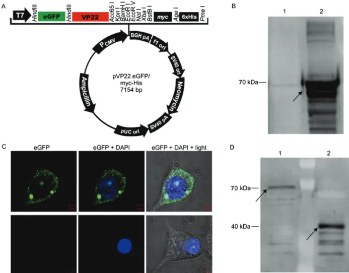
In order to test the ability of VP22 to enter cultured stem cells, we generated an expression vector containing VP22 coupled to eGFP (pVP22.eGFP; Figure 1A). To confirm its ability to yield the desired fusion protein (VP22.eGFP of,70 kDa), this construct was transfected
into 293T cells, and these cells were subjected to analysis by both Western blotting (Figure 1B) and confocal microscopy (Figure 1C), revealing the production of the desired fusion protein.
Presence of the VP22 protein in the culture medium of the producing cells
To document the capacity of VP22 to penetrate and cross membranes, we first analyzed whether the VP22 protein and the VP22.eGFP fusion protein were secreted by the cells and could be detected in the culture medium. We found that the VP22 protein and the newly con-structed VP22.eGFP fusion protein were able to trans-pose the membrane of their overproducing cells and could be detected in the culture medium by Western blotting (Figure 1D).
VP22 transduction into stem cells
no fluorescence being found inside these cells (Figure 2A; lower panels).
To estimate the number of cells that incorporated the VP22.eGFP protein and the period of time during which these proteins could be shown inside the cells, we treated the hDPSCs with cell extracts obtained from 293T cells transiently expressing eGFP alone or VP22.eGFP. After 24 h, we analyzed the eGFP protein present in these cells by flow cytometry. The results showed that 1.1% of the cells treated with the cellular extract containing eGFP were fluorescent, whereas, 10.3% of fluorescent cells were observed upon treatment with the cellular extract
containing VP22.eGFP (Figure 2C).
For mESCs, we used wide field confocal microscopy, and observed eGFP fluorescence only in cells treated with the extract containing VP22.eGFP, while no fluorescence was found in cells treated with the extract containing only eGFP or in the control cells without any treatment (Figure 2C). A confocal z-stack scan was also performed on these cells in order to ensure that the VP22.eGFP fusion protein was inside the cell (data not shown). These results demonstrate that VP22 is capable of delivering an active protein, in this case eGFP, into both hDPSCs and mESCs.
Figure 1.A, Schematic drawing of the vector constructed and used in the present study, pVP22.eGFP.B, The expression of the VP22.eGFP fusion protein was confirmed by Western blot using an anti-VP22 antibody. The 293T cells transfected with pLPCX.eGFP were used as a negative control (lane 1) and the extract obtained from 293T cells transfected with pVP22.eGFP (lane 2) showed a band of,70 kDa corresponding to the fusion of eGFP (,30 kDa) to VP22 (,40 kDa); the arrow indicates the band.C, The 293T cells
Figure 2.VP22.eGFP transduction into stem cells.A, hDPSCs showed green fluorescence after 30 min of incubation with protein extracts containing VP22.eGFP (upper panels); the negative control, incubated for the same period of time with the protein extracts obtained containing eGFP alone showed no fluorescence inside the cell (lower panels). hDPSCs were visualized by confocal microscopy, and optical slides of,0.5mm were obtained. The Figure shows a representative cell for each condition on three different optical slides.B, The mESCs were observed under a confocal microscope in a wide field. The cells treated with protein extracts containing VP22.eGFP showed green fluorescence; the cells incubated with the protein extracts obtained containing eGFP revealed no fluorescence. Also, mESCs with no treatment were visualized by confocal microscopy as a control for the possible auto-fluorescence of the cell (control). DAPI staining was used to define the nucleus of the cell. The Figure shows a representative cell for each condition in a wide field.C, Flow cytometry analysis shows the quantitation of hDPSCs that incorporated eGFP after 24-h incubation with protein extracts obtained from 293T cells after 48 h of transfection with eGFP- or VP22.eGFP-expressing vectors.
Maintenance of the stem cell phenotype
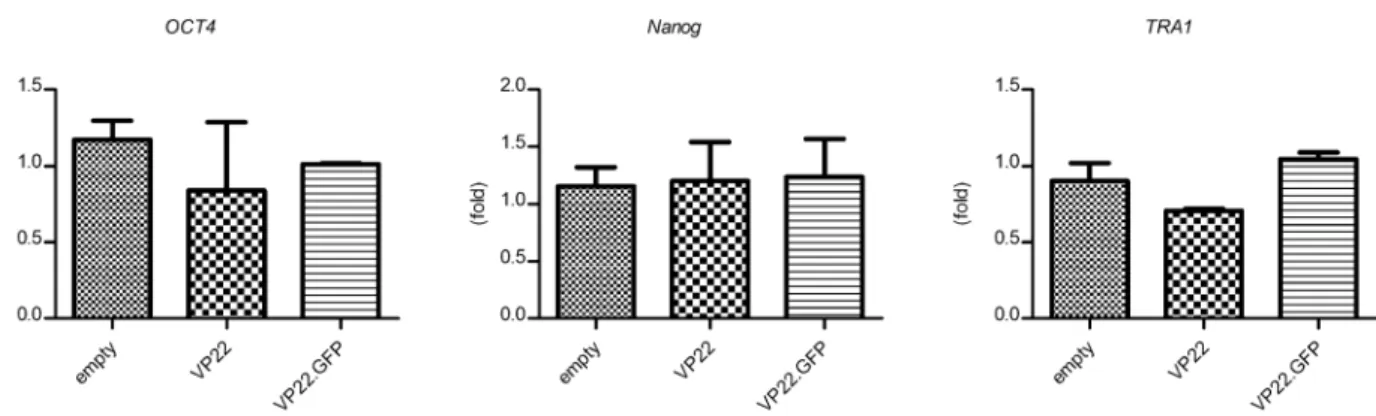
Since stem cells are sensitive to exogenous stimuli, we decided to investigate whether the VP22 or the VP22.eGFP fusion proteins could affect the phenotype of hDPSCs. To this end, we co-cultured the hDPSCs with CHO cells stably expressing VP22 or VP22.eGFP proteins for 6 days. We showed that none of the stem cell markers analyzed were affected by VP22 alone or by the VP22.eGFP fusion protein (Figure 3). Thus, no significant difference was found in the expression of OCT4, Nanog and TRA1 stem cell markers when compared to hDPSCs co-cultured with parental CHO cells or to the ones co-cultured either with CHO cells expressing VP22 or VP22.eGFP. These results also indicate that co-culturing of hDPSCs with CHO cells does not affect the stem cell phenotype of the former.
Discussion
Despite the controversy concerning the VP22 trans-duction capacity (4,16), we have demonstrated for the first time in living stem cells the ability of VP22 to enter and deliver other proteins into two different types of stem cells, namely hDPSC and mESC, proving the viability of employing VP22 as a tool in stem cell research and differentiation.
Some of the arguments against the VP22 transduction ability are that the fluorescence observed inside the cell was only visible after fixation. According to this argument, the fixation procedure would open some channels in the membrane and allow the VP22.eGFP protein attached to the membrane to enter the cell and migrate to the nucleus due to its positive charge (16).
To avoid any fixation artifacts, we developed a protocol to test the capacity of VP22 to enter stem cells using live/unfixed stem cells, and were able to demon-strate that VP22 is, in fact, capable of entering these cells and not simply adsorbing to the membrane. Moreover, we undertook confocal microscopy to probe the cells through different optical sections in order to demonstrate that the VP22.eGFP fusion protein was indeed inside the cells and not only adsorbed to the cell membrane, a situation that could not be distinguished using other techniques such as flow cytometry. The confocal microscopy results confirm that VP22 is, in fact, capable of transducing stem cells (Figure 2).
Here we show that the VP22.eGFP fusion protein is expressed by the producing cells and exported to the culture medium (Figure 1), being capable of entering the stem cells. Although we showed fluorescence only in the cytoplasm of the receptor cells (Figure 2A) we were able to visualize the eGFP fluorescence also in the nucleus of some other receptor cells (data not shown). It is also known that the VP22 present in the cytoplasm of receptor cells is able to reach the nucleus when these cells undergo mitosis (11,12), allowing any protein fused to VP22 to exert its
function in the nucleus of the cell, initiating a signaling cascade without any genome alteration.
Having established that, we used flow cytometry analysis to estimate the efficacy and durability of the ectopic protein expression using this method. We found that even after 24 h of treatment with the cellular extract containing VP22.eGFP we could observe fluorescence in 10.3% of the cells analyzed (Figure 2C). Regarding the method used, we believe that a reasonable percentage of fluorescent cells was achieved, showing that the VP22 protein may be considered an efficient tool to deliver exogenous proteins into stem cells. We also believe that this efficacy may be increased when the cells are treated with the purified protein.
In the present study, we used a protein transduction protocol to demonstrate that the VP22 protein may be used as a tool to transduce stem cells without altering their genome. We adopted cellular extracts based on simplicity; however, we believe that, in order to deliver a protein aimed at changing the stem cell phenotype, the purified protein or co-culturing of the stem cells with cells producing the desired protein should be adopted.
In the co-culture method, the stem cells would be constantly receiving the protein fused to VP22. One issue is whether the VP22 protein may be considered an innocuous molecule when transduced into stem cells. To address this question, we co-cultured the hDPSCs with CHO cells expressing either VP22 or VP22.eGFP for 6 days and observed that after this period the cells were still expressing the OCT4, Nanog and TRA1 stem cell markers, showing that neither VP22 nor any other protein produced by the CHO cells seemed to interfere with the stem cell phenotype (Figure 3).
Several cell types have been used in VP22 studies, with satisfactory results (6,8,9,13-15). However, to date, no reports have addressed the capacity of VP22 to enter any type of stem cell, which would make VP22 a valuable tool to be used in stem cell research and differentiation.
In a study seeking a possible treatment for Parkinson’s disease, another type of PTD protein, the HIV virus TAT protein, successfully used the PTD system to promote neuronal differentiation in murine neural stem cells (20), supporting the fact that the PTD system may be used to drive stem cell differentiation into the desired cell type. However, it is possible that different types of PTD proteins, such as VP22, TAT or Antennapedia, may have different mechanisms of translocation (3). Therefore, since different types of stem cells may behave differently when subjected to the same treatment, it is important to ensure the capacity of VP22 to enter different kinds of stem cells in order to confirm that this protein indeed constitutes a possible tool to be used in stem cell differentiation.
results obtained in our laboratory indicate that this VP22 system is also valuable to deliver active proteins into stem cells for in vitro differentiation, since the addition of the VP22.Pax4 fusion protein to a cell differentiation protocol, which leads to insulin producing cells, seems to protect these cells, leading to a higher outcome of insulin-producing clusters and improving the expression of some important genes related to Pax4 (Kossugue PM, Gabanyi I, Lojudice FH, Sogayar MC, unpublished results).
Upon demonstrating the presence of eGFP fluores-cence inside two different types of stem cells, namely hDPSCs and mESCs, the present study offers a practical demonstration that VP22 may indeed be a valuable tool to transduce proteins into different types of stem cells. In
addition, VP22-mediated protein transduction may be used in stem cell differentiation without disturbing their genome, thus improving the safety of these cells and the reliability of cell therapy protocols.
Acknowledgments
We are grateful to Dr. Stuart Perkins (Biomedical Sciences Department, Wiltshire, UK) for kindly donating the anti-VP22 antibody. We also acknowledge the excellent technical support of Zizi de Mendonc¸a, Sandra Regina de Souza, De´bora Cristina da Costa, Ricardo Krett de Oliveira, and Marluce Mantovani. Research supported by FAPESP, FINEP, CNPq, and BNDES.
References
1. Phillips MI, Tang YL. Genetic modification of stem cells for transplantation.Adv Drug Deliv Rev2008; 60: 160-172, doi: 10.1016/j.addr.2007.08.035.
2. Ford KG, Souberbielle BE, Darling D, Farzaneh F. Protein transduction: an alternative to genetic intervention?Gene Ther2001; 8: 1-4, doi: 10.1038/sj.gt.3301383.
3. Schwarze SR, Hruska KA, Dowdy SF. Protein transduction: unrestricted delivery into all cells?Trends Cell Biol2000; 10: 290-295, doi: 10.1016/S0962-8924(00)01771-2.
4. Lundberg M, Wikstrom S, Johansson M. Cell surface adherence and endocytosis of protein transduction domains.Mol Ther2003; 8: 143-150, doi: 10.1016/S1525-0016(03)00135-7.
5. Nishi K, Saigo K. Cellular internalization of green fluores-cent protein fused with herpes simplex virus protein VP22 via a lipid raft-mediated endocytic pathway independent of caveolae and Rho family GTPases but dependent on dynamin and Arf6.J Biol Chem2007; 282: 27503-27517, doi: 10.1074/jbc.M703810200.
6. Derer W, Easwaran HP, Leonhardt H, Cardoso MC. A novel approach to induce cell cycle reentry in terminally differ-entiated muscle cells.FASEB J2002; 16: 132-133. 7. Ribeiro MM, Klein D, Pileggi A, Molano RD, Fraker C,
Ricordi C, et al. Heme oxygenase-1 fused to a TAT peptide transduces and protects pancreatic beta-cells. Biochem Biophys Res Commun2003; 305: 876-881, doi: 10.1016/ S0006-291X(03)00856-8.
8. Perkins SD, Flick-Smith HC, Garmory HS, Essex-Lopresti AE, Stevenson FK, Phillpotts RJ. Evaluation of the VP22 protein for enhancement of a DNA vaccine against anthrax. Genet Vaccines Ther2005; 3: 3, doi: 10.1186/1479-0556-3-3. 9. Xiong F, Xiao S, Yu M, Li W, Zheng H, Shang Y, et al. Enhanced effect of microdystrophin gene transfection by HSV-VP22 mediated intercellular protein transport. BMC Neurosci2007; 8: 50, doi: 10.1186/1471-2202-8-50. 10. Noguchi H, Matsumoto S. Protein transduction technology:
a novel therapeutic perspective.Acta Med Okayama2006; 60: 1-11.
11. Elliott G, O’Hare P. Cytoplasm-to-nucleus translocation of a herpesvirus tegument protein during cell division.J Virol2000; 74: 2131-2141, doi: 10.1128/JVI.74.5.2131-2141.2000.
12. Martin A, O’Hare P, McLauchlan J, Elliott G. Herpes simplex virus tegument protein VP22 contains overlapping domains for cytoplasmic localization, microtubule interaction, and chromatin binding. J Virol 2002; 76: 4961-4970, doi: 10.1128/JVI.76.10.4961-4970.2002.
13. Brewis N, Phelan A, Webb J, Drew J, Elliott G, O’Hare P. Evaluation of VP22 spread in tissue culture.J Virol2000; 74: 1051-1056, doi: 10.1128/JVI.74.2.1051-1056.2000. 14. Lemken ML, Wolf C, Wybranietz WA, Schmidt U, Smirnow I,
Buhring HJ, et al. Evidence for intercellular trafficking of VP22 in living cells. Mol Ther 2007; 15: 310-319, doi: 10.1038/sj.mt.6300013.
15. Saha S, Yoshida S, Ohba K, Matsui K, Matsuda T, Takeshita F, et al. A fused gene of nucleoprotein (NP) and herpes simplex virus genes (VP22) induces highly protective immunity against different subtypes of influenza virus. Virology2006; 354: 48-57, doi: 10.1016/j.virol.2006.04.015. 16. Fang B, Xu B, Koch P, Roth JA. Intercellular trafficking of
VP22-GFP fusion proteins is not observed in cultured mammalian cells. Gene Ther 1998; 5: 1420-1424, doi: 10.1038/sj.gt.3300741.
17. Wang R, Liang J, Jiang H, Qin LJ, Yang HT. Promoter-dependent EGFP expression during embryonic stem cell propagation and differentiation.Stem Cells Dev2008; 17: 279-289, doi: 10.1089/scd.2007.0084.
18. Lindroos B, Ma¨enpa¨a¨ K, Ylikomi T, Oja H, Suuronen R, Miettinen S. Characterisation of human dental stem cells and buccal mucosa fibroblasts. Biochem Biophys Res Commun 2008; 368: 329-335, doi: 10.1016/j.bbrc.2008. 01.081.
19. Sukoyan MA, Kerkis AY, Mello MR, Kerkis IE, Visintin JA, Pereira LV. Establishment of new murine embryonic stem cell lines for the generation of mouse models of human genetic diseases.Braz J Med Biol Res2002; 35: 535-542, doi: 10.1590/S0100-879X2002000500004.