CLINICAL
ASPECTS OF HUMAN
VENEZUELAN
EQUINE
ENCEPHALITIS
IN TEXAS1
G. Stephen Bowen, M.D.; 2 Thomas R. Fashinell, M.D.;3 Paul B. Dean, M.D.;4 and Michael B. Gregg, M.D.5
The 1971 VEE epidemic in Texas provided new information about the clinical syndrome of that disease. The present article analyzes this information, offeen’ng a detailed description of findings concewzing incubation periods, physical symptoms, clinical I laboratory data, sequelae, and other matters relating to manifestations of VEE in the United States.
Introduction
The etiologic agent of Venezuelan equine encephalitis (VEE) was first isolated and characterized from specimens obtained during an equine epizootic in Venezuela in
1936 (1,Z). Since that time, outbreaks in man and equine animals have occurred in Trini- dad (3, 4), Ecuador (5), Peru (6), Colombia (7), and Venezuela (8, 9). Human illness has also occurred in areas where VEE viruses are endemically maintained (10-12) by transmis- sion cycles usually involving rodents and Culex mosquitoes of the subgenus Melanoco- nion.
Human VEE infections may be so mild that they escape detection (13, 14), or they may
‘From the Bureau of Laboratories and Bureau of Epidemiology, Center for Disease Control, United States Public Health Service, Department of Health, Educa- tion, and Welfare, Atlanta, Georgia 30333, U.S.A. Also appearing in Spanish in the BoletZn de la Oficina
Sanitaria Panamericana, 1976.
2Vector-Borne Disease Division, Center for Disease Control (CDC), P.O. Box 2087, Fort Collins, Colorado 8052 1.
3Formerly Epidemic Intelligence Service Officer, CDC: present address: Resident in Medicine, Sacramento Medical Center, Sacramento, California 95817.
4Tormerly Epidemic Intelligence Service Officer, CDC; present address: Resident in Dermatology, School of Medicine, University of Colorado Medical Center, Denver, Colorado 80220.
‘Chief. Viral Diseases Division, Bureau of Epidemio- logy, CDC.
produce a clinical syndrome of varying severity (7-9, 12). The proportion of severe cases resulting in neurologic sequelae or death has varied widely from outbreak to outbreak. Moreover, in the midst of a large outbreak it may be difficult to know how much of the morbidity and mortality to ascribe to VEE virus, since laboratory documentation is required in each case to distinguish this disease from others with similar symptoms (15).
Laboratory-acquired infections have re- sulted in mild to moderately severe disease, but no deaths or permanent sequelae have been reported from such cases (16-19).
The spread of the latest VEE epidemic from Ecuador to Texas has been described by Guticrrez, et aE. (ZO), Franck and Johnson (21), Lord (22), Martin, et al. (23) and Sudia and Newhouse (24). The outbreak was recognized in Ecuador in the winter and spring of 1969, and in Guatemala and El Salvador in June and July of that same year (25, 26). In 1970 the virus spread both south and north, and after passing through the Atlantic coastal lowlands of Mexico it reached the southern border of Texas by 1 July 19’71. The course of the epidemic in humans and equines in the United States has been described by Zehmer, et al. (27). The purpose of the present article is to report on the
Bowen et al. l HUMAN VEE IN TEXAS 47
clinical syndrome of VEE in the U.S. and to describe some of the outbreak’s epidemiologic parameters. Virologic and serologic studies performed at the Center for Disease Control (CDC) will be described elsewhere (15).
Materials and Methods
Except for five cases discovered by the senior author during a six-week clinical follow-up of the outbreak in Cameron County, all cases reported here were found as a result of a hospital-based surveillance system set up by the Texas State Department of Health and the Bureau of Epidemiology, CDC, Atlanta, Georgia. Thirty-eight hospitals in 20 cities participated in this VEE surveillance. One person at each participating hospital (a nurse, admissions clerk, or records librarian) made daily reports to county and state health officials on persons who were admitted with suspected cases of VEE. Laboratory directors and attending private physicians were contacted to facilitate collec- tion of paired blood specimens from all such persons.
During the peak of the epidemic, addi- tional clinical reports and blood specimens were sent from the offices of interested physicians and from emergency rooms to participating hospital laboratories. This pro- cedure permitted VEE cases in outpatients to be diagnosed.
Blood specimens and case reports were obtained from the hospital laboratories by county health personnel and were sent to the Texas State Department of Health or the CDC, Atlanta, for diagnostic evaluation.
Clinical follow-up was attempted on all the patients from Cameron and Hidalgo counties four to six weeks after their initial illness. An examination was performed on all who could be located and a blood specimen was obtained. Those who were symptomatic at four to six weeks were examined again nine to twelve months after their illness.
The VEE cases reported here were labora- tory-confirmed by the Arbovirology Section, CDC, and by the Communicable Diseases
Diagnostic Laboratory of the Texas State Department of Health. Confirmation was based on at least one of the following: (1) isolation of VEE virus from the patient‘s serum; (2) serologic conversion, as demon- strated by a change in hemagglutination- inhibition (HI) test results from negative to positive (a titer 2 20); or (3) serologic conversion, as shown by a change in weaned-mouse neutralization test re- sults-from a log neutralization index (LNI) of 0 to a LNI greater than 1.7. More detailed descriptions of these procedures are given elsewhere (25).
Results
Eighty-eight officially reported cases occur- red in July and August 1971, seventy of them in Cameron and Hidalgo, the two southern- most Texas counties. All but one of the other cases occurred around Corpus Christi (in Nueces, San Patricia, Kleberg, Aransas, and Refugio counties), the only exception being a case in Maverick County along the Mexican border further west. Twenty-two additional infections were also discovered, five by the forementioned follow-up of known human cases in Cameron County and 17 by an illness-and-blood survey of Port Isabel, Texas, conducted by Drs. Dean, Lawrence, Schoen- berger, Gregg, and other staff members of the CDC Bureau of Epidemiology.
The epidemic curve, sex ratios, and attack rates presented here are based on analysis of all 88 officially reported cases. However, because detailed case reports were not available for some patients, only 79 cases are analyzed in the section on clinical history.
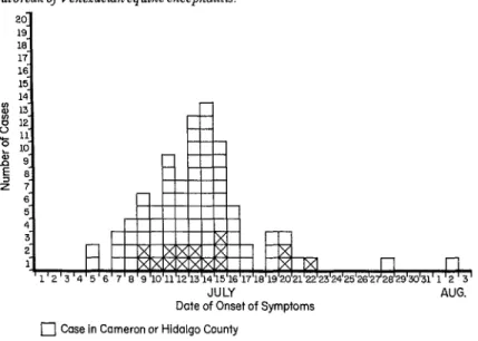
An epidemic curve of the 88 VEE cases, all of which occurred between 5 July and 2 August, is shown in Figure 1. A peak rate of 13 cases per day was reported for 13 and 14 July. Although the first cases in the Corpus Christi area began four days later than the first ones in Cameron and Hidalgo counties, peak activity in both areas took place in the seven-day period of 9-15 July.
48 PAHO BULLETIN l Vol. X, No. 1, 1976
JULY
Date of Onset of Symptoms
c] Case in Cameron or Hidalgo County
q
Case in Corpus Christi AreaAUG.
ed cases occurred either in Cameron and With regard to case rates, there was no Hidalgo counties or in four counties in and evidence of any significant difference in the around Corpus Christi (Nueces, San Patricia, rate at which the disease affected inhabitants Kleberg, and Aransas) , and some 647,676 with Spanish versus Anglo surnames, but a people were at risk. The respective male: male:female case ratio of 2:l was observed. female ratio of this at-risk population was 48.3 Although about the same number of people to 51.7. Spanish surnames predominated in were at risk in Cameron and Hidalgo as in the this population, the respective percentages of Corpus Christi area, the attack rate in the Spanish and Anglo surnames being 63 and 37 former area was much higher (21 cases per per cent. Table 1 shows the age distribution of 100,000 vs. 4.9 per lOO,OOO), as shown in the at-risk populations in the two principal Table 1. In Cameron and Hidalgo counties,
areas affected. the highest age-specific attack rate observed
TABLE 1- VEE attack rates, by age group, among the populations at risk in Camemn and Hidalgo counties and in the Corpus Christz area, July-August 1971.
Corpus Christi area (Nueces, San Patricia, Kleberg,
Cameron ana Hidalg0 counties ana Aransas counties) Total (six counties)
Age Pop. at No. of Attack rate
group POP. at NO. of Attack rate POP. at No. of Attack rate
(wars) risk cases per 100,000 risk cases per 100,000 risk cases per 100,000
o-9 75,088 5 6.6 67,948
10-19 78,453 18 22.9 72,499
20-29 40,480 18 44.5 50,980
30-39 31,766 11 34.6 35,553
40-49 33,335 10 30.0 37,659
50-59 25,422 3 11.8 28,832
60-69 21,532 1 4.6 19,310
70 16,024 4 25.0 12,795
5.9 143,036 9 6.3
8.3 150,952 24 15.9
2.0 91,460 19 20.8
8.4 67,319 14 20.8
2.7 70,994 11 15.5
0 54,254 3 5.5
0 40,842 1 2.4
7.8 28,819 5 17.3
Bowen, et al. l HUMANVEEINTEXAS 49
(44.5/100,000) occurred in the 20-29 age group, whereas in the Corpus Christi area the lo-19 and 30-39 age groups were the ones most affected.
Incubation Period
Eleven of the 79 patients were exposed at one of two high-risk beach areas, and the time of their exposure was known. These circum- stances provided an opportunity to estimate incubation periods for naturally-acquired epidemic VEE in a more precise fashion than was ever possible before. These incubation periods could be determined for those of the eleven who were not Texas residents because they all became ill two to four days after ar- riving in south Texas. The periods could also be determined for those who were Texas resi- dents because they went to a high-risk beach in one of two groups, stayed only a few hours, and became ill within 24 hours of at least one other member of their group. All persons listed in Table 2 were infected at Boca Chica Beach or on South Padre Island. Some of the cases could have had shorter incubation periods than has been indicated, since the patients may have been exposed to infected mosquitoes at any time between their arrival and the onset of symptoms. There was no evidence of any rela- tion between the length of the incubation period and the severity of disease symptoms.
Clinical History
The patients’ most common early com- plaints were of headache and of suddenly feeling hot; over half experienced myalgia initially, and 38 per cent had chills. A greater variety of symptoms were manifested later in the course of the illness (see Table 3), but again the pattern of headache, of feeling hot, and of myalgia stood out. Overall, 40 per cent vomited, 10 per cent had chills, 23 per cent had a sore throat, and 22 per cent had mild diarrhea with three or four loose stools per day.
There was no discernible difference in the frequency of each of these symptoms in children as compared to adults. But probable central nervous system involvement was seen more frequently in persons under 17 years of age. Six of the 25 patients under 17 (four boys and two girls of ages 1, 3, 4, 6, 14 and 16) experienced one or more grand ma1 seizures, temporary paralysis, or coma. These cases represented 24 per cent of the cases in patients under 17 and 7.6 per cent of the total cases.
Less dramatic signs of central nervous system involvement were seen in four children and six adults. These milder signs consisted of confusion, hallucinations, or gait abnormality in association with excessive drowsiness. If these additional mild cases are added to the six more severe cases, it may be seen that 16 of
TABLE 2-E&mated incubation periods of VEE in I1 persons with naturally acquired cases (Texas, 1971).
Arrival at Boca Chica Beach or onset of
Patient Age South Padre Island symptoms
Estimated
number (years) Sex Date of arrival Time of arrival Date Time incubation period
1 29 M 7113
2 31 F 7111
3 21 M 7112
4 I.5 F 7/11
5 48 F 7/10
6 31 F 7/8
7 15 M 7/12
8 47 F 7/2
9 20 F 7/10
10 67 F 7/10
11 16 M 716
6:00 PM 7/14 9:30 PM 27.5 hours
7:oo PM 7113 6-7:00 AM 36 hours
12:OO Noon 7114 2:oo AM 38 hours
7:oo PM 7112 3:oo AM 56 hours
7112 2 days
7110 2 days
Not 7115 Not 3 days
KllOWll 715 KIlOWIl 3 days
7113 3 days
7/14 4 days
50 PAHO BULLETIN l VoZ. X, No. I, 1976
TABLE 3-Symptoms reported by VEE patients dunng Sequelae their illness (Texas, 1971).
Symptoms reported % of patients affected Headache
Feeling hot Myalgia Vomiting Chills Drowsiness Diarrhea Weakness Sore throat Neck pain Eye pain Arthralgia Ataxia - History of sei;! Hallucinations Confusion
:ure
89 84 66 39 33 29 22 20 20 19 15 11 9 6 6 5
Paresthesias 4
the 79 VEE cases (20 per cent) showed evidence of “encephalitis.” Overall, 10 of 28 young people (36 per cent) showed signs of central nervous system involvement, whereas only six of 51 adults (11 per cent) did so, and none of the adults had seizures or were comatose.
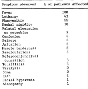
Physical Findings
Table 4 shows the physical symptoms observed in patients acutely ill with VEE. All of the cases were febrile, a temperature between 102°F and 105°F representing the maximum fever observed in 75 per cent of the cases. This fever lasted anywhere from one to five days, having a duration of one.and a half to four days in 80 per cent of the cases.
TABLE 4- Phystcal findings in acute VEE cases (Texas, 1971)
Symptoms observed % of patients affected Fever
Lethargy Pharyngitis Nuchal rigidity Palatal ulceration
or petechiae Confusion Seizure Agitation
Muscle tenderness Fasciculations Scleroconjunctival
congestion Tonsillitis Paralysis Coma Rash
Facial hyperemia Adenopathy
100 43 22
10
Twenty-three per cent of the patients had nonexudative pharyngitis, and three persons had ulcerations l-2 mm in diameter with sharp borders on the uvula and soft palate. There were no abnormal neurological find- ings in most cases except for lethargy, which was present in 40 per cent. Eleven per cent had nuchal rigidity, 6 per cent were observed having a seizure, 2 per cent had paralysis, 2 per cent were comatose,, and 2 per cent had hyperactive but symmetrical reflexes.
3
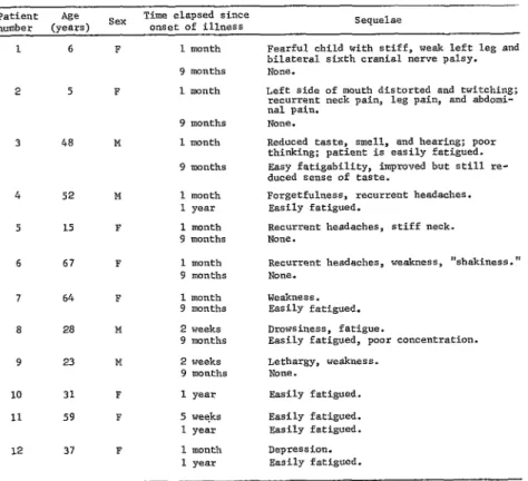
Most patients had recovered completely a week after the onset of symptoms. One patient, a 15-year-old girl, had a recurrence of her complete clinical syndrome four days after all initial symptoms had disappeared. She recovered again three days later without sequelae. Table 5 shows the 12 patients who still had persistent symptoms one month after their illness. Nine adults complained of one or more recurrent symptoms, including head- aches, weakness, myalgia, and tiring easily. Two patients had diplopia; one 37-year-old man complained of reduced ability to taste, smell, and hear in addition to tiring easily and experiencing mental slowness.
Bowen, et al. l HUMANVEEINTEXAS 51
TABLE 5-Sequelae in 12 of 48 persons infected with VEE (Texas, 1971).
Patient Age sex Time elapsed since
number (years) onset of illness seque1ae
1 6
2 5
3 48
4 52
5 15
6 67
7 64
a 28
9 23
10 31
11 59
12 37
1 month
9 months 1 month
9 months 1 month
9 months
1 month 1 year 1 month 9 months
1 month 9 months
1 month 9 months
2 weeks 9 months
2 weeks 9 months
F 1 year
F 5 weeks
1 year
F 1 month
1 year
Fearful child with stiff, weak left leg and bilateral sixth cranial nerve palsy. NO"%
Left side of mouth distorted and twitching; recurrent neck pain, leg pain, and abdomi- nal pain.
None.
Reduced taste, smell, and hearing; poor thinking; patient is easily fatigued. Rasy fatigabilfty, impmved but still re- duced sense of tastes
Forgetfulness, recurrent headaches. Easily fatigued.
Recurrent headaches, stiff neck. None.
Recurrent headaches, weakness, "shakiness." NO"=.
WCSk"eSs. Easily fatigued. Drowsiness, fatigue.
Easily fatigued, poor concentration. Lethargy, weakness.
None. Easily fatigued.
Easily fatigued. Easily fatigued. Depression. easily fatigued.
knee and ankle, and decreased power of the flexors and extensors of the left leg. She fell to the left when forced to walk.
Eleven of these 12 patients were reexam- ined nine months to a year after their illness. The two girls with paralysis had completely recovered. Seven of the nine adults examined still complained of tiring easily, but all the other symptoms had disappeared.
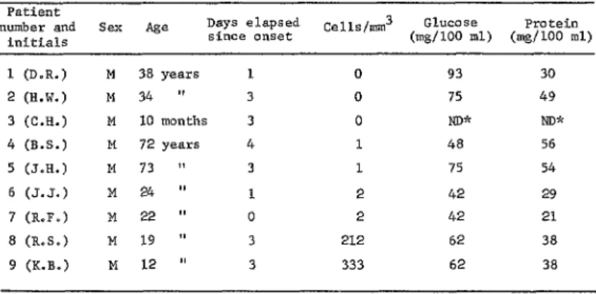
Clinical Laboratory Data
Laboratory studies are available from 46 VEE patients, but a complete blood count was the only laboratory test performed on 89 per cent of these persons. Data are available for only nine patients from whom cerebrospinal fluid (CSF) was collected during the first four days after the onset of symptoms (see Table 6). Seven of these nine CSF samples were
found to have O-2 white blood cells (WBC) /mm3, but the remaining two fluids (obtain- ed three days after onset) showed 212 and 333 WBC/mm3. All the white blood cells observed were lymphocytes. Several fluids showed moderate depression of CSF glucose or elevation of CSF protein. Data on blood chemistries are so sparse that no meaningful conclusions can be drawn.
52 PAHOBULLETIN ' Vol. X, No. 1, 1976
TABLE 6-Results of examination of cerebrospinalfluidfrom nine confirmed VEE cmes (Texas, 1971)
Patient
number and Sex Age Days elapsed Cells/mm3 Glucose Protein
initials since onset (mg/lOO ml) (mg/lOO ml)
1 (D.R.) M 38 years
2 (W.W.) M 34 ”
3 (C.H.) M 10 months
4 (B.S.) M 72 years
5 (J.H.) M 73 ”
6 (J-J.) M 24 ”
7 (R.F.) M 22 ”
a (R.s.) M 19 "
9 (K.B.) M 12 ”
0 93 30
0 75 49
0 ND* NW
1 48 56
1 75 54
2 42 29
2 42 21
212 62 38
333 62 38
aNot done.
FIGURE Z-Total white blood cell counts in VEE pa- FIGURE 3 -Total segmented neutrophil counts in
tients, Texas, 1971. VEEpatzents, Texas, 1971.
14.000
12,000
10,000
mg 8,000
; g 6,000
4,000
2,000
C
. :
. .
I .
. ! . . .
ii.! . *
. i . : l ’
, !. .
1 123456789
DAY OF ILLNESS
FIGURE 4-Absolute lymphocyte counts in VEE pa- tients, Texas, 1971,
.
. .
.
. .
. * .
0
0123456789
DAY OF ILLNESS
14,000
% 12,000
2 10,000 4 2 g 8,000 5 g 6,000 B 5 : 4,000
W
y 2,000
. . . .
I I I I I 1 1 I 1
1 23456789
DAY OF ILLNESS
Bowen, et al. l HUMAN VEE IN TEXAS 53
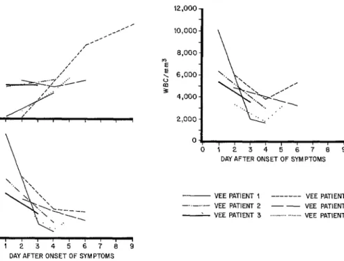
1,500/mm3. From day 1 to day 4 over 80 per cent of these values were between 150 and 1,490/mm3, and nine were between 150 and 500/mm3. In contrast, from day 5 to day 9, most absolute lymphocyte values were above 1,500/mm3. However, since relatively few determinations were made late in the course of illness, it is difficult to draw valid conclusions concerning recovery of these leukocyte levels.
Plots of white blood cell (WBC) and absolute segmented neutrophil (ASN) counts are nearly parallel until day 5 or 6 (see Figures 2, 3, and 5). Normal WBC counts on days 1 and 2 depended on normal initial values for ASN. When ASN values began to fall on the third day of symptoms, total WBC counts also fell. Though these ASN values were still abnormally low as late as day 8 after onset of symptoms, some recovery was evident begin- ning on day 6 or 7. In addition, total leukocyte counts began to rise on days 5 and
6, a development which reflected the fore- mentioned rises in absolute lymphocyte values. Thus it is evident that VEE virus does, in some manner, cause depression in the blood values of both segmented neutrophils and lymphocytes.
Since VEE symptoms can be so similar to those of other acute infectious diseases, paired sera from ill persons who were shown not to have had VEE were tested for rises in antibodies against enteroviruses and leptos- pires. Two cases of Coxsackie B infection and two cases of leptospirosis were discovered; these patients’ clinical signs and symptoms were indistinguishable from those of the VEE cases. In addition, the Texas StateDepartment of Health isolated type 4 Echovirus from the cerebrospinal fluid of three patients from Hidalgo County who had been ill just before the VEE outbreak with clinical syndromes identical to those observed in VEE cases.
FIGURE 5 -Absolute lymphocyte, absolute segmented neutmphil, and total white blood cell counts in six VEE pa- tients, Texas, 1971.
3,000- 12,000 -
“E 2,400-
E
DAY AFTER ONSET OF SYMPTOMS
- VEE PATIENT 1 --- VEE PATIENT 4
-“-..- VEE PATIENT 2 - - VEE PATIENT 5
d VEE PATIENT 3 . . . . .._.... VEE PATlENT 6
0123456789
54 PAHO BULLETIN l Vol. X, No. 1, 1976
Discussion
The 2:l male-female case ratio and the higher age-specific attack rates observed in young adults (ages 20-49) in the Cameron- Hidalgo area probably indicate that exposure occurred outside the home and was occupa- tionally related. Men working outdoors, espe- cially in agriculture or ranching, would be more likely to come in contact with infected mosquitoes than would children and women staying at home. A similar point has been made by Martin, et al. (23), who observed a 33:l maleifemale case ratio during a VEE outbreak in a Costa Rican village.
The incubation periods observed in the Texas outbreak (27.5 hours to four days) correspond closely to incubation periods cited in published reports of laboratory-acquired infections, which were found to range from 36
hours to six days (15-18). The clinical syndrome seen in the U. S. during this outbreak does not differ dramatically from previously reported accounts of laboratory- acquired ( 15-18) or naturally acquired VEE (7-9, 14, 24). Most of the illnesses, especially those of adults, were relatively mild. They were nearly identical to the illnesses described by Sanmartin, et al. (7) in 1954. The clinical relapse in a 15-year-old girl was similar to recurrences previously observed in laboratory- acquired cases (lb). One man complained of greatly reduced ability to hear, smell, and taste. Anosmia and ageusia, but not hearing loss, were previously reported by Koprowski in connection with artificially acquired VEE
(18).
Previous observations (9, 2.5, 28, 29, 30) have emphasized the greater severity of VEE in children than in adults. This observation was borne out in the recent U.S. epidemic. Only 25 of the patients described in this paper were under 17 years old, but all five patients with severe encephalitis were in this group. Four of the five, including the two girls that experienced residual paralysis, were less than 7 years of age.
Severe sequelae did not occur among the VEE patients studied. This absence of deaths
or serious sequelae in the U.S. experience contrasts markedly with the experience of other countries during VEE outbreaks. There are several possible explanations for this difference, the most likely being that there were too few pediatric cases in the United States. That is, because deaths or severe sequelae occur in only a small proportion of childhood cases, there were insufficient cases to make such outcomes likely. Serosurveys in Port Isabel, Cameron County, Texas, indi- cated that 120 to 150 cases of VEE occurred in this town of 4,000, so that the attack rate observed was on the order of 3.0-3.8 per cent. However, tests for VEE performed on sera from three other sources (from Mercy Hospital, Brownsville, Cameron County; from a serosurvey of family members and neighbors of Brownsville cases; and from public health clinics in Cameron and Hidalgo counties) indicated that infection rates for these counties as a whole were less than 0.5 per cent. This suggests that if hundreds of cases had occurred among children in the United States, deaths and more severe sequelae might have been recognized. Other less likely explanations for the absence of deaths or severe sequelae in this outbreak could involve specific characteristics of the population at risk, such as general health status, nutritional status, or genetic dif- ferences.
It is also possible that the viral strains causing disease in the United States were less pathogenic to man than were strains causing outbreaks elsewhere. Monath, et al. (31) has reported a small amount of evidence pointing to differences in the virulence of VEE strains in rhesus monkeys. That is, one monkey infected with VEE subtype IC showed neurologic signs, but none of the monkeys infected with subtypes IA or IB did so. Also, Corristan (32) has demonstrated differences in the pathogenicity of VEE epidemic subtypes for several mammalian species.
Bowen, et al. l HUMAN VEE IN TEXAS 5.5
neutrophils not mentioned in those earlier reports. These results contrast with those obtained by Gutierrez (30) and Madalengoi- tia, et al. (33), who found leukopenia in a very low percentage of VEE cases. Overall, our results indicate that VEE virus may cause profound depressions in absolute blood values of both segmented neutrophils and lympho- cytes .
There is a little experimental evidence that tends to corroborate these observed altera- tions in leukocyte counts. About 40 per cent of a group of people inoculated with the TC-83 strain of attenuated VEE virus vaccine experienced leukopenia (< 4,500 WBC/ mm3), usually between the third and fifth day after inoculation. The cells predominantly affected were segmented neutrophils (34). If an incubation period of two to three days is assumed for epidemic VEE, the appearance of leukopenia and neutropenia on the second and third day after the onset of symptoms corresponds closely to the time sequence observed for VEE TC-83. Likewise, a majority of 10 rhesus monkeys inoculated with VEE subtypes IA to IE experienced leukopenia on day five after inoculation. Total WBC counts
as low as 2,000/mm3 were frequent (31). Both lymphocytes and neutrophils were affected. Again, if an incubation period of two to three days is assumed for man, the day of appearance of leukopenia in man is in close agreement with the monkey data. Leukopenia in rhesus monkeys does seem to be more transitory than in man.
In conclusion, it must be emphasized that neither the clinical picture nor routine hospital laboratory tests clearly differentiate VEE from other illnesses. In fact, virologic and serologic testing of sera from suspected VEE cases demonstrated that only one-third of such cases actually were VEE (1.5). In other outbreaks where laboratory confirmation of suspected cases was attempted, percentages of 0 (35), 33 (36), 34 (37), and 77 (29) were confirmed as actually being cases of VEE. Our findings demonstrate once again that adequate laboratory documentation of all suspected cases during epidemics provides the only accurate basis for comparing the severity of illness, incidence of central nervous system complications, seriousness of sequelae, and mortality resulting from diverse outbreaks.
ACKNOWLEDGMENTS The authors wish to thank a number of people
whose help in collecting and testing specimens, reviewing medical records, and examining patients made this study possible. We are indebted to the physicians, nursing staffs, administrators, medical record librarians, and laboratory staffs of Mercy Hospital (Brownsville), Dolly Vinsant Memorial Hospital (San Benito), Valley Baptist Hospital (Harlingen), Knapp Memorial Hospital (Weslaco), McAllen General Hospital, Edinburg General Hospital, and Mission Municipal Hospital. Special thanks are due to Sister Mary Alfonso, Sister Marjorie Marie, Mr. Ortiz, Mr. Murray, Mr. Flores, Mrs. Valverde, and Mrs. Flores, who assisted us in many ways both during and after nr aal working hours.
We are likewise very grateful to Dr. John Copenhaver, Mrs. Louise Fisher, Mrs. MacDonald, Mrs. Mary Luico, Mrs. Nancy McFall, Sister Sylvia Cirdenas, Mr. Bob Guzmln, and many other members of the staffs of the Cameron and Hidalgo county health departments; and to Dr. Jesse Irons and Dr. Lois Leffingwell of the Texas State Department of Health.
56 PAHO BULLETIN l Vol. X, No. 1, 1976
The Venezuelan equine encephalitis epidemic which occurred in Texas in 1971 produced a wide range of predominantly mild clinical symptoms. This epidemic, which peaked on 13-14 July, was most intensely felt in the far-south counties of Cameron and Hidalgo. In all, 88 laboratory-con- firmed human cases were reported to the U.S. Center for Disease Control by the Texas State Department of Health.
The ratio of male to female cases was about two to one. An attack rate of 20.8 cases per 100,000, observed in both the 20-29 and 30-39 age groups, was higher than attack rates experienced by other age groups and by the population at large. Together, Cameron and Hidalgo counties expe- rienced a much higher overall attack rate (21.7 cases por 100,000) than did affected counties in the Corpus Christi area (4.9 cases per 100,000).
Knowledge about when various patients were first exposed points to an incubation period ranging from 27.5 hours to four days. In those 79 cases for which clinical data were available, the most common clinical manifestations were foynd to be fever, severe headache, myalgia, and chills. Evidence of mild to moderate central nervous system involvement was found in 10 out of 25 children and young people under 17 years of age, and in six out of 54 adults. Two children still had residual paralysis six weeks after onset of illness, but by 10 months these sequelae had disappeared. Seven of the 54 adults, however, still complained of tiring easily a year after onset of illness. Leukopenia, as demonstrated by a count of less than 4,500 white blood cells per cubic millimeter, was observed in 75 per cent of the patients examined.
REFERENCES
(I) Beck, E. C., and R. W. Wyckoff. Venezue- lan equine encephalomyelitis. Science 88: 530, 1938.
(2) Kubes, V., and F. A. Rios. The causative agent of infectious equine encephalomyelitis in Venezuela. Science 90: 20-21, 1939.
(3) Gilyard, R. I. A clinical study of Venezue- lan virus equine encephalomyelitis in Trinidad, B.W.I.JAm l’eet Med Assoc 106: 267-277, 1945.
(4) Tigertt, W. D., and W. G. Downs. Studies on the virus of Venezuelan equine encephalomyeli- tis in Trinidad, W.I.: I. The 1943-1944 Epizootic. Am J Trap Med Hyg 11: 822-834, 1962.
(5) Sotomayor, C. G. A study of the virus of equine encephalomyelitis in Ecuador. J Am Vet Med Assoc 109: 478-480, 1946.
(6) Smith, H. P., and L. F. Contreras. La encephalomyelitis equina. Rev Inst Nat Biol Animal (Lima, Peru) 4: 3-9, 1953.
(7) Sanmartin-Barberi, C., H. Groot, and E. Osorno-Mesa. Human epidemic in Colombia caused by the Venezuelan equine encephalomyeli- tis virus. Am J Trap Med Hyg 3: 283-293, 1954.
(8) Sellers, R. J., G. H. Bergold, 0. M. Suarez, and A. Morales. Investigations during Venezuelan equine encephalitis outbreaks in Venezuela, 1962-1964. Am J Trap Med Hyg 14: 460-469, 1965.
(9) Bricefio Rossi, A. L. Rural epidemic encephalitis in Venezuela caused by a Group A arbovirus (VEE). ProgMed Viral 9: 176-203, 1967.
(10) Johnson, K. M., A. Shelokov, P. H. Peralta, G. J. Dammin, and N. A. Young. Recovery of Venezuelan equine encephalomyelitis virus in Panama: A fatal case in man. Am J Trap Med Hyg 17: 432-440, 1968.
(II) Ehrenkranz, N. J., and M. C. Sinclair. The natural occurrence of Venezuelan equine encepha- litis in the United States. New Engl J Med 282: 298-302, 1970.
(12) Franck, P.T., and K. M. Johnson. An outbreak of Venezuelan equine encephalitis in man in the Panama Canal Zone. Am J Trap Med Hyg 19: 860-865, 1970.
(13) De Mucha-Macias, J., I. Sanches-Spendo- la, and C. Campillo-Sainz. Venezuelan equine encephalomyelitis antibodies in human beings of southeastern Mexico. Am J Trap Med Hyg 15: 364-368, 1966.
(14) Work, T. H. Serological evidence of arbovirus infection in the Seminole Indians of southern Florida. Science 145: 270-272, 1964.
(15) Bowen, G. S., and C. H. Calisher. Virologic and serologic studies of Venezuelan encephalitis in man-Texas, 1971. Manuscript in preparation.
Bowen, et al. l HUMAN VEE IN TEXAS 57
(17) Casals, J., E. C. Curnen, and L. Thomas. Venezuelan equine encephalomyelitis in man. J Exp Med 77: 521-530, 1943.
(18) Koprowski, H., and H. R. Cox. Human laboratory infection with Venezuelan equine encephalomyelitis virus: Report of four cases. New Eng J Med 236: 647-654, 1974.
(19) Sutton, L. S., C. C. Brooke, and M. D. Frederick. Venezuelan equine encephalomyelitis due to vaccine in man. JAMA 155: 1473-1476, 1954.
(20) Gutierrez, V. E., T.P.C. Monath, A. Alava, B. Uriguen, R. W. Chamberlain, and R. M. Arzube. Epidemiological investigations of the 1969 epidemic of Venezuelan encephalitis in Ecuador. AmJEpidemiol 102:400-413, 1975.
(21) Franck, P. T., and K. M. Johnson. An outbreak of Venezuelan equine encephalomyelitis in Central America: Evidence for an exogenous source of a virulent virus subtype. Am J Epidemiol 94: 487-495, 1971.
(22) Lord, R. D. History and geographic distribution of Venezuelan equine encephalitis. Bull Pan Am Health Organ 8 (2): 100-l 10; 1974. (23) Martin, D. H., G. A. Eddy, W. D. Sudia, W. C. Reeves, V. F. Newhouse, and K. M. Johnson. An epidemiologic study of Venezuelan
equine encephalomyelitis in Costa Rica, 1970. Am J Epidemiol 95: 565-578, 1972.
(24) St&a, W. D., and V. F. Newhouse. Venezuelan equine encephalitis in Texas. 1971: Informational report. Mosquito News 31: 350-351, 1971.
(25) H&ran, A. R., J. E. McGowan, Jr., and B. E. Henderson. Venezuelan equine encephalo- myelitis: Surveys of human illness during an epizootic in Guatemala and El Salvador. Am J Epidemiol93: 130-136, 1971.
(26) Sudia, W. D., R. D. Lord, V. F. Newhouse, D. L. Miller, and R. F. Kissling. Vector-host studies of an epizootic in Guatemala and El Salvador. Am J Epidemiol 93: 137-143, 1971.
(27) Zehmer, R. B., P. B. Dean, W. D. Sudia, C. H. Calisher, G. E. Sather, and R. Parker. Venezuelan equine encephalitis epidemic-Texas. J Am Vet Med Assoc 162: 777-779, 1971.
(28) Suarez, 0. M., and G. H. Bergold. Investigations of an outbreak of Venezuelan equine
encephalitis in towns of eastern Venezuela. Am J Trap Med Hyg 17: 875-880, 1968.
(29) Sanmartin, C. “Diseased hosts: Man.” In: Venezuelan Encephalitis: Proceedings of the Workshop-symposium on Venezuelan Encephalitis Virus, Washington, D.C., 14-17 September 1971. Pan American Health Organization, Washington, D.C., 1972, pp. 186-188 (PAHO Scientific Publication No. 243).
(30) Gutierrez, E. In: Ykzezuelan Encephalitis. Pan American Health Organization, Washington, D.C., 1972, pp. 195-197. (PAHO Scientific Publication No. 243.)
(31) Monath, T. P., C. H. Calisher, M. Davis, and G. S. Bowen. Experimental studies of rhesus monkeys infected with epizootic and enzootic subtypes of Venezuelan encephalitis virus. J Infect Dti 129: 194-200, 1974.
(32) Corristan, Edwin C. Personal communica- tion, 1974.
(33) Madalengoitia, J., 0. Palacios, J. Comejo Ubiliuz, and S. Alva. “An outbreak of Venezuelan encephalitis in man in the Tumbes Department of Peru.” In: Venezueiun Encephalitis. Pan Amer- ican Health Organization, Washington, D.C., 1972, pp. 198-201. (PAHO Scientific Publication No. 243.)
(34) Alevizatos, A. L., R. W. McKinney, and R. D. Feigin. Live attenuated Venezuelan equine encephalomyelitis virus vaccine: I. Clinical effects in man. Am J Trofi Med Hyg 16: 762-768, 1967. (35) Sanmartin, C. In: Venezuelan Encefihali- tti. Pan American Health Organization, Washing- ton, D.C., 1972, p. 219. (PAHO Scientific Publication No. 243.)
(36) Scherer, W. F., J. V. Ordofiez, P. B. Jahrling, B. A. Pancake, and R. W. Dickerman.
Observations of equines, humans, and domestic and wild vertebrates during the 1969 equine epizootic and epidemic of Venezuelan encephalitis in Guatemala. Am JEpidemiol95: 255-266, 1972.