ENZOOTIC RODENT LEISHMANIASIS IN TRINIDAD, WEST INDIES’ Elisha S. Tikasingh, B.Sc., M.A., Ph.D.’
Human cutaneous leishmaniasis is a zoonotic drsease widely distributed in Central and South America. Small mammals play important roles in the natural history of the disease. This article attempts to define more precisely the roles that these mammals play in the ecology of the parasite.
Introduction
Human cutaneous leishmaniasis is widely distributed in several countries of Central and South America. In recent years, workers in Mexico, Belize, Panama, and Brazil have shown quite convincingly that the disease exists as a zoonosis which only accidentally infects man with the parasite, and that wild animals (espe- cially rodents) serve as the primary hosts. Three excellent papers have recently been published reviewing both the epidemiology in these coun- tries (II) and taxonomic problems (12, 13).
Although Ashcroft’s survey of helminthic and protozoan infections of the West Indies (1) does not mention the existence of leishmaniasis in Trinidad, the human disease seems to have been recognized there in the late 1920’s. The . administrative reports of the Surgeon-General for Trinidad and Tobago covering that period (16-22) cite the following hospital admissions for cutaneous leishmaniasis:
Year No.
of
admissions1925. Not listed
1926 5
1927 264
1928 184
1929 Not listed
1930 136
1931 Not listed
‘Also appearing in Spanish in Boletin de la Ojkina Sanitaria Panamericana, Volume LXXVII, 1974.
2Senior Lecturer, Trinidad Regional Virus Labora- tory, University of the West Indies.
It is difficult to explain the sudden appear- ance of the disease after 1925 and its sudden disappearance after 1930, but there may have been significant reporting irregularities.
During studies on arboviruses, Worth, et al. (24) observed lesions at the base of the tail in specimens of Marmosa spp., Heteromys anoma- lus and Zygodontomys brevicauda caught in Bush Bush Forest, Trinidad, and suggested that these lesions were similar to those caused by Leishmania mexicana in rodents of Belize (14). This was the first suggestion that rodent leish- maniasis might be present in Trinidad. It was not until 1968, however, that Tikasingh (IS) reported the presence of amastigotes in lesions found on tails of the rice rat Oryzomys laticeps3 and two murine opossums (Marnwsa jkscata and M. mitis) captured in Turure
Forest.
The present status of the taxonomy of rice rats is unsatisfactory. Among other things, the type previously referred to as Oryzomys laticeps at this laboratory has been identified by Dr. Guy Musser (American Museum of Natural History) as 0. capito velutinus. Thus, the report by Tikasingh (1.5) that Leishmania amastigotes were found in 0. laticeps (sub- sequently quoted by Lainson and Shaw-II) is incorrect, since the rodent was actually 0. capito velutinus.
The discovery of Leishmania in Trinidadian rodents stimulated further study of the para- site’s ecology. This paper summarizes observa- tions along these lines that were made at various localities. Particular emphasis was given
Tikmingh
lRODENT LEISHMANIASIS
IN TRINIDAD
233
PLATE l-Above:
A clearing in the mora forest at Vega de Oropouche near the sandfly trapping station at
VdO-2. Note the buttress of a mature mora tree and the large number of surrounding immature trees. Below: The
secondary forest at Aripo-Waller Field, showing both timite (Manicaria saccifera) and tirite (Ix/znosiphon
arouma) in the foreground. Timite, a palm, is found in the seasonal marsh forests of Trinidad and may grow to a
234 PAHO BULLETIN * Vol. VIII, No. 3, 1974
to the Vega de Oropouche area, where the Leishmania work was combined with arbovirus studies.
Materials and Methods Description
of
Areas StudiedVega de Oropouche. As shown in Figure 1, this area is about 8 km northeast of Sangre Grande, Trinidad’s fifth-largest town, and some 4-6 km west of the sea. The land is roughly eight meters above sea level and is situated in an area of heavy rainfall which receives an average of over 255 cm of rain per year. The soil is sandy, so that drainage is not normally im- peded, but many small temporary ground pools do form at the height of the rainy season.
The forest is classified as an Evergreen Seasonal Forest of the crappo-guatecare: mora {Carapa-Eschweilera: Mora excelsa) type (2). There is a continuous canopy layer at 24-27 meters, and the forest floor is dominated by numerous seedling mora trees. According to Bell (3) the larger canopy trees lose all or nearly all the leaves in their canopy branches, an event which occurs mainly in April and October. As a result, there is a large quantity of leaf litter on the forest floor at these times. The
larger and more desirable mora trees were removed by logging operations in the early 1950’s; lumbering activities were again under- taken in May 1971 (see Plate 1).
Field studies on Leishmania were carried out at Vega de Oropouche from November 1969 through September 1971 at two arbovirus stations (VdO-2 and VdO-3); the work was finally discontinued because of the lumbering operations.
Turure Forest. (See Figure 1.) This is an Evergreen Seasonal Marsh Forest in which the indicator species, “palma real” (Jessenia oligo- carpa) and timite (Manicaria saccifera) form a secondary canopy at 3-9 meters. Because of logging over the years, the upper canopy at 18-24 meters is not as continuous as when it was studied by Beard (2) in the 1940’s. Poor drainage and the existence of many depressions result in a great deal of standing water during the rainy season. Rainfall in this area averages over 255 cm per year.
Aripo- WaZZer Field. (See Figure 1.) This area is separated from Turure Forest by the Eastern Main Road, and is part of the Northern Plain of Trinidad. The area includes some unique stretches of Savannah that are of natural origin. As noted by Beard (2) the border areas of the savannahs contain palm-marsh merging FIGURE l-Map of Trinidad showing localities surveyed for enzootic cutaneous
leishmaniasis. The scale at upper left is in miles.
N
i
MILES
dZ==TO
Tikusingh * RODENT LEISHMANIASIS IN TRINIDAD 235
“islands.” The vegetation of the marsh islands is quite similar to that of Turure Forest. There are large patches of tirite (Monotagma spicatum) on the forest floor. The mean annual rainfall over a period of seven years at El Suzan Estate, Cumuto, was 261.6 cm. Three stations (A2, A3, and AlO) used in the arbovirus programme (Tikasingh, in preparation) were utilized in the work on leishmaniasis.
Santa
Cruz. (See Figure 1.) Activities in the Santa Cruz Valley were limited to an area near Cantaro Village, just eight km northeast of the capital city of Port-of-Spain. The valley floor is planted mainly with citrus, cocoa and coffee, but the hilly areas are covered by seasonal deciduous forest. The mean annual rainfall over 23 years of observation was 195.5 cm.North Coast Road. This is essentially a hilly area north of the Santa Cruz Valley, about 5 km along the North Coast Road (see Figure 1). Its elevation of approximately 427 meters above sea level makes it the highest area surveyed. The forest is of the low montane type (2), with a closed canopy at 2 l-24 meters and no true lower stratum. Because of the hilly nature of the area, there are no water-logging operations. Rainfall is about 195 cm per annum.
Chaguaramas. (See Figure 1.) Chaguaramas is located on the northwestern peninsula of the island. It is essentially a hilly area cut by four main valleys. The study sites were situated in the valleys, most of which are dominated by secondary forest; the exception is Tucker Valley, where the forest is replaced by a large citrus estate. Rainfall is about 195 cm per year.
Capture and Processing
of
MammalsMammals were live-trapped using small and medium-sized Hav-a-hart traps. Coconuts and occasionally bananas were used as bait. Depend- ing on the site, trapping operations were carried out anywhere from one to four nights per week.
All captured mammals wer’e sent to the Trinidad Regional Virus, Laboratory at Port of Spain, where they were examined under ether
for dermal glterations such as lesions and discrete swellings. When a lesion or swelling was found, a partial incision was made around it, the skin was folded back and an impression smear was taken. The slide was then fixed with methanol and stained with Giemsa’s stain. Microscopic examinations for Leishmania amas- tigotes4 were made with a high-power (dry) objective and an oil-immersion objective. If no lesion or swelling were found the animal was considered negative. Captured animals were marked by toe-clipping and were released at the site of capture. At Vega de Oropouche the animals were usually released at weekly inter- vals, while at other locations this was usually done one to three days after capture. Pregnant animals were usually held at the laboratory for delivery of their litters and then returned to the forest after their young were weaned.
Attempts were also made to culture the leishmania parasites in NNN medium. This was done as follows: pieces of skin around a lesion were finely triturated in 2.5 ml of a saline solution containing antibiotics. As recom- mended by Herrer, et al (7), the material was placed in a refrigehtor for one or two days to reduce bacterial contamination, after which 0.2-0.4 ml was used to inoculate the NNN medium. Cultures were incubated in an air- conditioned room and examined at 5, 10, and 30 days; if no promastigotes5 were seen after 30 days the culture was considered negative and discarded. It was also discarded (usually between five and ten days after inoculation) if it were heavily contaminated with fungi.
When a strain of promastigotes was isolated, it was immediately inoculated into hamsters’ noses and occasionally into the dorsal portion of one or both hind feet. The inoculated animals were examined for characteristic leish- mania1 swellings at weekly intervals over a one-month period and at monthly intervals thereafter for one year. In addition, one Leish- 4The amastigote (unflagellated) stage of the leish- mania parasite is found in vertebrate hosts, including man. -
236 PAHO BULLETIN l Vol. VIII, No. 3, 1974
mania strain was inoculated into the dorso-basal portion of the tail of two mice (MUS musculus) and another was inoculated into the same region of the rice rat Oryzomys capita.
Results: Small Mammal Survey
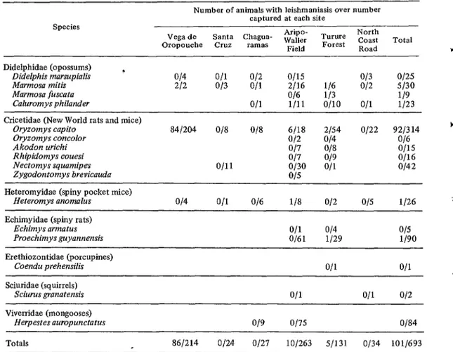
Sixteen species of small mammals from six localities in northern Trinidad were trapped and examined for leishmanial infections. Of 693 animals caught (exclusive of recaptures) one species, the rice rat 0. capita, accounted for 45.3 per cent of the total. Six species and a total of 101 animals were found to harbor amastigotes. 0 capito was the most commonly infected species, showing infection rates of
41.2, 33.3, and 3.7 per cent, respectively, at Vega de Oropouche, Aripo-Waller Field, and Turure Forest.
f Ecological Notes and Rodent Distribution
Vega de Oropouche. Only four species of small mammals were trapped at Vega de Oro- pouche (see Table l), and 96.3 per cent of the
214 animals collected were 0. capita. A total of c. 2,327 trap nights6 yielded an average of 20.6
animals per 100 trap nights (this figure includes 275 recaptures-see Table 2). The peak months ‘Operation of a single trap for one night consti-
tutes one trap night. f
TABLE 1-Leisbmania infections found on animals captured at various localities in Trinidad, August 1968-December 1971.
Species
Number of animals with leishmaniasis over number captured at each site
Vega de Santa Chagua- Aripo- Wailer Turure North
Oropouche Crll7. ramas Field Forest Coast Road Total *
Didelphidae (opossums) ~ Didelphis marsupialis
Marmosa mitis Marmosa fuscata
Caluromys philander
?-
014 O/l 012 o/15 O/3 O/25
212 O/3 O/l 2116 116 o/2 5130
O/6 113 119
O/l l/11 o/10 011 l/23 :
Cricetidae (New World rats and mice) Oryzomys capito
Oryzomys concolor Akodon urichi Rhipidomys couesi Nectomys squamipes Zygodontomys brevicauda
84/204 O/8 O/8 6118 2154 o/22 9213 14 c
o/2 O/4 016
O/7 ‘318 o/15 P
O/7 019 O/16
o/11 o/30 O/l o/42
015 Heteromyidae (spiny pocket mice)
Heteromys anomalus O/4 O/l O/6 l/8 o/2 015 l/26
Echimyidae (spiny rats) Echimys armatus
Proechimys guyannensis O/61 O/l O/4 l/29 015 i/90
Erethiozontidae (porcupines)
Coendu prehensilis O/l O/l 4
Sciuridae (squirrels)
Sciurus granatensis O/l O/l 012
Viverridae (mongooses)
Herpestes auropunctatus o/g o/75 O/84
Tikusingh * RODENT LEISHMANUSIS IN TRINIDAD 237
TABLE 2-Oryzomys capito specimens trapped at Vega de Oropouche, Trinidad, November 1969Sep- tember 1971.
1969 1970 1971 Totals
New captures Recaptures
Total No. of all captures Percentage of captures
that were recaptures
;: 170 27 204 232 43 275
7 402 70 479
0 57.7 61.4 57.4 Trap nights
New captures/l00 trap nights Recaptures/100
trap nights Total captures/100
trap nights
96 1,481 7.50 2,327 7.3 * 11.5 3.6 8.8 0 15.7 5.7 11.8 7.3 27.2 9.3 20.6
of collection were August and September 1970, when animals were captured at the respective rates of 39 and 33 per 100 trap nights. However, the peak months for new captures (excluding recaptures) were February, March, and April 1970 (see Figure 2).
Seventeen pregnant 0. capito captured at Vega de Oropouche were kept in the laboratory for delivery of their litters. Figure 3 shows the month in which the litters were born, together with the numbers of immature individuals trapped over the 23-month period. This infor- mation strongly implies that breeding occurs throughout the year.
Aripo-Walk Field. Here the variety of mam- mals trapped was greater, as shown in Table 1. The mongooses (Herpestes auropunctatus) were trapped in the Savannah area at Waller Field in FIGURE 2-Fluctuations in the number of Oryzomys capito captured at Vega de Oropouche, Trinidad, November 1969-1971.
39 36
association with another program; none were found to be positive for Leishmania. This species was therefore not included in any further analysis or discussion.
The spiny rat (Proechimys guyannensis) was the most abundant animal captured, accounting for 32.5 per cent of the total catch. The water rat (Nectomys squamipes) was next (15.9 per cent), followed by 0. capito (9.5 per cent).
Approximately 31 per cent of the mammals trapped (excluding mongooses) belonged to one of seven arboreal species; however, a majority of these were caught in traps placed on the ground.
FIGURE 3-Monthly captures of immature Oryzomys capifo correlated with the number of litters produced in the laboratory by wild-caught pregnant females. Except for one immature individual, captured at Aripo-Wailer Field in August 1970, all the animals were caught at Vega de Oropouche.
,MMATlJRES
CAPTURED
238 PAHO BULLETIN l Vol. VIII, No. 3, 1974
Location of Lesions
Nearly all the lesions found were located on the dorsal part of the tail. Usually they were situated at the base of the tail, but they were also found occasionally in the mid-tail region. The entire tail of one 0. capito was covered with lesions consisting of swellings and depres- sions. Another 0. capito had a lesion on the left ear, and another had a swelling on one of its toes on the right hind foot that was found to contain abundant amastigotes. Similar instances of parasite distribution and appearance have been described by other workers (8, 9).
Duration of infection
The marking and releasing program allowed us to observe the duration of natural infections. Out of 36 0. capito that were recaptured and another five that were kept in captivity, 24 maintained their infections for at least 30 days. Of the 24, six possessed amastigotes for at least 100 days. One animal, designated TRVL 12602, was kept in the laboratory after its eighth capture. When it eventually died, it had maintained its infection for 285 days (see Table 3). Herrer, et aZ. (8) found similar long-lasting infections in four 0. capito with recorded durations of 164, 266, 267, and 286 days. One naturally infected murine opossum (Marmosa
mitis) that was kept in the laboratory main- tained its infection for at least 41 days.
Results: NNN Cultures
Skin snips from 80 animals were inoculated into NNN media. Of these, two (both from 0. capita) were positive for promastigotes; 2i were negative and 57 were contaminated. The promastigotes appeared between 5 and 10 days and can therefore be considered “fast-growing” strains. These promastigotes failed to grow when inoculated into vertebrates (Mesocricetus auratus, Mus musculus, and 0. capita).
Scrapings were taken from the tail of a Marmosa mitis and inoculated intradermally
into the nose and right hind foot of a hamster. An inconspicuous lesion found on the inocu- lated foot after three months contained abun- dant amastigotes. Material taken from this lesion and inoculated into NNN medium pro- duced promastigotes in five days.
Results: Seasonal Incidence
The seasonal infection data for 0. capita at Vega de Oropouche is shown in Figure 4. The number of 0. capito examined is also plotted, but where an animal was positive for Leish- mania on more than one occasion it was only counted once. The most animals were examined
TABLE 3-Duration of leishmania infections in Oryzomys capito infected naturally and recaptured (Vega de Oropouche, Trinidad).
No. of infected
animals
No. of times each animal was
recaptured
Minimum duration of infection in days for each
infected animal
20 1 7, 8,9,13,14,16,16, 17,20,
20, 21,22,28,36,37,41,43, 50,70,70
9 2 7, 29,41,47,56,70,91,97,
153
1 4 83
1 5 159
3 6 91,103,117
1 7 285
Tikasingh l RODENT LEISHMANIASIS IN TRINIDAD 239
Y
l
FIGURE 4-Seasonal incidence of cutaneous leish- mania& in specimens of Oryzonzys capito captured at Vega de Oropouche, Trinidad, November 1969-1971.
in March 1970, but the peak number of infections was not recorded until June 1970, the beginning of the rainy season.
Fewer animals were examined in 197 1, but a higher proportion of these were shown to harbor Leishmania infections. Only 35.7 per cent of the animals were positive (10 of 28 examined) in the period January-March 1971, but this proportion rose to 85.7 per cent (12 of 14 examined) in the April-September period; the difference between these figures is highly significant (0.00 1 < p < 0.0 1).
Discussion
The foregoing data suggest that Oryzomys capito is the reservoir host for the species of Leishmania under investigation. Not only is it one of the most common rodents found in the areas studied, but overall (taking recaptures into consideration) some 30 per cent of the 0. capito trapped were found to be infected. At Vega de Oropouche the infection rate for this species on first capture was even higher (41 per cent). Moreover, high infection rates have been found for the same species in Panama (36 per cent-S); in Brazil’s Mato Gross0 State (54 per cent-l@; and in the Amazon Basin (10 per cent, reported by Nery-Guimaraes, cited by Lainson and Shaw-9, and 18 per cent, reported by Lainson and Shaw-9).
The fact that some 0. capito maintained their infections for long periods means they
have the potential to serve as constant sources of amastigotes for vectors. The long-lasting infections could also allow the parasite to survive through the dry season (January-May) when vector densities are generally low. These additional factors further suggest that 0. capito is the reservoir host of the parasite in the area studied.
During nine months of trapping at Vega de Oropouche in 1971 the capture rate for Oryzomys was relatively low-only about four animals per 100 trap nights as compared to 11 per 100 trap nights in 1970. The maximum was recorded in March 1971, after which there was a steady decline until the Leishmania program at Vega de Oropouche was terminated in September. It is difficult to say whether this decline reflected either a true population “crash” or emigration.
In May 1971 heavy lumbering started in the area. The felling of trees with a height of 25 meters or more does considerable damage to the smaller ones. This circumstance, together with the opening of several tracks in the forest to remove logs, could have altered the habitat of the forest floor to such an extent that small mammals in the area were affected.
In October 1970 and again in January 1971 it was discovered that the traps were being interfered with by unknown agents (possibly a dog, which was seen in the area on several occasions). This could account for the decline in the numbers of rodents caught in these two months. Even if this was the case, however the sudden decline to a very low level in the period May-September could well have been caused by a true population “crash” or by emigration.
240 PAHO BULLETIN - Vol. VIII, No. 3, 19 74
normal population levels in certain Trinidadian forests.
Since it appears that 0. capito breeds throughout the year, it seems probable that susceptible animals are constantly available and that Leishmania occurs regularly (subject to the availability of adequate numbers of vectors). Therefore, the peak appearance of infected animals in June 1970 could possibly be corre- lated with the gradual buildup of susceptible immature individuals over the preceding months (see Figures 2 and 4).
The murine opossums Marmosa fuscata and M. mitis seem to be secondarily involved, since some 1.5 per cent were found to be infected. Although they are arboreal species, the fact that 39 individuals were captured in ground- placed traps indicates they often descend to the ground, where they may come in contact with the vector. Other workers have found low rates of infection for opossums-one out of 35 in Panama (8); one out of 13 examined in Brazil’s Mato Gross0 State (10); none out of 13 in the Amazon Basin (9); and more recently, one out of 40 along the Trans-Amazon highway (13; Lainson, personal communication). These inves- tigators may be working with a different species of Leishmania, or the behavior of the species of opossums investigated may be different. In Belize, opossums examined for Leishmania were found not to be infected (5, 22).
The presence of amastigotes in a sore on a single’ Caluromys philander opossum constitutes the first record of the parasite in this mammal.
The low infection rate found for Proechimys guyannensis could reflect the methods and criteria used to tell whether an animal was infected or not. Animals whose skin appeared normal were considered negative; but Lainson (13) has reported isolating Leishmania mexi- cana amazonensis from 2 1 P. guyannensis in the
Amazon Basin with apparently normal skin. In Panama, Herrer, et al. (7) also isolated a species of Leishmania from normal skin biopsies of the porcupine Coendu rothschildi and found in another study (II) that 40 per cent of a group of infected animals showed no signs of gross dermal alterations. No P. guyannensis were captured at Vega de Oropouche during our recent work, but it was the most common rodent collected at Aripo-Waller Field, where it could play a more important role in the ecology of the parasite than indicated by the investiga- tion reported here.
Species of Leishmania Involved
Recent papers by Lainson and Shaw (II, 13) have helped put the large assortment of New World leishmanias into some kind of order. These authors recognize two complexes (L. mexicana and L. brasiliensis) each with a number of subspecies. Using this classification, it seems reasonably clear that the subspecies we have been investigating is L. mexicana ama- zonensis. This is indicated by the reservoir hosts involved, as well as by the behavior of the parasite in NNN medium and hamsters.
Human Involvement
Lainson and Shaw (II, 13) have only rarely found L. mexicana amazonensis infecting man in north Brazil. They consider this due to the disinclination of the vector to bite people. Human leishmaniasis appears rare in Trinidad, and the occasional case may not be detected. One human case, attributed to L. m. mexicana, was recently reported (4) from Cedros in southwestern Trinidad, but where the patient became infected is not known.
SUMMARY
c Tikasingh * RODENT LEISHMANIASIS IN TRINIDAD 241
infections were Marmosa mitis, M. fuscata, Caluromys philander, Heteromys anomalus, and Proechimys guyannensis.
Recapture of previously released specimens t showed that the 0. capito infections were long-lasting; in one case an infection persisted for 285 days.
NNN cultures of promastigotes were ob- tamed by culturing material from the leish- mania1 lesions of wild rodents and from the foot lesion of a hamster inoculated with -I
scrapings from naturally infected Marmosa mitis. The cultures became positive in five to ten days.
At one locality (Vega de Oropouche) the largest number of 0. capito were found in- fected in June (1970), at the beginning of the rainy season.
It was concluded that the subspecies of Leishmania under investigation wasL. mexicana amazonensis.
ACKNOWLEDGMENTS The studies and observations on which this
paper is based were conducted with the support and under the auspices of the Governments of * Trinidad and Tobago, Barbados, Guyana, Jamaica, and the Eastern Caribbean Territories, and of the Overseas Development Administra- tion of the United Kingdom and the Rocke- feller Foundation. Grateful acknowledgment is also made to the Standing Advisory Committee for Medical Research in the British Caribbean for providing a grant (R 218 1) to carry out a portion of this work.
Appreciation is extended to Drs. R. Lainson
and J. J. Shaw of the Wellcome Parasitology Unit, Belem, Brazil, and to Dr. R. Lumsden of the London School of Hygiene and Tropical Medicine for reading the manuscript and making several suggestions.
I want to thank Mr. C.O.R. Everard for allowing me to examine the mongooses and some of the other mammals which were trap- ped in the course of his projects, and also to thank the following technicians who assisted at various times: Miss Denise Wilson, Mr. R. Martinez, Mr. A. Guerra, and Mrs. L. Bhagwandin.
REFERENCES
l
(I) Ashcroft, M. T. A history and general survey of the helm&b and protozoal Infections of the West Indies. Ann Trop Med Parasitol 59:478-493,1965.
(2) Beard, J. S. The Natural Vegetation of Trimdad. Oxford, England; Clarendon Press, 1946. (Oxford Forestry Memoirs, No. 20).
(3) Be& T.I.W. Management of the Trinidad Mora Forests with Special Reference to the Matura Reserve. Port of Suain, Government of Tri- nidad and Tobago, Forestry Division, 1971. (4) Bhaskar, A. G. Fist reported case of cutaneous
leishmaniasis in Trinidad. (In press.) (5) Disney, R.H.L. Observations on a zoonosis:
leishmaniasis in British Honduras. Journal of Applied Ecology 5: l-59, 1968.
(6) Everard, C.O.R., and E. S. Tikasingh. Ecology of the rodents Proechimys guyannensis trinitatis and Oryzomys capito velutinus, on Trinidad. J Mammal 54: 815-886, 1973.
(7) Herrer, A., V. E. Thatcher, and C. M. Johnson. Natural infections of Leishmania and trypano- somes demonstrated by skin culture. J Para- sit01 52: 954957,1966.
(8) Herrer, A., S. R. Telford, and H. A. Christensen. Enzootic cutaneous leishmaniasis in eastern Panama: I. Investigation of the infection among forest mammals. Ann Trop Med Para- sitol65: 349-358, 1971.
(9) Lainson, R., and J. J. Shaw. Leishmaniasis in Brazil: I. Observations on enzootic rodent leishmaniasis: Incrimination of Lutzomyia fkviscutellata (Mang.) as the vector in the lower Amazon basin. Tram R Sot Trop Med Hyg 62: 385-395,196s.
(10) Lainson, R., and J. J. Shaw. Leishmanlasis in Brazil: V. Studies on the epidemiology of cutaneous leishmaniasis in Mato Gross0 State and observations on two distinct strains of Leishmania isolated from man and forest animals. Trans R Sot Trop Med Hvg 64: _- 654-667, 1970.
(II) Lainson, R., and J. J. Shaw. Epidemiological considerations of the leishmanias with par- ticular reference to the new world. In: Ecology and Physiology ofparasites. Toronto, University of T&onto&e&, 1971.
(12) Lalnson, R., and J. J. Shaw. Leishmaniasis in the New -World: Taxonomic problems. Br Med
Bull 28: 4448,1972.
(13) Lainson, R., and J. J. Shaw. Leishmanias and Leishmaniasis of the New World, with par- ticular reference to Brazil. Bulletin of the Pan American Health Organization, Volume 8, No.
3, 1973, pp. 1-19.
242 PAHO BULLETIN - Vol. VIII, No. 3, 1974 4
Leishmania mexicuna among the forest rodents. Trans R Sot Trop Med Hyg 58: 136-153, 1964.
(15) Tikasingh, E. S. Leishmaniasis in Trinidad. A preliminary report. Trans R Sot Trop Med Hvn63: 411,1969.
(16) T&dad and Tobago. Administration Report of
the Surgeon-General for the Year 1925. Port of Spain, Government Printer, 1926. (17) Trinidad and Tobago. Administration Report of
the Surgeon-General for the Year 1926. Port of Spain, Government Printer, 1927.
(IS) Trinidad and Tobago. Administration Report of
the Surgeon-General for the Year 19i7. Port of Spain, Government Printer, 1928.
(19) Trinidad and Tobago. Administration Report of
the Surgeon-General for the Year 1928. Port of Spain, Government Printer, 1929.
(20) Trinidad and Tobago. Administration Report of
the Surgeon-General for the Year 1929. Port of Spain, Government Printer, 1930.
(21) Trinidad and Tobago. Administration Report of
the Surgeon-General for the Year 1930. Port
of Spain, Government Printer, 1931. t (22) Trinidad and Tobago. Administration Report of
the Surgeon-General for the Year 1931. Port of Spain, Government Printer, 1932.
(23) Williams, P. Phlebotomine sandflies and Ieish- maniasis in British Honduras (Belize). Trans R Sot Trop Med Hyg 64: 317-368,197O. (24) Worth, C. B., W. G. Downs, T.H.G. Aitken, and
E. S. Tikasingh. Arbovirus studies in Bush t Bush Forest, Trinidad, W.I., September
1959-December 1964. IV. Vertebrate popula- tions. Am J Trop Med Hyg 17: 269-275, 1968.
c