R E S E A R C H
Open Access
Trypanosoma cruzi
Discret Typing Units
(TcII and TcVI) in samples of patients from two
municipalities of the Jequitinhonha Valley, MG,
Brazil, using two molecular typing strategies
Maykon Tavares de Oliveira
1, Girley Francisco Machado de Assis
2, Jaquelline Carla Valamiel Oliveira e Silva
1,
Evandro Marques Menezes Machado
1, Glenda Nicioli da Silva
3,4, Vanja Maria Veloso
3,4, Andrea Mara Macedo
6,
Helen Rodrigues Martins
5and Marta de Lana
1,3,4*Abstract
Background:Trypanosoma cruziis classified into six discrete taxonomic units (DTUs). For this classification, different biological markers and classification criteria have been used. The objective was to identify the genetic profile ofT. cruzi
samples isolated from patients of two municipalities of Jequitinhonha Valley, MG, Brazil.
Methods:Molecular characterization was performed using two different criteria forT. cruzityping to characterize 63T. cruzisamples isolated from chronic Chagas disease patients. The characterizations followed two distinct methodologies. Additionally, the RAPD technique was used to evaluate the existence of genetic intragroup variability.
Results:The first methodology identified 89 % of the samples as TcII, but it was not possible to define the genetic identity of seven isolates. The results obtained with the second methodology corroborated the classification as TcII of the same samples and defined the classification of the other seven as TcVI. RAPD analysis showed lower intra-group variability in TcII.
Conclusions:The results confirmed the preliminary data obtained in other municipalities of the Jequitinhonha Valley, showing a predominance of TcII, similar to that verified in northeast/south axis of Brazil and the first detection of TcVI in the study region. The second protocol was more simple and reliable to identify samples of hybrid character.
Keyword:Trypanosoma cruzi, Genotyping,T. cruziDTUs, Chronic patients, Jequitinhonha Valley, MG, Brazil
Background
At present approximately 6 to 7 million people are esti-mated to be infected worldwide withTrypanosoma cruzi,
the etiologic agent of Chagas disease, mostly in Latin America where Chagas disease is endemic [1]. T. cruzi,
is a flagellate digenetic protozoan belonging to the order Kinetoplastida, family Trypanosomatidae [2], dispersed throughout the American continent from Argentina and
Chile to the southern United States of America. Several studies have demonstrated that this protozoan is heteroge-neous, consisting of several sub-populations of parasites that circulate in both, domestic and wild environments, with a high rate of biological and genetic diversity [3–5].
Currently, according to the second taxonomic consensus forT. cruziapproved during the XXV Protozoology Meet-ing held in Buzios, RJ, Brazil, the species is subdivided into six discrete typing units (DTU) named TcI, TcII, TcIII, TcIV, TcV and TcVI [6], related to several previous classifications based on different molecular markers. Regarding the geo-graphical distribution of theT. cruzigenotypes, it has been demonstrated that TcI has the largest distribution in all America. In Colombia, Mexico, Guatemala, Venezuela, Panama and Bolivia there is evidence of a predominance of * Correspondence:delana@nupeb.ufop.br
1Núcleo de Pesquisas em Ciências Biológicas (NUPEB), Universidade Federal de Ouro Preto (UFOP), Campus Universitário Morro do Cruzeiro, CEP: 35400-000, Ouro Preto, MG, Brazil
3Departamento de Análises Clínicas, Escola de Farmácia, UFOP, CEP: 35400-000 Campus Universitário Morro do Cruzeiro, CEP: 35400-000, Ouro Preto, MG, Brazil
Full list of author information is available at the end of the article
this DTU circulating in the sylvatic [7] and domestic cy-cles, associated in some cases to cardiac clinical forms in humans [8–10]. In the Southern Cone countries, both DTUs (TcI and TcII) were observed in the sylvatic cycle [8–10]. However, only TcII was predominantly associated with human infection, while TcI was rarely found in humans [11, 12].T. cruziIII was detected in human infec-tions [8] and both, TcIII and TcIV [8, 9] are mainly en-countered in the sylvatic and domestic cycles. TcII, TcV and TcVI are frequently isolated from infected individuals in the south of America but rarely isolated from sylvatic transmission cycles [13, 14].
Although few studies have been accomplished in Brazil concerning lesserT. cruzisubdivisions, there is evidence that the majority of the strains isolated from patients be-long to TcII [11, 15, 16] and less frequently to TcV; except in the Amazon Basin where TcI is the most prevalent DTU infecting humans while TcIII and TcIV DTUs were occasionally recorded [17, 18]. At present in Brazil, the TcII strains seem to be more associated with human infec-tions responsible for tissue damage, and consequently with several clinical forms of Chagas disease, while cases of human infections caused by TcI strains are still rare and usually asymptomatic [19], despite the recording of some symptomatic cases of Chagas disease in the Amazon with cardiac manifestations [20, 21].
Due to the scarcity of publications regarding the geo-graphic distribution of the newly classified T. cruzi
DTUs, including in Brazil, this study proposed to characterize genetically samples of this parasite isolated from patients with chronic Chagas disease living in an important endemic area of Brazil named Jequitinhonha Valley. We aimed to highlight that the knowledge of the distribution and intragroup variability of the newly categorizedT. cruzi genotypes in the domestic cycle of Chagas disease in this region, where all severe clinical forms of the disease are present, may provide additional contributions to further investigation of the association between the T. cruzi genotype and the pathophysio-logical aspects of this disease, not evaluated yet, con-tinuously researched by several authors [16, 22].
Methods
Patients and samples ofT. cruzi
The samples of T. cruzi (n = 63) evaluated in this study were isolated from patients in the early (7/63 patients with less than 14 years old) and later chronic phases (56/63) of Chagas disease, all born and living in the municipalities of Berilo (62 patients) and José Gonçalves de Minas (only one patient), distant 24 km, both of the Jequitinhonha Valley, MG, Brazil. There were 19 male and 44 female patients, aged between 7 and 73 years. For isolation of the parasites the hemoculture technique [23] was used. In addition, the reference clones of the six T. cruzi DTUs, kindly provided by Dr. Michel Tibayrenc (IRD, France), were also characterized in parallel: TcI (P209 cl1, 92101601P cl1 and Cutia cl1), TcII (MAS cl1 and Tu18 clI), TcIII (CM-17 and X-109/2), TcIV (CAnIII cl1 and 92122102R), TcV (BUG2148 cl1 and SO3 cl5) and TcVI (P63 cl1 and Tula-huen cl2).
Preparation ofT. cruzicell pellets
After isolation by hemoculture parasites were maintained in growth by successive addition of LIT (Liver Infusion Tryptose) medium up 35 ml of culture. Then, the cultures were subjected to four cycles of washing and centrifugation, using sterile phosphate buffered saline (PBS) at 3500 rpm, 4 °C to prepare damp mass.
Extraction of DNA
DNA extraction was processed after thawing and homogenization of the wet mass of each T. cruzi
sample. DNA from the samples was obtained using the WizardTM Genomic DNA Purification Kit (Pro-mega, Madison, WI, USA), following the manufactur-er's instructions. For molecular analysis, the DNA samples were diluted at a concentration of 3 ng/μL.
Control DNA and reagent-free samples were proc-essed in parallel.
Criteria forT. cruzigenotyping
The criteria [24] and [16] showed in Table 1 were used for
T. cruzi genotyping, both recommended by the expert
Table 1Genotyping ofTrypanosoma cruziisolates into DTUs (TcI–TcVI) according to the methodologies of [24] and [16]
T. cruzi
DTU
24sαrDNA
Souto et al. (1996)
RFLP-HSP60
Sturm et al.(2003)
RFLP-GPI
Westenberger et al. (2005)
RFLP-COXII
Freitas et al. (2006) SL-IRBurgos et al. (2007) DNA fragments in base pairs and the number of bands expected
TcI 110 bp 1 band 2 bands Haplotype A (262 bp + 81 bp + 30 bp) ~150/157 bp
TcII 125 bp 1 band 3 bands Haplotype C (212 bp + 81 bp) ~150/157 bp
TcIII 110 bp 2 bands 2 bands Haplotype B (294 bp + 81 bp) 200 bp
TcIV ~120 bp 1 band 3 bands Haplotype B (294 bp + 81 bp) 200 bp
TcV 110 bp + 125 bp 3 bands 4 bands Haplotype B (294 bp + 81 bp) ~150/157 bp
committee [4]. In the genotyping protocol [24] the samples were subjected to a PCR algorithm for DTU genotyping which combines the analyses of the polymorphism of the
24sα-LSU rDNA gene as well as the profile of bands
ob-tained after PCR-RFLP of theHSP60andGPIgenes. In the protocol [16] the24Sα-LSU rDNA miniexons and the
pro-file of bands observed after PCR-RFLP of subunit II of the Cytochrome oxidase gene polymorphism were analyzed.
Amplification of the 3' region of the rDNA gene24Sα-LSU rDNA
All DNA samples were subjected to three successive PCR amplifications of the divergent domain D7 of24Sαsubunit
rDNA (LSU rDNA) according to methodology [25] using a thermocycler (Biocycler MJ96G). The PCR products were subjected to electrophoresis in 6 % polyacrylamide gel and revealed by silver staining [26].
PCR-RFLP (restriction fragment length polymorphism) of
HSP60(heat shock protein) andGPI(glucose 6-phosphate
isomerase) genes
The polymorphism of HSP60 (heat shock protein) and GPI (glucose 6-phosphate isomerase) genes for the popula-tions ofT. cruzi was evaluated according to the protocol [27] using the primersHSP60-1andHSP60-2described in [28] forHSP60and the primers SO1 and SO2 described in [29] for GPI.
PCR was performed using the thermocycler Biocycler MJ 96G, and the digestion reaction with the restriction en-zymes was performed using EcoRV for HSP60 and HhaI
for GPI according to the manufacturer’s instructions. The products were subjected to electrophoresis in a 1.5 % agarose gel and revealed by staining with ethidium bromide.
PCR of the mitochondrial gene cytochrome oxidase subunit II
For amplification of the gene region comprising a fragment of subunit II of the mitochondrial enzyme Cytochrome oxi-dase (COII) the protocol [30] using TcMit10 and TcMit21 primers was used. Subsequently, 10 μL of the amplified
products were digested employing the restriction enzyme
AluI(Invitrogen, USA, 4 to 12 U/μL) according to the
man-ufacturer’s instructions. The fragments generated were vi-sualized in 6 % polyacrylamide gel stained with silver [26].
Intergenic spacer miniexon genes ofT. cruzi(SL-IR) For amplification of the intergenic region of the miniexon genes the protocol [31] was used employing TcIII and UTCC primers. The analysis of the amplified products was performed in 1.5 % agarose gel stained with ethidium bromide.
RAPD (Random Amplified Polymorphic DNA)
RAPD analysis following the [32] methodology was used with the objective of verifying the intra-group genetic vari-ability using 10 different primers: A10 - 5’ GTGATCGC AT 3’, A7 - 5’GAAACGGGTG 3’, A15 - 5’TTCCGAACCC 3’, B15 - 5’GGAGGGTGTT 3’, B19 - 5’ACCCCCGAAG 3’, F13 - 5’ GGCTGCAGAA 3’, F15 - 5’CCAGTACTCC 3’, N9 - 5’ TGCCGGCTTG 3’, N19 - 5’ GTCCGTACTG 3’, U7 - 5’CCTGCTCATC 3’.
The RAPD profiles were used to construct a matrix of presence/absence of each band visualized, from which an analysis of similarity was constructed [33] using NTSYSpc software [34]. The relationships be-tween T. cruzi strains were estimated using a dendro-gram representative of the RAPD data. They were constructed based on DICE coefficient [33] and un-weighted pair group analysis (UPGMA) using the Mega 6.04 Beta software. In order to estimate Shannon diver-sity, cophenetic correlation coefficient, heterozygosity per locus (He) and principle coordinates analysis (PCoA) were performed using GenAlEx 6.5 software. In this case, a genetic distance matrix was constructed. This calculation of pairwise genetic distances for binary data followed the method [35], in which any compari-son with the same state yields a value of 0 (both 0vs.0 comparisons and 1 vs. 1 comparisons), while any com-parison of different states (0 vs. 1 or 1 vs. 0) yields a value of 1.
Ethics
The isolation of the T. cruzi samples here characterized was obtained of the patients by blood collection performed after obtaining of a signed consent form approved by the Ethics Committee for Research in Humans from the Centro de Pesquisas René Rachou (CPqRR), FIOCRUZ, Belo Horizonte, MG (Process Number 007/02).
Results
Genotyping according to the methodologies24SαrDNA, PCR-RFLPHSP60and GPI genes [24]
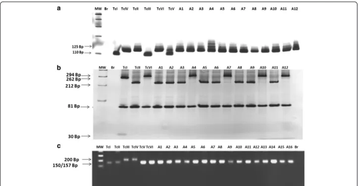
According to the criterion [24] 56 out of 63 isolates showed band profiles compatible with TcII DTU: 125 bp (rDNA type I) for24Sα rDNA gene products (Fig. 1); a
single band for PCR-RFLP HSP60/EcoRV (Fig. 1 and Table 2) and three bands for PCR-RFLPGPI/HhaI(Fig. 1 and Table 2) compatible with the reference clone of the TcII DTU (MAScl1). However, with seven isolates it was not possible to reach a consensus following this method-ology. Although these seven samples gave the 125 bp fragment for analysis of the polymorphism of D7 24Sα
(triple fragments ranging from 100 to 432 bp) character-istic of hybrid lineages or TcV or TcVI (Fig. 1 and Table 2), and double bands, characteristic of the lineages TcI and TcIII for PCR-RFLP of the GPI gene (Fig. 1 and Table 2).
Genotyping according to the methodologies24SαrDNA, PCR-RFLP COII gene and Miniexon SL-IR [16]
Due to results obtained above all samples were also ana-lyzed by the protocol proposed by [16]. The fifty-six sam-ples previously identified as TcII by the protocol [24] had Fig. 1a- Profiles obtained by DNA genotyping ofTrypanosoma cruzisamples isolated from patients of the Jequitinhonha Valley, Minas Gerais, Brazil, resultant from the amplification of the 3' region of the24SαrDNA gene. MW - Molecular weight of 100 bp, Br - negative reaction control,
TcI - representative clone of TcI lineage (P209 cl1, 110 bp), TcIV - representative clone TcIV lineage (CANIII, 120 bp), TcII - representative clone of TcII lineage (MAS cl1, 125 bp), TcIII - representative clone of the TcIII lineage (CM-17, 110 bp), TcVI - representative clone of the hybrid lineage TcVI (Tulahuen cl2, 125 bp) and TcV - representative clone of TcV lineage (Bug2148, 110 bp + 125 bp). A1–sample 229, A2–sample 452, A3–sample 748, A4 - sample 798, A5–sample 205, A6–sample 1918, A7–sample 2535, A8–sample 2491, A9–sample 264, A10–sample 376, A11–sample 646 and A12–sample 211.b- Profiles obtained with DNA ofTrypanosoma cruzisamples isolated from chagasic patients of the Jequitinhonha Valley, Minas Gerais, Brazil, by the analysis of theHSP60gene polymorphism after PCR-RFLP. MW - Molecular weight of 100 bp, A1–sample 1918, A2–sample PSF060, A3–sample 438, A4–sample 543, A5–sample 229, A6–sample 2119. Br–PCR negative control; TcI–TcVI samples profile of the reference clones of the 6 lineages ofT. cruzi(TcI - P209 cl1, single band of 432–462 bp; TcII–MAS cl1, single band of 432–462 bp; TcIII–CM-17 double bands; TcIV–CAN III cl1, single band; TcV–Bug 2148, triple bands; TcVI–Tulahuen cl2, triple bands).c- Profiles of DNA genotyping ofT. cruzi
samples isolated from chagasic patients of the Jequitinhonha Valley, Minas Gerais, Brazil, by the analysis ofGPI/HhaIgene polymorphism via PCR-RFLP. MW - Molecular weight of 100 bp, BR–PCR negative control, A1–sample 2014, A2–sample 646, A3–sample 453, A4–sample 2119, A5–sample 440, A6–sample 501, A7–sample 523, A8–sample 2478, A9–sample PSF060, A10–sample 1100, A11–sample 820, and A12–sample 2535.TcII–(clone TU18 cl1 representative of DTU TcII), TcV–(clone SO3 cl5, DTU TcV), TcI–(clone 92101601P cl1, DTU TcI), TcIII–(Clone X109/2, DTU TcIII), TcVI–(clone Tulahuen, DTU TcVI) and TcIV–(CANIII cl1 DTU TcIV)
Table 2Trypanosoma cruzigenotyping of samples isolated from patients of the Jequitinhonha Valley, MG, Brazil Molecular typing
criteria
T. cruziSamples
DTU TcII DTU TcVI
Lewis et al., (2009) PSF060, 103, 264, 299, 376, 438, 440, 452, 467, 479, 493, 501, 523, 525, 529, 543, 595, 646, 653, 701, 728, 791, 795, 806, 817, 818, 820, 829, 839, 845, 855, 860, 896, 914, 953, 1100, 1107,1113, 1315, 1442, 1536, 1661, 1662, 1663 1635, 1918, 2014,2336,2337, 2339, 2405, 2408, 2478, 2491, 2495, 2497
?????
D’Ávilla et al., (2009) PSF060, 103, 264, 299, 376, 438, 440, 452, 467, 479, 493, 501, 523, 525, 529, 543, 595, 646, 653, 701, 728, 791, 795, 806, 817, 818, 820, 829, 839, 845, 855, 860, 896, 914, 953, 1100, 1107, 1113, 1315, 1442, 1536, 1661, 1662, 1663 1635, 1918, 2014, 2336, 2337, 2339, 2405, 2408, 2478, 2491, 2495, 2497
229, 748, 798, 1205, 1337, 2118, 2119
Total (63 samples)
56 samples DTU TcII
the typing confirmed: 125 bp (rDNA type I) for amplified products of24SαrDNA; 212 bp + 81 bp, characteristic of
mitochondrial haplotype C lineage and 150 bp −157 bp for amplified products of intergenic spacer of the mini-exon (SL-IR) ofT. cruzi, typical of TcII MAScl1 reference clone. The seven samples with previous controversial identification (229, 748, 798, 1205, 1337, 2118, 2119) showed a double profile of bands of 294 bp and 81 bp (Fig. 2 and Table 2) for PCR-RFLPCOII, similar to the ref-erence clones of the haplotype B (CM-17 and Tulahuen cl2) or strains belonging to TcIII, TcIV and TcVI lineages. Regarding the amplification of the DNA fragment of the intergenic miniexon spacer (SL-IR) ofT. cruzi, all samples showed band profiles of approximately 150–157 bp, char-acteristic of the TcI, TcII, TcV and TcVI lineages [31] (Fig. 2 and Table 2).
Therefore, according to the protocol [16] it was verified that the seven samples with inconclusive results in the
protocol proposed by [24] were all classified as belonging to DTUTcVI because they presented a profile of 125 bp fragment for the rDNA gene, approximately 150–157 bp fragment for the intergenic spacer of the miniexon and 294 bp and 81 bp (mitochondrial haplogroup B) which allowed the classification of the T. cruzi samples as belonging to TcVI compatible with the Taluhen cl2 refer-ence clone (Table 1) of thisT. cruziDTU.
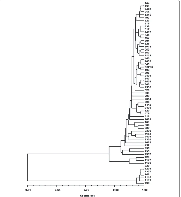
RAPD analysis
For RAPD analysis only bands showing sharp fragments were selected. After amplification 4731 fragments were detected in the two different DTUs. The size of bands ranged between 100 and 1400 bp.
The RAPD profiles were complex, with a total of 81 bands of which only 12 (14.8 %) were shared among all the samples. The proportion of polymorphic loci was 72.84 %. The cophenetic correlation coefficient that checks that a
Fig. 2a–Profiles obtained by DNA genotyping ofTrypanosoma cruziisolated from patients of the Jequitinhonha Valley, Minas Gerais, Brazil, of the region 3' of the24SαrDNA gene. MW - Molecular weight of 100 bp, Br–PCR negative control. TcI–TcI representative clone (P209 cl1 profile
of 110 bp), TcIV–representative clone of TcIV (CANIII, 120 bp), TcII - representative clone of TcII(MAS cl1, 125 bp), TcIII–representative clone of the lineage TcIII (CM-17, 110 bp), TcVI–representative of the hybrid lineage TcVI (Tulahuen cl2, 125 bp) and TcV–representative clone of lineage TcV (Bug2148, 110 bp + 125 bp); A1–sample 229, A2–sample 452, A3–sample 748, A4–sample 798, A5–sample 1205, A6–sample 1918, A7–
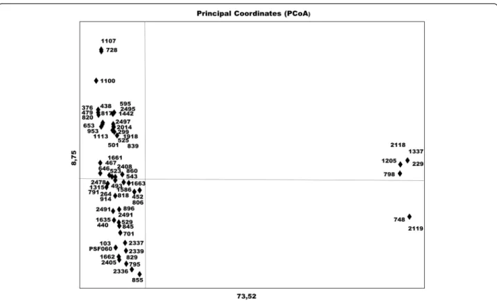
dendrogram faithfully preserves the pairwise distances between the original unmodeled data points gave a coeffi-cient value of 0.989, confirming good representation of the similarity matrices in the form of the dendrogram. The UPGMA dendrogram distinguished the samples into three distinct clusters (Fig. 3). PCoA for the first two axes con-solidated 82.27 % of the variation between the components (Fig. 4) and the total formed three groups, as shown in the
UPGMA dendrogram. The expected mean heterozygosity per locus (He) was 0.181, indicative of low genetic diversity among samples. Moreover, the Shannon diversity index (0.284), which is a quantitative measure that reflects how many different types there are in a dataset was also consid-ered low. This evaluation has been used to describe the species richness. When this index is closer to 1.0, greater is the species diversity.
Discussion
T. cruzi, the etiologic agent of Chagas disease, is com-posed of heterogeneous subpopulations circulating in both sylvatic and domestic cycles [36], and this diversity can be detected at morphological [37], biological [3], antigenic [38], epidemiological [4] and genetic levels [36, 39].
Therefore, to better understand the disease it is import-ant to study the molecular epidemiology of this parasite, which naturally is related with the characteristics men-tioned. So, the present study was developed with the pur-pose of identification of the genetic lineages of T. cruzi
samples isolated from chronic chagasic patients of the Jequitinhonha Valley, MG, considered an area of intense transmission of Chagas disease in the 1980s [40] and (ii) to evaluate the molecular descriptive epidemiology of T. cruziwithin this population.
For molecular typing it was firstly used the protocol proposed by [24]. Considering the 63 samples evaluated, a total of 56T. cruzisamples showed a profile consistent with TcII as previously demonstrated in patients of Virgem da Lapa municipality, located only 28 km from Berilo and 48 km from José Gonçalves de Minas, where the isolates were analyzed by isoenzyme profiles [41]. The first work that used the criteria proposed by [24] for
T. cruzigenotyping was the study [42] as mentioned by [4]. They identified 18 isolates from domestic cats and vectors in an endemic region of Chagas disease in Bahia,
all belonging to the TcII group. Our results corroborate the obtained in this study, indicating that this method is efficient in identifying TcII DTU. However, seven sam-ples (229, 748, 798, 1205, 1337, 2118 and 2119) revealed different patterns or combinations of bands to those expected for any DTU. The results obtained by protocol [24] already indicated genetic variation in the profiles of some clones used as reference in their work. The clone Saimiri3 cl1, for example, showed a profile of TcIV in RAPD analysis. However, the triple assay showed incon-clusive identification closer to the profile of TcII than any other genetic group. The most important issue that may hinder the identification of the isolates is the occur-rence of mixed infections or multiconality already docu-mented in vectors, several reservoirs and human [38], but fortunately not identified in our samples since the profiles of bands detected where exactly the same veri-fied in clones of reference for eachT. cruziDTU and be-cause the pattern of bands for each one is intra-DTU excludent [6]. Additionally, when a mixture of TcIII and TcII clones occurred in [43] these authors found similar profiles to TcV and TcVI, which could result in errone-ous identification or characterization of the isolates, since the samples studied by them were not cloned pre-viously to clarify this problem.
geographic areas are typed using this protocol. For our seven samples that showed different patterns of bands for the expected by the protocol [24] a process may be occurring, such as nucleotides deletion or insertion of the GPI gene sequence, which prevented the enzyme to cut the DNA sequence at the specific site. On the other hand, [44, 45] publications do not have problems with the use of GPI gene as a marker, however employing dif-ferent protocols. The isoenzyme analysis of these isolates for GPI also showed a profile of bands distinct from that expected for the TcVI stocks (data not shown). Our ana-lysis of GPI showed profiles corresponding to those expected for TcIV and TcIII DTUs, which could suggest a more extensive variation in the gene, but for others (G6PD and IDH) the profile was compatible with TcVI (data not shown). Regarding the use of the HSP60gene as a marker, there are not many publications that deal with this methodology, except the works [27] and [28]. In this study we did not observe differences between the representative samples of the same lineage for these two genes using the PCR-RFLP. However, it is important to note those authors used clones of references previously genotyped by others markers, as well as different restric-tion enzymes to cut the HSP60 and GPI gene fragments, which could explain the absence of differences observed compared to the present study that genotyped new sam-ples isolated from humans.
To solve the typing of these seven samples the protocol provided by [16] was also employed in the characterization of all samples. According to [31] when incongruity occurs in the identification ofT. cruziisolates, with probable pres-ence of a hybrid profile, the use of PCR-RFLP of theCOII
gene described by [30] is important for the characterization of the isolate to guarantee the right profile identification because after cutting the PCR products with the AluI enzyme, samples associated with the mitochondrial hap-logroup B profile can be considered as belonging to the DTUs TcIII, TcIV, TcV or TcVI. However, employing this methodology combined with the evaluation of the poly-morphism of the 3' rDNA gene region and of the inter-genic region of the miniexon gene ofT. cruzi(SL-IR) also suggested a quick, cheap and effective solution for identify-ing strains of T. cruzi as suggested by [6] and [4]. Here, seven isolates from chronic chagasic patients (229, 748, 798, 1205, 1337, 2118 and 2119) after COII/Alu RFLP showed profiles of representative clones of mitochondrial haplotype B (294 bp + 81 bp) and that the DTU observed may correspond to hybrids DTUs V and VI. The SL-IR identified the group with a band of 200 bp, characteristic of the lineages TcIII and TcIV. Thus, all samples analyzed in this study amplified by this methodology showed a frag-ment of approximately 150–157 bp, excluding the possibil-ity of the isolates being identified as TcIII or TcIV, which confirms that these samples are actually hybrids belonging
to DTU TcVI, due also to the results obtained in the rDNA analysis. The other 56 samples were confirmed as TcII genotype, including the only sample of José Gonçalves de Minas, which corroborated the classification of [24].
that clones ofT. cruziare able to establish stronger infec-tions in certain hosts and such host-parasite relainfec-tionships could work as filters for some sub-populations of the para-site [5, 49, 60]. Maybe the prolonged infection in these patients may have provided a selection of the parasites better adapted to the Berilo population or the ecoepide-miological conditions of the study area. This hypothesis justifies the similarity between the isolates grouped in a particular branch of the phenogram.
Phenomena ofT. cruzihybridization and ecogeographi-cal distribution of the parasite were also described by other authors referent to TcI in Bolivia, employing loci variability by microsatelites and mitochondrial DNA [7] and between TcI with TcIII and TcIV, respectively [61] using MLST and SNP as molecular markers. However, the study of parasites isolated from reservoirs and vectors of the same region would be necessary to better understand the eco-epidemiology ofT. cruziin the region studied.
Conclusions
The results obtained in this study agree with data in the lit-erature and demonstrate the predominance of the lineage TcII in human Chagas disease in more one endemic region of Brazil, as well as its association with genotype TcVI, now firstly described in human infection in the Jequitinhonha Valley, MG, Brazil, which may have been masked in the pi-oneer classification here used due to the limitations of the markers used which probably erroneously identified those samples as possibly TcII.
The RAPD analysis showed low variability of the genetic profiles betweenT. cruzisamples of the same lineage.
The results still suggest variation among samples from different geographic regions, and more, that the correct identification ofT. cruziDTUs should be made with cau-tion and based on a larger number of markers, mainly when hybrid samples are present.
Abbreviations
DTU:Discrete Typing Unit; TcI:Trypanosoma cruziI DTU; TcII:Trypanosoma cruziII DTU; TcIII:Trypanosoma cruziIII DTU; TcIV:Trypanosoma cruziIV DTU; TcV:Trypanosoma cruziV DTU; TcVI:Trypanosoma cruziVI DTU; MG: Minas Gerais state, Brazil; RJ: Rio de Janeiro state, Brazil; RAPD: Randomly amplified Polymorphic DNA; IRD: Institute de la Recherche pour le Dévelopment; LIT: Liver Infusion Tryptose; PBS: Phophate Buffer Solution; Rpm: Rotation per minute; DNA: Desoxiribonucleotide; RNA: Ribonucleotide; PCR: Polymerase chain reaction; RFLP: Restriction fragment length polymorphism; HSP60: Heat shock protein; GPI: Glucose phosphate isomerase;AluI: Restriction endonuclease from
Arthrobacter luteus; UPGMA: Unweighted pair group analysis; bp: Base pair; MW: Molecular weight.
Competing interests
The authors declare that they have no competing interests.
Authors’contributions
Maykon Tavares Oliveira: involved with all laboratorial activities related to the genotyping of the parasites; Girley Francisco Machado de Assis: isolation of the parasites samples; Jaquelline Carla Valamiel de Oliveira e Silva: maintenance, growth and preparation of damp mass of the parasites; Evandro Marques Menezes Machado: technical assistance to problems related to the methodologies of genotyping; Glenda Nicioli da Silva: interpretation and
analyses of RAPD data; Vanja Maria Veloso: technical assistance in RAPD technique; Andrea Mara Macedo: training in genotyping protocol [11]; Helen Rodrigues Martins: help and training of genotyping protocol [20]; Marta de Lana: coordinator of the study and collaborations. All authors have participated in this study and agree with the final version of the manuscript.
Acknowledgements
We would like to thank FAPEMIG and CNPq agencies and the Universidade Federal de Ouro Preto (UFOP) for financial support, as well as CNPq for the author’s research fellowship and Global Science Editing Ltd., UK for English language revision.
Author details
1Núcleo de Pesquisas em Ciências Biológicas (NUPEB), Universidade Federal de Ouro Preto (UFOP), Campus Universitário Morro do Cruzeiro, CEP: 35400-000, Ouro Preto, MG, Brazil.2Departamento- Básico de Saúde, Universidade Federal de Juiz de Fora (UFJF), CEP: 35010-177, Campus Governador Valadares, Governador Valadares, MG, Brazil.3Departamento de Análises Clínicas, Escola de Farmácia, UFOP, CEP: 35400-000 Campus Universitário Morro do Cruzeiro, CEP: 35400-000, Ouro Preto, MG, Brazil. 4Programa de Pós-Graduação em Ciências Farmacêuticas (CiPHARMA), Escola de Farmácia, UFOP, Campus Universitário Morro do Cruzeiro, 35400-000, Ouro Preto, MG, Brazil.5Departamento de Farmácia, Faculdade de Ciências Biológicas e da Saúde, Universidade dos Vales do Jequitinhonha e Mucuri (UFVJM), 39100-000 Diamantina, MG, Brazil.6Departamento de Bioquímica e Imunologia, Instituto de Ciências Biológicas (ICB), Universidade Federal de Minas Gerais (UFMG), 6627, Belo Horizonte 31270-901MG, Brazil.
Received: 9 July 2015 Accepted: 9 October 2015
References
1. WHO - Chagas disease (American trypanosomiasis). Fact sheet N°340. Updated March 2015.
2. Chagas C. Nova tripanozomiase humana. Estudos sobre a morfolojia e o ciclo evolutivo doSchizotrypanum cruzin. gen., n. sp., ajente etilógico de nova entidade morbida do homem. Mem Inst Oswaldo Cruz. 1909;159–218. 3. Andrade SG. Caracterização de cepas deTrypanosoma cruziisoladas no
Recôncavo Baiano. Rev Pat Trop. 1974;65–121.
4. Zingales B, Miles AM, Campbell AD, Tibayrenc M, Macedo AM, Teixeira MMG, et al. The revisedTrypanosoma cruzisubspecific nomenclature: Rationale, epidemiological relevance and research applications. Infect Genet Evol. 2012;12:240–53.
5. Macedo AM, Machado CR, Oliveira RP, Pena SD.Trypanosoma cruzi: genetic structure of populations and relevance of genetic variability to the pathogenesis of Chagas disease. Mem Inst Oswaldo Cruz. 2004;99:1–12.
6. Zingales B, Andrade SG, Briones MRS, Campbell DA, Chiari E, Fernandes O, et al. A new consensus forTrypanosoma cruziintraspecific nomenclature: second revision meeting recommends TcI to TcVI. Mem Inst Oswaldo Cruz. 2009;104:1051–4.
7. Messenger LA, Garcia L, Vanhove M, Huaranca C, Bustamante M, Torrico M, et al. Ecological host fitting of Trypanosoma cruzi TcI in Bolivia: mosaic population structure, hybridization and a role for humans in Andean parasite dispersal. Mol Ecol. 2015;24(10):2406–22.
8. Ramírez JD, Guhl F, Rendón LM, Rosas F, Marin-Neto JA, Morillo CA. Chagas cardiomyopathy manifestations andTrypanosoma cruzigenotypes circulating in chronic Chagasic patients. PLoS Negl Trop Dis. 2010;4(11), e899. 9. Carrasco HJ, Segovia M, Llewellyn MS, Morocoima A, Urdaneta-Morales S,
Martínez C, et al. Geographical distribution ofTrypanosoma cruzigenotypes in Venezuela. PLoS Negl Trop Dis. 2012;6(6), e1707.
10. Guhl F, Ramírez JD. Retrospective molecular integrated epidemiology of Chagas disease in Colombia. Infect Genet Evol. 2013;20:148–54.
11. Barnabé C, Brisse S, Tibayrenc M. Population structure and genetic typing of
Trypanosoma cruzi, the agent of Chagas disease: a multilocus enzyme electrophoresis approach. Parasitol. 2000;120:513–26.
12. Lages-Silva E, Ramírez LE, Pedrosa AL, Crema E, Galvão LMC, Pena SDJ. Variability of kinetoplast DNA gene signatures ofTrypanosoma cruziII strains from patients with different clinical forms of Chagas’disease in Brazil. JClinicalMicrobiol. 2006;44:2167–71.
and population genetics of DTUs TcV and TcI in Bolivia and Peru. Infect Genet Evol. 2011 Oct;11(7):1752-60.
14. Ramírez JD, Montilla M, Cucunubá ZM, Floréz AC, Zambrano P, Guhl F. Molecular epidemiology of human oral Chagas disease outbreaks in Colombia. PLoS Negl Trop Dis. 2013;7, e2041.
15. Steindel M, Kramer Pacheco L, Scholl D, Soares M, de Moraes MH, Eger I, et al. Characterization ofTrypanosoma cruziisolated from humans, vectors, and animal reservoirs following an outbreak of acute human Chagas disease in Santa Catarina State. Brazil Diagn Microbiol Infect Dis. 2008;60:25–32.
16. D’Ávila DA, Andréa MM, Helder MSV, Eliane DG, Ana Maria C, Carlos RM, et al. Probing Population Dynamics ofTrypanosoma cruziduring Progression of the Chronic Phase in Chagasic Patients. J Clin Microbiol. 2009;1718–1725. 17. Coura JR, Junqueira AC, Fernandes O, Valente SA, Miles MA. Emerging Chagas
disease in Amazonian Brazil. Trends Parasitol. 2002;18:171–6.
18. Meza SK, Kaneshima EN, Silva Sde O, Gabriel M, de Araújo SM, Gomes ML. Comparative pathogenicity in Swiss mice ofTrypanosoma cruziIV from northern Brazil andTrypanosoma cruziII from southern Brazil. Exp Parasitol. 2014;146:34–42.
19. Dias JC. Community participation and control of endemic diseases in Brazil: problems and possibilities. Cad Saúde Pública. 1998;2:19–37.
20. Magalhães BM, Coelho LI, Maciel MG, Ferreira JM, Umezawa ES, Coura JR, et al. Serological survey for Chagas disease in the rural areas of Manaus, Coari, and Tefé in the Western Brazilian Amazon. Rev Soc Bras Med Trop. 2011;44:697–702.
21. Coura JR, Viñas PA, Brum-Soares LM, de Sousa AS, Xavier SS. Morbidity of Chagas heart disease in the microregion of Rio Negro, Amazonian Brazil: a case–control study. Mem Inst Oswaldo Cruz. 2013;108:1009–13. 22. Pinto AY, Valente VC, Coura JR, Valente SA, Junqueira AC, Santos LC, et al.
Clinical follow-up of responses to treatment with benznidazol in Amazon: a cohort study of acute Chagas disease. PLoS One. 2013;27:64450. 23. Chiari E, Dias JC, Lana M, Chiari CA. Hemocultures for the parasitological
diagnosis of human chronic Chagas' disease. Rev Soc Bras Med Trop. 1989;22:19–23.
24. Lewis MD, Jonathan MA, Yeo M, Hernán JC, Martin SL, Miles MA. Genotyping of Trypanosoma cruzi: Systematic Selection of Assays Allowing Rapid and Accurate Discrimination of All Known Lineages. Am J Trop Med Hyg. 2009;81:1041–9.
25. Souto RP, Fernandes O, Macedo AM, Campbel DA, Zingales B. DNA markers define two major phylogenetic lineages ofTrypanosoma cruzi. Mol Biochem Parasitol. 1996;83:141–52.
26. Santos FR, Pena SD, Epplen JT. Genetic and population study of a Y-linked tetranucleotide repeat DNA polymorphism with a simple non-isotopic technique. Hum Genet. 1993;90:655–6.
27. Westenberger SJ, Barnabe C, Campbell DA, Sturm NR. Two hybridization events define the population structure ofTrypanosoma cruzi. Genetics. 2005;171:527–43.
28. Sturm NR, Vargas NS, Westenberger SJ, Zingales B, Campbell DA. Evidence for multiple hybrid groups inTrypanosoma cruzi. Int J Parasitol. 2003;33:269–79. 29. Gaunt MW, Yeo M, Frame IA, Stothard JR, Carrasco HJ, Taylor MC, et al.
Mechanism of genetic exchange in American trypanosomes. Nature. 2003;421:936–9.
30. de Freitas JM, Augusto-Pinto L, Pimenta JR, Bastos-Rodrigues L, Gonçalves VF, Teixeira SM, et al. Ancestral genomes, sex, and the population structure of
Trypanosoma cruzi. PLoS Pathog. 2006;2(3), e24.
31. Burgos JM, Altcheh J, Bisio M, Duffy T, Valadares HM, Seidenstein ME, et al. Direct molecular profiling of minicircle signatures and lineages ofTrypanosoma cruzibloodstream populations causing congenital Chagas disease. Int J Parasitol. 2007;37(12):1319–27.
32. Brisse S, Barnabe C, Tibayrenc M. Identification of sixTrypanosoma cruziphylogenetic lineagens by random amplified polymorphic DNA and multilocus enzymeelectrophoresis. Int J Parasitol. 2000;30:35–40. 33. Dice LR. Measures of the amount of ecologic association between species.
Ecology. 1945;26:297–302.
34. Rohlf FJ. On applications of geometric morphometrics to studies of ontogeny and phylogeny. Syst Biol. 1998;47:147–58.
35. Huff DR. RAPD variation within and among natural populations of outcrossing buffalograss Buchloe dactyloides (Nutt) Engelm. Theor Appl Genet. 1993;86:927–34.
36. Macedo AM, Martins MS, Chiari E, Pena SD. DNA fingerprinting ofTrypanosoma cruzi: a new tool for characterization of strains and clones. Mol Biochem Parasitol. 1992;55:147–53.
37. Brener Z, Chiari E. Morphological variations observed in different strains of
Trypanosoma cruzi. Rev Inst Med Trop Sao Paulo. 1963;5:220–4. 38. Bhattacharyya T, Falconar AK, Luquetti AO, Costales JA, Grijalva MJ, Lewis
MD1. Development of peptide-based lineage-specific serology for chronic Chagas disease: geographical and clinical distribution of epitope recognition. PLoS Negl Trop Dis. 2014;8(5), e2892.
39. Barnabé C, Brisse S, Tibayrenc M. Phylogenetic diversity of bat trypanosomes of subgenus Schizotrypanum based on multilocus enzyme electrophoresis, random amplified polymorphic DNA, and cytochrome b nucleotide sequence analyses. Infect Genet Evol. 2003;2:201–8.
40. Dias JC, Loyola CC, Brener S. Chagas' disease in Minas Gerais: current status and perspectives. Rev Bras Malariol Doencas Trop. 1985;37:7–28. 41. Schlemper Jr BR. Caracterização de cepas deTrypanosoma cruziisoladas de
pacientes com diferentes formas clínicas da doença de Chagas. Tese de Doutorado: Universidade Federal do Rio de Janeiro; 1982.
42. Rimoldi A, Tomé AR, Ambrósio DL, Fernandes MZ, Martinez I, De Araújo RF, et al. Morphological, biological and molecular characterization of three strains ofTrypanosoma cruziChagas, 1909 (Kinetoplastida,
Trypanosomatidae) isolated fromTriatoma sordida(Stal) 1859 (Hemiptera, Reduviidae) and a domestic cat. Parasitol. 2012;139:37–44.
43. Yeo M, Lewis MD, Carrasco HJ, Acosta N, Llewellyn M, da Silva Valente SA, et al. Resolution of multiclonal infections ofTrypanosoma cruzifrom naturally infected triatomine bugs and from experimentally infected mice by direct plating on a sensitive solid medium. Int J Parasitol. 2007;37:111–20.
44. Yeo M, Mauricio IL, Messenger LA, Lewis MD, Llewellyn MS, Acosta N, et al. Multilocus sequence typing (MLST) for lineage assignment and high resolution diversity studies inTrypanosoma cruzi. PLoS Negl Trop Dis. 2011;5, e1049. 45. Lauthier JJ, Tomasini N, Barnabé C, Rumi MM, D'Amato AM, Ragone PG, et al.
Diosque Candidate targets for Multilocus Sequence Typing ofTrypanosoma cruzi: validation using parasite stocks from the Chaco Region and a set of reference strains. Infect Genet Evol. 2012;12(2):350–8.
46. D’Ávila DA, Gontijo ED, Lages-Silva E, Meira WSF, Chiari E, Galvão LMC. Random amplified polymorphic DNA profiles ofTrypanosoma cruziisolates from chagasic patients with different clinical forms. Parasitol Res. 2006;98:455–61.
47. Abolis NG, Marques de Araújo S, Toledo MJO, Fernandez MA, Gomes ML.
Trypanosoma cruziI-III in southern Brazil causing individual and mixedinfections in humans, sylvatic reservoirs and triatomines. Acta Trop. 2011;120:167–72.
48. Fernandes O, Mangia RH, Lisboa CV, Pinho AP, Morel CM, Zingales B, et al. The complexity of the sylvatic cycle ofTrypanosoma cruziin Rio de Janeiro state (Brazil) revealed by the non-transcribed spacer of the mini-exon gene. Parasitol. 1999;118:161–6.
49. Zingales B, Stolf BS, Souto RP, Fernandes O, Briones MR. Epidemiology, biochemistry and evolution ofTrypanosoma cruzilineages based on ribosomal RNA sequences. Mem Inst Oswaldo Cruz. 1999;1:159–64.
50. Câmara AC, Varela-Freire AA, Valadares HM, Macedo AM, D'Avila DA, Machado CR, et al. Genetic analyses ofTrypanosoma cruziisolates from naturally infected triatomines and humans in northeastern Brazil. Acta Trop. 2010;115:205–11.
51. Solari A, Muñoz S, Venegas J, Wallace A, Aguilera X, Apt W, et al. Characterization of Chilean, Bolivian, and ArgentinianTrypanosoma cruzipopulations by restriction endonuclease and isoenzyme analysis. Exp Parasitol. 1992;75:187–95.
52. Rozas M, Botto-Mahan C, Coronado X, Ortiz S, Cattan PE, Solari A. Coexistence ofTrypanosoma cruzigenotypes in wild and periodomestic mammals in Chile. Am J Trop Med Hyg. 2007;77(4):647–53.
53. Coronado X, Zulantay I, Albrecht H, Rozas M, Apt W, Ortiz S, et al. Variation inTrypanosoma cruziclonal composition detected in blood patients and xenodiagnosis triatomines: implications in the molecular epidemiology of Chile. Am J Trop Med Hyg. 2006;74(6):1008–12.
54. Vazquez-Prokopec GM, Cecere MC, Canale DM, Gürtler RE, Kitron U. Spatiotemporal patterns of reinfestation byTriatoma guasayana(Hemiptera:
Reduviidae)in a rural community of northwestern Argentina. J Med Entomol. 2006;42:571–81.
55. Valadares HM, Pimenta JR, de Freitas JM, Duffy T, Bartholomeu DC, Oliveira Rde P, et al. Genetic profiling ofTrypanosoma cruzidirectly in infected tissues using nested PCR of polymorphic microsatellites. Int J Parasitol. 2007;38:839–50.
56. Cardinal MV, Lauricella MA, Ceballos LA, Lanati L, Marcet PL, Levin MJ, et al. Molecular epidemiology of domestic and sylvaticTrypanosoma cruzi
57. Burgos JM, Diez M, Vigliano C, Bisio M, Risso M, Duffy T, et al. Molecular identification ofTrypanosoma cruzidiscrete typing units in end-stage chronic Chagas heart diseaseand reactivation after heart transplantation. Clin Infect Dis. 2010;51:485–95.
58. Andrade SG, Campos RF, Steindel M, Guerreiro ML, Magalhães JB, Almeida MC, et al. Biological, biochemical and molecular features ofTrypanosoma cruzistrains isolated from patients infected through oral transmission during a 2005 outbreak in the state of Santa Catarina, Brazil: its correspondence with the newT. cruziTaxonomy Consensus 2009. Mem Inst Oswaldo Cruz. 2011;106:948–56.
59. Macedo AM, Oliveira RP, Pena SD. Chagas disease: role of parasite genetic variation in pathogenesis. Expert Rev Mol Med. 2002;4:1–16.
60. Macedo AM, And Pena SDJ. Genetic variability of Trypanosoma cruzi: implications for the pathogenesis of Chagas disease. Parasitol Today. 1998;14:119–23. 61. Llewellyn MS, Rivett-Carnac JB, Fitzpatrick S, Lewis MD, Yeo M, Gaunt MW,
et al. ExtraordinaryTrypanosoma cruzidiversity within single mammalian reservoir hosts implies a mechanism of diversifying selection. Int J Parasitol. 2011;41:609–14.
Submit your next manuscript to BioMed Central and take full advantage of:
• Convenient online submission
• Thorough peer review
• No space constraints or color figure charges
• Immediate publication on acceptance
• Inclusion in PubMed, CAS, Scopus and Google Scholar
• Research which is freely available for redistribution
![Table 1 Genotyping of Trypanosoma cruzi isolates into DTUs (TcI – TcVI) according to the methodologies of [24] and [16]](https://thumb-eu.123doks.com/thumbv2/123dok_br/15696394.628181/2.892.86.808.918.1098/table-genotyping-trypanosoma-cruzi-isolates-dtus-according-methodologies.webp)