PÓS-‐GRADUAÇÃO EM CIÊNCIAS FISIOLÓGICAS
UNIVERSIDADE FEDERAL DE SÃO CARLOS
CENTRO DE CIÊNCIAS BIOLÓGICAS E DA SAÚDE
DEPARTAMENTO DE CIÊNCIAS FISIOLÓGICAS
LABORATÓRIO DE FISIOLOGIA DO EXERCÍCIO
EFEITO DO TREINAMENTO RESISTIDO E REPOSIÇÃO DE HORMÔNIO
SOBRE O TRANSPORTADOR DE GLICOSE HEPÁTICO (GLUT2) DE RATAS
OVARIECTOMIZADAS
.
Tese de doutorado de Luciane Magri Tomaz
Orientador: Prof. Dr. Sérgio E. A. Perez
Co-‐Orientador: Prof. Dr. Jean Marc Lavoie
São Carlos -‐ SP
Outubro de 2016
UNIVERSDIDADE FEDERAL DE SÃO CARLOS
PROGRAMA INTERINSTITUCIONAL DE
PÓS-‐GRADUAÇÃO EM CIÊNCIAS FISIOLÓGICAS
EFEITO DO TREINAMENTO RESISTIDO E REPOSIÇÃO DE HORMÔNIO
SOBRE O TRANSPORTADOR DE GLICOSE HEPÁTICO (GLUT2) DE RATAS
OVARIECTOMIZADAS
.
Tese apresentada ao Programa Interinstitucional de Pós-‐Graduação em Ciências Fisiológicas, do Centro de Ciências Biológicas e da Saúde da Universidade Federal de São Carlos, como parte dos requisitos para obtenção do título de Doutor em Ciências Fisiológicas. Área de Concentração: Fisiologia.
14 de Outubro de 2016.
Processamento Técnico
com os dados fornecidos pelo(a) autor(a)
T655e
Tomaz, Luciane Magri
Efeito do treinamento resistido e reposição de hormônio sobre o transportador de glicose hepático (GLUT2) de ratas ovariectomizadas / Luciane Magri Tomaz. -- São Carlos : UFSCar, 2016.
65 p.
Tese (Doutorado) -- Universidade Federal de São Carlos, 2016.
Centro de Ciências Biológicas e da Saúde
Programa Interinstitucional de Pós-Graduação em Ciências Fisiológicas
Folha de Apryóvação
Assinaturas dos membros da comissão examinadora que avaliou ^-a^rpvou a Defesa de Tese de Doutorado da candidata Luciane Magri Tomaz, realizada em 14/10/2016:
Prof. Dr. Serái ardold^Andrade Perez llJFSCír
Prof. Dr. Fabio Santos de Lira UNES^i
Profa. Ora. Fúlvia de Barros IVIancradõ Gobatto UNiCAIVIIP
Profa. Dra. Márcia Regina Cominetti
História acadêmica
Iniciei a prática de esportes aos 10 anos, me encontrei na natação. Esporte este que tomei como paixão e profissão por 12 anos. Desde que me encontrei na natação, descobri uma graduação, motivada pelo meu primeiro treinador Fabiano Carvalho. Acho que naquela época eu nem sabia o que era uma graduação, mas a admiração pelo trabalho e o quanto ele me transformou era tão grande que quando me perguntavam o que eu gostaria de ser quando crescesse, logo respondia; -‐ “Profissional de Educação Física!” Ao longo destes 12 anos nadando e estudando, terminei a graduação em Educação Física. – “E agora? Qual o caminho?” Tantas possibilidades. De novo, depois de tanto tempo, meu grande primeiro treinador me ajudou a encontrar o caminho, mais que isso, me mostrou que o caminho eu já sabia, mas era preciso acreditar que eu era capaz de fazer o que quer que eu me propusesse a fazer, se fosse com a mesma vontade e disciplina que eu tive por 12 anos de natação!
Tomei coragem e mergulhei nos mares da fisiologia do exercício, não foi nada fácil, tive de dar alguns “Sprints de 100 m nos estudos” para conseguir acompanhar a 9a turma do
curso de fisiologia do exercício da UFSCar. Foi em 2009 que iniciei a especialização em fisiologia do exercício e me deparei com um oceano de informações e possibilidades. Mais que isso, em pouco tempo descobri que apesar das dificuldades de viajar final de semana, assistir aula do professor Vilmar Baldissera sobre fisiologia cardiorrespiratória, fazer o teste de lactato mínimo, sair da piscina praticamente carregada, de novo descobri o que eu queria! Estava apaixonada pela fisiologia do exercício. Estava pronta para novas braçadas. Mestrado será? Eu gostei muito de fisiologia, mas fazer um mestrado era um empenho muito grande! Deixar tudo e mudar para São Carlos? Será que dou conta? Sabia que dava, só não sabia que tão rápido e tão intenso.
Terminei a especialização em Julho de 2010 e em Agosto deste mesmo ano me mudei para a cidade de São Carlos. Cinco meses de muito estudo, com muita disciplina e foco, passei em primeiro lugar com o professor Sérgio Perez e em 60 na classificação geral de bolsas do
por fim, a apresentação do trabalho também foi! Apesar do empenho do Sérgio em me ajudar a apresentar o trabalho, foi ruim, muito ruim e voltando desta viagem incrível, eu fiz uma promessa pessoal e verbalizei a ele; -‐ “Próximo congresso internacional que eu for, meu inglês será bom, pelo menos!” Em pouco tempo terminei o mestrado e já estava com o projeto do doutorado em andamento. Uma etapa tão importante concluída, mas que passou tão rápido quanto as provas de 50 m.
Tive tempo de analisar meus erros, acertos e em Outubro de 2013 me matriculei no doutorado. Com uma visão mais ampla sobre a importância da pós-‐graduação para a pesquisa científica, procurei me aprofundar na linha de pesquisa que estava trabalhando durante o mestrado. Neste período, estavam abertos editais do governo federal brasileiro para o desenvolvimento de parte do projeto de doutorado no exterior. Por que não? Tentar é o mínimo. Arrumei tudo o que era preciso e 6 meses depois eu estava embarcando para o Canadá! Um pouco de tudo eu levava na minha mala, menos o inglês, em contrapartida, tinha muita força de vontade e disciplina, mas muita, muita ansiedade também! Primeiro dia na Universidade de Montreal, ao ir até a sala do professor Jean-‐Marc Lavoie, sétimo andar, um cheiro de cloro que eu não conseguia me conter, fui em direção ao cheiro e lá estava a porta da arquibancada daquela piscina gigantesca. Respirei fundo muitas vezes antes de ir falar com o professor. Acho que se eu entendi ⅓ do que ele me disse foi muito. O tempo passou, muitas braçadas eu dei naquela piscina e em pouco tempo eu estava falando inglês, aprendendo e pesquisando muito! Encerrei meu período no Canadá apresentando meus dados em um congresso Canadense e uma apresentação do meu projeto para graduandos, pós-‐graduandos e professores do departamento, em inglês! Foi uma experiência incrível.
De volta ao Brasil, ao laboratório, muita vontade de aplicar tudo que vi e experimentei. Cursei as disciplinas oferecidas no programa com um olhar muito mais profundo e crítico. Senti o quão enriquecedor foi ter esta experiência no Canadá. Foi sensacional entender como as coisas funcionam no exterior e assim, conseguir valorizar o que o Brasil e o Canadá tem de melhor.
Nutrition and Biochemistry”. Felicidade indescritível, acho que naquela linguagem da natação, seria algo como aquele momento em que você fica em 20 lugar por alguns décimos,
mas abaixou alguns segundos do seu melhor tempo. Só quem acompanhou do início ao fim para entender a felicidade que é isso! Em fim alguns dos projetos em que participei no decorrer desta minha formação de professora e pesquisadora em ciências fisiológicas, estão nos itens anexos da tese, assim como o Artigo!
Agora estou aqui, em mais uma etapa desafiante. Apresentando a tese em inglês, no meu país, e o que vai ser daqui pra frente não sei. Quanto mais me aprofundo, percebo que cada braçada é como uma gota no oceano, insignificante talvez, mas essencial. Nestes anos todos, me alegra lembrar que depois que aprendi a nadar, seja em um rio de águas calmas ou em mar aberto, a direção a ser tomada está em minhas mãos. Hoje posso dizer que não me preocupo em saber até onde vou chegar, preciso apenas de oportunidades!
AGRADECIMENTOS
Tão desafiante quanto a produção desta tese, foi utilizar duas página para agradecer as pessoas que fizeram parte desta minha trajetória. Sempre achei muito difícil esta parte dos agradecimentos, talvez porque a vida não se coloca em análise de regressão e não é pelo valor de P que descobrimos a significância das pessoas na nossa trajetória. Mas vamos lá, este é o momento.
Primeiramente agradeço a Deus por me guiar, iluminar e me dar serenidade para seguir em frente e não desanimar, digo, persistir apesar das dificuldades. Agradeço aos meus pais Aparecido Tomaz de Brito e Tereza Martins Magri Tomaz, e familiares pela paciência, carinho, atenção e estímulos por que não é fácil. Obrigada Natalia, Carolina e Rafaela, minhas irmãs!
Agradeço imensamente meu marido, assim que tudo der certo por aqui, oficializaremos o que já é muito oficial pra nós. João Paulo você é a melhor companhia que pude imaginar em ter. Esta tese com certeza não é só minha, se tem alguém que faz parte disso, com certeza é você. É até engraçado quando as pessoas falam que eu tenho facilidade com tecnologia ou que sou bem dinâmica com algumas coisas, mal sabem não é mesmo? Não só pelo direcionamento “tecnológico” que você me proporciona, mas principalmente pelas discussões acerca do metabolismo hepático e treinamento que você se esforça tanto pra entender e me ajudar. Obrigada pela tão gigantesca paciência e carinho que você tem por mim!
Dentro da pós-‐graduação fiz sinceras amizades, Marina Barbosa minha mentora desde o mestrado, obrigada pelas discussões, ensinamentos e companhia. Luísa Cedin que chegou no laboratório como IC foi ficando e cativando com seu bom humor e sinceridade! Gustavo Canevazzi, nossa loucura de tentar mudar o mundo, independente do caminho que trilharmos, sabemos qual será nossa estratégia pra isso! Vocês são muito importantes, muito obrigada pela amizade.
Aos meus orientadores; Sérgio Perez, agradeço por me dar carta branca e suporte em tudo que eu inventava de fazer, ainda bem que tudo deu certo não é mesmo?! Agradeço imensamente ao professor Jean Marc que também abriu as portas do seu laboratório, pelo acolhimento, amizade e todo o suporte que me proporcionou enquanto eu estava em Montreal e até por aqui no Brasil. Muito Obrigada mesmo!
aprendizado, obrigada Natalia Santanielo, Maria Fernanda Rodrigues, Danilo Bertucci, Cristiani Lagoeiro, Júlio Cesar, Giovanna Togashi, Jeferson Teixeira, Guilherme Borges, José Neto, Leandro Ruffoni, Daiana Viana, Anderson Lino, Fabiano Ferreira, Mateus Domingos, Rodrigo Magosso, Thiago, João, Markus Campos e Fernanda Daniele.
A Zahra Farahnak que me acolheu no laboratório em Montreal, me ensinou de tudo, desde técnicas de laboratório, pesquisas e até que o “outro lado do sol” era a sombra! Ótimos momento, ótimos aprendizados e amizade, meu muitíssimo obrigada por tudo que fez por mim.
Agradeço aos técnicos de laboratório, Pierre Corriveau, da UdeM que só de olhar minhas amostras sabia o que eu tinha feito certo ou errado, e a Tatiana Passos pela ajuda no andamento das análises por aqui.
Ao Programa Interinstitucional de Pós-‐Graduação em Ciências Fisiológicas UFSCar/UNESP. A todos os professores do departamento de Ciências Fisiológicas que sempre nos auxiliaram tanto no processo de aprendizado e formação quanto no andamento e viabilidade das nossas pesquisas. À Secretaria de Pós-‐Graduação do Departamento de Ciências Fisiológicas da UFSCar, Alexandre e a Carmen. Ao departamento de Cinesiologia da Universidade de Montreal, por facilitar a parceria e disponibilizar infraestrutura e equipamentos para o desenvolvimento dessa tese de doutorado. A todas estas instituições a baixo por, de alguma forma, viabilizar a conclusão desta tese. Aos membros da banca examinadora, por aceitarem o convite em participar da defesa e promover importantes contribuições acadêmica e científica.
De tudo ficaram três coisas A certeza de que estamos começando A certeza de que é preciso continuar A certeza de que podemos ser interrompidos... antes de terminar. Façamos da interrupção um caminho novo Da queda, um passo de dança Do medo, uma escada Do sonho, uma ponte Da procura, um encontro!
Fernando Sabino
SÍNTESE DA TESE EM LÍNGUA PORTUGUESA
Tanto a menopausa quanto a retirada do hormônio estradiol de ratas, pela
ovariectomia (Ovx), são associadas com várias alterações metabólicas, tais como a perda de
densidade mineral óssea, diabetes, perturbações da função muscular, doença cardíaca
coronária, (LEITE et al., 2009; MALTAIS, 2009; JELENIK, 2013), incluindo o aumento da
deposição de gordura no fígado (NEMOTO et al, 2000; PAQUETTE et al, 2007; VÖLZKE et al,
2007). O acúmulo de gordura no fígado é conhecida como um componente de síndrome
metabólica hepática (MARCHESINI et al., 2005). A esteatose hepática é duas vezes mais
comum no período pós-‐menopausa do que no período pré-‐menopausa (BARSALANI et al,
2008; CLARK, 2002). O mecanismo geral do acumulo de gordura no fígado envolve um
desequilíbrio entre a disponibilidade e eliminação de lipídios. O acúmulo excessivo de
gordura no fígado pode ocorrer como um resultado do aumento da entrega de gordura para
o fígado e o aumento da síntese de gordura no fígado, reduzido a oxidação das gorduras e
reduziu a exportação de gordura sob a forma de triglicerídeos de lipoproteínas de baixa
densidade. Neste contexto, é importante considerar estratégias para prevenir ou reverter
essas mudanças metabólicas no período pós-‐menopausa. Existem estratégias consolidadas
para tentar solucionar estes problemas; a terapia de reposição hormonal e exercícios são
ótimas estratégias (SAENGSIRISUWAN et al., 2009). A reposição hormonal (E2) é uma das
terapias mais eficazes utilizados para reduzir essas complicações pós-‐menopáusicas
desfavoráveis a saúde da mulher. Mas apesar desta eficácia na prevenção de muitas doenças
metabólicas, há um maior risco de seus efeitos adversos, tais como o aumento da incidência
de câncer de mama e um aumento transitório de tromboembolismo venoso (SKOUBY et al.,
2005; SCARABIN; OGER; PLUMESA, 2003). A prática de exercício de endurance também tem
sido importante estratégia para evitar a acúmulo de gordura e a regulação positiva de
2010). Há também evidências de que o treinamento resistido (RT) em ratas poça ser uma
alternativa para o treinamento de endurance por impedir ou reduzir o conteúdo de gordura
hepático (CORRIVEAU et al, 2008; LEITE et al, 2009) sem aparente evidências de efeitos
adversos. Se, de facto, a lipogênese de novo é aumentada no fígado das ratas Ovx, as
moléculas de glicose adicionais devem estar disponíveis como substratos para a síntese de
lipídios. Mas, ainda há pouca informação sobre como o fígado gorduroso pode afetar o
metabolismo de glicose hepático.
Assim, o objetivo deste estudo foi avaliar o efeito da Ovx e 12 semanas de
treinamento resistido (RT) sobre a expressão gênica e proteica do transportador de glucose
2 (GLUT2), o principal transportador de glicose hepático, e sobre o fator de transcrição
PPARγ, conhecido por modular a expressão de GLUT2. Para esta proposta, foram utilizadas
quarenta ratas da espécie Holstman, separadas em 5 grupos: Sham-‐Sedentário (Sed), Sham
treinamento resistido (RT), Ovx-‐Sed, Ovx-‐RT, e Ovx-‐reposição hormonal (E2). O protocolo de
RT foi constituído por sessões a cada 72 h, durante 12 semanas, o qual os animais realizavam
de 4 a 9 escaladas verticais (1,1 m) com pesos progressivos (até 30 g), atados à cauda e com
intervalos de 2 min. A reposição do estradiol foi realizado por meio do implante de uma
cápsula Silastic (5% de 17β-‐estradiol em 10μl de solução) no dorso das ratas.
Além da gordura hepática, a concentração proteica de GLUT2 e a transcrição de
PPARγ foram aumentados (P < 0,05) em comparação entre animais Ovx e Sham-‐SED,
sugerindo o aumento da absorção de glucose hepática decorrente a retirada do estrogênio. O
RT, e a E2 em ratas Ovx reduziu o acúmulo de gordura no fígado, bem como a expressão de
GLUT2 e PPARγ para valores próximos ao do grupo controle Sham-‐Sed. Os resultados deste
estudo sugerem que o acúmulo de gordura no fígado de ratas Ovx é acompanhada por um
aumento da captação de glicose proporcionando assim, um aumento da lipogênese de novo.
EFEITO DO TREINAMENTO RESISTIDO E REPOSIÇÃO DE HORMÔNIO SOBRE O TRANSPORTADOR DE GLICOSE HEPÁTICO (GLUT2) DE RATAS OVARIECTOMIZADAS.
RESUMO
A retirada do estrogênio através da ovariectomia (Ovx) em ratas, tem sido constantemente associado a distúrbios do metabolismo lipídico, dentre eles, o acúmulo de gordura hepático que já é bem documentado. Por outro lado, ainda há pouca informação sobre como o fígado gorduroso pode afetar o metabolismo de glicose hepático. Assim, o objetivo deste estudo foi investigar o efeito da Ovx e 12 semanas de treinamento resistido sobre a expressão gênica e proteica do transportador de glucose 2 (GLUT2), o principal transportador de glicose hepático, e sobre o fator de transcrição PPARγ, conhecido por modular a expressão de GLUT2. O treinamento resistido (RT) em ratas é uma categoria de exercícios que recebia pouca atenção, mas tem mostrado resultados positivos sobre as alterações metabólicas de ratas Ovx, bem como em mulheres pós-‐menopausadas. Quarenta ratas da espécie Holstman foram separadas em 5 grupos: Sham-‐Sedentário (Sed), Sham treinamento resistido (RT), Ovx-‐Sed, Ovx-‐RT, e OVX-‐ reposição hormonal (E2). O protocolo de RT foi constituído por sessões a cada 72 h, durante 12 semanas, o qual os animais realizavam de 4 a 9 escaladas verticais (1,1 m) com pesos progressivos (até 30 g), atados à cauda e com intervalos de 2 min. Além da gordura hepática, os níveis proteicos de GLUT2 e a transcrição de PPARγ foram aumentados (P < 0,05) em comparação entre animais Ovx e Sham-‐SED, sugerindo o aumento da absorção de glucose hepática decorrente a retirada do estrogênio. O treinamento resistido, bem como a reposição do hormônio em ratas Ovx reduziu o acúmulo de gordura no fígado, bem como a expressão de GLUT2 e PPARγ para níveis próximos ao do grupo controle Sham-‐Sed. Os resultados deste estudo sugerem que o acúmulo de gordura no fígado de ratas Ovx é acompanhada por um aumento da captação de glicose proporcionando assim, um aumento da lipogênese de novo. RT parece ser um modelo de exercício apropriado para contornar estes efeitos.
Palavras chaves: Acúmulo de gordura, PEPCK, Glicogênio Hepático, Exercício e Glicose.
INTERINSTITUTIONAL PROGRAM OF POST GRADUATION IN PHYSIOLOGICAL SCIENCES
FEDERAL UNIVERSITY OF SÃO CARLOS BIOLOGICAL SCIENCES AND HEALTH CENTER
DEPARTMENT OF PHYSIOLOGICAL SCIENCES EXERCISE PHYSIOLOGY LABORATORY
Doctoral Thesis of Luciane Magri Tomaz
Supervisor: Professor. Ph.D. Sérgio E. A. Perez
Co-‐Supervisor: Professor. Ph.D. Jean Marc Lavoie
EFFECTS OF RESISTANCE TRAINING AND HORMONE REPLACEMENT ON HEPATIC GLUCOSE TRANSPORTER 2 (GLUT2) IN OVARIECTOMIZED RATS.
ABSTRACT: Oestrogens withdrawal in rats, through ovariectomy (Ovx), has been repeatedly associated with disturbances in lipid metabolism. Among them, liver fat accumulation is well substantiated. On the other hand, there is few information on how liver fat in Ovx rats may affect glucose metabolism in liver. The purpose of the study was to investigate the effects of Ovx and 12 weeks resistance training (RT) program on gene expression of GLUT2 the main glucose transporter in liver and on PPARγ a transcription factor known to target GLUT2 gene expression. RT in rat is an exercise training model that has received little attention but that has been shown to result in metabolic adaptations in Ovx rats as well as in post-‐menopausal women. Holstman rats were divided into 5 groups: Sham-‐sedentary (Sed), Sham-‐resistance trained (RT), Ovx-‐Sed, Ovx-‐RT, and Ovx-‐Sed with hormone replacement (E2). The RT protocol consisted of resistance training sessions held every 72 h for 12 weeks, during which session the animals performed 4 to 9 vertical climbs (1.1 m) with progressive weights (up to 30 g) tied to the tail at 2 min intervals. In addition to liver fat, GLUT2 protein levels and PPARγ transcripts were all increased (P < 0.05) in Ovx compared to Sham-‐Sed animals, suggesting an increase hepatic glucose uptake under oestrogens withdrawal. Resistance training as well as oestrogens replacement in Ovx rats decreased liver fat accumulation as well as GLUT2 and PPARγ gene expression to the level of Sham-‐Sed group. The present results suggest that liver fat accumulation in Ovx rats is accompanied by an increased glucose uptake thus providing substrate for an increased de novo lipogenesis. RT appears to be an appropriate exercise model to circumvent these effects.
Key Words: Liver fat, PEPCK, Hepatic glycogen, Exercise and Glucose.
FIGURE LIST
Figure 1: Effects of estradiol in many tissue about glucose and lipid metabolism. ... 18
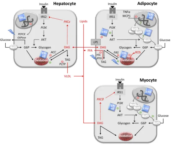
Figure 2: Metabolic effects of E2 in hepatocyte, adipocyte and myocyte. ... 20
Figure 3: Schematic representation of E2 signaling in liver metabolism. ... 22
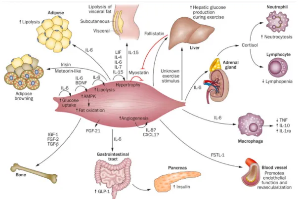
Figure 4: Resistance training effects in different tissues. ... 23
Figure 5. Organogram of the experimental groups ... 27
Figure 6. Materials used to construct the silastic capsule. ... 28
Figure 7. Rat climbing the ladder during the training session. ... 29
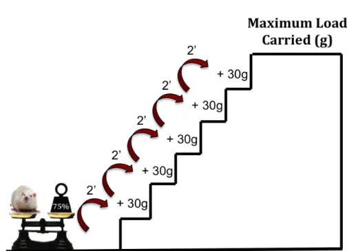
Figure 8. Maximum load determination from 75% of rat’s body mass. ... 31
Figure 9. Carried load per training session. ... 31
Figure 10. Graphical simulation of agarose gel and integrity number ... 34
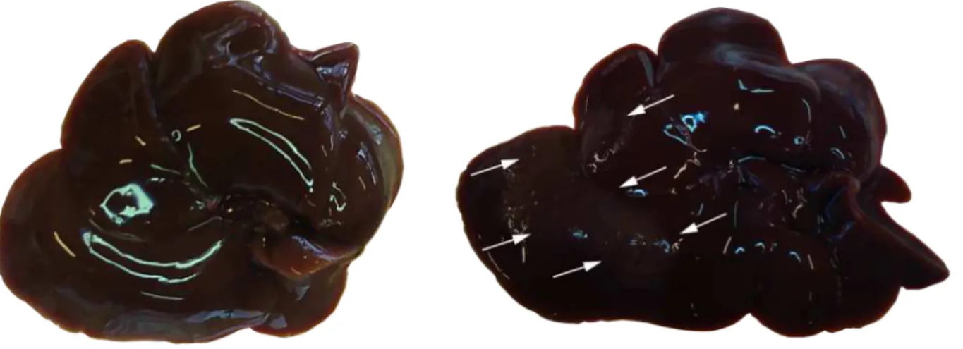
Figure 11. Color and size of liver. ... 38
Figure 12. Carried Load on trained groups throughout training. ... 39
Figure 13. Hepatic mRNA and protein levels of the GLUT2. ... 40
Figure 14. Liver TAG and hepatic mRNA expression of PPARγ. ... 41
Figure 15 Glycogen levels and mRNA expression of PEPCK. ... 42
Figure 16. Coefficient correlation and linear regression of GLUT2 and PPARγ gene expression. ... 43
TABLE LIST
Table 1. Training schedule. ... 29
Table 2. Oligonucleotides primers used to RT-‐PCR. ... 34
Table 3. Anthropometric parameters and food intake. ... 39
ABBREVIATION LIST
GLUT2: Glucose transporter 2
PPARγ: Peroxisome proliferator-‐activated receptor gamma
Ovx: Ovariectomy
RT: Resistance training
Sed: Sedentary
E2: Estrogen Hormone replacement
PEPCK: Phosphoenolpyruvate carboxykinase
SREBP-‐1c: sterol regulatory element-‐binding protein 1c
SCD-‐1: stearoyl-‐CoA desaturase-‐1
VLDL-‐TG: very low density lipoprotein triglycerides
cDNA: complementary DNA
RIN: RNA integrity number
GAPDH: Glyceraldehyde 3-‐phosphate dehydrogenase
ACTB: Beta-‐actin
Liver TAG: Hepatic Triacylglycerol
NAFLD: Non-‐alcoholic fatty liver disease
SUMMARY
1. INTRODUCTION ... 18
2. OBJECTIVE ... 24
2.1 Especific Objective ... 24
3. HYPOTHESIS ... 25
4. METHODS ... 26
4.1 Ethics Committe Approve ... 26
4.2 Animal Care ... 26
4.3. Experimental Groups ... 26
4.4 Surgical procedures of Ovariectomy and Sham ... 27
4.5 Hormone Replacement (E2) ... 28
4.6 Resistance training ... 28
4.6.1 Apparatus to training sessions ... 28
4.6.2 Familiarization Step. ... 30
4.6.3 Maximal load determination step. ... 30
4.6.4 Training Session. ... 30
4.7 Food intake and body weight control ... 32
4.8 Animals euthanasia and tissue sampling ... 32
4.9 17β-‐estradiol plasma level ... 32
4.10 Quantitative Real Time Polymerase Chain Reaction (RT-‐PCR) ... 33
4.11 Protein Assay ... 35
4.12 Liver Triglycerides Assay ... 36
4.13 Hepatic Glycogen Assay ... 36
4.14 Statistical Analysis ... 36
5. RESULTS ... 38
6. DISCUSSION ... 44
6.1 Ovariectomy ... 44
6.2 Estrogen Replacement ... 46
6.3 Resistance training ... 47
7. CONCLUSION ... 48
8. STUDY LIMITATIONS ... 49
9. FUTURE PERSPECTIVES ... 50
10. ATTACHMENTS ... 51
10.1 Published Article ... 51
10.2 Published Articles and in submission process related to this Project ... 58
10.3 Abstracts Published in International Conferences ... 59
10.4 Ethics Committee's opinion on the Animal’s Use ... 60
11. REFERENCE ... 61
1. INTRODUCTION
Estrogens are steroid hormones primarily known for their role in promotion of
female sex characteristics and reproductive capability. Estrogens are produced by the
ovaries and adrenal glands and circulate throughout the body, where they have effects on
most organ systems, including brain, breast, cardiovascular (heart andvasculature) immune,
reproductive (ovaries and uterus), bladder, skin, and bone (JELENIK; RODEN, 2013;
MESSIER et al., 2011). There are three forms of estrogens in the female body: estrone (E1),
estradiol (E2), and estriol (E3). During a woman’s reproductive years, the principal
circulating estrogen is 17β-‐estradiol (E2). 17β-‐estradiol is involved in gene regulation and
has an important role in several physiological and pathological states in both men and
women (BARROS; MACHADO; GUSTAFSSON, 2006)
Figure 1: Effects of estradiol in many tissues about glucose and lipid metabolism. From JELENIK; RODEN, (2013) Tissue-‐specific stimulatory (+) and inhibitory (-‐)
Around 40s age the estrogens level starts to decrease and menstrual cycles start to be
irregular, this period is called climacteric. In this period many hormonal changes happen,
predominantly caused by a marked decline in the ovarian follicle number. When there are
the cessation of follicular activity associated to the cessation menstruation it marks the end
of natural female reproductive life and the postmenopausal period (MESSIER et al., 2011).
Both menopause and estrogen withdrawal in rats, through ovariectomy or other
animal models to simulate menopause, are associated with several metabolic changes, such
as loss of bone mineral density, diabetes, impairment of muscle function, coronary heart
disease, (LEITE et al., 2009; MALTAIS; DESROCHES; DIONNE, 2009; JELENIK; RODEN, 2013)
including increase of liver fat deposition (NEMOTO et al., 2000; PAQUETTE et al., 2007;
VÖLZKE et al., 2007). Liver fat accumulation is recognized as the hepatic component of
metabolic syndrome (MARCHESINI et al., 2005).
Hepatic steatosis is twice as common in post-‐menopausal period as in pre-‐
menopausal time (BARSALANI et al., 2008; CLARK; BRANCATI; DIEHL, 2002). The general
mechanism of liver fat accumulation involves an imbalance between lipid availability and
lipid disposal. Excessive fat accumulation in liver can occur as a result of increased fat
delivery into liver and increased fat synthesis in liver, reduced fat oxidation, and reduced fat
exportation in the form of very low density lipoprotein triglycerides (VLDL-‐TG) (LAVOIE;
PIGHON, 2012; MUSSO; GAMBINO; CASSADER, 2009). Measurements of molecular markers
in different studies suggest a reduction in lipid oxidation and in the production of VLDL and
an increase in de novo lipogenesis (BARSALANI et al., 2008; PAQUETTE et al., 2009).
The specific origin of the lipids that accumulate in rat liver under estrogen
withdrawal remains unclear. However, several molecular markers of de novo lipogenesis in
the liver, including the transcription factor sterol regulatory element-‐binding protein 1c
(SREBP-‐1c) and its downstream enzyme stearoyl-‐CoA desaturase-‐1 (SCD-‐1), have been
2011; PAQUETTE et al., 2008). If, indeed, de novo lipogenesis is increased in the livers of Ovx
rats, then extra glucose molecules must be available as substrates for lipid synthesis.
When glucose concentration outside the liver rises, glucose is rapidly taken up into
hepatocytes via GLUT2. GLUT2 is known to transport glucose across the hepatic plasma
membrane in a bidirectional manner (WOOD; TRAYHURN, 2003) and are not sensitive to
insulin. Thus, the process of glucose uptake and phosphorylation by the hepatocyte depends
on the glucose concentration outside of the cell. Glucose is temporarily ‘trapped’ by
phosphorylation to glucose-‐6-‐phosphate and is unaffected by insulin, at least in the short
term (CHOUKEM; GAUTIER, 2008). However, insulin is crucial in the subsequent steps of the
storage process, like glycogen synthase and inhibition of glycogen phosphorylate
(PETERSEN et al., 1998).
Figure 2: Metabolic effects of E2 in hepatocyte, adipocyte and myocyte.
Several studies have shown that liver GLUT2 expression is regulated under different
metabolic states. Liver GLUT2 expression is decreased during starvation and is restored to
normal levels by a high-‐carbohydrate diet (BURCELIN et al., 1992; THORENS; CHARRON;
LODISH, 1990) and GLUT2 are increased in streptozotocin-‐diabetic rats associated with
SREBP-‐1c expression increased in liver (IM et al., 2005b). In addition, we also targeted the
transcription factor PPARγ.
PPARγ acts as a critical transcription factor in the regulation of adipose
differentiation, lipid storage, and of genes involved in energy storage and utilization. It can
also affect insulin sensitivity by regulating hormones, cytokines, and proteins that are
involved in insulin resistance (STAELS; FRUCHART, 2005). Gene expression of PPARγ is low
in liver tissue, it represent only 10% -‐ 30% of the level in adipose tissue (MATSUSUE et al.,
2003; ROGUE et al., 2010). Nevertheless, there is some evidence that liver GLUT2 is a direct
target of PPARγ, thus contributing to glucose transport into the liver (IM et al., 2005a).
PPARγ also targets genes involved in glucose metabolism as the phosphoenolpyruvate
carboxykinase (PEPCK) (NAKAMURA; YUDELL; LOOR, 2014). PEPCK is an enzyme in the lyse
family used in the metabolic pathway of gluconeogenesis. It converts oxaloacetate into
phosphoenolpyruvate and carbon dioxide. Pyruvate dehydrogenase catalyzes an irreversible
degradation step of pyruvate to acetyl CoA, however PEPCK induction switches the
metabolic fate of pyruvate from oxidation to G3P synthesis and has been almost exclusively
linked to gluconeogenesis to the point that changes in the levels of PEPCK mRNA or its
activity are associated with the control of hepatic glucose output with alterations in life span
(YANG; KALHAN; HANSON, 2009).
Thus, it is important to consider strategies to prevent or revert these metabolic
changes in postmenopausal period. Nowadays there are consolidated strategies to try to
2009). Hormone replacement therapy is one of the most effective therapies used to reduce
unfavorable postmenopausal complications such as osteoporosis (STEVENSON, 2005),
hypercholesterolemia (ŽEGURA et al., 2006), coronary heart disease (COLLINS et al., 2006),
and risk of developing type 2 diabetes mellitus (SKOUBY et al., 2005), despite its adverse
effects, such as an increased incidence of breast cancer and a transient increase in venous
thromboembolism (SCARABIN; OGER; PLU-‐BUREAU, 2003). In Ovx rodents, estrogen
replacement decreases fat accumulation, improves serum lipid profiles (SHINODA; LATOUR;
LAVOIE, 2002), and restores insulin action on muscle glucose transport.
Figure 3: Schematic representation of E2 signaling in liver regulation of glucose and lipid metabolism.
Figure from (ZHU et al., 2013).
Furthermore, there is accumulating evidence that endurance exercise training
overcomes several of the metabolic effects of ovariectomy in rats. For instance, Ovx animals
can benefit from exercise training by a reduction in fat gain (RICHARD; ROCHON; DESHAIES,
1987; SHINODA; LATOUR; LAVOIE, 2002) and insulin resistance in skeletal muscle
(SAENGSIRISUWAN et al., 2009).
Endurance exercise training has also been reported to prevent liver fat accumulation
in rats (PIGHON et al., 2010) as well as the up regulation of several genes involved in de novo
lipogenesis (PIGHON; GUTKOWSKA; JANKOWSKI, 2010). There is also evidence that
decreases liver fat content (CORRIVEAU et al., 2008; LEITE et al., 2009). Thus, RT has been
reported to promote muscle strength and hypertrophy in Ovx animals providing protection
against menopause-‐associated sarcopenia and osteopenia (CORRIVEAU et al., 2008; LEITE et
al., 2009).
Figure 4: Resistance training effects in different tissues. Figure from (BENATTI; PEDERSEN, 2015).
More recently, it was reported that resistance training restored the gene expression
of key molecules involved in de novo lipogenesis in livers of ovariectomized rats
(DOMINGOS; RODRIGUES; STOTZER, 2011). We, therefore, used a resistance training
program, which can be considered a model of strength training, to test the hypothesis that
resistance training ameliorates the increase in GLUT2 expression in livers of Ovx rats.
2. OBJECTIVE
The aim of this study was to use the resistance training program and estrogen
replacement to promote alterations on hepatic GLUT2 and liver TAG of ovariectomized rats.
2.1 Especific Objective
Analyze the gene and protein expression of GLUT2 in trained ovariectomized rats or
with hormone replacement.
Analyze the lipid content and PPARγ gene expression in trained ovariectomized rats
or with hormone replacement.
Quantify the liver glycogen and PEPCK gene expression in trained ovariectomized
rats or with hormone replacement.
Present a correlation of GLUT2 and PPARγ gene expression results in ovariectomized
rats trained, sedentary or with hormone replacement.
3. HYPOTHESIS
We postulated that Ovx-‐induced liver fat accumulation would also result in increased
hepatic glucose uptake through increased expression of GLUT2. The first purpose of the
present study was to test the hypothesis that hepatic expression of GLUT2 is increased in
Ovx rats. Thus, higher liver TAG as well PPARγ will be presented by Ovx rats.
To test the treatment of Ovx condition, we used the RT and E2 in Ovx rats to test the
hypothesis that both will revert the Ovx effects in liver.
The RT as well E2 will act antagonistically to the effects of ovariectomy. Both
interventions decrease GLUT2 and liver TAG and PPARγ expression and increase gene
expression of PEPCK and glycogen amount in ovariectomy rats.
4. METHODS
4.1 Ethics Committe Approve
All experiments described in the present report were conducted according to the
(Guide for the Care and Use of Laboratory Animals, 1996) and approved by the Ethics
Committee on Animal Use from the Federal University of São Carlos (CEUA-‐UFSCar) Protocol
n° 005/2013.
4.2 Animal Care
Female Holtzman rats (n = 40) from the animal facility of the University of São Paulo
State (UNESP, Araraquara, Brazil) weighting ~ 220 g upon arrival were housed in collective
cages. The animals had ad libitum access to food and tap water. All animals were fed with
commercial rodent chow. Their environment was controlled in a reverse light cycle (12 h
dark starting at 08:00 AM). Food intake was monitored daily over the entire experimental
period. Body mass was measured 3 times/week at the same time of day.
Upon arriving at Exercise Physiology Laboratory from Federal University of São
Carlos, (UFSCar, São Carlos, Brazil) the animals have remained for two weeks in
acclimatization condition and inverted cycle. After this period, the animals were divided into
five groups according to Figure 5.
4.3. Experimental Groups
Rats were randomly distributed into five experimental groups (n = 8/group). Rats
first underwent a bilateral ovariectomy (n = 24) or a bilateral sham operation (Sham, n =
group of Ovx and one group of Sham rats were submitted to a 12-‐week resistance training
program. The third group of Ovx rats remained sedentary and was given 17β-‐estradiol
replacement. Altogether, five groups were compared: Sham-‐sedentary (Sed), Sham-‐
resistance trained (RT), Ovx-‐Sed, Ovx-‐RT, and Ovx-‐Sed with hormone replacement (Ovx-‐E2).
Figure 5. Organogram of the experimental groups Adapted from BARBOSA et al., 2016
4.4 Surgical procedures of Ovariectomy and Sham
Ovariectomy and sham surgeries were performed when the rats reached 250 g of
body mass, according to the technique described by KALU (1991). To perform this
procedure, rats were first anesthetized with a mixture of ketamine-‐zylazine (61.5 -‐ 7.6
mg/kg, ip). A bilateral incision (1.0 -‐ 1.5 cm) was made through the skin and muscle layers.
The ovaries were removed and the skin and muscles were sutured. The sham surgery was
performed using the same procedure but the ovaries were not removed. The analgesic
tramadol hydrochloride (20 mg/kg) was injected every 24 h for five days after surgery. The
rats were allowed to recover from surgery for seven days, which also permitted us to ensure
second surgical procedure to insert Silastic capsules under the skin of the neck to provide
hormonal replacement.
4.5 Hormone Replacement (E2)
Silastic capsules (15 mm) with internal and external diameter of 1.02 mm and 2.16
mm, respectively (Dow Corning VWR International, Buffalo Grove, IL, USA) were used for E2
replacement. Sunflower oil was used as the vehicle, with a 5% concentration of 17β-‐estradiol
(50 mg/ml oil). Ten μL of this solution was pipetted into each capsule and then both sides
were sealed with Silastic glue. The capsules were stored for 24 h to allow the glue to dry.
After drying, the capsules were kept in saline (0.9%) solution (CAMPOS et al., 2014).
Figure 6. Materials used to construct the Silastic capsule.
4.6 Resistance training
4.6.1 Apparatus to training sessions
The training sessions consisted of climbing a vertical wooden and metal ladder with
the following dimensions: 80° of inclination, 110 cm height x 18 cm width, and 2 cm spacing
between the ladder rungs. At the top of the ladder, the animals reached a 20 x 20 x 20 cm
cage, which allowed them to rest between climbing sessions. The first three training sessions
maximum possible weight load. The load consisted of fishing sinkers that were previously
weighed, labeled, and stored in falcon tubes. The falcon tubes were tied to proximal portion
of the rats’ tails. The present RT protocol is an adaptation of the program described by
Hornberg & Farrar (2004).
Figure 7. Rat climbing the ladder during the training session. From Leite, R.D. Thesis, 2009.
The training sessions were conducted every 72 hours (Table 1) with no fixed weekly
days for training. The training schedule was 12 weeks, totaling 27 strength training sessions.
Table 1. Training schedule.
Familiarization, maximum load determination and training sessions every 72 hours. (X) Days recovery.
4.6.2 Familiarization Step.
The animals were first familiarized with the training procedures over two
consecutive days. The overload apparatus was attached to the rat's tail without any weight.
The rats were placed at the bottom of the ladder and familiarized with the climb. Once they
reached the cage at the top of the ladder, the rats were allowed a 2 min rest period. If
necessary, a physical stimulus with fingers or tweezers was applied to the animal's tail to
initiate climbing movements. The rats were considered to have adapted to the ladder
protocol when voluntary climbing was performed three consecutive times without any
stimulus.
4.6.3 Maximal load determination step.
After 2 days of familiarization to training session, the third day was the maximal load
determination. It was initiated with a load equivalent to 75% of the rat’s body mass. After
each successful climb, the rats recovery for 2 minutes and 30 g were added to the apparatus
until the rat was unable to climb the entire length of the ladder even after three successive
stimulus at the tail. The highest carried load was considered the maximum carrying capacity
of each rat as shown figure 8.
4.6.4 Training Session.
The training sessions consisted of 4 to 9 climbs with progressively increasing weights. The
size of the ladder required the animals to perform 8 -‐ 12 movements per climb. The first four
climbs were carried out with loads corresponding to 50, 75, 90, and 100% of the rat’s
maximal carrying capacity as determined in the last training session. For the 5 subsequent
entire ladder as shown figure 9. The new maximal load determined during the training
program was used for the next session.
Figure 8. Maximum load determination from 75% of rat’s body mass.
4.7 Food intake and body weight control
Food intake (grams per cage) was daily monitored always at determined hour. Body
mass was checked every Monday, Wednesday and Friday also at determined hour. Both
procedures were conducted during all the experimental period. These controls were
performed to certify the possible changes caused by the three different performed
interventions.
4.8 Animals euthanasia and tissue sampling
Rats were euthanized between 9:00 AM and 12:00 PM after removal of food at 7:00
AM. Rats in the trained groups were euthanized 48 h following the last training session. The
animals were euthanized by decapitation with the same period of experimental
interventions. Immediately after decapitation, blood samples were collected and centrifuged
at 3000 rpm for 10 minutes at 4°C, and then stored at -‐20°C.
The livers were weighed and then washed with saline (0.9%). The median of lobe
each liver was selected by anatomical visualization. This lobe was separated from the others
and divided into 3 aliquots in microtube being; 1) to genetic analysis; 2) to protein analysis
and 3) to reserve for other analyzes. The other liver lobes were stored in aluminum foil in a
freezer -‐80˚ C.
4.9 17β-‐estradiol plasma level
Measurement of plasma levels was performed by the ELISA technique (MA; SHIEH;
LEE, 2006) using commercial kits (ADI-‐900-‐174, Enzo Life Sciences, Farmingdale, NY, USA)
samples were tested in duplicate. Quantification was given in relation to the standard curve
using logistic 4-‐parameter nonlinear regression.
4.10 Quantitative Real Time Polymerase Chain Reaction (RT-‐PCR)
The hepatic tissue homogenization was performed in liquid nitrogen in a crucible, a
porcelain pestle. Without defrosting the tissue was macerated until form a powder. This
powder was collected and stored in a new microtube. Approximately 20 mg of powder
hepatic tissue was weighed and placed in a new microtube. The samples were lysed with
Lysis Buffer containing 1% 2-‐mercaptoethanol (10 μl / 1 ml) and shaken quickly (3 min 30 s
/ sec) on a rotor with small metal spheres. It was centrifuged at 2600 g for 5 minutes at
room temperature and 250 μl of the supernatant was transferred to a RNAse free microtube
from a mini kit of Invitrogen according to the manufacturer’s protocol. Then, the RNA was
treated with DNAse (Invitrogen) in order to avoid genomic contamination.
All next steps, to obtain the Ct values were performed in the IRIC -‐ Institute for
Research in Immunology and Cancer at the University of Montreal -‐ Canada. The RNA
integrity analysis was performed by electrophoresis of RNA micro capillary separation. This
method generates the value of RIN (RNA integrity number) by an integrity algorithm
assigned the RNA value. The RIN value between 10:08 imparts integrity of the sample. A
graphical simulation of agarose gel was represented figure 10. The samples with protein
contamination or integrity number below 8 values were discarded (SCHROEDER et al.,
Figure 10. Graphical simulation of agarose gel and integrity number
Total RNA (2 pg) was reverse-‐transcribed into complementary DNA (cDNA) using
high-‐capacity cDNA reverse transcription kits (Applied Biosystems). Reverse-‐transcribed
samples were stored at -‐20°C. GAPDH gene expression was determined using a pre-‐validated
Taqman Gene Expression Assay (Applied Biosystems, Rn01462d61, Foster City, CA). The
gene expression levels of target genes were determined using assays designed with the
Universal Probe Library from Roche. The primer sets and UPL probe numbers are presented
in Table 2.
Table 2. Oligonucleotides primers used to RT-‐PCR.
GLUT2: Glucose transporter 2; PEPCK: Phosphoenolpyruvate carboxykinase; PPARγ: Peroxisome proliferator-‐ activated receptor gamma GAPDH: Glyceraldehyde 3-‐phosphate dehydrogenase; ACTB: Beta-‐actin.
To validate the efficiency of the qPCR assays, we used a mix of the samples tested in