UNIVERSIDADE FEDERAL DO CEARÁ
FACULDADE DE FARMÁCIA, ODONTOLOGIA E ENFERMAGEM PROGRAMA DE PÓS-GRADUAÇÃO EM ODONTOLOGIA
MARIA DENISE RODRIGUES DE MORAES
EFEITO DO ENVELHECIMENTO DE RESTAURAÇÕES DE CIMENTO DE IONÔMERO DE VIDRO NA DESMINERALIZAÇÃO DO ESMALTE E DENTINA:
ESTUDO in situ
MARIA DENISE RODRIGUES DE MORAES
EFEITO DO ENVELHECIMENTO DE RESTAURAÇÕES DE CIMENTO DE IONÔMERO DE VIDRO NA DESMINERALIZAÇÃO DO ESMALTE E DENTINA: ESTUDO in situ
Di sser t ação apresentada ao Programa de Pós Gr aduação em Odontologia da Faculdade de Farmácia, Odontologia e Enfermagem da Universidade Federal do Ceará como um dos requisitos para a obtenção do Título de Mestre em Odontologia.
Área de concentração: Clínica Odontológica
Orientadora: Profa. Dra. Lidiany Karla Azevedo Rodrigues
MARIA DENISE RODRIGUES DE MORAES
EFEITO DO ENVELHECIMENTO DE RESTAURAÇÕES DE CIMENTO DE IONÔMERO DE VIDRO NA DESMINERALIZAÇÃO DO ESMALTE E DENTINA: ESTUDO in situ
Dissertação apresentada à Coordenação do Programa de Pós-graduação em Odontologia da Universidade Federal do Ceará como requisito parcial para obtenção do Título de Mestre em Odontologia. Área de concentração: Clínica Odontológica
Apr ovada em: ___/ ___/___
BANCA EXAMINADORA
_____________________________________
Profa. Dra. Lidiany Karla Azevedo Rodrigues (Orientadora) Universidade Federal do Ceará-UFC
_______________________________________ Prof. Dr. Haroldo César Pinheiro Beltrão
Universidade Federal do Ceará- UFC
Dedico este trabalho a Deus, pela minha vida. Aos meus pais, Josualdo e Raimunda, exemplo de amor, dedicação e humildade e por terem me ensinado sempre a importância dos valores. Aos meus i r mãos, Samara, André e Moisés pelo amor compreensão e carinho.
AGRADECIMENTOS
À minha orientadora, professora Dra. Lidiany Karla Azevedo Rodrigues, pela confiança, dedicação, atenção e oportunidades dadas. Por sempre estimular a união do grupo de pesquisa e o desenvolvimento de projetos de destaque. Por ser um exemplo de determinação e auto-confiança.
Aos professores da Disciplina de Dentística Operatória Clínica, Prof. Dr. Haroldo César Pinheiro Beltrão, Prof. Dr. Sérgio Lima Santiago e Prof. Dr. Carlos Augusto de Oliveira Fernandes, pelos importantes ensinamentos profissionais, acolhimento e paciência.
A todos os professores dos cursos de Graduação em Odontologia da UFC que contribuíram para o meu desenvolvimento pessoal, profissional e científico.
Ao professor Dr. Vicente Paulo Aragão Sabóia, pela maneira determinada como se dedica à pesquisa. Por ter sido o primeiro professor da UFC a me estimular pela pesquisa científica.
À todos os professores da Pós-Graduação pelo exemplo de competência, disponibilidade, atenção e contribuição ao meu aprendizado durante as disciplinas do Mestrado.
Ao Programa de Pós-Graduação em Odontologia, pela oportunidade de realizar o curso de Mestrado. Ao professor Dr. Sérgio Lima Santiago, coordenador do Programa de Pós-Graduação em Odontologia da UFC, pelo exemplo de determinação para o sucesso do programa.
Ao Prof. Dr. Jaime Aparecido Cury e Profa. Dra. Lívia Maria Tenuta pelo acolhimento e ensinamentos durante o período de Mestrado sanduíche no Laboratório de Bioquímica da Faculdade de Odontologia de Piracicaba- Unicamp.
Ao Sr. Waldomiro Vieira Filho, técnico de laboratório da FOP, pelas análises realizadas no Laboratório de Bioquímica da Faculdade de Odontologia de Piracicaba- Unicamp.
Aos funcionários da Odontologia, em especial Maria Marta de Lima Texeira (Martinha) e Marta Ferreira do Nascimento (Martinha) e aos funcionários Rui Lino de Sousa, Tatiana Abreu de Sousa, José Augusto Sousa e José Williame Silva por toda colaboração para as minhas atividades durante o mestrado.
Ao técnico em prótese dental do curso de Odontologia, Antônio Carlos de Oliveira Filho (Carlinhos), por ser tão prestativo e pela confecção dos dispositivos intra-orais.
Às estimadas amigas Juliana Paiva Marques Lima, Mary Anne Sampaio de Melo, pelo aprendizado mutuo e divisão de trabalhos no vários momentos vividos. Agradeço pelo empenho em cada momento e principalmente pela sicera amizade.
Aos alunos de iniciação científica, Diego Martins de Paula, Diego Góes e João Paulo Saraiva Wenceslau, pela presteza e ajuda indispensáveis em etapas da pesquisa.
A todos os colegas de mestrado, Alrieta Henrique Teixeira, Ana Patrícia Souza de Lima, André Mattos Brito de Souza, Daniela da Silva Bezerra, Françoise Parahyba Dias, Gabriela Eugênio de Sousa Furtado, George Táccio de Miranda Candeiro, Isabela Alves Pacheco, Jorgeana Abrahão Barroso, José Luciano Pimenta Couto, Marília Mota Silva, Mirela Andrade Campos, Regina Cláudia Ramos Colares, Saulo Hilton Botelho Batista, Virgínia Régia Souza da Silveira, pelo colaboração em alguma fase deste trabalho.
Aos secretários do Programa de Pós-Graduação em Odontologia da UFC, Sr. Germano Mahlmann Muniz Filho e Sra. Lúcia Ribeiro Marques Lustosa, pela atenção, cuidado e apoio sempre prestados.
Á bibliotecária Sra. Rosane Maria Costa pela atenção com as padronizações bibliográficas.
À CAPES pela concessão de bolsa e ao CNPq pela concessão de bolsa e de auxílio financeiro (Nº processo 477070/2008-6).
Meus sinceros agradecimentos aos voluntários desta pesquisa, a qual sem os mesmos não teria sido realizada.
A todos que de maneira direta ou indireta ajudaram na elaboração desta pesquisa.
MUITO OBRIGADA!
RESUMO
O benefício de restaurações de cimento de ionômero de vidro modificado por resina (RMGIC) na inibição de cáries pode ser questionado na presença de dentifrício fluoretado e de restaurações envelhecidas. Objetivo: Avaliar o efeito do processo de envelhecimento em cáries ao redor de restaurações na junção amelo-cementária, na presença de flúor proveniente de RMGIC ou dentifrício. Métodos: Um estudo in situ randomizado duplo cego e cruzado foi realizado em duas fazes de 14 dias. Desesseis voluntários utilizaram um dispositivo intraoral contendo blocos de dentes restaurados com resina composta (CR) ou RMGIC, tendo sofrido ou não o processo de envelhecimento. Os blocos foram expostos a uma solução de 20% de sacarose, 10x/dia e os voluntários usaram dentifrício com ou sem flúor 3x/dia. O biofilme formado sobre os blocos foi analizado para determinar a contagem de streptococos totais, estreptococos mutans e lacatobacilos, assim como a concentração de flúor. Resultados: A desmineralização do esmalte foi determinada por meio de microdureza longitudinal na margem da restauração. Foram utilizados os testes de Kruskal–Wallis e ANOVA fatorial, seguidos pelo teste de Tukey (p<0.05). Não foram encontradas diferenças estatisticamente significantes na microbiota cariogência. A concentração do flúor no biofilme foi maior para restaurações de RMGIC. Para o esmalte, independente do uso do dentifrício ou do processo de envelhecimento, houve maior desmineralização ao redor de restaurações de CR. Para a dentina, na ausência do dentifrício fluoretado, houve maior desmineralização ao redor de restaurações de resina composta envelhecidas. Conclusão: Os resultados sugerem que restaurações RMGIC promovem proteção contra cárie em esmalte e dentina, enquanto que o processo de envelhecimento afeta o desenvolvimento de cárie em dentina, aumentando a progressão da cárie.
ABSTRACT
The beneficial of resin modified glass ionomer cement (RMGIC) restorations in caries inhibition may be questioned due to F-dentifrice use and ageing of the restorations. Objective: To evaluate the effect of an ageing process on caries around cemento-enamel junction restorations in the presence of fluoride from RMGIC or dentifrice. Methods: A randomized double-blind crossover in situ study was performed in 2 phases of 14 days. Sixteen volunteers wore palatal devices containing dental slabs restored with composite resin (CR) or RMGIC, either aged or unaged. The slabs were exposed to a 20% sucrose solution, 10x/day and the volunteers used a non-F or an F-dentifrice 3x/day. The biofilm formed over the slabs was analyzed to determine the counts of total streptococci, mutans streptococci and lactobacilli as well as F-concentration. Results: Enamel demineralisation was determined by cross-sectional microhardness (CSMH) at the margin of the restoration. Kruskal–Wallis and factorial ANOVA, followed by Tukey test, were used to data evaluation (p<0.05). No statistically significant differences were found in the cariogenic microbiota. F-concentration in biofilm was higher for RMGIC restorations. For enamel, higher demineralization around CR restorations was observed regardless dentifrice or ageing process. For dentine, higher demineralization was observed around aged RC restorations without F-dentrifice use. Conclusions: These results suggest that RMGIC restorations provided protection against secondary caries either for enamel and dentine, while ageing process affects caries development in dentine, increasing caries progression.
LISTA DE ABREVIATURAS, SIGLAS E SÍMBOLOS
B / PLB Biofilme / Plaque like biofilm
CIV / GI Cimento de Ionômero de vidro / Glass ionomer cement
CIVMR /
RMGIC
Cimento de ionômero de vidro modificado por resina / Resin modified glass ionomer cement
DNF / NF Dentifrício não fluoretado / Non fluoride dentifrice ET / TS Estreptococos totais / Total streptococcus
EM / MS Estreptococos mutans / Mutans streptococci LB / LB Lactobacilos / Lactobacilli
ML / CSMH Microdureza em corte longitudinal / Cross section microhardness RC / CR Resina Composta / Composite Resin
SUMÁRIO
1 INTRODUÇÃO GERAL 14
2 PROPOSIÇÃO 17
3 CAPÍTULO 18
4 CONCLUSÃO GERAL 35
REFERÊNCIAS 36
APÊNDICES 39
1 INTRODUÇÃO GERAL
A durabilidade das restaurações depende de vários fatores, tais como: condição do risco de cárie e idade do paciente, tipo e tamanho da restauração, bem como características do material restaurador (MJÖR et al., 2000). Assim, a escolha do tipo de material restaurador torna-se relevante, visto que a capacidade de liberação de íons anti-cárie (fluoretos e metais pesados) é determinante no desenvolvimento de lesão secundária (DIONYSOPOLOUS et al., 1994). Isso é relevante quando se observa que boa parte do tempo clínico dos cirurgiões-dentistas é gasto com a troca de restaurações, devido a cáries secundárias, e ainda que esta troca inevitavelmente ocasiona uma remoção adicional da estrutura dentária remanescente (KIDD et al., 1992; MJÖR, 2000).
Segundo Kidd (2001), cárie secundária é uma cárie primária adjacente a uma restauração. A área do tecido dental onde ocorre a desmineralização ao longo da parede da cavidade é denominada de lesão de parede. Assim, o material restaurador deve ser considerado um meio de destaque para prevenção de cárie secundária pois se encontra adjacente ao sitio de desenvolvimento da lesão. Dessa forma, a presença de agentes cariostáticos nos materiais restauradores pode reduzir ou eliminar desmineralização, inibindo a desmineralização e/ou atividades bacterianas e facilitando o processo de remineralização (FEATHERSTONE, 1994; HSU et al., 1998; WIEGAND et al., 2007). Cenci e colaboradores (2008a) mostraram que a desmineralização adjacente a restaurações de cimentos de ionômero de vidro modificado por resina foi maior na dentina distante da parede da cavidade do que perto dela. Isso pode ser explicado pelo fato de haver alta concentração de Fˉ no biofilme presente nesta área (BENELLI et al., 1993; HARA et al., 2006; CENCI et al., 2008a), possibilitando a incorporação no mineral do dente na forma de fluoroapatita (ten Cate et al., 2003). Dessa forma, os fluoretos tornam-se aliados na prevenção de cárie, pois estando presente na composição dos materiais restauradores, tais como o ionômero de vidro, e na cavidade oral, podem inibir a formação de cárie secundária (SOUSA et al., 2009).
equivalente ou maiores quantidades de fluoretos que os cimentos modificados por resina (HARA et al., 2002; GJORGIEVSKA et al., 2009). Além disso, segundo Verbeeck et al., (1993) e Miller et al., (1995), os ionômeros que apresentam formulações em cápsulas pré-dosadas e são manipulados mecanicamente oferecem maior liberação de flúor quando comparados com os cimentos manipulados manualmente e dosados de acordo com a orientação do fabricante. Acredita-se que o pré-encapsulamento leva a uma padronização dos constituintes da mistura, que se apresentam em uma correta proporção. Além disso, a trituração mecânica permite uma maior reação entre as partículas do pó e o líquido, resultando em maior quantidade de matriz e menor percentual de partículas não reagidas, e assim o flúor preso na matriz é então liberado em maior quantidade.
Contudo, segundo alguns estudos in situ, os materiais restauradores contendo fluoretos tem demonstrado capacidade de inibir o desenvolvimento de cáries adjacentes às restaurações em esmalte e dentina (BENELLI et al., 1983; TENUTA et al., 2005, YAMAMOTO et al., 2005, CENCI et al., 2008a). Entretanto, este efeito não se confirma quando da utilização regular do dentifrício com flúor (SOUSA et al., 2009, CENCI et al., 2008a). Um estudo mostrou que, na presença de dentifrício fluoretado, apenas o ionômero encapsulado, que é manipulado de maneira bem controlada, foi capaz de inibir cárie secundária in situ (SOUSA et al., 2009).
Por outro lado, Cenci e colaboradores (2008b) observaram que o envelhecimento de restaurações de resina composta aumenta a rugosidade superficial das restaurações e diminui o selamento marginal no esmalte, o que poderia favorecer o aparecimento de novas lesões cariosas. Além disso, o envelhecimento de restaurações de cimento de ionômero de vidro pode reduzir o efeito inibitório de cárie in situ, visto que essa característica tende a ser maior imediatamente após a realização da restauração (HAYACIBARA et al., 2003).
Vale ressaltar que a ação do flúor liberado por matérias restauradores atua no processo desmineralização-reminelarização no esmalte e dentina de forma mais eficaz do que a quando proveniente de dentifrícios fluoretados. Isso pode ser explicado pelo fato de os fluoretos liberados da restauração estarem em um microambiente formado pelo fluido e estroma do biofilme, durante a dinâmica do processo carioso, e que a concentração deste íon nesse ambiente é responsável pela saturação mineral do dente (TENUTA et al., 2005; 2010).
envelhecimento, o possível efeito antibacteriano dos cimento de ionômero de vidro sobre os microorganismos patogênicos da cárie dentária. Além disso, é importante verificar se a ação desse material na manutenção da dureza do esmalte e dentina e na liberação de flúor para o microambiente do biofilme se conserva após um período tempo.
Entretanto, devido a razões éticas e às vantagens de um melhor controle experimental das variáveis, além de maior relação custo efetividade, parece desejável a utilização de modelos in situ para testar materiais e técnicas restauradoras e sua capacidade de inibir o desenvolvimento de cáries recorrentes, antes da realização de extensos e dispendiosos estudos clínicos (BENELLI et al., 1993; TENUTA et al., 2005). Além disso, o estresse térmico que ocorre in vivo é freqüentemente reproduzido in vitro através de regimes de termociclagens (CENCI et al., 2008b).
2 PROPOSIÇÃO
Este estudo objetiva avaliar o efeito do processo de envelhecimento no desenvolvimento de cárie in situ ao redor de restaurações de cimento de ionômero de vidro em esmalte e dentina em relação ao percentual de desmineralização dos tecidos dentários ao redor das restaurações, à composição bioquímica, por análise de flúor no estroma do biofilme formado sobre as restaurações, e à ação antibacteriana nos microorganismos formados sobre as restaurações.
3 CAPÍTULO
Esta dissertação está baseada no Artigo 46 do Regimento Interno do Programa de Pós Graduação em Odontologia da Universidade Federal do Ceará, que regulamenta o formato alternativo para dissertações de Mestrado e permite a inserção de artigos científicos de autoria e co-autoria do candidato (Anexo A). Por se tratarem de pesquisas envolvendo seres humanos, ou parte deles, o projeto de pesquisa deste trabalho foi submetido à apreciação do Comitê de Ética em Pesquisa da Faculdade de Medicina da Universidade Federal do Ceará, tendo sido aprovado sob protocolo nº 143/06 (Anexo B). Assim sendo, esta dissertação contém um artigo, que será submetido para a publicação no periódico “Journal of Dentistry”, tendo sido previamente analisado e corrigido por um corretor da língua inglesa.
Capítulo 1
In situ effects of aged glass ionomer cement restorations on biofilm and enamel-dentine
demineralization.
In situ effects of aged glass ionomer cement restorations on biofilm and enamel-dentine
demineralization.
De-Moraes MDR, Bezerra DS, Rodrigues LKA
Faculty of Pharmacy, Dentistry and Nursing, Federal University of Ceara, Fortaleza, Brazil Running Title – Anticaries effect of aged glass ionomer cement.
Key words – Secondary Caries, Glass Ionomer, Fluoride, Thermal Cycling.
Full address of the author to whom correspondence should be sent:
Lidiany Karla Azevedo Rodrigues
Faculdade de Farmácia, Odontologia e Enfermagem Dentística Operatória Clínica
Rua Monsenhor Furtado nº 1061
Bairro- Rodolfo Teófilo - CEP 60430-170 Phone- #558533668403
Fax- #558533668232 Fortaleza-CE
Abstract
The beneficial of resin modified glass ionomer cement (RMGIC) restorations in caries inhibition may be questioned due to F-dentifrice use and ageing of the restorations. Objective: To evaluate the effect of an ageing process on caries around cemento-enamel junction restorations in the presence of fluoride from RMGIC or dentifrice. Methods: A randomized double-blind crossover in situ study was performed in 2 phases of 14 days. Sixteen volunteers wore palatal devices containing dental slabs restored with composite resin (CR) or RMGIC, either aged or unaged. The slabs were exposed to a 20% sucrose solution, 10x/day and the volunteers used a non-F or an F-dentifrice 3x/day. The biofilm formed over the slabs was analyzed to determine the counts of total streptococci, mutans streptococci and lactobacilli as well as F-concentration. Results: Enamel demineralisation was determined by cross-sectional microhardness (CSMH) at the margin of the restoration. Kruskal–Wallis and factorial ANOVA, followed by Tukey test, were used to data evaluation (p<0.05). No statistically significant differences were found in the cariogenic microbiota. F-concentration in biofilm was higher for RMGIC restorations. For enamel, higher demineralization around CR restorations was observed regardless dentifrice or ageing process. For dentine, higher demineralization was observed around aged RC restorations without F-dentrifice use. Conclusions: These results suggest that RMGIC restorations provided protection against secondary caries either for enamel and dentine, while ageing process affects caries development in dentine, increasing caries progression.
Keywords: Secondary Caries, Glass Ionomer Cement, Fluoride, Thermal cycling.
Introduction
specific site at risk of secondary caries occurrence, fluoride releasing restorative materials were developed. The anticaries effect of resin modified glass ionomer cements (RMGIC) has been reported in in situ studies.2,3,4 Nevertheless, the RMGIC capability of inhibiting recurrent caries is more evident in the absence of other sources of fluoride and in well-controlled manipulation conditions. 4
Previous study exhibited conflicting data as to whether or not these materials significantly prevent or inhibit secondary caries and affect the growth of caries-associated bacteria compared to non-fluoridated restoratives.5 Another thing to be considered is that the rate of fluoride releasing from these materials is not constant but it exhibits a relatively rapid initial rate, which decreases with time.6 Consequently, the cariostatic potential of glass ionomer cements (GIC) restorations could be reduced as their fluoride releasing rate declines through the ageing process.7 This fact may be particularly relevant mainly in situations where there is no use of fluoridated toothpaste, since the lack of this ion may affect the fluoride recharge presented by GIC.
Thus, this study aimed to evaluate in situ the effect of an ageing process on caries around cemento-enamel junction restorations in the presence of fluoride from RMGIC or dentifrice, or by a combination of them. Also, the microbiological and biochemistry composition, by analysis of fluoride, of the plaque-like biofilm (PLB) formed on the restoration was evaluated. The null hypotheses tested were that there would not be any effect of (1) presence of induced ageing, (2) type of restorative material, or (3) dentifrice use on the response of the assessed variables.
Materials and Methods
Study Population and Ethical Aspects
the volunteers were excluded from the study. Three potential individuals refused to participate, leaving 17 volunteers who initiated the study. One volunteer gave up after using the palatal device for 72 h. Then, the sample was composed by 16 healthy adults (12 females and 4 males), from graduate and pos graduate of Faculty of Pharmacy, Dentistry and Nursing, Federal University of Ceará, Fortaleza, Brazil, aged from 19 to 36 years old who were able to comply with the experimental protocol. Consent forms were signed prior to enrollment in the study.
Experimental Design
This was a cross-over randomized double-blind split-mouth in situ study for caries induction by sucrose exposure and biofilm accumulation performed in 2 phases of 14 days. The factors under study as a factorial 2x2x2 design were: (1) Ageing status in 2 levels: with ageing (A+) and without ageing induction (A–). (2) Restorative material in 2 levels: composite resin (CR) and resin-modified glass ionomer cement (RMGIC). (3) F treatment in 2 levels: non fluoride dentifrice (NF; placebo) and fluoride dentiftice (FD; 1,100 µg F/g as NaF, silica-based). Therefore, the 8 experimental subsets obtained from the association of these factors were assigned to the volunteers.
Specimen Preparation
Sixty extracted impacted human third molars, with roots more than two-thirds formed, free of apparent enamel defects, macroscopic cracks, abrasions and staining (as assessed by visual examination) were used to perform this in situ study. The teeth were stored in a 0.01% (v/v) thymol solution at 4ºC for thirty days and refrigerated until use. A total of 120 enamel-dentine slabs were obtained using a water-cooled diamond saw and a cutting machine (IsoMet Low Speed Saw, Buehler, Lake Bluff, IL, USA). The enamel-dentine (4 x 4 x 2 mm) slabs were prepared from the cervical region of nonerupted human third molars (2 mm above and 2 mm below the cementum-enamel junction) (figure 1). The tooth preparation was carried out according to Sousa et al.4 with the difference that the occlusal margin of the cavity was located on enamel, while the gingival margin was located in dentine. After cavity preparation, all slabs were sterilized by autoclaving according to Amaechi et al.8 and stored in 100% humidity until being inserted into the palatal appliances. The cavities were restored with one of the following materials: Filtek-Z-250/ Single Bond composite resin (3M ESPE Dental Products, St. Paul, MN, USA), as a control group, and Fuji II LC encapsulated resin-modified glass ionomer (GC America Incorporation, Alsip, IL, USA), according to the manufacturers’ recommendations (for details see Sousa et al.4). The slabs were then polished with aluminum oxide discs (Sof-lex disk system 3M ESPE Dental Products Division) with each disk applied for 15 s. Sixty slabs were restored, half with CR and half with RMGIC, and submitted to accelerated ageing by thermal cycling (10,000 cycles, 5 - 55ºC, 60 seconds, 14-days ageing period)9 by using a thermocycler equipament (THE 1100, SD Mechatronik GmbH, Feldkirchen-Westerham, Germany). The last 60 slabs were restored, half with CR and half with RMGIC, only two days before finish the ageing process of others slabs. Next, all slabs were stored in 100% humidity for 24 h and put in the palatal appliances for in situ cariogenic challenge.
Palatal appliance preparation
In situ phase
Cariogenic challenge to the restored specimens was provided by dripping 20% sucrose solution onto all slabs, 10 times/day, during 14 days.3 No restrictions were made in regard to the volunteers' diet, but they were instructed to remove the appliances during meals11,12 and using fluoridated water (0.7 mg F/L). Volunteers were instructed to remove the appliance and drip one drop of 20% sucrose solution onto each mesh that was above the slabs, 10 x/day at predetermined times (until 8:00 a.m. from 9:30 p.m.). When removed, the devices were kept wet in plastic boxes to keep the bacteria biofilm viable.13 Before replacing the palatal appliance in the mouth, a 5-min waiting time was standardized for sucrose diffusion into the dental biofilm. During the lead-in and washout periods (1-week) as well as throughout each experimental phase, the volunteers brushed their teeth using the phase-designed dentifrice (NF or FD - Fórmula & Ação Dentistry Product, São Paulo, SP, Brazil). The dentifrice treatment was performed 3 x/day, after mealtimes when volunteers’ habitually performed their oral hygiene. The appliances were extra-orally brushed, except the slab area, and volunteers were asked to brush carefully over the covering meshes, to avoid disturbing the biofilm.
Microbiological analysis
in CFU/mg dental biofilm (wet weight).
Cross-sectional microhardness testing (CSMH)
Enamel-dentine slabs were longitudinally sectioned through the center of the restoration. The segments were embedded in acrylic resin and serially polished. Cross-sectional microhardness measurements were made with a microhardness tester (Future Tech Corp FM-ARS 9000; Tokyo, Japan) with a Knoop diamond under a 25 g/5 s load on enamel and 5g/5 s on dentine. One lane of 11 indentations was made, being 30 and 120 µm from the preparation margin. The indentations were made at the following depths: 10, 20, 30, 40, 50, 60, 80, 100, 120, 140 and 180 µm from the outer enamel and dentine. Integrated demineralization (D S) was calculated according Sousa et al.4
Fluoride analysis of the biofilm
Finished the cariogenic challenge, the biofilms over the slabs were colected with sterile plastic spatula, put in sterile pre-weighed microcentrifuge tubes, identified, and dehydrated for a 24-hour period. After this period the weight of dry biofilm was obtained by using a digital weighing-machine of 5 digits. The samples were treated with 100 µL of 0.5 M HCl for each 1 mg of biofilm, stirred for 3 h at room temperature, centrifuged, and the supernatant collected was neutralized with 2 M NaOH (0.125 mL/10 mg biofilm wet weight) and kept frozen until analysis.2 After this period it was added to the samples the same volume of TISSAB II (containing 20 g NaOH / L, pH 5.0). Quantification of fluoride in the solution was done with an ion selective electrode connected to an ion analyser (Orion EA-940), which was previously calibrated with a series of 8 standard solutions (from 0.025 to 32.0 ppm Fˉ), in triplicate. The readings of the samples were expressed in millivolt (mV) and transformed into μgF ˉ/m (ppm F ˉ) by linear regression of the calibration curve and calculated according the weight of the biofilm in milligrams.12
Statistical Analysis
variances, normal distribution of errors and absence of outliers were not satisfied, thus it was not possible to normalize these data. Therefore, a non-parametric test for comparing multiple independent samples (Kruskal–Wallis) was applied. For the other variables, Tukey’s test was applied. SAS System 9.1.3 software (SAS Institute) was used as a statistical program. The significance level was set at 5%.
Results
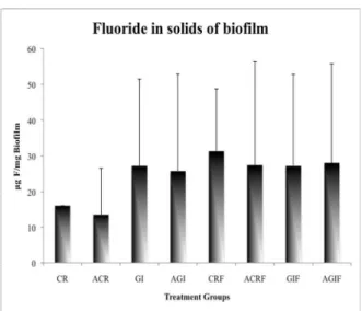
In regard to microbiological composition of the biofilm formed on the slabs restored with the different materials, no significant differences were found between the treatments (p > 0.05, table 1) by any studied factor. In regard to fluoride concentration of the biofilm, the effects of restorative material was significant, fluoride levels were higher for the groups restored with RMGIC, regardless of toothpaste and ageing (p < 0.05, table 3). There was a interaction between dentifrice and material showing higher fruoride levels (Figure 2).
According to our results, the first null hypothesis that there is no effect of the presence of induced ageing was partially accepted, inasmuch significant differences were found for the D S parameter only for dentine, where aged restorations presented higher demineralization despite the type of material (p < 0.05, table 2). In addition, the second null hypothesis that stated the effects of restorative material was partially accepted, the groups restored with RMGIC presented lower demineralization than CR regardless the ageing process, only in enamel substrate (p < 0.05, table 2). The third null hypothesis tested were rejected.
Discussion
The in situ model used has been shown to be adequate to study secondary caries.10,16 In addition, subjecting the specimen to thermal cycling can give some insight into the temperature - dependent degradation of the material.17 Thermal cycling induces stress between a tooth substrate and a restorative material at the bonding interface5 and, by simulating intra-oral conditions, 10,000 thermal cycles might represent one year of in vivo functioning. However, there is no concrete evidence that failures in practice occur because of thermal stresses.9
Microbiological analysis from oral biofilm revealed no differences between
treatments (Table 1). These results are supported by other in situ studies,3,4 who demonstrated
Fluoride analysis from biofilm showed that treaments performed in the presence
of fluoride from restorative material presented higher amounts of this ion in solids of biofilm.
Since no F-dentifrice effect was found it can be suggested that F-releasing from dental
material was similar to that found for F-dentifrice and that it was high enough to inhibit
demineralization in enamel.
Secondary caries are lesions present at the margin of an existing restoration. This
kind of lesion may be a result from microleakage18, marginal gap or discrepacies between
tooth and restoration. In regard to the lower demineralization found in dentine for unaged
fluoride restorations, it can be speculated that the ageing process caused a reduction of marginal fit inducing microleakage19 and or the presence of marginal gaps20 and more secondary caries. However, to the best of our knowledge no other study has correlated ageing processes with wall lesion or recurrent caries.
Another thing to be emphasized is that thermal cycling was only relevant for caries induction in dentine, showing that water storage affects the bond stability and simulate hydrolytic-degradation processes.21 While enamel is predominantly mineral, dentine contains a significant amount organic material, and this humid and organic nature of dentine makes this tissue very difficult to bonding.22 The the stability of the bonded interface over time is related to the generation of a compact and homogenous hybrid layer, that can reduce the integrity with phisical changes.21 Additionally, ageing causes an increase in surface roughness of composite restorations, leading to an increase accumulation of biofilm, thereby increasing secondary caries.19
lower the demineralization found in groups that submitted to the F-dentifrice use.
In situ studies have shown that restorative materials containing fluoride can inhibit caries adjacent to restorations in enamel10,24,25,4 and dentine3. For enamel, our results are supported by these previous studies, however this effect was observed in dentine in the presence of until one source of fluoride. It can be suggested that the likelihood of caries development around restorations may be more common in root dentine, since the rate of mineral loss can be twice as fast from root as it is from enamel.26
Conclusions
These results suggest that RMGIC restorations provided protection against secondary caries either for enamel and dentine, while ageing process affects caries development in dentine, increasing caries progression
Acknowledgments
Table 1. Microbiological analysis of dental biofilm according to treatment (Mean values with their
standard deviation).
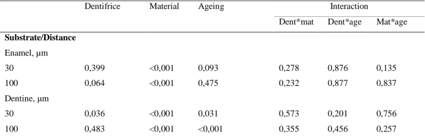
Table 2. P values of mineral loss.
Dentifrice Material Ageing Interaction
Dent*mat Dent*age Mat*age
Substrate/Distance
Enamel, µm
30 0,399 <0,001 0,093 0,278 0,876 0,135
100 0,064 <0,001 0,475 0,232 0,877 0,837
Dentine, µm
30 0,036 <0,001 0,031 0,573 0,201 0,756
100 0,483 <0,001 <0,001 0,355 0,456 0,257
Tabela 3. ANOVA Results (p values) of fruoride levels on biofilm.
Dentifrice Material Ageing Interaction
Dent*mat Dent*age Mat*age
Level of Fluoride
ugF/g biofilm 0,206 0,005 0,569 0,028 0,918 0,392
Figure 1- Blocks human tooth, enamel-dentine, prepared from the cervical region of nonerupted human third
molars. The occlusal margin of the cavity was located in enamel, while the gingival margin was located in
References
1. Mjör IA, Toffenetti F. Secondary caries: a literature review with case reports. Quintessence International 2000; 31:165-79.
2. Tenuta LMA, Del Bel Cury AA, Bortolin MC, Vogel GL, Cury JA. Ca, Pi, and F in the Fluid of Biofilm Formed under Sucrose. Journal of Dental Research 2006; 85:834-838.
3. Sousa RP, Zanin ICJ, Lima JPM, Vasconcelos SMLC, Melo MAS, Beltrão HCP, et al. In situ effects of restorative materials on dental biofilm and enamel demineralisation. Journal of Dentistry 2009; 37:44-51.
4. Wiegand A, Buchalla W, Attin T. Review on fluoride-releasing restorative materials-Fluoride release and uptake characteristics, antibacterial activity and influence on caries formation. Dental Materials 2007; 23:343-362.
5. Burke FM, Ray NJ, McConnell RJ. Fluoride-containing restorative materials. International Dental Journal 2006; 56:33-43.
6. Carvalho AS, Cury JA. Fluoride release from some dental materials in different solutions. Operative Dentistry 1999; 24:14-19.
8. Gale MS, Darvell BW: Thermal cycling procedures for laboratory testing of dental restorations. Journal of Dentistry 1999; 27:89-99.
9. Benelli EM, Serra MC, Rodrigues AL Jr, Cury JA. In situ anticariogenic potential of glass ionomer cement. Caries Research 1993; 27:280-284.
10.Cenci MS, Tenuta LMA, Cenci TP, Del Bel Cury AA, Cury JA. Effect of Microleakage and Fluoride on Enamel-Dentine Demineralization around Restorations. Caries Research 2008a; 42:369-79.
11.Cury JA, Rebelo MAB, Del Bel Cury AA. In situ relationship between sucrose exposure and the composition of dental plaque. Caries Research 1997; 31:356-360.
12.Cury JA, Rebelo MAB, Del Bel Cury AA, Derbyshire MTVC, Tabchoury CPM. Biochemical composition and cariogenicity of dental plaque formed in the presence of sucrose or glucose and fructose. Caries Research 2000; 34:491-7.
13.Zero TD. Dentifrices, mouthwashes, and remineralization/caries arrestment strategies. BMC Oral Health 2006; 6:1-13.
14.Lobo MM, Gonçalves RB, Ambrosano GM, Pimenta LA. Chemical or microbiological models of secondary caries development around different dental restorative materials. Journal of Biomedical Materials Research Part B: Applied Biomaterials 2005; 74:725-31.
Journal of Oral Sciences 2005; 113:245-250.
16.Hara AT, Turssi CP, Ando M, González-Cabezas C, Zero DT, Rodrigues AL Jr, et al. Influence of fluoride-releasing restorative material on root dentine secondary caries in situ. Caries Research 2006; 40:435-439.
17.De Munck J, Van Landuyt K, Peumans M, Poitevin A, Lambrechts P, Braem M, et al. A critical review of the durability of adhesion to tooth tissue: Methods and results. Journal of Dental Research 2005; 84:118-132.
18.Kidd EAM. Diagnosis of Secondary Caries. Journal of Dental Education 2001; 65: 997-1000.
19.Cenci MS, Cenci T-P, Donassollo TA, Sommer L, Strapasso A, Demarco FF. Influence of thermal stress on marginal integrity of restorative materials. Journal of Applied Oral Science 2008b; 16:106-10.
20.Coelho-de-Souza FH, Camacho GB, Demarco FF, Powers JM. Fracture Resistance and Gap Formation of MOD Restorations: Influence of Restorative Technique, Bevel Preparation and Water Storage. Operative Dentistry 2008; 33:37-43.
21.Saboia VPA, Silva FCFA, Nato F, Mazonni A, Cadenaro M, Mazzoti G, Giannini M, Breschi L. Analysis of differential artificial ageing of the adhesive interface produced by a two-step etch-and-rinse adhesive. Eur J Oral Sci 2009; 117: 618–624.
23.Duggal MS, Toumba KJ, Amaechi BT, Kowash MB, Higham SM. Enamel demineralization in situ with various frequencies of carbohydrate consumption with and without fluoride toothpaste. Journal of Dental Research 2001; 80:1721-1724.
24.Tenuta LM, Ribeiro CC, Gonçalves NC, Del Bel Cury AA, Aires CP, Tengan C, et al. The short-term in situ model to evaluate the anticariogenic potential of ionomeric materials. Journal of Dentistry 2005; 33:491-7.
25.Yamamoto K, Arai K, Fukazawa K, Fukui K, Nagamatsu K, Kato K, et al. Effect of plaque uoride released from a glass-ionomer cement on enamel remineralization in situ. Caries Research 2005; 39:157-60.
4 CONCLUSÃO GERAL
Nas condições deste estudo in situ:
REFERÊNCIAS
BENELLI, E. M.; SERRA M. C.; RODRIGUES, A. L. JR; CURY, J. A. In situ anticariogenic potential of glass ionomer cement. Caries Res., v. 27, n. 4, p. 280-4, 1993.
CENCI, M. S.; TENUTA, L. M. A.; PEREIRA-CENCI, T.; CURY, A. A.; CURY, J. A. Effect of Microleakage and Fluoride on Enamel-Dentine Demineralization around Restorations. Caries Res., v. 42, n. 5, p. 369-79, 2008a.
CENCI, M. S.; PEREIRA-CENCI, T.; DONASSOLLO, T. A.; SOMMER, L.;
STRAPASSON, A.; DEMARCO, F. F. Influence of thermal stress on marginal integrity of restorative materials. J Appl Oral Sci., v. 16, n. 2, p. 106-10, 2008b.
DIONYSOPOLOUS, P.; KOTSANOS, N.; KOLINIOTOU-KOUBIA, E.;
PAPADOGIANNIS, Y. Secondary Caries Formation In Vitro around Fluoride-releasing Restorations. Clinical Relevance: Fluoride releasing restorations inhibit caries-like lesions. Oper Dent., v. 19, n. 5, p. 183-8, 1994.
FEATHERSTONE, J. D. B. Fluoride, remineralisation and root caries. Am J Dent., v. 7, n. 5. p. 271-4, 1994.
GJORGIEVSKA E.; NICHOLSON, W. J.; ILJOVSKA, S.; SLIPPER, I. The potential of fluoride-releasing dental restoratives to inhibit enamel demineralization: an SEM study. Prilozi, v. 30, n. 1, p. 191-204, 2009.
HAYACIBARA, M. F.; ROSA, O.P.; KOO, H.; TORRES, S. A.; COSTA, B.; CURY, J. A. Effects of fluoride and aluminum from ionomeric materials on S. mutans biofilm. J Dent Res. v. 82, n. 4, p. 267-71, 2003.
HARA, A. T.; TURSSI, C. P.; SERRA, M. C.; NOGUEIRA, M. C. Extent of the cariostatic effect on root dentin provided by fluoride-containing restorative materials. Oper Dent., v. 27, n. 5, p. 480-7, 2002.
restorative material on root dentine secondary caries in situ. Caries Res., v. 40, p. 435-439, 2006.
HSU, C. -Y. S., DONLY, K. J., DRAKE, D. R., WEFEL, J. S. Effects of Aged Fluoride-containing Restorative Materials on Recurrent Root Caries. J Dent Res., v. 77, n. 2, p. 418-425, 1998.
KIDD E. A.; TOFFENETTI, F.; MJöR, I. A. Secondary caries. Int Dent J., v. 42, n. 3, p. 127-38, 1992.
KIDD, E. A. M. Diagnosis of Secondary Caries. J Dent Educ., v. 65, n. 10, p. 997-1000, 2001.
LING, L.; XU, X.; CHOI, G. -Y.; BILLODEAUX, D.; GUO, G.; DIWAN, R. M. Novel F-releasing Composite with Improved Mechanical Properties. J Dent Res., v. 88, n. 1, p. 83-88, 2009.
MILLER, B. H.; KOMATSU, H.; NAKAJIMA, H.; OKABE, T. Effect of glass ionomer manipulation on early fluoride release. Am J Dent., v.8, n. 4, p.182-6, 1995.
MJÖR, I. A.; TOFFENETTI F. Secondary caries: a literature review with case reports. Quintessence Int., v. 31, n. 3, p. 165-79, 2000.
SABOIA, V. P. A.; SILVA, F. C. F. A.; NATO, F.; MAZONNI, A.; CADENARO, M.; MAZZOTI, G.; GIANNINI, M.; BRESCHI, L. Analysis of differential artificial ageing of the adhesive interface produced by a two-step etch-and-rinse adhesive. Eur J Oral Sci., v. 117, n. 1, p. 618–624, 2009.
SOUSA, R. P.; ZANIN, I. C. J.; LIMA, J. P. M.; VASCONCELOS, S. M. L. C.; MELO, M. A. S.; BELTRÃO, H. C. P.; RODRIGUES, L. K. A. In situ effects of restorative materials on dental biofilm and enamel demineralisation. J Dent., v. 37, n. 1, p. 44-51, 2009.
TEN CATE, J. M.; LARSEN, M. J.; PEARCE, E. I. F.; FEJERSKOV, O. Chemical
TENUTA, L. M.; RIBEIRO, C. C.; GONÇALVES, N. C.; DEL BEL CURY, A. A.; AIRES, C. P.; TENGAN, C.; TAGLIAFERRO, E. P.; PECHARKI, G. D.; NAPIMOGA, M. H.; TABCHOURY, C. P.; CURY, J. A. The short-term in situ model to evaluate the
anticariogenic potential of ionomeric materials. J Dent., v. 33, n. 6, p. 491-7, 2005.
VERBEECK, R.M.; DE MOOR, R. J.; VAN EVEN, D. F.; MARTENS, L. C. The short-term fluoride release of a hand-mixed vs. capsulated system of a restorative glass-ionomer cement. J Dent Res., v.72, n. 3, p.577-81, 1993.
WIEGAND, A.; BUCHALLA, W.; ATTIN, T. Review on fluoride-releasing restorative materials-Fluoride release and uptake characteristics, antibacterial activity and influence on caries formation. Dent Mater., v. 23, n. 3, p. 343-362, 2007.
APÊNDICE A - TERMO DE DOAÇÃO DE DENTES
Eu,____________________________________________,CRO_______, estou doando ____ dentes terceiros molares retidos, extraídos em meu consultório localizado
a_____________________________________________(Rua e Cidade), por razões independentes da pesquisa e sob consentimento do paciente, para a pesquisa intitulada “EFEITO DO ENVELHECIMENTO DE RESTAURAÇÕES DE CIMENTO DE IONÔMERO DE VIDRO NA DESMINERALIZAÇÃO DO ESMALTE E DENTINA: ESTUDO in situ.”, que será realizada na Faculdade de Farmácia, Odontologia e Enfermagem da Universidade Federal do Ceará pela pesquisadora: Maria Denise Rodrigues de Moraes.
Fortaleza, ____ de ______ de 200__.
_______________________________________
APÊNDICE B - TERMO DE CONSENTIMENTO LIVRE E ESCLARECIDO
Título da Pesquisa- EFEITO DO ENVELHECIMENTO DE RESTAURAÇÕES DE
CIMENTO DE IONÔMERO DE VIDRO NA DESMINERALIZAÇÃO DO ESMALTE E DENTINA: ESTUDO in situ.
Objetivo da Pesquisa: avaliar in situ o potencial anticárie de materiais restauradores quando
submetidos a uma situação de envelhecimento acelerado e de alto desafio cariogênico in situ.
Você está sendo convidado a contribuir com a realização de uma pesquisa. Leia atentamente as informações abaixo e faça qualquer pergunta que desejar, para que todos os questionamentos sejam esclarecidos.
Convidamos você a participar da pesquisa intitulada EFEITO DO ENVELHECIMENTO DE RESTAURAÇÕES DE CIMENTO DE IONÔMERO DE VIDRO NA DESMINERALIZAÇÃO DO ESMALTE E DENTINA: ESTUDO in situ.
A cárie dental ainda está entre as doenças mais relevantes em termos de saúde pública, principalmente nos países em desenvolvimento. Grupos de crianças continuam apresentando alta atividade da doença. Em termos mundiais, cerca de 20 a 25% das crianças e adolescentes apresenta 60 a 80% do total de cárie da população. Tal fato enfatiza a necessidade de se aperfeiçoar métodos preventivos já existentes, com a introdução de técnicas inovadoras que possam agir como coadjuvantes na prevenção e controle da cárie dental neste segmento da população.
Desta forma, novos materiais são frequentemente lançados no mercado odontológico com o apelo de conseguirem controlar, ainda que de uma forma localizada, o aparecimento de novas lesões de cárie.
modelos in situ para testar materiais e técnicas restauradoras e sua capacidade de inibir o desenvolvimento de cáries recorrentes antes da realização de extensos e dispendiosos estudos clínicos. No entanto, há na literatura científica uma escassez de estudos in situ que testem todas as categorias de materiais restauradores existentes visando uma maior objetividade na ocasião de sua indicação.
Procedimentos
Será realizado um estudo do tipo cruzado que compreenderá 4 tratamentos, sendo duas fases de 14 dias, durante a qual você utilizará um dispositivo intra-oral palatino contendo blocos de esmalte e dentina dental humano. Os 4 tratamentos serão:
Tratamento 1: Restauração com Resina Composta (Z-250) envelhecida Tratamento 2: Restauração com Resina Composta (Z-250) não envelhecida
Tratamento 3: Restauração com Ionômero de vidro modificado por resina encapsulado (Fuji II) envelhecido
Tratamento 4: Restauração com Ionômero de vidro modificado por resina encapsulado (Fuji II) não envelhecido
Em um período anterior ao início das fases do experimento (7 dias), você deverá fazer uso do dentifrício pré-determinado a fim de padronizar as concentrações de flúor na saliva.
Instruções:
a) Na fase clínica: Todos os blocos contidos no dispositivo deverão ser gotejados com a solução de sacarose a 20% dez vezes ao dia respeitando os horários pré-determinados pelo pesquisador (8:00, 9:30, 11:00, 12:30, 14:00, 15:30, 17:00, 18:30, 20:00, 21:30 hor as).
Após cinco minutos do gotejamento (uma gota sobre cada bloco), o dispositivo deverá ser recolocado na boca sem ser lavado;
b) utilizar o dispositivo intra-oral palatino diariamente, inclusive para dormir;
d) fazer uso do dentifrício padronizado três vezes ao dia durante a escovação. Durante a escovação, o dispositivo deverá ser removido e os voluntários deverão limpar seus aparelhos cuidadosamente para evitar a remoção do biofilme dental formado sobre os blocos. O tempo de escovação do dispositivo e dos dentes não deve exceder a 3 minutos e a região da telinha deve ser escovada delicadamente para evitar remoção ou perturbação da placa bacteriana. e) fazer uso de água fluoretada de abastecimento de Fortaleza (0,7 ppm F).
Desconfortos e Riscos
Vocês poderão apresentar discreta halitose apenas durante o período experimental, o que poderá ser resolvido com adequada higiene dental. Mesmo com remotas possibilidades, caso esta halitose persista após o período experimental será realizada uma profilaxia dentária, bem como será lhe fornecido enxaguatório bucal com clorexidina até que o problema seja resolvido. O uso das soluções será apenas como gotas sobre os blocos presentes nos dispositivos intra-orais, não implicando em qualquer aumento de cárie dental nos voluntários. No entanto, caso haja o surgimento de alguma lesão de carie inicial a mesma receberá tratamento adequada com compostos fluoretados anticariogênicos. O dispositivo intra-oral pode causar um leve desconforto, que é, entretanto, semelhante ao desconforto causado por um aparelho ortodôntico móvel. Durante todo o período da pesquisa, acompanhamentos semanais serão realizados, para verificar as condições do aparelho e da sua saúde bucal. Cabe ressaltar que não haverá consumo direto da substância, pois a mesma será gotejada sobre os blocos.
O benefício que vocês terão será um auxílio indireto, contribuindo para a realização deste projeto e o conhecimento que vocês adquirirão sobre o potencial anticárie de materiais restauradores. Este conhecimento poderá ser utilizado futuramente em prol da população alto risco à cárie.
Forma de acompanhamento e assistência
Os pesquisadores envolvidos na pesquisa estarão à disposição de vocês para ajuste no aparelho intra-oral a fim de minimizar qualquer desconforto.
Garantia de esclarecimento
pesquisa. Também os pesquisadores supracitados assumem o compromisso de proporcionar informação atualizada obtida durante o estudo, ainda que esta possa afetar a vontade do indivíduo em continuar participando. Qualquer dúvida ou problema com o dispositivo intra-oral, por favor, comunicar-nos com a maior brevidade possível.
Tel: 3366-8410 (Clínica de Dentística) Formas de ressarcimento
Vocês serão ressarcidos de eventuais despesas com o transporte-alimentação para a retirada das amostras contidas nos dispositivos.
Formas de indenização
Não há danos previsíveis decorrentes desta pesquisa. Garantia de sigilo
Os pesquisadores asseguram a sua privacidade quanto aos dados confidenciais envolvidos na pesquisa.
Liberdade para se recusar em participar da pesquisa
Tendo compreendido perfeitamente tudo o que me foi informado sobre a minha participação no mencionado estudo e estando consciente dos meus direitos, das minhas responsabilidades, dos riscos e dos benefícios que a minha participação implicam, concordo em dele participar e par isso DOU O MEU CONSENTIMENTO SEM QUE PARA ISSO EU TENHA SIDO FORÇADO OU OBRIGADO.
__________________________________________________ Assinatura do voluntário
Endereço do Voluntário: Documento (RG): Telefones:
__________________________________________________
Assinatura do Profissional que aplicou o TCLE
Assinatura do Responsável pelo estudo Universidade Federal do Ceará
Rua Cap. Francisco Pedro s/n. Rodolfo Teófilo. CEP. 60430-170 Fone: 33668410/33668426
Departamento de Odontologia Restauradora
ATENÇÃO: A SUA PARTICIPAÇÃO EM QUALQUER TIPO DE PESQUISA É VOLUNTÁRIA. EM CASO DE DÚVIDAS REALIZAR CONTATO COM O COMITÊ DE ÉTICA EM PESQUISA DA UFC.
APÊNDICE C - INSTRUÇÕES AOS VOLUNTÁRIOS
Cada voluntário receberá:
- 2 tubos de dentifrício,1 escova dental, 7 frascos conta-gotas com solução de sacarose a 40%, isopor 500mg a fim de manter em congelador os frascos com solução de sacarose, estojo de aparelho ortodôntico (acomodação do dispositivo), 1 pacote de gaze estéril.
Os voluntários deverão seguir as seguintes instruções:
a) Na fase clínica: todos os blocos contidos no dispositivo deverão ser gotejados com a solução de sacarose a 20% dez vezes ao dia respeitando os horários pré-deter minados pelo pesquisador (8:00, 9:30, 11:00, 12:30, 14:00, 15:30, 17:00, 18:30, 20:00, 21:30 hor as).
Após cinco minutos do gotejamento (uma gota sobre cada bloco), o dispositivo deverá ser recolocado na boca sem ser lavado;
b) utilizar o dispositivo intra-oral palatino diariamente, inclusive para dormir;
c) remover o dispositivo intra-oral somente durante as refeições ou ingestão de qualquer bebida ácida, durante este período o mesmo deve ser conservado no estojo fornecido e em ambiente úmido com o objetivo de manter as bactérias da placa viáveis;
d) fazer uso do dentifrício padronizado três vezes ao dia durante a escovação. Durante a escovação, o dispositivo deverá ser removido e os voluntários deverão limpar seus aparelhos cuidadosamente para evitar a remoção do biofilme dental formado sobre os blocos. O tempo de escovação do dispositivo e dos dentes não deve exceder a 3 minutos e a região da telinha deve ser escovada delicadamente para evitar remoção ou perturbação da placa bacteriana. e) fazer uso de água fluoretada de abastecimento de Fortaleza (0,7 ppm F).
APÊNDICE E – Representação esquemática do método de extração de flúor para análise dos dentifrícios