A.C. Marques, L.V.C. Lotufo, P.C. Paiva, P.T.C. Chaves & S.N. Leitão (Guest Editors) DOI: 10.3856/vol41-issue2-fulltext-12
Research Article
Cytotoxicity of actinomycetes associated with the ascidian
Eudistoma vannamei
(Millar, 1977), endemic of northeastern coast of Brazil
Paula C. Jimenez1,2, Elthon G. Ferreira1,2, Luana A. Araújo1, Larissa A. Guimarães1, Thiciana S. Sousa3 Otília Deusdenia L. Pessoa3, Tito M. C. Lotufo1 & Letícia V. Costa-Lotufo1,2
1
Instituto de Ciências do Mar (LABOMAR), Universidade Federal do Ceará Av. Abolição, 3207, 60165-081, Fortaleza, Brazil
2
Departamento de Fisiologia e Farmacologia, Faculdade de Medicina Universidade Federal do Ceará, Av. da Universidade, 2853 Benfica
Fortaleza, CE, 60020-181, Brazil
3
Departamento de Química Orgânica e Inorgânica, Universidade Federal do Ceará Av. da Universidade, 2853, Benfica, Fortaleza, CE, 60020-181, Brazil
ABSTRACT. Previous studies demonstrated that the crude extract of the ascidian Eudistoma vannamei,
endemic from northeasttern Brazil, strongly hinders growth of tumor cells in vitro by inducing apoptosis due to tryptophan derivatives, which are commonly found in bacteria. This study presents a bioactivity-guided screening among actinomycetes, associated with E. vannamei, aiming at recognizing active principles with biological relevance. Twenty strains of actinomycetes, designated as EVA 0101 through 0120, were isolated from colonies of E. vannamei among which 11 were selected for cytotoxicity evaluation. The extracts from EVA 0102, 0103, 0106, 0109 and 0113 were the most active, and were further studied for IC50 determination
and chemical analysis by 1H NMR. IC50 values obtained ranged from 3.62 µg mL -1
(for EVA 0109 in leukemia cells) to 84.65 µg/mL (for EVA 0106 in melanoma cells). All active extracts exhibited the same TLC and spectroscopic profiles, suggesting the presence of quinones and other related secondary metabolites. Furthermore, these strains were identified and compared based on their respective 16S rRNA sequences. The results herein identified the five strains as Micromonospora spp. while phylogenetic analysis suggests that they are possibly two different Micromonospora species producing the cytotoxic compounds.
Keywords: marine biotechnology, Eudistoma vannamei, marine microorganisms, Micromonospora sp.,
Brazil.
Citotoxicidad de actinomicetos asociada a la ascidia
Eudistoma vannamei
(Millar, 1977), endémica de la costa noreste de Brasil
RESUMEN. Estudios previos demostraron que el extracto crudo de la ascidia Eudistoma vannamei, endémica de la costa noreste de Brasil, obstaculiza fuertemente el crecimiento de células tumorales in vitro por inducir apoptosis. El análisis químico del extracto sugirió la presencia de derivados de triptófano, comúnmente encontrados en bacterias. El estudio presenta un screening de citotoxicidad en actinomicetos asociados con E.
vannamei, para reconocer principios activos biológicamente relevantes. Veinte cepas de actinomicetos,
designados como EVA 0101 hasta 0120, fueron aisladas de colonias de E. vannamei y 11 fueron seleccionadas para la evaluación de citotoxicidad. Los extractos de EVA 0102, 0103, 0106, 0109 y 0113 resultaron las más activas y fueron estudiadas para determinación de la CL50 y perfiles de CCF y
1
H RMN. Las CL50 obtenidas
oscilaron entre 3,62 (EVA 0109 en las células de leucemia) y 84,65 µg mL-1 (EVA 0106 en las células de melanoma). Los extractos activos presentan el mismo perfil de CCF y espectroscópico, lo que sugiere la presencia de quinonas y metabolitos secundarios relacionados. Además, las cinco cepas fueron identificadas y comparadas sobre la base de sus secuencias de 16S ARNr. Las que se identificaron como Micromonospora
Palabras clave: biotecnología marina, Eudistoma vannamei, microorganismos marinos, Micromonospora sp., Brasil.
___________________
Corresponding author: Letícia Lotufo (lvcosta@secrel.com.br)
INTRODUCTION
Colorful soft-bodied sessile marine invertebrates have been a rich source of natural products with biomedical properties for the past 60 years (Baily, 2009; Molinski et al., 2009; Mayer et al., 2010). Among them, the tunicates are one of the most prolific in terms of novel drug prototypes for the anticancer pharmaceutical arsenal (Blunt et al., 2010, 2011). For instance, trabectedin, a tetrahydroisoquinoline alkaloid obtained from Ecteinascidia turbinata, was recently approved for the treatment of soft tissue sarcoma, with the commercial name Yondelis® (Cuevas & Francesh, 2009; D’Incalci & Galmarini, 2010). Additionally, plitidepsin, a cyclodepsipeptide isolated from Aplidium albicans is in advanced trial phase for the therapy of multiple myeloma and other malignant tumors, but has already received orphan drug status for the treatment of multiple myeloma and acute lymphoblastic leukemia (Ocio et al., 2008; Singh et al., 2008).
There are mounting evidences that the associated microorganisms are the authentic suppliers for most of the relevant compounds (Zhang et al., 2005; Imhoff et al., 2011). For the ascidians, in particular, there is a compelling amount of information pointing into this direction (Schmidt & Donia, 2010). Despite the oceans are acknowledged as a rich and highly complex microbiological environment, they have only shared their wealth as a natural drug source for the past 10 years. Thus, the study of marine microor-ganisms as sources of new drugs is still at its youth (Jensen et al., 2005; Fenical & Jensen, 2006; Liu et al., 2010).
This study takes part in a pioneer venture aiming to the exploration of the biotechnological potentials of microorganisms, mainly actinomycetes, from the coast of Ceará. The actinomycetes have proven to be a prolific source of new and bioactive compounds (Fenical & Jensen, 2006). The purpose of the present investigation is to find biologically active strains of actinomycetes and evaluate their respective cytoto-xicity, while gathering information on the microbiota associated with Eudistoma vannamei. This species is endemic from the northeastern coast of Brazil and is the most abundant ascidian recorded for the state of Ceará. Previous studies have revealed that its crude extract strongly hinders growth of tumor cells in vitro,
promoting further apoptosis (Jimenez et al., 2003, 2008).
MATERIALS AND METHODS
Sampling and isolation of bacteria strains
Specimens of E. vannamei were hand collected at Taiba Beach (03°30'21.23”S, 038°53'40.16”W), located on the northeastern coast of Brazil. Sampling was carried out with the slightest contamination possible, as the material was gathered and stored with sterile utensils. For isolation of microorganisms, five colonies were homogenized in sterile seawater 1:5 (m/v), and aliquots of 20 L were stroked onto Petri dishes containing different agar based media, which may favor actinomycetes growth (modified from Jensen et al., 1991). The following agar-based media were used herein, all added with cycloheximide 0.1 mg mL-1: SCA (Starch Casein Agar, HIMEDIA, prepared with distilled H2O), AIA (Actinomycetes
Isolation Agar, HIMEDIA, prepared in 75% water), SWA (Sea-water Agar, 1.8% agar in 75% sea-water) and GYM (glucose 0.4%, yeast extract 0.4%, malt extract 1%, CaCO3 0.2%, 1.8% agar in 75%
sea-water).
Purity of strains were resolved by sequential re-strikes onto brand new agar plates and pure strains were inoculated in liquid A1 medium (10% soluble starch, 4% yeast extract and 2% peptone in 75% sea-water) for culture maintenance and up-scaled growth (Jensen et al., 1991). Grown media for each pure strain was sampled into cryovials and supplemented with 25% glycerol for storage at -70oC.
Up-scaled growth and extraction
Initially, pure cultures were inoculated in 500 mL Erlenmeyer flasks filled with 100 mL of liquid A1 medium. Flasks were kept at 28oC and 200 rpm for 5 days. Next, 10 mL of grown media were added to newly assembled Erlenmeyer flasks with 100 mL of fresh media and kept at 28oC and 200 rpm agitation for 10 days.
remove excessive salt, and fully dried under compressed air, yielding the crude extracts. At this point, extracts were resuspended in DMSO to a final concentration of 10 µg mL-1 and were ready to be evaluated for their biological activity.
Screening for cytotoxicity-MTT assay
Cytotoxic activity of extracts was evaluated against four human tumor cell lines obtained from the National Cancer Institute (Bethesda, MD, USA): HL-60 (promyelocytic leukemia), MDA-MB-435 (mela-noma), SF-295 (glioblastoma) and HCT-8 (colon carcinoma). Cells were grown in RPMI-1640 medium supplemented with 2 mM glutamine, 10% fetal calf serum, 100 µg mL-1 strep-tomycin and 100 U mL-1 penicillin and housed at 37ºC with a 5% CO2
atmosphere. The cell cultures were regularly split to keep them in a logarithmic phase of growth.
Firstly, in a qualitative approach, cell lines were exposed to 100 µg mL-1 of the extracts for 72 h and the effect on cell proliferation was evaluated in vitro using the MTT [3-(4,5-dimethyl-2-thiazolyl)-2,5-diphenyl-2H-tetrazolium bromide] assay, as described by Mosmann (1983). The extracts that reduced over 80% of cell growth, when compared to non-treated cells, were tested again, in a quantitative manner, however, at concentrations varying serially from 0.02 to 100 µg mL-1 to determine the IC50 (the
concentration that inhibits growth in 50%), which was calculated, along with the respective 95% CI (confidence interval), by a non-linear regression using the software GraphPad Prism 5.0.
Preliminary chemical analysis
The active extracts (EVA 0102, EVA 0106, EVA 0109 and EVA 0113) were submitted to a preliminary chemical characterization. Initially, these extracts were compared by thin layer chromatography (TLC) on precoated silica gel aluminum sheets (Kieselgel 60 F254, 0.20 mm, Merck) using two different solvent
systems (CHCl3 and CHCl3: EtOAc 1:1) and the
compounds were detected by exposure to UV (312 and 365 nm) light and by spraying with vanillin/ perchloric acid/EtOH solution followed by heating at 100°C. The extracts were also analyzed by 1H NMR spectroscopy. The 1H NMR spectra were acquired on a Bruker Avance DRX-500 spectrometer equipped with 5 mm inverse detection z-gradient probe. The chemical shifts, given on the δ scale, were referenced to the residual undeuterated CHCl3 (δH 7.27) fraction.
The methodology was based in a previous work published by Jimenez et al. (2008).
Molecular identification: nucleic acid extraction, 16S rRNA gene amplification, and sequencing
The selected microorganisms were characterized using a molecular approach based on the protocol described by Gontang et al. (2007) with slight modifications. Genomic DNA was extracted from a fresh, 3 to 5 day grown, bacterial culture following the DNeasy protocol (QIAGEN Inc., USA). Amplification of the 16S rRNA gene was carried out with Ilustra™ PureTaq Ready-To-Go PCR beads (GE Healthcare, UK) using the universal eubacterial primers 27F AGAGTTTGATCCTGGCTCAG-3’) and 1492R (5’-TACGGCTACCTTGTTACGACTT-3’). PCR pro-ducts were purified with MiniElute PCR purification kit (QIAGEN Inc., USA).
DNA sequencing was performed by Macrogen Inc. (Seoul, Korea) using the ABI PRISM BigDyeTM Terminator Cycle Sequencing kit (Applied Biosys-tems, USA) and the same primers used for amplification, following the protocols supplied by the manufacturer.
The nucleotide sequences were assembled, analyzed and manually edited using the BioEdit Sequence Alignment Editor (Hall, 1999) and compared to sequences within the NCBI database (http://www.ncbi.nlm.nih.gov) using the Basic Local Alignment Search Tool (BLAST).
The identity of the strain was confirmed after an evaluation of the phylogenetic relationships with the most similar sequences obtained from the EzTaxon server (Chun et al., 2007). The sequences were aligned using Muscle ver. 3.6, and the reconstruction was performed using a combined bootstrapping and maximum likelihood approach through RAxML (Stamakis, 2006) on the CIPRES portal (Miller et al., 2010). The tree was produced using Archaeopterix ver. 0.957 (Han & Zmasek, 2009). After the phylogenetic analysis, the sequences were submitted to NCBI GenBank.
RESULTS
Isolation and cytotoxicity screening of bacteria associated with E. vannamei
Eva 0101
Eva 0102
Eva 0103
Eva 0105
Eva 0106
Eva 0109
Eva 0110
Eva 0111
Eva 0113
Eva 0114
Eva 0118
0 25 50 75 100
HCT-8
cell growth inhibition (%)
0 25 50 75 100
MDA MB-435
cell growth inhibition (%)
0 25 50 75 100
SF-295
cell growth inhibition (%)
Figure 1. Qualitative cytotoxicity screening of EtOAc extracts from liquid medium (dark gray) and MeOH extracts from biomass (light gray) of bacterial strains isolated from E. vannamei. Extracts were tested (100 µg mL-1) against HCT-8, MDA-MB-435 and SF-295 cell lines by the MTT assay after 72 h incubation. Data were obtained from two independent experiments and presented as a percentage of growth inhibition relative to untreated cells.
Table 1. Cytotoxic activity of EtOAc extracts obtained from bacterial strains isolated from Eudistoma vannamei eva-luated by the MTT assay after 72 h incubation. IC50 values and their respective 95% confidence intervals were obtained
by nonlinear regression on the GraphPad Prism 5.0 software (Intuitive Software for Science, San Diego, CA). N.T.: not tested.
Strain Isolation media
IC50 (µg mL-1)
95% CI
HL-60 HCT-8 MDA MB-435 SF-295
EVA 0102 SCA 24.05 14.81 - 39.06
16.48 13.22 - 20.55
60.41 49.20 - 74.17
26.87 17.77 - 40.65
EVA 0106 AIA 61.92 32.85 - 116.7
14.66 7.831 - 27.44
84.65 64.17 - 111.7
14.06 8.243 - 23.96
EVA 0109 GYM 3.88 2.43 - 6.20
3.62 0.44 - 0.67
23.78 19.86 - 28.48
5.12 4.43 - 5.91
EVA 0113 SWA 30.07 17.71 - 51.06
10.90
6.83 - 17.38 N.T.
9.97 6.92 - 14.38
The IC50 was determined for the extracts obtained
from EVA 0102, EVA 0106, EVA 0109 and EVA 0113, since the yield of the extract from EVA 0103 was insufficient for further analyses. The tested extracts showed a similar activity profile, with little variation in potency. As shown on Table 1, EVA 0109 yielded the most cytotoxic extract, with IC50 values
varying from 3.62 to 23.78 µg mL-1. EVA 0113
followed, with IC50 ranging from 9.97 to 30.07 µg
mL-1.
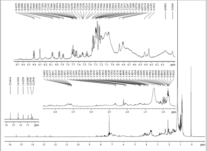
Chemical analysis of active extracts
depicted (Fig. 2). After a detailed analysis, it was clear the presence of chelated hydroxyl groups (δ 15.44-12.29), aromatic and/or olefine hydrogens (δ 8.41-5.92), hydrogens bound to oxygenated carbon (δ 3.64-3.54), as well as methylene, methyne and methyl protons (δ 2.34-0.83) (Silverstein et al., 1991).
The aforementioned data were compatible with the presence of quinones or related secondary metabolites.
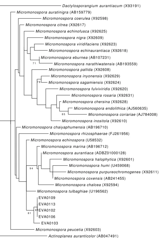
Identification and phylogenetic analyses
The analysis of the 16S rRNA from the 5 EVA bacterial strains revealed a high degree of similarity among them (Fig. 3). When included in a reconstruction of the phylogeny along with the most similar sequences found on Genbank, the EVA clade came out as the sister group of a clade formed by Micromonospora echinospora, M. marina, M. auran-tiaca, M. halophytica, M. humi, M. purpureo-chromogenes, M. coxensis, M. chalcea and M.
tulbagihiae. The EVA clade was formed with a relatively high support value and EVA 0109, 0113, 0102 and 0106 showed a higher degree of overall similarity. For this clade a single contig was then generated and deposited on NCBI GenBank under accession number JN797618. The sequence from EVA 0103 is slightly different, so it was deposited with a separate accession number (JN797619), despite its 16S rRNA sequence is still more than 99.9% similar to the other EVA strains.
DISCUSSION
The study herein began with a screening for cytotoxicity among actinomycetes isolated from the tunicate E. vannamei. The active extracts showed identical 1H NMR profiles and the respective strains were identified based on a molecular biology approach, where all five isolates were identified within the genus Micromonospora. The results pointed
Dactylosporangium aurantiacum (X93191)
Micromonospora auratinigra (AB159779)
Micromonospora coerulea (X92598)
Micromonospora citrea (X92617)
Micromonospora echinofusca (X92625)
Micromonospora nigra (X92609) Micromonospora viridifaciens (X92623)
Micromonospora echinaurantiaca (X92618)
7 1
Micromonospora eburnea (AB107231)
Micromonospora narathiwatensis (AB193559) Micromonospora pallida (X92608)
7 8
Micromonospora inyonensis (X92629)
Micromonospora sagamiensis (X92624)
Micromonospora fulviviridis (X92620) Micromonospora rosaria (X92631)
Micromonospora chersina (X92628)
8 5
Micromonospora endolithica (AJ560635) Micromonospora coriariae (AJ784008) Micromonospora inositola (X92610)
Micromonospora chaiyaphumensis (AB196710)
Micromonospora rhizosphaerae (FJ261956) Micromonospora echinospora (U58532)
9 4
Micromonospora marina (AB196712)
Micromonospora aurantiaca (ADBZ01000128)
Micromonospora halophytica (X92601) Micromonospora humi (U459068)
Micromonospora purpureochromogenes (X92611)
Micromonospora coxensis (AB241455)
Micromonospora chalcea (X92594)
Micromonospora tulbaghiae (U196562)
8 1
EVA0109
8 1
EVA0113
EVA0102
EVA0106
EVA0103
Micromonospora peucetia (X92603)
Actinoplanes auranticolor (AB047491)
Figure 3. Maximum likelihood tree of the Micromonospora lineages isolated from E. vannamei and most similar species. Dactylosporangium and Actinoplanes were used as outgroups.
to at least two species of Micromonospora producing cytotoxic compounds associated with E. vannamei. Micromonospora are a well-known group of Gram-positive, spore forming microbes, and are considered the most abundant actinobacteria, along with Rhodococcus and Streptomyces in marine environ-ments (Maldonado et al., 2005). Additionally, they are
yielded many compounds with biomedical relevance. Thiocoraline is produced by a strain of M. marina isolated from a soft coral collected near the coast of Mozambique (Romero et al., 1997). This cyclic thiodepsipeptide showed strong cytotoxic activity against various tumor cell lines, with IC50 around
2nM. Moreover, thiocoraline induced cell cycle perturbations due to inhibition of DNA-polymerase activity (Pérez-Baz et al., 1997; Romero et al., 1997; Erba et al., 1999). Other example is diazepinomicin, a dibenzodiazepine alkaloid with antimicrobial proper-ties isolated from a Micromonospora strain recovered from Didemnum proliferum, a tunicate collected off the Japanese coast (Charan et al., 2004).
In fact, associations of Micromonospora and other actinomycetes with tunicates have been documented elsewhere (Menezes et al., 2010). The Fijian tunicate Polysyncraton lithostrotum is the host to the then unidentified species M. lomaivitiensis, the producer of the potent anticancer compounds lomaivitins A and B (He et al., 2001). These molecules also have antimicrobial activity, which is thought to scare off other bacteria (He et al., 2001).
The results presented here, showing the cytoto-xicity of a Micromonospora strain and descri-bing its association with the Brazilian endemic ascidian E. vannamei, highlight their potential as producers of relevant molecules. The chemical profile of Micromo-nospora extracts was different from the one observed by Jimenez et al. (2008) for E. vannamei extracts. While the active compounds presented in ascidian extracts correspond to tryptophan derivatives, spectra data generated for the bacterial extracts herein hinted on the presence of quinoid-like compounds, such as anthracyclines or anthracyclinones (Pretsch et al., 2000; Laatsch & Fotso, 2008). This data suggested that the isolated Micromonospora strain is not responsible for the production of ascidian bioactive compounds. Nonetheless, the activity was similar for both ascidian extract (IC50 ranging from bellow 2.00
to 23.80 µg mL-1, Jimenez et al., 2008) and bacterial ones (IC50 ranging from 3.62 to 84.65 µg mL-1).
The occurrence of anthracyclines or related compounds withholding some sort of bioactivity is rather common within the Micromonospora genus. M. lupini yielded the anti-cell invasion anthraquinones lupinacidins A, B and C (Igarashi et al., 2007, 2011). The anthracyclines micromonomycin (Yang et al., 2004) and spartamicins A and B (Nair et al., 1992), produced by different strains of Micromonospora, have antimicrobial and antifungal properties, respecti-vely. Moreover, the previously mentioned lomaivitins are hybrid molecules flanked with naphtoquinone moieties (He et al., 2001).
Finally, it can be concluded that acnitomycetes isolated from the Brazilian endemic ascidian E. vannamei are a promising source of bioactive compounds. Therefore, additional studies are in progress to resolve the active principles and to characterize the means by which they may exert cytotoxicity.
ACKNOWLEDGMENTS
This work has been supported by Conselho Nacional de Desenvolvimento Científico e Tecnológico (CNPq), Financiadora de Estudos e Projetos (FINEP) and International Foundation for Science (IFS). The authors also wish to thank Silvana França, for technical assistance with the cell cultures, and M.Sc. José Gustavo de A. Lima for generating and editing the 1H NMR spectra.
REFERENCES
Baily, C. 2009. Ready for a comeback of natural products in oncology. Biochem. Pharmacol., 77(9): 1447-1457.
Bérdy, J. 2005. Bioactive microbial metabolites: a per-sonal view. J. Antibiot., 58: 1-26.
Blunt, J.W., B.R. Copp, W.P. Hu, M.H. Munro, P.T. Northcote & M.R. Prinsep. 2010. Marine natural products. Nat. Prod. Rep., 27: 165-237.
Blunt, J.W., B.R. Copp, W.P. Hu, M.H. Munro, P.T. Northcote & M.R. Prinsep. 2011. Marine natural products. Nat. Prod. Rep., 28: 196-268.
Charan, R.D., G. Schlingmann, J. Janso, V. Bernan, X. Feng, & G.T. Carter. 2004. Diazepinomicin, a new antimicrobial alkaloid from a marine Micromonos-pora sp. J. Nat. Prod., 67(8): 1431-1433.
Chun, J., J.H. Lee, Y. Jung, M. Kim, S. Kim, B.K. Kim & Y.W. Lim. 2007. EzTaxon: a web-based tool for the identification of prokaryotes based on 16S ribosomal RNA gene sequences. Int. J. Syst. Evol. Microbiol., 57: 2259-2261.
Cuevas, C. & A. Francesch. 2009. Development of Yondelis (trabectedin, ET-743). A semisynthetic process solves the supply problem. Nat. Prod. Rep., 26(3): 322-337.
D'Incalci, M. & C.M. Galmarini. 2010. A review of trabectedin (ET-743): a unique mechanism of action. Mol. Cancer Ther., 9(8): 2157-2163.
Fenical, W. & P.R. Jensen. 2006. Developing a new resource for drug discovery: marine actinomycete bacteria. Nature Chem. Biol., 2: 666-673.
Gontang, E.A., W. Fenical & P.R. Jensen. 2007. Phylogenetic diversity of gram-positive bacteria cultured from marine sediments. Appl. Environ. Microbiol., 73(10): 3272-3282.
Hall, T.A. 1999. BioEdit: a user-friendly biological sequence alignment editor and analysis program for Windows 95/98/NT. Nucl. Acids Symp., 41: 95-98. Han, M.V. & C.M. Zmasek. 2009. PhyloXML: XML for
evolutionary biology and comparative genomics. BMC Bioinformatics, 10: 356.
He, H., W.D. Ding., V.S. Bernan, A.D. Richardson, C.M. Ireland, M. Greenstein, G.A. Ellestad & G.T. Carter. 2001. Lomaiviticins A and B, potent antitumor antibiotics from Micromonospora lomaivitiensis. J. Am. Chem. Soc., 123: 5362-5363.
Igarashi, Y., M.E. Trujillo, E. Martínez-Molina, S. Yanase, S. Miyanaga, T. Obata, H. Sakurai, I. Saiki, T. Fujita & T. Furumai. 2007. Antitumor anthraquinones from an endophytic actinomycete Micromonospora lupini sp. nov. Bioorg. Med. Chem. Lett., 17(13): 3702-3705.
Igarashi, Y., S. Yanase, K. Sugimoto, M. Enomoto, S. Miyanaga, M.E. Trujillo, I. Saiki & S. Kuwahara. 2011. Lupinacidin C, an inhibitor of tumor cell invasion from Micromonospora lupini. J. Nat. Prod., 74(4): 862-865.
Imhoff, J.F., A. Labes & J. Wiese. 2011. Bio-mining the microbial treasures of the ocean: new natural products. Biotechnol. Adv., 29(5): 468-482.
Jensen, P.R., R. Dwight & W. Fenical. 1991. Distri-buition of actinomycetes in near-shore tropical marine sediments. Appl. Environ. Microbiol., 57(4): 1102-1108.
Jensen, P.R., T.J. Mincer, P.G. William & W. Fenical. 2005. Marine actinomycetes diversity and natural product discovery. Antoine van Leeuwenhoek, 87(1): 43-48.
Jimenez, P.C., S.C. Fortier, T.M.C. Lotufo, C. Pessoa, M.E.A. Moraes, M.O. Moraes & L.V. Costa-Lotufo. 2003. Biological activity in extracts of ascidians (Tunicata, Ascidiacea) from the northeastern Brazilian coast. J. Exp. Mar. Biol. Ecol., 287(1): 93-101.
Jimenez, P.C., D.V. Wilke, R. Takeara, T.M Lotufo, C. Pessoa, M.O. Moraes, N.P Lopes & L.V. Costa-Lotufo. 2008. Cytotoxic activity of a dichloromethane extract and fractions obtained from Eudistoma vannamei (Tunicata: Ascidiacea). Comp. Biochem.
Physiol., Part A Mol. Integr. Physiol., 151(3): 391-398.
Laatsch, H. & S. Fotso. 2008. Naturally occurring anthracyclines. Top. Curr. Chem., 282: 3-74.
Liu, X., E. Ashforth, B. Ren, F. Song, H. Dai, M. Liu, J. Wang, Q. Xie & L. Zhang. 2010. Bioprospecting microbial natural product libraries from the marine environment for drug discovery. J. Antibiot., 63: 415-422.
Maldonado, L.A., J.E. Stach, W. Pathomaree, A.C. Ward, A.T. Bull & M. Goodfellow. 2005. Diversity of cultivable actinobacteria in geographically widespread marine sediments. Antonie van Leeuwen-hoek, 87(1): 11-18.
Mayer, A.M.S., K.B. Glaser, C. Cuevas, R.S. Jacobs, W. Kem, R.D. Little, J.M. McIntosh, D.J. Newman, B.C. Potts & D.E. Shuster. 2010. The odyssey of marine pharmaceuticals: a current pipeline perspective. Trends Pharmacol. Sci., 31(6): 255-265.
Menezes, C.B.A., R.C. Bonugli-Santos, P.B. Miqueletto, M.R.Z. Passarini, C.H.D. Silva, M.R. Justo, R.R. Leal, F. Fantinatti-Garboggini, V.W. Oliveira, R.G. Berlinck & L.D. Sette. 2010. Microbial diversity associated with algae, ascidians and sponges from the north coast of São Paulo State, Brazil. Microbiol. Res., 165(6): 466-482.
Miller, M.A., M.T. Holder, R. Vos, P.E. Midford, T. Liebowitz, L. Chan, P. Hoover & T. Warnow. 2010. The CIPRES Portals. CIPRES. 2010-12-20. URL: http://www.phylo.org/-sub_sections/portal. Reviewed: 20 December 2010. (Archived by WebCite(r) at http://www.web-citation.org/5imQlJeQa).
Molinski, T.F., D.S. Dalisay, S.L. Lievens & J.P. Saludes. 2009. Drug development from marine natural products. Nat. Rev. Drug Disc., 8(1): 69-85. Mosmann, T.J. 1983. Rapid colorimetric assay for
cellular growth and survivor: application to proli-feration and cytotoxicity assays. J. Immunol. Methods, 65: 55-63.
Nair, M.G., S.K. Mishra, A.R. Putnam & R.C. Pandey. 1992. Antifungal anthracycline antibiotics, spartana-micins A and B from Micromonospora spp. J. Antibiot., 45(11): 1738-1745.
Ocio, E.M., M.V. Mateos, P. Maiso, A. Pandiella & J.F. San-Miguel. 2008. New drugs in multiple myeloma: mechanisms of action and phase I/II clinical findings. Lancet Oncol., 9(12): 1157-1165.
Pretsch, E., P. Bühlmann & C. Affolter. 2000. Structure determination of organic compounds. Tables of spectral data. Springer-Verlag, New York, 436 pp. Romero, F., F. Espliego, J. Pérez-Baz, T. García de
Quesada, D. Grávalos, F. de la Calle & J.L. Fernández-Puentes. 1997. Thiocoraline, a new depsipeptide with antitumor activity produced by a marine Micromonospora. I. Taxonomy, fermentation, isolation, and biological activities. J. Antibiot., 50(9): 734-737.
Schmidt, E.W. & M.S. Donia. 2010. Life in cellulose houses: symbiotic bacterial biosynthesis of ascidian drugs and drug leads. Curr. Opin. Biotechnol., 21: 827-833.
Singh, R., M. Sharma, P. Joshi & D.S. Rawat. 2008. Clinical status of anti-cancer agents derived from marine sources. Anticancer Agents Med. Chem., 8(6): 603-617.
Received: 16 May 2011; Accepted: 22 October 2012
Silverstein, R.M., G.C. Bassler & T.C. Morrill. 1991. Spectrometric identification of organic compounds. John Wiley & Sons, New York, pp. 289-314.
Stamakis, A. 2006. RAxML-VI-HPC: maximum like lihood-based phylogenetic analyses with thousands of taxa and mixed models. Bioinformatics, 22(21): 2688-2690.
Yang, S.W., T.M. Chan, J. Terracciano, R. Patel, D. Loebenberg, G. Chen, M. Patel, V. Gullo, B. Pramanik & M. Chu. 2004. A new anthracycline antibiotic micromonomycin from Micromonospora sp. J. Antibiot., 57(9): 601-604.