A Novel Aldo-Keto Reductase (AKR17A1) of
Anabaena
sp. PCC 7120 Degrades the Rice
Field Herbicide Butachlor and Confers
Tolerance to Abiotic Stresses in
E
.
coli
Chhavi Agrawal, Sonia Sen, Shivam Yadav, Shweta Rai, Lal Chand Rai*
Molecular Biology Section, Centre of Advanced Study in Botany, Banaras Hindu University, Varanasi-221005, India
*lcrbhu15@gmail.com
Abstract
Present study deals with the identification of a novel aldo/keto reductase, AKR17A1 from Ana-baenasp. PCC7120 and adds on as 17thfamily of AKR superfamily drawn from a wide variety of organisms. AKR17A1 shares many characteristics of a typical AKR such as—(i) conferring
tolerance to multiple stresses like heat, UV-B, and cadmium, (ii) excellent activity towards known AKR substrates (isatin and 2-nitrobenzaldehyde), and (iii) obligate dependence on NADPH as a cofactor for enzyme activity. The most novel attribute of AKR17A1, first reported in this study, is its capability to metabolize butachlor, a persistent rice field herbicide that adversely affects agro-ecosystem and non-target organisms. The AKR17A1 catalyzed- deg-radation of butachlor resulted into formation of 1,2-benzene dicarboxylic acid and 2,6 bis (1,1, dimethylethyl) 4,-methyl phenol as the major products confirmed by GC-MS analysis.
Introduction
Paddy fields are naturally endowed with plenty of biological nitrogen fixers like cyanobacteria which continue to present a self regenerating system important for soil health and productivity. Currently, the world's increasing population vis-à-vis accelerated food demand calls for aug-mented paddy cultivation which requires sustainable pest management through judicious use of agrochemicals. Rice paddies are often subjected to heavy herbicide application which nega-tively affects non-target organisms including cyanobacteria. Our earlier work on butachlor, a common rice field herbicide vividly demonstrated its negative impact on the physiology and metabolism ofAnabaenaspp. [1]. Though butachlor is a pre-emergent herbicide that controls annual grasses and broadleaf weeds, its ill-effects on environment and non-target organisms [1–7] cannot be overlooked.
Butachlor is a highly stable and persistent herbicide [8] with a typical field half-life of 18–19 days [9,10]. Hence there is a need to underscore the factors which regulate its dissipation from soil. There are various reports cataloguing the influence of soil types [11], moisture condition
OPEN ACCESS
Citation:Agrawal C, Sen S, Yadav S, Rai S, Rai LC (2015) A Novel Aldo-Keto Reductase (AKR17A1) of
Anabaenasp. PCC 7120 Degrades the Rice Field Herbicide Butachlor and Confers Tolerance to Abiotic Stresses inE.coli. PLoS ONE 10(9): e0137744. doi:10.1371/journal.pone.0137744
Editor:Franck Chauvat, CEA-Saclay, FRANCE
Received:June 19, 2015
Accepted:August 21, 2015
Published:September 15, 2015
Copyright:© 2015 Agrawal et al. This is an open access article distributed under the terms of the Creative Commons Attribution License, which permits unrestricted use, distribution, and reproduction in any medium, provided the original author and source are credited.
Data Availability Statement:All relevant data are within the paper and its Supporting Information files.
Funding:LCR received funds from Indian Council of Agricultural Research, New Delhi, India (Grant no. P27/130).
[12], organic matter content [12,13], and microbial activity [14,15] on butachlor's persistence in soil. However, microbial transformation/degradation is considered the most promising route for the minimization of pesticides from soil [16]. The role of some bacterial isolates such asStenotrophomonas acidaminiphila[17],Rhodococcussp. strain B1 [18],Catellibacterium caenisp. [19],Mycobacteriumsp. J7A andSphingobiumsp. J7B [20] in butachlor degradation has been witnessed. However, nothing is known about cyanobacterial strains or their enzymes in degradation of butachlor. Since cyanobacteria are directly affected by repeated herbicide application, how they manage to survive under butachlor stress is a question worth asking. Organisms usually possess a battery of enzymes which metabolize toxic substances produced under stress. Our proteomic study [1] demonstrated enhanced accumulation of a number of stress responsive proteins including aldo-keto reductase (All2316) inAnabaenasp. PCC 7120 when exposed to butachlor stress.
Aldo-keto reductases (AKRs), in general, are a group of structurally related proteins with similar kinetics and wide distribution across all phyla ranging from prokaryotes, protozoans, yeast, plants, animals to humans. These proteins together form a growing aldo-keto reductase superfamily consisting of 15 families (<40% amino acid identity with other families,>60%
amino acid identity among subfamilies) with diverse metabolic functions (http://www.med. upenn.edu/akr/). All members of AKR family known till date possess a conserved (β/α)8or
TIM (triosephosphate isomerase) barrel motif which provides a common scaffold for an NAD (P)(H)-dependent catalytic activity and a number of variable loops and helixes that determine substrate specificity [21]. The spectrum of AKR’s substrate is wide catalyzing redox transfor-mation of a variety of carbonyl compounds including glucose, glucocorticoids, smaller car-bonyl metabolites, glutathione conjugates, lipid peroxidation products, drugs, environmental pollutants, pesticides and xenobiotics [21–24]; however, their precise physiological function is still unclear. A number of plants AKRs are known to be stress regulated and have been exploited to produce stress resistant transgenic lines [25–28]. Positive role of AKR in detoxifi-cation of reactive carbonyl species (RCS) produced under oxidative stress, such as methyl-glyoxal, HNE and TBARS has also been witnessed [29,30] and linked with its ability to confer tolerance against abiotic stresses [27,31,32]. A few recent proteome based studies in cyanobac-teria also demonstrated enhanced accumulation ofAnabaenaAKR under a variety of stresses such as butachlor [1], salt [33], UV-B [34] and cadmium [35] hence indicating its physiological role in abiotic stress management. However, in diazotrophic cyanobacteria these important gene family candidates have never been investigated.Anabaenasp. PCC 7120 (also known as Nostocsp. PCC 7120, from here onwordsAnabaena7120) represents a suitable model for plant and agriculture based research, because in addition to being photoautotrophic, it pos-sesses the most desirable agricultural trait e.g., N2fixation. In view of having a fully sequenced
genome, it was selected for the present study. This study first time examines the potential of a butachlor responsive aldo-keto reductase (All2316) ofAnabaena7120 [1] in (i) metabolism of an agriculturally relevant chloroacetanilide herbicide butachlor, and (ii) development of stress resistant transgenic lines. Based on our experimental findings we demonstrate that ORF all2316 encoding aldo-keto reductase ofAnabaena7120 not only has potential to combat abi-otic stresses but also plays important role in butachlor degradation, thus identifying it as a promising gene to produce transgenics capable of stress tolerance and butachlor degradation.
Experimental Procedures
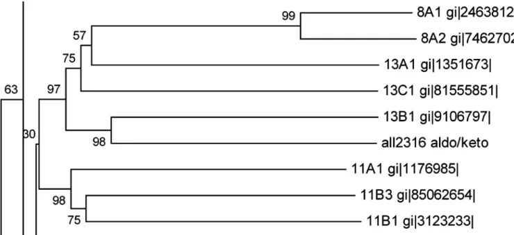
1. Homology search and evolutionary relationships
(All2316) ofAnabaena7120 by using BLASTP [36] algorithm. Evolutionary analyses were con-ducted in MEGA6 [37] using the Neighbor-Joining method [38]. The optimal tree with the sum of branch length = 27.84402135 is shown. The percentage of replicate trees in which the associated taxa clustered together in the bootstrap test (1000 replicates) is shown next to the branches [39]. The tree is drawn to scale, with branch lengths in the same units as those of the evolutionary distances used to infer the phylogenetic tree. The evolutionary distances were computed using the p-distance method [40] and are in the units of the number of amino acid differences per site. The analysis involved 156 amino acid sequences. All ambiguous positions were removed for each sequence pair. There were a total of 1125 positions in the final dataset.
2. Cyanobacterial and bacterial strains and plasmids
Anabaena7120 was grown photoautotrophically in BG-11medium [41] buffered with 10 mM HEPES-NaOH, pH 7.5 at 24±2°C under day light fluorescent tubes emitting 72μmol photon
m−2s−1PAR (photosynthetically active radiation) light intensity with a photoperiod of 14:10 h.
E.colistrains DH5αand BL21 (DE3) (Novagen) were used as host for cloning and over-expres-sion respectively.E.colicultures were stored as 10% (v/v) glycerol stocks at−80°C and
main-tained on Luria–Bertani (LB) plates at 37°C containing 1.5% (w/v) agar. Cells harboring recombinant plasmids were grown and maintained on LB medium supplemented with 100μg/
ml ampicillin [42]. Plasmid pET21a (Novagen) was used as a vector for cloning.
3. AKR transcript analysis
Total RNA was extracted from 50 mL culture (OD750nm0.6) ofAnabaena7120 before and
after 1 day of butachlor (32μM) [1], Cd (10μM) [43], NaCl (100mM) [33], As (40mM) [44],
heat (42°C for 1 h) [45], UV-B (12.9 m Wm-2nm-1for 30 min) [46], or desiccation (30°C for 10h) [47] treatment using the RNASure mini kit (Nucleo-pore). One microgram of total RNA was reverse transcribed in a 20μl reaction mixture using the iScript cDNA synthesis kit
(BioRad). For the transcript analysis, following gene specific primer sets were designed using
primer3 software: 5’-GGAATCCAGAACATCTGCGT-3’and 5’-CCCACAAATCGAAT
CAAACC-3’forakrand 5’-CACACTGGGACTGAGACAC-3’and 5’-CTGCTGGCACG
GAGTTAG-3’for reference 16S rRNA gene. 15ng of cDNA extracted from each sample was used for quantitative real-time PCR (qRT-PCR). Reactions were performed in a total volume of 20μl including 10 pmol of forward and reverse primers and 1x Sso fast evagreen qPCR
supermix (BioRad). CFX-96 (Bio-Rad) was used for PCR and for detection of fluorescence change. Transcript levels were normalized to 16S transcript and calculated relative to 0 h using the 2−ΔΔCtmethod. The comparativeΔΔCt method was used to evaluate the relative quantities
of each amplified product in the samples. The threshold cycle (Ct) was automatically deter-mined for each reaction by the system set with default parameters. The specificity of the PCR was determined by melting curve analysis of the amplified products.
4. Cloning of aldo/keto reductase in
E
.
coli
BL21
For cloning and over-expression analysis, genomic DNA fromAnabaena7120 was extracted following the protocol of Srivastava et al. [48]. The open reading frameall2316, encoding aldo/ keto reductase was amplified by polymerase chain reaction using genomic DNA as the
tem-plate. The PCR primer pairs used were Pf: 50GGAGGAGCATATGGAAACTACACAGCTAG
30and Pr: 50CCAGTCTCGAGAATTTTCTGGACTTCTTCG 30(the Nde1 and Xho1
5μl of 5× Phusion HF Buffer (NEB), 200μM dNTPs, 0.5μM forward and reverse primer and
0.5U Phusion DNA polymerase (NEB) in an iCycler (Bio-Rad, USA). The amplified product was gel purified using a QIAquick gel extraction kit (Qiagen). The purified PCR product was digested with Nde1 and Xho1 (NEB), and the resultant DNA fragment was cloned into the pET-21a expression vector (Novagen), digested with the same restriction enzymes. pET21a incorporates a C-terminal histidine (6x His) tag to aid purification. After ligation the construct pET-21a–AKR and empty vector pET-21a were introduced intoE.colistrain BL21 (DE3) for expression studies. The recombinant plasmid was isolated and the DNA sequence of AKR was confirmed by sequencing. Prior to use in further experimental procedures, transformation was confirmed by colony PCR analysis for the targeted AKR gene and by Nde1 and Xho1 double digestion on plasmid isolated from the cell transformed with empty and recombinant vector.
5. Over-expression and purification of recombinant protein
BL21 (DE3) cells transformed with recombinant plasmid pET-21a–AKR were grown overnight in Luria-Bertani (LB) medium containing 100μg/ml ampicillin. Overnight culture was diluted
1:100 in fresh LB broth (5x 100ml culture) containing 100μg/ml ampicillin and then incubated
at 37°C for 3–4 h (or till OD600reached ~0.7). Once the desired optical density reached the
cul-tures were cooled at RT and induced with a final concentration of 0.05mM IPTG by incubating at 16°C for 16/17 h and shaking at 200 rpm. After incubation, cells were harvested by centrifu-gation, resuspended in lysis buffer containing 20mM sodium phosphate (pH 7.4), 0.5M NaCl and 40mM imidazole, and disrupted by grinding in the presence of 10μM PMSF (Sigma),
under liquid nitrogen. The cell debris was pelleted by centrifugation at 4°C and 20,000g for 30 min. The supernatant containing the recombinant His-tagged fusion protein was purified using PureProteome Nickel Magnetic Beads (Merck Millipore) by following the manufacturer’s instructions. To assess the protein purity, collected fraction was visualized on 12% SDS-PAGE stained with Coomassie Brilliant Blue R250. The purified protein was subjected to MALDI--TOF MS/MS analysis to confirm its identity. The relative protein concentration was measured by determination of its absorbance at 280 nm.
6. Western blotting
Western blotting was performed as described by Pandey et al [49]. Whole cell lysate ofE.coli cells containing the pET-21a-AKR before and after IPTG induction was loaded on a 12% SDS– PAGE gel and blotted onto Immobilon-P PVDF membrane (Millipore, Billerica MA). The membrane was probed with penta-His antibody (mouse monoclonal, Qiagen) as primary and peroxidase-labelled anti-mouse antiserum as secondary antibody and visualized by ECL West-ern Blotting Analysis System (Amersham Bioscience).
7. Measurement of multiple stress tolerance by spotting and liquid assay
The effect of abiotic stresses onE.coliBL21(DE3) strains transformed with empty pET 21a and pET 21a-AKR was analyzed using spot assay in different treatments of cadmium (CdCl2), heat,
UV-B, arsenic (Na3AsO4), mannitol (for drought) or NaCl. The transformed cells were grown
in LB medium supplemented with 100μg/ml ampicillin at 37°C. When OD600 reached 0.6,
IPTG was added to a final concentration of 0.05 mM, and allowed to grow for additional 5–6 h at 16°C. Thereafter, the cultures were serially diluted to three levels (10−1, 10−2and 10−3). 3
μl
from each dilutions was spotted on LB-ampicillin plates containing CdCl2(0.1, 0.2 and 0.3
mM), Na3AsO4(4, 6 and 8mM), mannitol (0.4, 0.6 and 0.8 M) or NaCl (0.4, 0.6 and 0.8 M). All
intervals (30m, 1h and 2h for heat and 10m, 15m and 20m for UV-B), then spotted on LB-ampicillin plates and incubated at 37°C overnight.
Growth of transformedE.colicells was also examined by liquid culture assay as given in Shrivastava et al [50]. Briefly, single colony ofE.colicells transformed either with empty vector (pET 21a) or recombinant plasmid (pET 21a-AKR) was inoculated in tubes containing fresh LB medium spiked with 100μg ml−1ampicillin and grown overnight. 200μl inoculum (OD600
nm0.5) was added into 50 ml LB medium having CdCl2(0.3mM), NaCl (600 mM), mannitol
(600mM) or Na3AsO4(6mM) and 0.2 mM IPTG, and incubated at 37°C with shaking (200
rpm). For UV-B stress, the inoculum already exposed to UV-B radiation (12.9 m Wm-2nm-1) for 10 min was used. For heat stress, the 50 ml LB medium having 200μl inoculum (OD600 nm
0.5) and 0.2 mM IPTG was incubated at 50°C with shaking (200 rpm). The aliquots were removed from each treatment every 2 h and absorbance at 600 nm was recorded for 24 h.
8. Enzyme assay and measurement of kinetic parameters
Aldo-keto reductase activity was assayed spectrophotometrically as per the method of Smiley and Bolen [51]. Assay was performed at 37°C in a standard reaction mixture (1mL) containing 50mM potassium phosphate buffer (pH 7.4), 0.12mM NADPH, 10mM 2-mercaptoethanol, 4–20μg of purified protein and a range of substrates such as xylose (50 mM), arabinose (50
mM), benzaldehyde (10 mM), ethylpyruvate (10 mM), methylglyoxal (0.5 mM), butachlor (0.5 mM), isatin (0.5 mM), or o-nitrobenzaldehyde (0.5 mM). Activity was monitored by measur-ing the decrease in NADPH absorbance at 340 nm and the NADPH oxidation was calculated using a molar extinction coefficient of 6,200 M-1cm-1[49]. The kinetic parameters of the puri-fied AKR (Km, kcat, kcat/Kmvalues) were calculated by non-linear regression of
Michaelis–Men-ten data at a various concentrations of substrates (final concentrations: benzaldehyde, 1–50 mM; ethylpyruvate, 5–50 mM; methylglyoxal, 1–20 mM; isatin, 0.1–0.5 mM; o-nitrobenzalde-hyde, 0.1–1.0 mM; butachlor, 0.1-.05 mM), using the GraphPad PRISM 5.04 version program (http://www.graphpad.com). All measurements were performed in three replicates, and the experiment without AKR under the same condition was used as control.
9. Measurement of butachlor degradation
In order to monitor the butachlor degrading potential of AKR17A1, the reaction mixtures were prepared in four variants which contained along with 50mM potassium phosphate buffer pH 7.4, 10 mM 2-mercaptoethanol and 0.1 mM butachlor, (i) neither NADPH nor AKR, (ii) NADPH (0.12mM), (iii) AKR (5μg), or (iv) both NADPH (0.12mM) and AKR (5μg). Each
sample was scanned separately using UV-visible spectrophotometer (Hitachi U-2910) within the wave length range of 200–400nm against 50mM potassium phosphate buffer (pH 7.4) as baseline reference. The absorption spectra of each sample was measured in three replicates with a scanning speed of 3600 nm min-1and a band width of 2.0 nm equipped with 1cm matched quartz cell.
10. Gas chromatography
–
mass spectrometry (GC
–
MS) analyses
To further attest the role of AKR17A1 in butachlor degradation and identify the degradation products if any, the reaction mixtures were prepared as described above (see section 9 in Exper-imental procedures). Samples were prepared in 10ml reaction volume. After incubation, the mixture was extracted with 20ml hexane. The hexane fraction was then filtered using 0.45μmcarrier gas with a flow rate of 1.21 ml min-1. Oven parameters were 120°C for 3 min, raised to 280°C at the rate of 10°C min-1and held for 16 min. The injector was kept at 260°C. Pressure was 99.1 kPa, linear velocity 41.3 cm s-1, purge flow 3.0 ml min-1and split ratio 10.0. The MS was operated in electron ionization (EI) mode at 70 eV in an m/z range of 40–600 amu and ion source temperature as 230°C. Identification of metabolites was done by comparing their mass spectra with those of the spectrophotometer database using the NIST08, and WILEY8 Librar-ies. The identification of compounds was confirmed by comparison of the fragmentation pat-tern and their retention indices (RI).
Results
1. Identification of All2316 as an entirely new family protein AKR17A1
Aldo-keto reductase encoded by ORFall2316was previously identified as butachlor responsive protein in the proteome ofAnabaena7120. Sequence analysis revealed that All2316 contained AKR signature motifs such as active side residues and catalytic tetrad (Asp50, Tyr55, Lys88, and His130) which are generally conserved among aldo-keto reductases [21]. For homology search, the All2316 protein sequence having 284 amino acids was compared with the members of all existing AKR families (AKR1-15). Unexpectedly, no proteins belonging to the AKR superfamily showed more than 40% homology. Although, All2316 showed close evolutionary relationships with AKR13B1 and assembled together by forming sister clade with AKR13A1 and AKR13C1 (Fig 1), the sequence similarity in terms of percent identity revealed that All2316 is only 40% identical to the AKR13B1. While with 13A1 and 13C1, it shows only 29% and 27% identity respectively. Furthermore, the comparison of amino acid sequences with the members of AKR13 family indicated that the sequences of the N-termini and loop regions of All2316 are also different from AKR13(s) (Fig 2). In view of the above, All2316 cannot be grouped with AKR13 family hence assigned a new family. The amino acid sequence of All2316
Fig 1. A segment of phylogenetic tree representing the relationship of ORF All2316 encodingAnabaena7120 AKR to other closely related members of the AKR superfamily.Analysis involved all annotated AKRs (155 proteins) that fall into 15 families of AKR superfamily along with the new AKR
fromAnabaena7120. The complete tree and the sequence alignment of the entire AKR protein sequences are given in supporting information (S1andS2 Files). Full names for the sequences used in this figure are available on the AKR homepage (http://www.med.upenn.edu/akr).
was thus submitted to the AKR superfamily web site (http://www.med.upenn.edu/akr/). Owing to low sequence identity with any of the existing AKR members the website recommended that protein be assigned an entirely new family AKR17 and referred to as AKR17A1.
2. Response of AKR17A1 to a variety of abiotic stresses
To study the stress response of AKR17A1,Anabaena7120 was subjected to different abiotic stresses such as butachlor, cadmium, UV-B, salt, heat, drought and arsenic. Our previous proteomic studies showed increased accumulation of AKR under butachlor [1], Cd [35], UV-B [34] and salt stress [33] which is also reflected in quantitative PCR analysis of AKR17A1 dis-playing 3.8, 4.1, 4.2 and 2.2 fold induction respectively. In line with the above results, the tran-script level forAKR17A1was also elevated upto 4.2, 2.6 and 3.1 fold under heat, drought and arsenic stress respectively (Fig 3and Table A inS3 File).
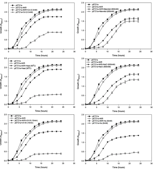
3. AKR17A1 confers tolerance against abiotic stresses
The stress tolerance potential of AKR17A1 was tested against a range of abiotic stressors such as cadmium (CdCl2), arsenic (Na2AsO4.7H2O), drought (mannitol), salinity (NaCl), heat
(50°C) and UV-B by spot and liquid culture assay. For spot assay, growth of bacterial colonies at dilution level 10−2producing the most significant differences has been displayed asFig 4.
TheE.colistrain BL21 (DE3) transformed with recombinant plasmid (pET21a-AKR) and vec-tor alone (control) following spotting on LB medium pretreated with cadmium, heat, and UV-B produced a significantly higher number of BL/pET21a-AKR colonies compared to BL/ pET21a (Fig 4). However, the growth of BL/pET21a-AKR was almost similar to BL/pET21a in arsenic, salt and mannitol containing media (Fig 4).
It is worth mentioning that the growth pattern of the above cells in LB liquid medium showed similar pattern as observed with spot culture assays. pET21a-AKR depicted 81, 34, 31.2, 28.3, 19 and 10.2% better specific growth rate and 45, 25.5, 23.7, 22, 16.2 and 9% decrease in doubling time under CdCl2, UV-B, heat (50°C), arsenic, mannitol, and NaCl stress
Fig 2. Sequence alignment ofAnabaena7120 aldo- keto reductase All2316 and members of AKR13 family using PROMALS3D multiple sequence and structure alignment server (prodata.swmed.edu/promals3d/).Representative sequences are colored according to predicted secondary structures (red: alpha-helix, blue: beta-strand). Consensus predicted secondary structure symbols: alpha-helix: h; beta-strand: e. Consensus amino acid symbols are: conserved amino acids are in bold and uppercase letters; aliphatic (I, V, L): l; aromatic (Y, H, W, F): @; hydrophobic (W, F, Y, M, L, I, V, A, C, T, H): h; alcohol (S, T): o; polar residues (D, E, H, K, N, Q, R, S, T): p; tiny (A, G, C, S): t; small (A, G, C, S, V, N, D, T, P): s; bulky residues (E, F, I, K, L, M, Q, R, W, Y): b; positively charged (K, R, H): +; negatively charged (D, E):-; charged (D, E, K, R, H): c.
respectively when compared to control. These results indicate that the expression of AKR17A1 gene significantly increased the Cd, UV-B, and heat tolerance ofE.colicells (Fig 5).
4. Butachlor metabolism and substrate specificity of AKR17A1
AKRs catalyze the reduction of aldehydes, ketones, dicarbonyls, steroids, and monosaccharides in an NAD(P)(H)-dependent manner [21]. To determine the substrate specificity and buta-chlor metabolizing potential of AKR17A1, the enzymatic activity of purified protein (Figures of coomassie stained 12% SDS-PAGE gel showing purified protein and immunoblot detection of recombinant protein AKR17A1 are given as Figs A and B inS3 File) against butachlor and other typical AKR substrates including aldehydes [52,30], ketones [53], bicarbonyl [29,54, 55], and sugars [56] was tested. Except for sugars, arabinose and xylose, AKR17A1 exhibited considerable reductase activity for all the substrates tested (Fig 6).
The best substrate studied in terms of highest specificity constant (Kcat/Km) was isatin, an
α-dicarbonyl tryptophan metabolite (Table 1). AKR17A1 also has ability to reduce o-nitro derivative of benzaldehyde with high efficiency (Table 1). Since, benzaldehyde as a substrate has high Kmand low Kcat/Kmwhen compared to o-nitro benzaldehyde, 2-nitro group seems Fig 3. Relative normalized expression of AKR17A1 inAnabaena7120 exposed to butachlor, cadmium, UV-B, heat, salt, drought and arsenic.
Transcript levels were determined by qRT-PCR. The 16S rRNA gene was used as an internal control for normalizing the variations in cDNA amounts. Biological triplicates were averaged. Bars indicate SE.
Fig 4. Spot assay ofE.colistrain BL21 (DE3) transformed with vector alone (BL/pET21a) and recombinant plasmid (BL/ pET21a-AKR) under different abiotic stresses.3μL of sequential dilutions (only growth at dilution level 10−2is shown here) were spotted on the Luria Bertani (LB) plates supplemented with varying concentration of (a) cadmium, (b) mannitol, (c) arsenic and (d) NaCl. For (e) UV-B and (f) heat (50°C),cells were pretreated for different time intervals and then spotted on the LB plates. All spot tests were performed in triplicate.
essential for high substrate activity. Methylglyoxal and ethyl pyruvate were poor substrates with high Kmand low Kcat/Km. Extremely low enzyme activity with the sugars arabinose and
xylose, suggests that these could be unlikely substrates of AKR17A1 in vivo. The most interest-ing observation was that AKR17A1 catalyzed the reduction of butachlor with the highest kcat
and considerably higher specificity constant (Kcat/Km) thereby indicating its propensity to
reduce chloroacetamide substrates as well (Table 1).
Fig 5. Growth behavior ofE.coliBL21 (DE3) cells transformed with pET21a and pET21a-AKR in response to (a) cadmium (0.3 mM) (b) mannitol (600 mM), (c) heat (50°C), (d) NaCl (600 mM), (e) UV-B (10 min) and (f) arsenic (6 mM) stress using liquid culture assay.The mean of three independent replicates is plotted with error bars indicating standard deviations. (a-f) represents the growth curves ofE.colistrains in LB medium
supplemented with 100μg ml−1ampicillin and 0.2 mM IPTG.
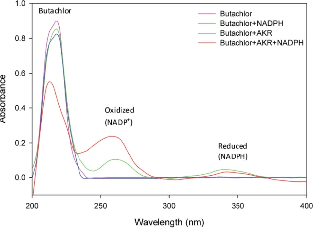
5. Spectroscopic analysis affirms butachlor degrading potential of
AKR17A1
Butachlor exhibited a characteristic absorption maximum in UV-C region at a wavelength near 215 nm. The presence of co-substrate NADPH or enzyme AKR17A1 alone did not have any significant effect on peak height of butachlor. Whereas in the presence of both NADPH and AKR17A1, the absorption peak of butachlor (at 215 nm) was decreased by 0.87 to 0.53 and of oxidized NADPH (near 261nm) was increased by 0.104 to 0.237 (Fig 7). This affirms the role of AKR17A1 in butachlor degradation and also indicates a strict requirement of cofactor NADPH for AKR17A1 because no significant activity was observed in its absence.
Fig 6. Specific activity of recombinant AKR17A1 on diverse substrates.Data represents the average of three measurements and bars indicate±SD.
doi:10.1371/journal.pone.0137744.g006
Table 1. Steady-state kinetic parameters of recombinant His-tagged AKR17A1.Kinetic constants are mean values of three independent experiments. Values in parentheses indicate standard error of the mean (SEM). The activity of AKR17A1 at various concentrations of substrates tested is given as Fig C in S3 File.
Substrate kcat(SEM) (min-1) Km(SEM) (mM) kcat/Km(M-1min-1)
Benzaldehyde 90.68 (4.23) 3.740 (1.098) 2.424×10⁴
o-nitro benzaldehyde 95.50 (3.57) 0.014 (0.002) 68.12×10⁵
Methylglyoxal 96.01 (1.24) 4.164 (0.973) 2.3×10⁴
Isatin 94.64 (8.25) 0.011 (0.005) 81.79×10⁵
Ethyl pyruvate 85.51 (2.47) 6.213 (2.214) 1.37×10⁴
Butachlor 126.4 (7.79) 0.548 (0.190) 23.06×10⁴
6. AKR17A1 metabolizes butachlor into benzenedicarboxylic acid and
phenol
To identify the degradation products formed following the reaction of AKR17A1 on butachlor, GC-MS analysis was performed in 4 independent reactions. In the absence of both NADPH and AKR17A1 (reaction 1), the chromatogram showed a single major peak‘A’(Rtmin= 14.9)
which corresponds to butachlor. NADPH alone (reaction 2) also showed a similar peak for butachlor (peak A). While, AKR17A1 alone (reaction 3) gave two major peaks of which peak A corresponds to butachlor and peak B to 1,2-benzene dicarboxylic acid; these constituted 80.56% and 4.49% of total peak area respectively. Interestingly, however, reaction 4 which con-tained both AKR and NADPH showed three major peaks (Fig 8) (i) a butachlor peak‘A’, (ii) a 1,2-benzene dicarboxylic acid peak‘B’, and (iii) peak‘C’corresponding to 2,6 bis (1,1, dimethy-lethyl) 4,-methyl phenol with respective peak areas of 50.89%, 33.91% and 5.07%. The percent composition of butachlor and products formed after each reaction is given inTable 2.
These results indicate that reaction 4 containing both enzyme AKR17A1 and cofactor NADPH exhibited 41.08, 40.64 and 38.03% decrease in the amount of butachlor when com-pared to reaction 1 (containing neither NADPH nor AKR17A1), 2 (only NADPH) and 3 (only AKR17A1), respectively. Whereas, the amount of 1,2-benzene dicarboxylic acid was increased upto 36.44, 35.0 and 32.45% compared to that in reaction 1, 2 and 3, respectively (Table 2). This indicates that AKR17A1 significantly contributes to metabolism/degradation of butachlor. Fig 7. Absorption spectrograms showing degradation of butachlor by NADPH dependent AKR17A1.The analysis was performed in triplicates. The absorption spectra for each samples was recorded in 1 mL sample volume containing 50 mM potassium phosphate buffer (pH 7.4), 10 mMβ
-mercaptoethanol, 0.1 mM butachlor and either 0.12 mM NADPH, 5μg AKR or both.
Fig 8. GC charts of the reaction products of AKR17A1 with butachlor used as substrate.(a) Control assay devoid of enzyme AKR17A1 and cofactor (NADPH); (b) reaction with NADPH alone; (c) enzymatic reaction with AKR17A1 alone; and (d) enzymatic reaction with both AKR17A1 and cofactor NADPH.
Discussion
To increase crop productivity, current agricultural practices are greatly dependent on acceler-ated pesticide usage. This not only causes environmental contamination through prolonged persistence but adversely affects various non-target organisms. Thus, there is a need to identify some eco-friendly systems to mitigate the ill-effects associated with excessive pesticide applica-tion. In this pursuit we identified a protein‘AKR17A1’fromAnabaena7120 an entirely new family member of AKR superfamily which degrades an extensively used rice field herbicide butachlor.
AKR17A1 primarily possess two novel attributes that validate its position as a new family member: (i) Although AKR17A1 shows close homology with AKR13B1 (Fig 1), its amino acid sequence displayed marked divergence particularly in the regions corresponding to loop B and C (Fig 2). Since, the key loops A, B and C contribute significantly to the substrate binding site [21]; AKR17A1 may not necessarily have similar function as AKR13B1 and therefore, cannot be placed in AKR13 family. (ii) AKR17A1 appears quite distinct from the only studied cyano-bacterial AKR, methylglyoxal reductase (AKR11B3) ofSynechococcussp. PCC 7002 [27], in terms of sequence identity (sharing 26.5% amino acid identity with AKR11B3) and substrate specificity. For example, unlike AKR11B3 which shows high activity towards methylglyoxal, AKR17A1 depicted high activity towards isatin and o-nitrobenzaldehyde. In fact the kcat/Km
values for isatin and o-nitrobenzaldehyde were much higher when compared with those of known AKRs such as AKR14A1 [29], AKR11B3 [30],AKR8A1 [52], AKR2E4 [55] and AKR4C8 [56] (Table B inS3 File). This further supports our claim for aldo/keto reductase (AKR17A1) ofAnabaena7120 to be a novel AKR protein.
Owing to low sequence identities with functionally characterized AKRs, the specific physio-logical functions for potential new members have not been established till date. However, many AKRs are known to play important role in stress defence. Abiotic stresses such as heat, heavy metal, UV-B irradiation, salinity, drought etc. enhance the intracellular ROS production which in turn cause lipid peroxidation and production of reactive carbonyl species considered as indirect effect of oxidative stress. AKRs have been known to detoxify not only certain lipid peroxidation derived reactive carbonyls (such as malondialdehyde (MDA) and 4-hydroxyno-nenal (4-HNE) etc.) but glycolysis-derived toxic intermediates (e.g methylglyoxal) also [26,27, 29–32]. Therefore, overexpression of many stress inducible AKRs have been found to be effec-tive in conferring tolerance against oxidaeffec-tive stress induced by heavy metal [25], drought [26], heat [27], salt [28], and UV-B [32] in many plant species. Our results showed elevated level of AKR17A1 transcript after exposure ofAnabaena7120 cells to different stresses such as cad-mium, butachlor, heat, UV-B, arsenic, salt and drought. These results are in tune with the pre-vious studies where several members of AKR superfamily are reported to be appreciably induced under various adverse conditions, such as AKR11B in anaerobic stress [57], AKR2B6 under osmotic, hydrogen peroxide and ionic stress [58], AKR3A1 and AKR3A2 in osmotic Table 2. Percent composition of butachlor and metabolites as determined by GC-MS analysis.The GC-MS measurements were carried out with the metabolites extracted from the reaction mix containing 50mM potassium phosphate buffer pH 7.4, 0.4 mM butachlor and 10mM 2-mercaptoethanol, (1) nei-ther NADPH nor AKR, (2) NADPH (0.12mM), (3) AKR (5μg), or (4) both NADPH (0.12mM) and AKR (5μg).
Reaction Mix Butachlor (%) 1,2-benzene dicarboxylic acid (%) 2,6 bis (1,1, dimethylethyl) 4,-methyl phenol (%)
1 98.7 1.294
-2 97.26 2.73
-3 95.71 4.28
-4 56.62 37.73 5.64
stress [58–60], AKR4C5 and AKR4C6 at elevated salt concentrations, and drought and heat-shock treatment [61]. Furthermore, heterologous expression of AKR17A1 inE.colishowed better growth under abiotic stresses such as heat, UV-B and cadmium. These results, thus, indi-cate that AKR17A1 probably offers stress tolerance by detoxifying reactive carbonyl species generated through heat, UV-B and cadmium treatments.
Furthermore, an increased transcript and moderately high affinity (low Km) and catalytic
efficiency (Kcat/ Km) of AKR17A1 for butachlor lead us to hypothesize its role in metabolism of
butachlor. This finds support from absorption spectrophotogram revealing AKR17A1 cata-lyzed NADPH dependent degradation of butachlor in vitro. The GC-MS results further attested AKR17A1 catalyzed butachlor degradation and production of 1,2-benzene dicarbox-ylic acid and 2,6 bis (1,1, dimethylethyl) 4,-methyl phenol as the major products. The GC-MS results are in agreement with the previous report where benzene dicarboxylic acid and phenol were the degradation products of butachlor during active growth of the cyanobacteriumNostoc muscorum[62].
Thus, aldo-keto reductase (AKR17A1) fromAnabaena7120 has emerged as a novel enzyme for degradation of an agriculturally relevant herbicide butachlor. The AKR17A1 seems to be a stress-responsive AKR and may offer multiple stress tolerance in vivo. Thus AKR17A1 emerges as a potential candidate for the development of transgenic cyanobacteria worth exploiting as effective biofertilizer in butachlor contaminated rice paddy fields.
Supporting Information
S1 File. The complete phylogenetic tree representing the relationship of ORF All2316 encodingAnabaenaAKR to all existing members of the AKR superfamily.
(EMF)
S2 File. The sequence alignment of the entire AKR protein sequences. (PDF)
S3 File.Real time quantitative RT PCR analysis results(Table A).SDS-PAGE (12%) analysis of recombinant protein inE.coliBL21 (DE3).(Fig A).Immunoblot detection of AKR17A1 protein inE.colitransformed with recombinant (R) and empty vectors (E)(Fig B). Aldo/keto reductase activity at various concentrations of (a) benzaldehyde, (b) ethyl pyruvate, (c) methyl glyoxal, (d) isatin, (e) o-nitro benzaldehyde and (e) butachlor(Fig C). GC-MS spectra of buta-chlor and metabolites produced after degradation(Fig D). Absorption spectra of NADPH in reaction mix without butachlor(Fig E). Comparison of AKR171A1 activity for isatin and o-nitrobenzaldehyde with other characterized AKRs(Table B).
(DOCX)
Acknowledgments
We thank AIRF Jawaharlal Nehru University, New Delhi, India for GC-MS analysis. We also thank Dr Trevor M. Penning of the Department of Pharmacology, University of Pennsylvania, for analysis and assignment of the AKR17A1 gene and Dr. Swati Mishra for her assistance in phylogenetic analysis.
Author Contributions
References
1. Agrawal C, Sen S, Singh S, Rai S, Singh PK, Singh VK, et al. Comparative proteomics reveals associa-tion of early accumulated proteins in conferring butachlor tolerance in three N2-fixingAnabaenaspp. J
Proteomics.2014; 96: 271–290. doi:10.1016/j.jprot.2013.11.015PMID:24291601
2. Kumari N, Narayan OP, Rai LC. Understanding butachlor toxicity inAulosira fertilissimausing physio-logical, biochemical and proteomic approaches. Chemosphere 2009; 77: 1501–7. doi:10.1016/j.
chemosphere.2009.10.005PMID:19879624
3. Ateeq B, Abul FM, Niamat AM, Ahmad W. Clastogenicity of pentachlorophenol, 2,4-D and butachlor evaluated byAlliumroot tip test. Mutat Res.2002; 514: 105–13. PMID:11815249
4. Chang SS, Ashton FM, Bayer DE Butachlor influence on selected metabolic processes of plant cells and tissues. J Plant Growth Regul.1985; 4: 1–9.
5. Liu WY, Wang CY, Wang TS, Fellers GM, Lai BC, Kam YC. Impacts of the herbicide butachlor on the
larvae of a paddy field breeding frog (Fejervarya limnocharis) in subtropical Taiwan. Ecotoxicology 2011; 20:377–84. doi:10.1007/s10646-010-0589-6PMID:21210217
6. Xu HD, Wang JS, Li MH, Liu Y, Chen T, Jia AQ. 1H NMR based metabolomics approach to study the toxic effects of herbicide butachlor on goldfish (Carassius auratus). Aquat Toxicol. 2015; 159: 69–80. doi:10.1016/j.aquatox.2014.11.020PMID:25528421
7. Dwivedi S, Saquib Q, Al-Khedhairy AA, Musarrat J. Butachlor induced dissipation of mitochondrial
membrane potential, oxidative DNA damage and necrosis in human peripheral blood mononuclear cells. Toxicology 2012; 302: 77–87. doi:10.1016/j.tox.2012.07.014PMID:22884430
8. Widmer SK, Spalding RF. Assessment of herbicide transport and persistence in ground water: a review. American chemical society, ACS symposium series: Herbicide metabolites in surface water and ground water. Washington, DC 1996; 630: 271–287.
9. Jayakumar R, Sree Ramulu US. Degradation and persistence of herbicide in transplanted rice.
Pro-ceeding of International Symposium, Indian Society of weed Science, Hissar, India. 1993: 123–124.
10. Sondhia S, Singh VP, Yaduraju NT. Persistence of butachlor in sandy clay loam soil and its residues in
rice grains and straw. Annals of Plant Protection Sciences 2006; 14: 206–209.
11. Pal R, Das P, Chakrabarti K, Chakraborty A, Chowdhury A.Butachlor degradation in tropical soils: Effect of application rate, biotic-abiotic interactions and soil conditions. J Environ Sci Heal.2006; 41:1103–1113.
12. Prakash NB, Suseeladevi L, Siddaramappa R. Effect of organic amendments on persistence of buta-chlor and pendimethalin in soil. Karnataka J Agril Sci. 2000; 13: 575–580.
13. Rao PC, Rama Lakshmi CS, Madhavi M, Swapna G, Sireesha A. Butachlor dissipation in rice grown soil and its residues in grain. Indian Journal of Weed Science 2012; 44: 84–87, 2012
14. Yu YL, Chen YX, Luo YM, Pan XD, He YF, Wong MH. Rapid degradation of butachlor in wheat
rhizo-sphere soil. Chemorhizo-sphere2003; 50: 771–774. PMID:12688489
15. Yang C, Wang M, Chen H, Li J. Responses of butachlor degradation and microbial properties in a
ripar-ian soil to the cultivation of three different plants. J Environ Sci.2011; 23: 1437–1444.
16. Sethunathan N, Yoshida T. AFlavobacteriumsp. That degrades diazinon and parathion. Can J Micro-biol.1973; 19:873–875. PMID:4727806
17. Dwivedi S, Singh BR, Al-Khedhairy AA, Alarifi S, Musarrat J. Isolation and characterization of butachlor catabolizing bacterial strainStenotrophomonas acidaminiphilaJS-1 from soil and assessment of its biodegradation potential. Lett Appl Microbiol. 2010; 51: 54–60. doi:10.1111/j.1472-765X.2010.02854.x PMID:20477958
18. Liu HM, Cao L, Lu P,Ni H, Li YX, Yan X, et al. Biodegradation of butachlor byRhodococcussp. strain B1 and purification of its hydrolase (ChlH) responsible for N-dealkylation of chloroacetamide herbi-cides. J Agric Food Chem. 2012; 60: 12238–12244. doi:10.1021/jf303936jPMID:23186529
19. Zheng J, Li R, Zhu J, Zhang J, He J, Li S, et al. Degradation of the chloroacetamide herbicide butachlor byCatellibacterium caenisp. nov DCA-1T. Int Biodeter Biodegr. 2012; 73: 16–22.
20. Kim NH, Kim DU, Kim I, Ka JO. Syntrophic biodegradation of butachlor byMycobacteriumsp. J7A and
Sphingobiumsp. J7B isolated from rice paddy soil. FEMS Microbiol Lett. 2013; 344: 114–120. doi:10.
1111/1574-6968.12163PMID:23617893
21. Jez JM, Bennett MJ, Schlegel BP, Lewis M, Penning TM. Comparative anatomy of the aldo–keto reduc-tase superfamily. Biochem J. 1997; 326: 625–636. PMID:9307009
22. Bohren KM, Bullock B, Wermuth B, Gabbay KH. The aldo-keto reductase superfamily. J Biol Chem
1989; 264: 9547–9551. PMID:2498333
23. Petrash JM. All in the family: aldose reductase and closely related aldo-keto reductases. Cell Mol Life
24. Barski OA, Tipparaju SM, Bhatnagar A. The aldo- keto reductase superfamily and its role in drug metabolism and detoxification. Drug Metab Rev.2008; 40: 553–624. doi:10.1080/
03602530802431439PMID:18949601
25. Hegedus A, Erdei S, Janda T, Toth E, Horvath G, Dudit D. Transgenic tobacco plants overproducing alfalfa aldose/aldehyde reductase show higher tolerance to low temperature and cadmium stress. Plant Sci.2004; 166: 1329–1333.
26. Oberschall A, Deák M, Török K, Sass L, Vass I, Kovács I, et al. A novel aldose/aldehyde reductase pro-tects transgenic plants against lipid peroxidation under chemical and drought stresses. Plant J. 2000; 24: 437–446. PMID:11115125
27. Turoczy Z, Kis P, Torok K, Cserhati M, Lendvai A, Dudits D, et al. Overproduction of a rice aldo–keto reductase increases oxidative and heat stress tolerance by malondialdehyde and methylglyoxal detoxi-fication. Plant Mol Biol. 2011; 75: 399–412. doi:10.1007/s11103-011-9735-7PMID:21246257
28. Kanayama Y, Mizutani R, Yaguchi S, Hojo A, Ikeda H, Nishiyama M, et al. Characterization of an uncharacterized aldo-keto reductase gene from peach and its role in abiotic stress tolerance. Phyto-chemistry 2014; 104: 30–36. doi:10.1016/j.phytochem.2014.04.008PMID:24837355
29. Grant AW, Steel G, Waugh H, Ellis EM. A novel aldo-keto reductase fromEscherichia colican increase resistance to methylglyoxal toxicity. FEMS Microbiol Lett. 2003; 218: 93–99. PMID:12583903
30. Xu D, Liu X, Guo C, Zhao J. Methylglyoxal detoxification by an aldo-keto reductase in the cyanobacte-riumSynechococcussp. PCC 7002. Mol Biol Evol. 2006; 152: 2013–2021.
31. Aguilera J, Prieto J. TheSaccharomyces cerevisiaealdose reductase is implied in the metabolism of
methylglyoxal in response to stress conditions. Curr Genet.2001; 39: 273–283. PMID:11525399
32. Hideg É, Nagy T, Oberschall A, Dudits D, Vass I. Detoxification function of aldose/aldehyde reductase during drought and ultraviolet-B (280–320 nm) stresses. Plant Cell Environ. 2003; 26: 513–522.
33. Rai S, Agrawal C, Shrivastava AK, Singh PK, Rai LC. Comparative proteomics unveils cross species variations inAnabaenaunder salt stress. J Proteomics 2014; 98: 254–270. doi:10.1016/j.jprot.2013.12.
020PMID:24406298
34. Shrivastava AK, Chatterjee A, Yadava S, Singh PK, Singh S, Rai LC. UV-B stress induced metabolic rearrangements explored with comparative proteomics in threeAnabaenaspecies. J. Proteomics2015; doi:10.1016/j.jprot.2015.05.014
35. Singh PK, Shrivastava AK, Chatterjee A, Pandey S, Rai S, Singh S, et al. Cadmium toxicity in diazo-trophicAnabaenaspp. adjudged by hasty up-accumulation of transporter and signaling and severe down-accumulation of nitrogen metabolism protein. J. Proteomics 2015; doi:10.1016/j.jprot.2015.05. 019
36. Altschul SF, Gish W. Local alignment statistics. Methods Enzymol.1996; 266: 460–480. PMID:
8743700
37. Tamura K, Stecher G, Peterson D, Filipski A, Kumar S. MEGA6: Molecular evolutionary genetics analy-sis version 6.0. Mol Biol Evol. 2013; 30: 2725–2729. doi:10.1093/molbev/mst197PMID:24132122
38. Saitou N, Nei M. The neighbor-joining method: A new method for reconstructing phylogenetic trees.
Mol Biol Evol.1987; 4: 406–425. PMID:3447015
39. Felsenstein J. Confidence limits on phylogenies: An approach using the bootstrap. Evolution 1985; 39:
783–791.
40. Nei M, Kumar S. Molecular Evolution and Phylogenetics. Oxford University Press, New York 2000.
41. Rippka R, Deruelles J, Waterbury JB, Herdman M, Stanier RY. Generic assignments, strain histories
and properties of pure cultures of cyanobacteria. J Gen Microbiol. 1979; 111: 1–61.
42. Sambrook J, Russell DW. Molecular cloning: a laboratory manual. Cold Spring Harbor, New York
2001.
43. Singh PK, Rai S, Pandey S, Agrawal C, Shrivastava AK, Kumar S, et al. Cadmium and UV-B induced changes in proteome and some biochemical attributes ofAnabaenasp. PCC7120. Phykos 2012; 42:
39–50.
44. Pandey S, Rai R, Rai LC. Proteomics combines morphological, physiological and biochemical attri-butes to unravel the survival strategy ofAnabaenasp. PCC7120 under arsenic stress. J Proteomics
2012; 75:921–37. doi:10.1016/j.jprot.2011.10.011PMID:22057044
45. Rajaram H, Apte SK. Nitrogen status and heat stress-dependent differential expression of thecpn60
gene chaperonin gene influences thermotolerance in the cyanobacteriumAnabaena. Microbiology
2008; 154: 317–325. doi:10.1099/mic.0.2007/011064-0PMID:18174150
46. Rai S, Singh S, Shrivastava AK, Rai LC. Salt and UV-B induced changes inAnabaenasp. PCC7120: Physiological, proteomic and bioinformatic perspectives. Photosynth Res 2013; 118: 105–114. doi:10.
47. Katoh H, Asthana RK, Ohmori M. Gene expression in the cyanobacteriumAnabaenasp. PCC 7120 under desiccation. Microb Ecol. 2004; 47: 164–174. PMID:14749909
48. Srivastava AK, Ara A, Bhargava P. A rapid and cost-effective method of genomic DNA isolation from cyanobacterial culture, mat and soil suitable for genomic fingerprinting and community analysis. J Appl Phycol2007; 19: 373–382.
49. Pandey S, Shrivastava AK, Rai R, Rai LC. Molecular characterization of Alr1105 a novel arsenate
reductase of the diazotrophic cyanobacteriumAnabaenasp. PCC 7120 and decoding its roles in abiotic stress management inEscherichia coli. Plant Mol Biol 2013; 83: 417–432. doi:
10.1007/s11103-013-0100-xPMID:23836391
50. Shrivastava AK, Pandey S, Singh PK, Rai S, Rai LC. alr0882 encoding a hypothetical protein of Ana-baenaPCC7120 protectsEscherichia colifrom nutrient starvation and abiotic stresses. Gene 2012; 511: 248–255. doi:10.1016/j.gene.2012.09.033PMID:23006586
51. Smiley KL, Bolen PL. Demonstration of D-xylose reductase and D-xylitol dehydrogenase inPachysolen tannophilus. Biotechnol Lett.1982; 4: 607–610.
52. Nakano M, Morita T, Yamamoto T, Sano H, Ashiuchi M, Masui R, et al. Purification, molecular cloning, and catalytic activity ofSchizosaccharomyces pombepyridoxal reductase: a possible additional family in the aldo-keto reductase superfamily. J. Biol. Chem. 1999; 274: 23185–23190. PMID:10438489
53. Ni Y, Li CX, Ma HM, Zhang J, Xu JH. Biocatalytic properties of a recombinant aldo-keto reductase with broad substrate spectrum and excellent stereoselectivity. Appl Microbiol Biotechnol. 2011; 89: 1111– 1118. doi:10.1007/s00253-010-2941-4PMID:20981419
54. Luccio EDI, Elling RA, Wilson DK. Identification of a novel NADH-specific aldo-keto reductase using sequence and structural homologies. Biochem J. 2006; 400: 105–114 PMID:16813561
55. Yamamoto K, Wilson DK. Identification, characterization, and crystal structure of an aldo–keto reduc-tase (AKR2E4) from the silkwormBombyx mori. Arch Biochem Biophys. 2013; 538: 156–163. doi:10.
1016/j.abb.2013.08.018PMID:24012638
56. Simpson PJ, Tantitadapitak C, Reed AM, Mather OC, Bunce CM, White SA, et al. Characterization of
two novel aldo-keto reductases from Arabidopsis: Expression patterns, broad substrate specificity, and an open active-site structure suggest a role in toxicant metabolism following stress. J Mol Biol. 2009; 392: 465–480. doi:10.1016/j.jmb.2009.07.023PMID:19616008
57. Marino M, Hoffmann T, Schmid R, Moebitz H, Jahn D. Changes in protein synthesis during the adpta-tion ofBacillus subtilisto anaerobic growth conditions. Microbiology 2000; 146: 97–105. PMID:
10658656
58. Norbeck J, Blomberg A. Metabolic and regulatory changes associated with growth ofSaccharomyces cerevisiaein 1.4 M sodium chloride: Evidence for osmotic induction of glycerol dissimilation via the dihy-droxyacetone pathway. J Biol Chem1997; 272: 5544–5554.
59. Ford G, Ellis EM. Three aldo-keto reductases of theyeast Saccharomyces cerevisiae. Chem Biol Inter-act.2001; 130–132: 685–698. PMID:11306086
60. Ford G, Ellis EM. Characterization of Ypr1p fromSaccharomyces cerevisiaeas a
2-methylbutyralde-hyde reductase. Yeast 2002; 19: 1087–1096. PMID:12210903
61. Gavidia I, Bermudez PP, Seitz HU. Cloning and expression of two novel aldo-keto reductases from Dig-italis purpurealeaves. Eur J Biochem. 2002; 269: 2842–2850. PMID:12071946
62. Anees S, Suhail S, Pathak N, Zeeshan M. Potential use of rice field cyanobacteriumNostoc muscorum