hnRNP K Coordinates Transcriptional
Silencing by SETDB1 in Embryonic Stem Cells
Peter J. Thompson1, Vered Dulberg1¤, Kyung-Mee Moon2, Leonard J. Foster2, Carol Chen1, Mohammad M. Karimi1,3, Matthew C. Lorincz1*
1Life Sciences Institute, Department of Medical Genetics, University of British Columbia, Vancouver, British Columbia, Canada,2Centre for High-Throughput Biology, University of British Columbia, Vancouver, British Columbia, Canada,3Biomedical Research Centre, University of British Columbia, Vancouver, British Columbia, Canada
¤ Current address: The Chaim Sheba Medical Center, Tel Hashomer, Israel *[email protected]
Abstract
Retrotransposition of endogenous retroviruses (ERVs) poses a substantial threat to ge-nome stability. Transcriptional silencing of a subset of these parasitic elements in early mouse embryonic and germ cell development is dependent upon the lysine methyltransfer-ase SETDB1, which deposits H3K9 trimethylation (H3K9me3) and the co-repressor KAP1, which binds SETDB1 when SUMOylated. Here we identified the transcription co-factor hnRNP K as a novel binding partner of the SETDB1/KAP1 complex in mouse embryonic stem cells (mESCs) and show that hnRNP K is required for ERV silencing. RNAi-mediated knockdown of hnRNP K led to depletion of H3K9me3 at ERVs, concomitant with de-repres-sion of proviral reporter constructs and specific ERV subfamilies, as well as a cohort of germline-specific genes directly targeted by SETDB1. While hnRNP K recruitment to ERVs is dependent upon KAP1, SETDB1 binding at these elements requires hnRNP
K. Furthermore, an intact SUMO conjugation pathway is necessary for SETDB1 recruitment to proviral chromatin and depletion of hnRNP K resulted in reduced SUMOylation at ERVs. Taken together, these findings reveal a novel regulatory hierarchy governing SETDB1 re-cruitment and in turn, transcriptional silencing in mESCs.
Author Summary
Retroelements, including endogenous retroviruses (ERVs), pose a significant threat to ge-nome stability. In mouse embryonic stem (ES) cells, the enzyme SETDB1 safeguards the genome against transcription of specific ERVs by depositing a repressive mark H3K9 tri-methylation (H3K9me3). Although SETDB1 is recruited to ERVs by its binding partner KAP1, the molecular basis of this silencing pathway is not clear. Using biochemical and genetic approaches, we identified hnRNP K as a novel component of this silencing path-way that facilitates the recruitment of SETDB1 to ERVs to promote their repression. HnRNP K binds to ERV sequences via KAP1 and subsequently promotes SETDB1 bind-ing. Together, our results reveal a novel function for hnRNP K in transcriptional silencing
OPEN ACCESS
Citation:Thompson PJ, Dulberg V, Moon KM, Foster LJ, Chen C, Karimi MM, et al. (2015) hnRNP K Coordinates Transcriptional Silencing by SETDB1 in Embryonic Stem Cells. PLoS Genet 11(1): e1004933. doi:10.1371/journal.pgen.1004933
Editor:Gregory S. Barsh, Stanford University School of Medicine, United States of America
Received:July 24, 2014
Accepted:December 2, 2014
Published:January 22, 2015
Copyright:© 2015 Thompson et al. This is an open access article distributed under the terms of the
Creative Commons Attribution License, which permits unrestricted use, distribution, and reproduction in any medium, provided the original author and source are credited.
Data Availability Statement:All relevant data are within the paper and its Supporting Information files
of ERVs and demonstrate a new regulatory mechanism governing the deposition of H3K9me3 by SETDB1 in ES cells.
Introduction
Long terminal repeat (LTR) retrotransposons, also called endogenous retroviruses (ERVs), are the relics of ancient and more recent germline retroviral integrations, comprising ~8–10% of the mouse and human genomes, respectively [1].De novoretrotransposition of these parasitic elements is responsible for ~10% of spontaneous mutations in mice [2]. Among the remaining transcriptionally competent ERVs in the mouse genome, many class I Moloney murine leuke-mia virus (MLV) and class II intracisternal A-type particle (IAP) and MusD elements are tran-siently expressed and subsequently silenced in the early embryo [3]. Distinct epigenetic mechanisms cooperate to maintain ERV silencing including DNA methylation, covalent his-tone modifications, chromatin remodelling and non-coding RNAs [4].
Although DNA methylation suppresses ERV transcription in differentiated somatic cells [5], pluripotent stem cell lines derived from the inner cell mass of the blastocyst, such as mu-rine embryonic stem cells (mESCs) utilize a DNA methylation-independent pathway to main-tain ERV silencing [6]. Key effectors in this silencing pathway are the conserved Krüppel-associated box zinc finger proteins (KRAB-ZFPs), the largest family of C2H2 zinc finger tran-scription factors in vertebrate genomes [7]. Earlier experiments utilizing the MLV-based retro-viral vectors harbouring a proline tRNA primer binding site (PBSPro) revealed that KRAB-ZFPs bind to specific proviral sequences such as the PBS, to direct the recruitment of a large si-lencing complex that includes the obligate co-repressor KAP1 (also called TRIM28/TIF1β) [8,9] and the lysine methyltransferase SETDB1 (also called ESET/KMT1E), which deposits H3K9me3 to maintain a repressive chromatin state [10,11]. Interestingly, the KRAB-ZFP/ KAP1 pathway also functions to protect the human genome against retroviral activity [12], in-dicating that this silencing pathway is conserved in primates. Although prototypical KRAB-ZFP candidates for this pathway have been identified, such as KRAB-ZFP809 and KRAB-ZFP819 [9,13], it remains unclear whether PBS binding is a general property of most KRAB-ZFPs or only a select few. Consistent with observations that PBS sequences alone are insufficient to confer SETDB1/ KAP1-mediated silencing [14], the transcription factor YY1 was shown to be required for si-lencing of the newly integrated MLV-based retroviruses in F9 embryonal carcinoma cells and mESCs [13], revealing that additional sequence-specific factors may collaborate with KRAB-ZFPs. In addition, KAP1 is apparently recruited to IAP elements via sequences in the 5’UTR downstream of the PBS [14].
In mESCs but not embryonic fibroblasts, both class I and II ERVs and newly integrated MLV-based retroviral vectors are marked with H3K9me3 by a SETDB1/KAP1-containing complex [11]. During DNA methylation reprogramming in E13.5 primordial germ cells (PGCs) ERVs are also marked by H3K9me3 and are silenced in a SETDB1-dependent manner [15]. Conditional knockout ofSetdb1in undifferentiated mESCs or E13.5 PGCs abolishes H3K9me3 at ERVs and leads to reduced levels of DNA methylation and increased
5-hydroxymethylation [16], concomitant with pervasive de-repression of distinct class I and II ERV families including MLV, IAP, MMERVK10C and MusD elements [11,15,17]. A similar phenotype is apparent upon deletion ofKap1in mESCs [14]. Indeed, KAP1 is required for SETDB1 recruitment, since depletion of KAP1 leads to a loss of SETDB1 binding and
H3K9me3 at ERVs and newly integrated MLV-based vectors [11,14]. The Small ubiquitin-like modifier (SUMO) paralogue SUMO1 is conjugated to KAP1 via the autocatalytic SUMO E3
hnRNP K Regulates SETDB1-mediated Transcriptional Repression
Columbia (BC) Knowledge Development Fund and the BC Proteomics Network. The funders had no role in study design, data collection and analysis, decision to publish, or preparation of the manuscript.
ligase activity of the plant homeodomain (PHD) zinc finger towards the bromodomain at the major lysine acceptor sites K554, K779 and K804 to direct SETDB1 recruitment and H3K9 methylation [18,19]. However, the role of SUMOylation in SETDB1-mediated repression of ERVs and the involvement of additional factors in SUMO-dependent SETDB1 targeting have not been addressed.
Here, we identified the RNA-binding protein and transcription co-factor heterogeneous nu-clear ribonucleoprotein K (hnRNP K) as a novel binding partner of the SETDB1/KAP1 com-plex in mESCs. Depletion of hnRNP K in these cells leads to a reduction of H3K9me3 and de-repression of class I and II ERVs, proviral reporter constructs and a cohort of germline-specific genes targeted by SETDB1. Strikingly, hnRNP K is required for SETDB1 but not KAP1 recruit-ment through its influence on SUMOylation levels at ERV chromatin. Taken together, our data reveal a novel RNA-independent role for hnRNP K in regulating recruitment of SETDB1 to KAP1-bound targets and in turn H3K9me3-dependent transcriptional repression in mESCs.
Results
hnRNP K interacts with the SETDB1/KAP1 complex in mESCs
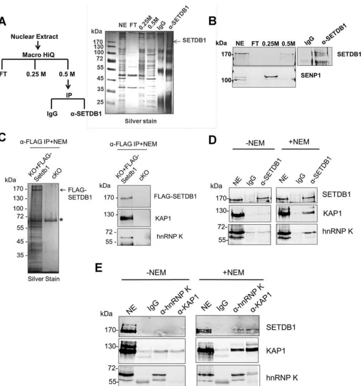
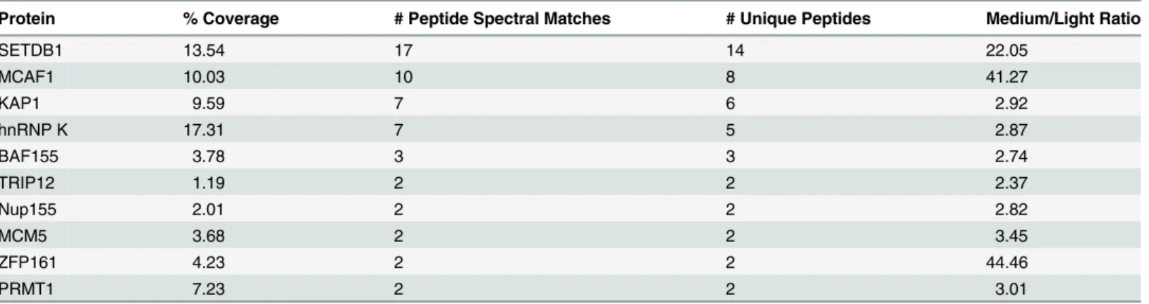
To identify novel factors involved in SETDB1-dependent transcriptional repression, we charac-terized endogenous SETDB1-containing complexes from mESCs by immunoprecipitation (IP) and mass spectrometry (MS), utilizing conditions that minimize de-SUMOylation of proteins given the SUMO-dependent interactions between SETDB1 and KAP1 [18]. Indeed, the Sen-trin/SUMO-specific proteases SENP1 and SENP7 can de-SUMOylate KAP1 [20,21] and are expressed in mESCs [22]. To enrich for candidate SUMO-dependent binding partners of SETDB1, we performed an anionic exchange step which efficiently depleted SENP1 followed by IP of endogenous SETDB1 with a specific N-terminal antibody [23] (Fig. 1A and 1B). MS analysis revealed the specific enrichment of KAP1 along with the previously described SETDB1 co-factor MCAF1 (also called mAM/ATF7IP) (Table 1), which directly interacts with SETDB1 independent of SUMOylation [18,24]. Detection of MCAF1 and KAP1 supports the validity of this approach to identify candidate SUMO-independent and SUMO-dependent binding partners. MS analysis of a SETDB1 IP without prior SENP depletion identified a differ-ent set of polypeptides associated with SETDB1 (S1A Fig.). While MCAF1 was identified in this direct IP approach, KAP1 was not (S1A Fig.), indicating that the presence of SENPs in mESC nuclear extracts precludes the association of SETDB1 with its SUMO-dependent bind-ing partners, includbind-ing KAP1. Among the novel SETDB1-associated proteins detected in the SENP-depleted but not the direct IP, we chose to focus on hnRNP K (Table 1), a ubiquitously expressed protein that functions as a DNA/RNA-binding transcriptional co-activator or co-re-pressor [25]. Notably,Hnrnpkis highly expressed in the inner cell mass and in mESCs relative to earlier stages of development in the preimplantation embryo [22] and was previously re-ported to directly interact with the KRAB-ZFPs Zik1 and Kid1 [25,26].
We further validated the interaction between hnRNP K and SETDB1 in mESCs using a combination of co-IP, immunostaining and co-sedimentation assays. Both KAP1 and hnRNP K were detected in FLAG-tagged SETDB1 complexes immunopurified from mESCs in the presence of the cysteine protease inhibitor N-ethylmaleimide (NEM), which blocks SENP ac-tivity [27] (Fig. 1C). Moreover, using a specific antibody raised against an internal epitope of SETDB1 [28], both KAP1 and hnRNP K co-precipitated with SETDB1 from mESC nuclear ex-tract only in the presence of NEM (Fig. 1D). The association of hnRNP K and SETDB1 was also apparent by immunostaining, which revealed that hnRNP K and SETDB1 colocalize in the nucleus and to a lesser extent the cytoplasm of mESCs upon a short incubation with NEM
Figure 1. hnRNP K is associated with SETDB1 and KAP1 in mESCs.(A) IP scheme to identify SUMO-dependent binding partners of SETDB1 and silver stained gel showing protein content of the indicated fractions. The nuclear extract input (NE), negative control IP (IgG), SETDB1 IP (α-SETDB1), and the flow-through (FT), 0.25 M KCl and 0.5 M KCl fractions from the anionic column are shown. (B) Western blot of SETDB1 and SENP1 in the indicated fractions and IP. (C) Silver stained gel of FLAG-SETDB1 IP and western blot of FLAG-SETDB1, KAP1 and hnRNP K in immunopurified FLAG-SETDB1 complexes isolated from tamoxifen-inducedSetdb1KO mESCs stably expressing 3XFLAG-Setdb1(KO+FLAG-Setdb1) or negative control uninducedSetdb1
conditional KO cells (cKO). Complexes were immunoprecipitated with FLAG antibodies and specifically eluted with 3XFLAG peptide. Asterisk marks a non-specific band. Cropped band at bottom of hnRNP K blot is IgG heavy chain (~55 kDa). (D) Co-IP assay of endogenous KAP1 and hnRNP K with SETDB1 from mESC nuclear extracts in the absence or presence of 10 mM SENP inhibitor (NEM). Note that hnRNP K is detected in mESCs as two bands at ~65 kDa and ~60 kDa, the larger of which represents full-length hnRNP K and the smaller is a splicing isoform hnRNP J, also produced from theHnrnpkgene [77]. The hnRNP K isoform is associated with SETDB1.‘NE’represents ~10% of nuclear extract input and‘IgG’is the negative control IP. (E) Co-IP assay of endogenous SETDB1 with KAP1 or hnRNP K from mESC nuclear extracts in the presence or absence of NEM, as in (D).
doi:10.1371/journal.pgen.1004933.g001
presence of NEM and hnRNP K and KAP1 also co-precipitated with each other (Fig. 1E), indi-cating that these proteins are present in a single complex. Notably, the IP of KAP1 was clearly more efficient in the presence of NEM (Fig. 1E), revealing that SENP inhibition may stabilize KAP1 oligomeric state, as KAP1 is known to form oligomers [29]. In addition, although hnRNP K binds to both DNA and RNA sequences [30,31], the interaction between SETDB1 and hnRNP K was not perturbed in the presence of RNAse A and DNase I (S1C Fig.) indicating that it is not dependent upon nucleic acid. Consistent with the finding that the KAP1 IP was more efficient in the presence of NEM (Fig. 1E), sucrose gradient ultracentrifugation of mESC nuclear extracts revealed that SENP inhibition promotes the stability of SETDB1/KAP1/ hnRNP K complexes, which migrated at higher density compared with the profile of purified GST-KAP1 and GST-hnRNP K (S1D–S1E Fig.). Although most of the hnRNP K remained uncomplexed with SETDB1/KAP1, a fraction of total nuclear hnRNP K clearly co-sedimented with SETDB1 and KAP1 at a higher density in fractions 9–11 in the presence of NEM
(S1E Fig), compared with GST-hnRNP K in fraction 5 (S1D Fig.). Together these results
con-firm that hnRNP K is associated with the SETDB1/KAP1 complex in mESCs.
hnRNP K directly interacts with KAP1
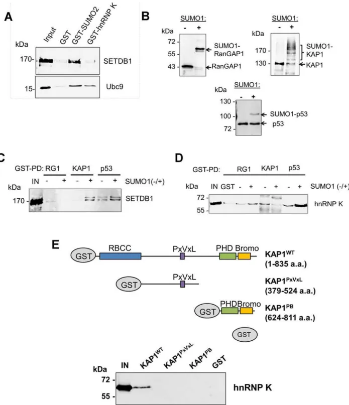
To determine whether hnRNP K directly binds to SETDB1, we performed GST pulldown as-says with recombinant SETDB1 or Ubc9 as a positive control protein for hnRNP K [32,33]. Al-though KAP1 is SUMOylated in the SETDB1 complex under standard tissue culture
conditions [18], hnRNP K was also reported to be SUMOylated but only following DNA dam-age [32,33]. Indeed, whereas SETDB1 complexes from mESCs contained both SUMOylated and unmodified KAP1, we found no evidence of SUMOylated hnRNP K (S2A Fig.) and thus used unmodified hnRNP K in subsequent pulldown assays. In contrast with Ubc9, which bound to SUMO2 and hnRNP K, SETDB1 bound to SUMO2 but not hnRNP K (Fig. 2A). In addition, no interaction was detected between FLAG-tagged SETDB1 and T7-tagged hnRNP K upon co-expression and FLAG IP from 293T cells (S2B Fig.). Together these data indicated that hnRNP K does not directly interact with SETDB1.
To determine whether hnRNP K directly binds SUMOylated and/or unmodified KAP1, we preparedin vitroSUMO1-conjugated GST-tagged KAP1, GST-p53 as a positive control bind-ing partner of hnRNP K [33], or a GST-tagged fragment of RanGAP1 as a model SUMO1 sub-strate for GST pulldown assays with recombinant SETDB1 or hnRNP K baits. Using purified SUMOylation cascade components, we achieved efficient mono-SUMOylation of RanGAP1 at K526 [34] and mono-, di-, tri- and tetra-SUMOylation of KAP1 (Fig. 2B) at its major SUMO Table 1. Mass spectrometric analysis of SETDB1 (medium) versus IgG (light) IPs from the 0.5 M anionic column fraction.
Protein % Coverage # Peptide Spectral Matches # Unique Peptides Medium/Light Ratio
SETDB1 13.54 17 14 22.05
MCAF1 10.03 10 8 41.27
KAP1 9.59 7 6 2.92
hnRNP K 17.31 7 5 2.87
BAF155 3.78 3 3 2.74
TRIP12 1.19 2 2 2.37
Nup155 2.01 2 2 2.82
MCM5 3.68 2 2 3.45
ZFP161 4.23 2 2 44.46
PRMT1 7.23 2 2 3.01
Figure 2. hnRNP K directly interacts with unmodified KAP1 in a region containing the RBCC domain.(A) GST pulldown assays using purified GST, GST-SUMO2 or GST-hnRNP K as baits with recombinant FLAG-SETDB1 or Ubc9 as prey proteins.‘IN’represents ~20% of pulldown prey protein input. (B) Western blot analysis ofin vitroSUMOylation reactions performed on GST-tagged RanGAP1 C-terminal fragment (residues 419–587), full-length wt KAP1 or p53. + or—indicates presence or absence of SUMO1 in the reaction. (C) GST pulldown assays using SUMOylated or unmodified GST-tagged baits from (B) with purified recombinant FLAG-GST-tagged SETDB1 as prey protein.‘IN’represents 10% of input SETDB1 prey protein. (D) GST pulldown assay as in (C) except using purified recombinant 6X-His-tagged hnRNP K as prey protein. (E) Schematic of wt KAP1 domain structure and KAP1 mutants used in GST pulldown assays. Unmodified GST-tagged wt KAP1, deletion mutants KAP1PxVxLand KAP1PBor GST alone were used as baits to pull down purified recombinant 6X-His-tagged hnRNP K.‘IN’represents 15% of the input hnRNP K protein.
doi:10.1371/journal.pgen.1004933.g002
acceptor lysines including K554, K676, K779 and K804 [18,19]. p53 was mono-SUMOylated at K386 [35] although this was less efficient in the absence of a SUMO E3 ligase (Fig. 2B). While SETDB1 directly bound to p53 independently of SUMOylation, it bound to KAP1 in a SUMO1-dependent manner (Fig. 2C), consistent with previous observations [18]. HnRNP K binding to p53 was enhanced by SUMOylation but surprisingly, its binding to KAP1 was de-creased upon SUMOylation (Fig. 2D).
KAP1 harbours several functional domains that participate in protein-protein interactions, including an N-terminal RING-B-box-coiled-coil (RBCC) domain, which mediates binding to KRAB-ZFPs and other proteins [36–38], a proline-x-valine-x-leucine (PxVxL) motif, which binds to HP1 proteins [39] and a C-terminal PHD finger-bromodomain that binds to Ubc9 and chromatin-modifying factors, including SETDB1 and CHD3 upon SUMOylation [10,18,
40]. While hnRNP K bound to wt full-length KAP1, it failed to bind to the deletion fragments containing only the PxVxL motif or only the PHD finger-bromodomain (Fig. 2E), revealing that hnRNP K binding requires the N-terminal RBCC domain. Taken together, these observa-tions indicate that hnRNP K and SETDB1 indirectly interact with each other via their binding to unmodified or SUMOylated KAP1 subunits. Consistent with this model, SETDB1 com-plexes in mESCs contain both unmodified and SUMOylated KAP1 (S2A Fig.), despite SETDB1 exhibiting binding affinity for only SUMOylated KAP1 (Fig. 2C). Furthermore, the interactions between hnRNP K and KAP1 in mESCs were unperturbed upon depletion of SETDB1 (S2C Fig), confirming that they interact in a SETDB1-independent manner. In addi-tion, endogenous KAP1 also co-precipitated with hnRNP K from 293T cell extracts (S2D Fig.), indicating that this interaction is not limited to mESCs. Finally, the observation that hnRNP K colocalized with KAP1 throughout the nucleus in mESCs in the absence of SENP inhibitor
(S2E Fig.) is consistent with the model that they form complexes in the absence of KAP1
SUMOylation in cells.
Depletion of hnRNP K disrupts SETDB1-dependent proviral silencing
We next investigated whether loss of hnRNP K compromises SETDB1-dependent transcrip-tional silencing of ERVs in mESCs. Using siRNA-mediated knockdown (KD), hnRNP K was efficiently depleted at the protein level by 24 h post-transfection (S3A Fig.). Notably, KD of hnRNP K in mESCs significantly reduced their proliferation by 72 h post-transfection
(S3B Fig). However, there was no gross effect on cell cycle distribution at this time-point and
only minimal effects on expression of the pluripotency marker SSEA1 (S3C–S3D Fig.). Fur-thermore, reduced proliferation was not associated with induction of apoptosis, as determined by Annexin V staining (S3D Fig.). Thus hnRNP K KD does not result in overt differentiation or apoptosis at this time-point.
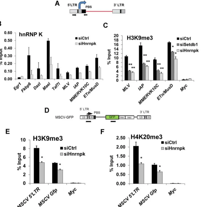
Figure 3. siRNA knockdown of hnRNP K results in de-repression of proviral reporters and ERVs.(A) Schematics of the MSCV (PBSGln)-GFP retroviral
vector in theSetdb1lox/-mESC line and the IAP LTR-PBS GFP reporter in the HA36 wt mESC line. (B) qRT-PCR analysis ofSetdb1andHnrnpkmRNA in
Setdb1lox/-33#6 MSCV-GFP cells transfected with control (siCtrl),Setdb1orHnrnpksiRNAs at 24 h post-transfection. Data are mean expression level
relative toGapdhmRNA and normalized to siCtrl. Data are means (+ s.d.) of three technical replicates. (C) Flow cytometry analysis on untransfected (-) cells and cells transfected with indicated siRNAs at 72 h post-transfection. Data represent mean percentage of SSEA1+, GFP+(double-positive) cells from three
biological replicates of 10,000 propidium iodide-negative (PI-) cells per sample. Error bars are s.d. (D) qRT-PCR analysis ofSetdb1,HnrnpkandMcaf1
mRNA in J1 wt orDnmt3a-/-; Dmnt3b-/-; Dnmt1-/-(DnmtTKO) cells transfected with indicated siRNAs at 24 h post-transfection. Data are mean expression
level (+ s.d.) normalized to expression levels in control siRNA transfected J1 wt mESCs, relative to level ofGapdhmRNA. (E) qRT-PCR analysis as in (D) of except for intact class I and II ERVs in J1 wt orDnmtTKO mESCs transfected with the indicated siRNAs at 96 h post-transfection.*p<0.01,**p<0.001,
Student’s two-tailed T-test, relative to siCtrl.
doi:10.1371/journal.pgen.1004933.g003
role of the SETDB1 co-factor MCAF1, which facilitates conversion of H3K9me2 to H3K9me3 by SETDB1 [24]. Interestingly,Setdb1KD cells showed a ~4-fold upregulation ofMcaf1 ex-pression (S4A Fig.), indicating that the level ofMcaf1expression is sensitive to the level of SETDB1. The MSCV proviral reporter was also de-repressed inMcaf1KD cells, (S4A–S4B Fig.), revealing that this catalytic co-factor of SETDB1 also plays a role in proviral silencing.
We next determined whether KD of hnRNP K disrupts silencing of ERVs. In contrast to the proviral reporter lines, KD of SETDB1 or hnRNP K in TT2 wt mESCs resulted in only modest de-repression (~2-fold) of class I and II ERVs by 72 h post-transfection (S4C–S4D Fig.), with the exception of robust induction of MMERVK10C elements in SETDB1 KD cells, despite effi-cient depletion of the protein (S4C Fig.). Surprisingly, class III MERVL elements, which are re-pressed by KAP1 in a SETDB1-independent manner [42], were strongly induced in hnRNP K KD cells (S4D Fig.).
We have shown previously that DNA methylation also plays a role in transcriptional repres-sion of ERVs, particularly of IAP elements, in mESCs cultured in serum [17]. To preclude the influence of DNA methylation, we knocked downSetdb1orHnrnpkinDnmt3a; Dnmt3b; Dnmt1triple KO (DnmtTKO) mESCs [43] (Fig. 3D), which are devoid of DNA methylation but maintain SETDB1 binding and H3K9me3 at ERVs [11] and thus solely rely on the SETDB1/H3K9me3 pathway for silencing of these elements. The absence of DNA methylation alone did not perturb silencing of MLV, MMERVK10C and MusD elements, but yielded a ~6-fold upregulation of IAP elements (Fig. 3E), consistent with the finding that IAP elements are modestly upregulated inDnmtTKO cells [11]. In contrast, KD ofSetdb1expression re-sulted in a substantial induction of ERVs in these cells (Fig. 3E). These ERVs were also de-repressed uponHnrnpkKD, with IAP elements showing an increase in expression of ~18-fold, ~3-fold greater than the control KD in theDnmtTKO line (Fig. 3E). Depletion of Mcaf1in theDnmtTKO cells (Fig. 3D) also resulted in upregulation of class I and II ERVs be-yond what was observed in theDnmtTKO line alone (S4E Fig.). Importantly, although IAP elements are strongly induced in DNA methylation-deficient, differentiatedDnmt1-/-; Oct4-negative mESCs [6], levels ofOct4mRNA was not appreciably reduced in any of these KD cultures compared to the control siRNADnmtTKO cells (S4F Fig.), indicating that ERVs were not induced as a secondary consequence of an increase in the number of differentiated cells in culture. Taken together, these results reveal that depletion of hnRNP K disrupts SETDB1/H3K9me3-mediated silencing of ERVs in mESCs.
hnRNP K is required for repression of a cohort of male germline genes
by SETDB1
Figure 4. hnRNP K plays a role in repression of SETDB1 target genes.(A) RNA-seq 2D scatterplots of gene expression (reads per kilobase per million mapped reads; log(RPKM) from two biological replicates of TT2 cells transfected with control orHnrnpksiRNAs at 72 h post-transfection. Genes upregulated 2-fold (‘UP’) in hnRNP K KD cells are labelled in red, whereas genes downregulated50% (‘DN’) are shown in blue. Data points correspond to n = 22,138 ENSEMBL annotated genes. (B) qRT-PCR analysis of selected lineage-specific genes identified as upregulated from the RNA-seq of hnRNP K KD cells.
Fkbp6,Dazl,Mael,Taf7lare direct SETDB1/H3K9me3 target genes. Data are relative mean expression level (+ s.d.) for three technical replicates. (C) UCSC genome browser screenshot including tracks for SETDB1 ChIP-seq in wt [28] and H3K9me3 ChIP-seq in wt andSetdb1KO mESCs and RNA-seq for wt and
Setdb1KO mESCs [17] along with total coverage tracks for hnRNP K KD replicates RNA-seq at theDazlgene. Numbers on the right indicate y-axis scale. (D) Venn diagram showing overlap of genes upregulated in hnRNP K KD cells (264) and genes upregulated in SETDB1 KO cells that are either bound by SETDB1 and/or marked by SETDB1-dependent H3K9me3 (134) according to [17]. p = 8.7×10⁻³⁰, Fisher’s exact test (n = 22,138 ENSEMBL-annotated
lineage transcription factorsGata6,Gata3,Tbx3,Tbx20,Foxa1,Nkx2–9andNkx2–2
(S1 Table). Previous ChIP-seq data indicates that these transcription factor genes harbour the
bivalent chromatin state of H3K4me3 and H3K27me3 [45–47] and are subject to polycomb re-pressive complex 2 (PRC2)-mediated silencing [48]. The de-repression ofGata6andGata3, which were upregulated ~15-fold and ~10-fold in hnRNP K KD cells, respectively (Fig. 4B), in-dicates that hnRNP K KD could eventually lead to a loss of pluripotency, since the overexpres-sion of these transcription factors is sufficient to drive endoderm lineage differentiation [49,50]. RNA-seq analysis of ERVs in the TT2 hnRNP K KD cells (S1 Table) generally con-firmed our qRT-PCR analysis from the same cells (S4D Fig.) in that class I and II elements were only modestly de-repressed (2-fold) while MERVL elements were strongly de-repressed (14-fold).
We next compared the list of genes upregulated in hnRNP K KD mESCs to our list of upre-gulated genes inSetdb1KO mESCs [17], which revealed 54 genes in common (S2 Table). We previously identified a cohort of 33 germline lineage genes that are directly repressed by SETDB1-dependent H3K9me3 and DNA methylation [17]. Notably, many of these direct SETDB1 target genes were consistently upregulated>2-fold in hnRNP K KD cells (15 of these genes are shown inS5B Fig.) For example, the promoter of the male germline geneDazl har-bours a peak of SETDB1 binding and SETDB1-dependent H3K9me3 and is upregulated in bothSetdb1KO and hnRNP K KD cells (Fig. 4C). In addition, of the 134 SETDB1-bound genes that are upregulated inSetdb1KO mESCs [17], 30 were consistently de-repressed in hnRNP K KD cells (Fig. 4DandS2 Table). Quantitative RT-PCR analysis of a subset of these genes, including the male germline-specific genesDazl,Fkbp6,MaelandTaf7lconfirmed that they are indeed upregulated in hnRNP K KD cells (Fig. 4B). Furthermore, levels of H3K9me3 at the promoters of these genes were reduced in hnRNP K KD cells to a similar extent as in SETDB1 KD cells (Fig. 4E). A comparison of the genes upregulated in hnRNP K KD andKap1 KO cells [14] also revealed a significant overlap (S5C Fig.) and included lineage-restricted genes such asGata6,Arg2andDkk1(S2 Table). We identified 33 genes that are commonly de-repressed inSetdb1KO,Kap1KO and hnRNP K KD mESCs, several of which were direct SETDB1 targets and were expressed in a lineage-dependent fashion, including the imprinted geneIgf2and liver-specific geneCml2(S2 Table). The promoter ofCml2lies immediately downstream of an intact ETn family retroelement that is bound by SETDB1 and marked by SETDB1-dependent H3K9me3, which spreads into theCml2promoter (S5D Fig.) indicating that this gene is silenced by the spreading of H3K9me3 from the intact ERV. In conclusion, these results support a role for hnRNP K in transcriptional repression of genes regulated by SETDB1 and KAP1 as well as PRC2, the latter via an undefined pathway.
H3K9me3 enrichment on proviral chromatin is reduced following
knockdown of hnRNP K
We next determined whether hnRNP K is bound at de-repressed ERVs. Since the presence of NEM increased the sensitivity of KAP1 and SUMO1 chromatin immunoprecipitation (ChIP), improving the enrichment of both at MLV and IAP 5’LTRs where KAP1 binding is high, but not at MERVL 5’LTRs where KAP1 binding is low [42] (S6A Fig.), we performed subsequent ChIP assays in the presence of NEM to preclude a refractory effect of SENP activity on the binding of these factors at ERV 5’LTRs and other loci (Fig. 5A). Under these conditions,
genes). (E) Native ChIP for H3K9me3 at the core promoters/TSS of the indicated genes on TT2 cells transfected with control,Setdb1orHnrnpksiRNAs at 72 h post-transfection. Data are mean enrichment relative to input from three technical replicates, error bars are s.d.*p<0.05,**p<0.005, Student’s
two-tailed T-test, relative to siCtrl.
Figure 5. hnRNP K is bound at ERVs and is required for H3K9me3 deposition at proviral chromatin.(A) Schematic of intact ERV structure and 5’ LTR-internal sequence amplified (primers shown as arrows). (B) Crosslinked ChIP of hnRNP K in TT2 cells transfected with control orHnrnpksiRNAs at 24 h post-transfection.Egr1core promoter and TSS (-50 to +50) was amplified as a negative control locus. Data shown are mean enrichment levels relative to input material from three technical replicates, error bars show s.d. (C) Native ChIP for H3K9me3 at the indicated ERV 5’LTR-internal regions in TT2 wt mESCs transfected with control,Setdb1, orHnrnpksiRNAs at 72 h post-transfection with the same primers as in (A). TheMyccore promoter and TSS (-50 to +50) was amplified as a negative control. (D) Schematic of the MSCV (PBSGln)-GFP vector with black bars showing the positions of ChIP amplicons for the MSCV 5’LTR-PBS region andGfpinternal sequence. (E) Native ChIP for H3K9me3 as in (C) except on siRNA KD ofHnrnpkin unsorted (GFP+and
GFP-) MSCV-GFP cells at 24 h post-transfection. (F) Native ChIP from the same KD cells as in (E) except for H4K20me3.
*p<0.001,**p<0.0001,
Student’s two-tailed T-test, relative to siCtrl.
doi:10.1371/journal.pgen.1004933.g005
hnRNP K was enriched at the promoters of the SETDB1-bound, H3K9me3-marked germline genesFkbp6,Dazl,MaelandTaf7l, with the highest level of enrichment detected atMael
(Fig. 5B), indicating that these loci are direct targets of hnRNP K in mESCs. Relative to the
germline gene promoters, the 5’LTRs of class I and II ERVs showed lower enrichment of hnRNP K, with ETn/MusD and MLV elements showing the highest and lowest levels, respec-tively (Fig. 5B). Importantly, the signal at ERVs and the germline gene promoters was specific, since it was reduced upon hnRNP K KD. In contrast, there was no enrichment of hnRNP K at theEgr1promoter (Fig. 5B), which is active in mESCs and was shown to be bound by hnRNP K only upon serum stimulation in the HCT116 colon cancer cell line [51,52]. Furthermore, RNAse did not perturb hnRNP K enrichment at ERVs (S6B Fig.), indicating that hnRNP K is recruited to class I and II ERVs in an RNA-independent manner.
We next determined whether hnRNP K is required for SETDB1-dependent H3K9me3 de-position at ERVs. As shown previously [11], SETDB1 KD resulted in depletion of H3K9me3 at MLV, IAP, MMERVK10C and ETn/MusD 5’LTRs (Fig. 5C). Strikingly, this effect was phe-nocopied upon KD of hnRNP K (Fig. 5C). Importantly, the reduction of H3K9me3 at ERVs was apparent in hnRNP K KD cells as early as 48 h post-transfection, similar to the kinetics of H3K9me3 perturbation in SETDB1 KD cells (S6C Fig.), indicating that this phenotype is not a secondary consequence of the loss of proliferation that commences at ~72 h post-transfection in hnRNP K KD cells (S3B Fig.). Furthermore, as early as 24 h after hnRNP K KD, there was a clear reduction of H3K9me3 at the MSCV 5’LTR-PBS andGfpregions (Fig. 5D and 5E). The levels of H4K20me3, a mark deposited by SUV420H1/2 enzymes in a SETDB1/H3K9me3-dependent manner [11], were also reduced at the MSCV provirus in hnRNP K-depleted cells
(Fig. 5F). H3K9me3 was dramatically reduced at both ERVs and the MSCV proviral reporter
in MCAF1 KD cells (S6C–S6D Fig.), consistent with its role as a catalytic co-factor of SETDB1 [24]. Importantly, siRNA-mediated depletion of hnRNP K or SETDB1 did not affect global H3K9me2 or H3K9me3 levels (S6E Fig.), indicating that the effect of hnRNP K depletion on H3K9me3 at ERVs is not the result of a general reduction of H3K9me2/3. Thus we concluded that hnRNP K is required for SETDB1-dependent H3K9me3 deposition at proviral chromatin.
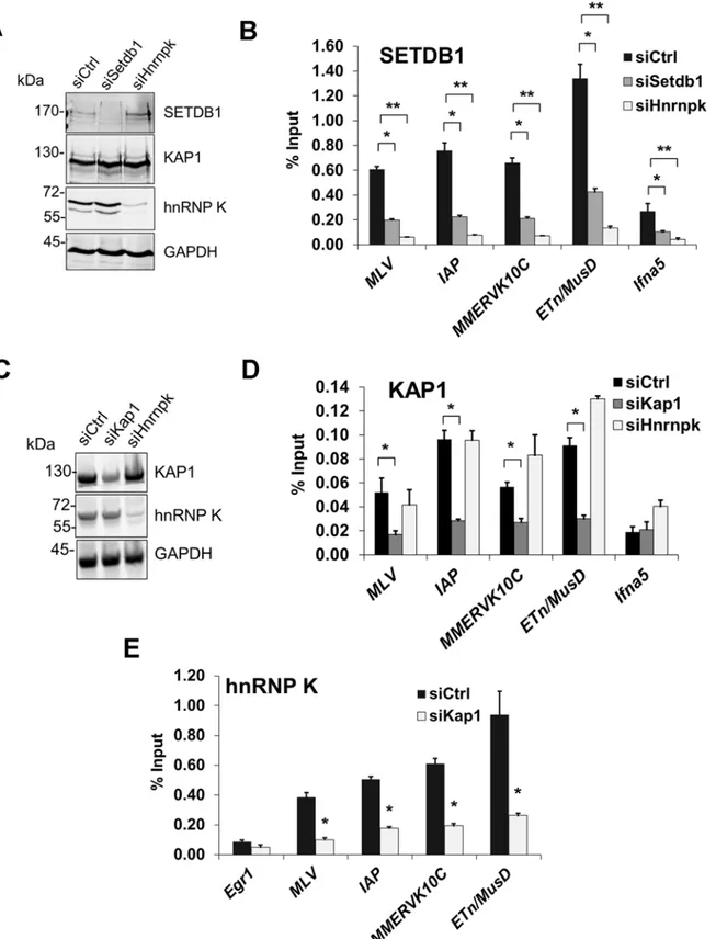
hnRNP K is required for SETDB1 recruitment to ERVs
To determine whether hnRNP K is required for SETDB1 recruitment, we next conducted ChIP analysis of SETDB1 in cells depleted of hnRNP K (Fig. 6A and 6B. A reduction of SETDB1 en-richment was apparent at all class I and II ERV LTRs in SETDB1 KD cells, confirming the specificity of our antibody (Fig. 6B). Strikingly, KD of hnRNP K also reduced the level of SETDB1 enrichment at ERVs (Fig. 6B), likely explaining the reduction of H3K9me3 observed at these loci following hnRNP K KD (Fig. 5C). SETDB1 enrichment was also reduced at the MSCV 5’LTR-PBS andGfpinternal region upon depletion of hnRNP K (S7A Fig.), revealing a link between loss of H3K9me3, de-repression of the MSCV proviral reporter and reduced SETDB1 recruitment. In contrast, KD of MCAF1 did not perturb SETDB1 enrichment at ERVs or the MSCV provirus (S7B Fig.). Importantly, neither SETDB1 nor KAP1 protein levels were reduced in hnRNP K KD cells (Fig. 6A) and SETDB1 was still localized to the nucleus in hnRNP K-depleted cells (S7C Fig.).
KAP1 is the only factor known to be required for SETDB1 recruitment to proviral chroma-tin [11,14]. While KAP1-depleted cells showed reduced levels of KAP1 enrichment at ERVs confirming antibody specificity, KD of hnRNP K did not affect KAP1 enrichment levels
(Fig. 6C and 6D). In contrast, KD of KAP1 substantially reduced hnRNP K enrichment at
Figure 6. hnRNP K is required for SETDB1 but not KAP1 recruitment to ERVs.(A) Western blot analysis of SETDB1, KAP1 and hnRNP K in TT2 cells transfected with control,Setdb1orHnrnpksiRNAs at 24 h post-transfection. GAPDH was detected as a loading control. (B) Crosslinked ChIP of SETDB1 in the same cells as in (B) except at 72 h post-transfection. All qPCR data are mean enrichment relative to input of three technical replicates, error bars are s.d. TheIfna5core promoter and TSS (-50 to +50) was amplified as a negative control locus. (C) Western blot analysis of KAP1 and hnRNP K in TT2 cells transfected with control,Kap1orHnrnpksiRNAs at 24 h post-transfection. (D) Crosslinked ChIP of KAP1 in the same cells as in (C) except at 72 h
SUMOylation on proviral chromatin is required for SETDB1 recruitment
and is reduced upon hnRNP K knockdown
Previous studies have shown that KAP1 SUMOylation is necessary for recruitment of SETDB1 and H3K9 methylation to promote silencing of heterologous promoters in transformed cell lines [18,19,53]. To determine whether a functional SUMOylation pathway is also necessary for SETDB1 recruitment to ERVs in pluripotent stem cells, we used either anacardic acid to inhibit SUMO E1 activating enzyme [54] or siRNAs to KDUbc9(also calledUbe2i) in the MSCV-GFP cell line and assayed for de-repression of the proviral LTR by flow cytometry (Fig. 7A). In accord with the inhibitory effect of anacardic acid on the activity of SUMO E1 activating enzyme Aos1/ Uba2 and histone H3 acetyltransferases such as p300 [55], this compound blocked both KAP1 SUMOylation and bulk histone H3 acetylation (Fig. 7B). While, anacardic acid treatment did not affect bulk H3K9me3 (Fig. 7B), it consistently de-repressed the proviral reporter in a dose-dependent manner, resulting in ~15% GFP+cells at 100μM (Fig. 7C).
Using siRNAs, we depletedUbc9mRNA to ~35% of the control (Fig. 7D, inset graph). As Ubc9is essential for early embryogenesis [56], we monitored changes in MSCV expression at 48 h post siRNA transfection. KD ofUbc9expression consistently de-repressed the proviral re-porter resulting in an average of 23% GFP+cells (Fig. 7D). Notably, ChIP analysis revealed that SUMO1 levels at the MSCV 5’LTR were dramatically reduced in Ubc9-depleted cells (S8A Fig.) indicating that the loss of SUMOylation on proviral chromatin correlates with de-repression. Furthermore, there was a reduction of SETDB1 enrichment at the MSCV provirus inUbc9KD cells (Fig. 7E), confirming that SUMOylation of chromatin proteins associated with ERVs enhances SETDB1 recruitment. Strikingly, SUMO1 levels were greatly reduced at the 5’LTRs of MLV, IAP, MMERVK10C and ETn/MusD elements by 24 h post-transfection of hnRNP K siRNAs (Fig. 7F). Moreover, this effect persisted in hnRNP K KD cells at 72 h post-transfection both at ERVs and the MSCV provirus (S8B–S8C Fig.), coinciding with the timeframe in which SETDB1 recruitment to proviral chromatin was compromised (Fig. 6B). Although the loss of SUMOylation at ERV chromatin upon hnRNP K KD could be a conse-quence rather than a cause of reduced SETDB1 recruitment, KD of SETDB1, which was suffi-cient to de-repress the MSCV LTR (S8D Fig.), did not concomitantly attenuate SUMOylation on proviral chromatin (S8E Fig.). Taken together these results are consistent with the model that hnRNP K is necessary for SUMOylation of proteins such as KAP1 on ERV chromatin, which is required for SETDB1 recruitment and in turn proviral silencing.
Discussion
KRAB-ZFP/KAP1 complexes [9,57] are thought to play a central role in repression of ERV transcription in pluripotent stem cells via SETDB1 recruitment [11,14]. In this work, we have identified hnRNP K as a novel co-factor, which is required for recruitment of SETDB1 to pro-viral chromatin and in turn for efficient propro-viral silencing. HnRNP K is a highly conserved, multi-functional protein involved in transcription regulation, mRNA splicing and translation [25]. Studies in flies, yeast and in mammalian cell lines reveal that hnRNP K plays important roles in development and gene regulation [58,59]. HnRNP K was reported to directly interact with chromatin regulatory proteins, such as the PRC2 subunit EED [60] and KRAB-ZFPs Zik1 and Kid1 [25,26], indicating that it may regulate Polycomb and/or KRAB-ZFP/
KAP1 complexes.
post-transfection. (E) Crosslinked ChIP as in (B) and (D) except of hnRNP K in TT2 cells transfected with control orKap1siRNAs at 72 h post-transfection.*p
<0.001,**p<0.0001, Student’s two-tailed T-test relative to siCtrl.
Figure 7. SUMOylation on proviral chromatin is required for SETDB1 recruitment and is compromised upon hnRNP K knockdown.(A) Illustration of the SUMO conjugation pathway including the activities of SUMO E1 activating heterodimer enzyme Aos1/Uba2 and SUMO E2 conjugating enzyme Ubc9. Anacardic acid was used to inhibit E1 activity while siRNAs were used to depleteUbc9transcripts to disrupt SUMO conjugation in the MSCV-GFP cell line. (B) Western blot analysis of KAP1, GAPDH, pan Histone H3 acetylation or H3K9me3 in MSCV-GFP cells incubated in varying concentrations of anacardic acid for 18 h prior to harvest. A mono-SUMOylated KAP1 band is detected at ~130 kDa (arrow). (C) Flow cytometry for GFP+cells in the MSCV-GFP line
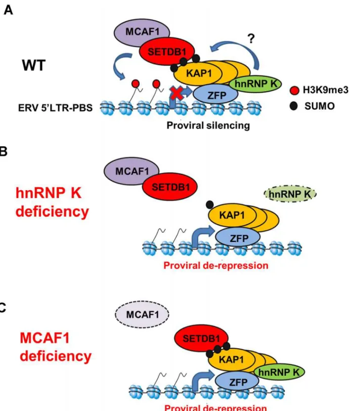
Our results reveal a role for hnRNP K in the KRAB-ZFP/KAP1-based silencing pathway acting on ERVs and retroviral vectors in pluripotent stem cells. Based on these findings, we propose a novel model for the SETDB1/KAP1 proviral silencing pathway incorporating hnRNP K (Fig. 8). In wt mESCs, KRAB-ZFPs recruit KAP1 in an oligomeric state, possibly as a homotrimer [29,36], to proviral chromatin and unmodified KAP1 may recruit hnRNP K. HnRNP K may promote KAP1 SUMOylation on chromatin, which then serves as a ligand for the SETDB1/MCAF1 complex [18,61], eliciting SETDB1-dependent H3K9me3 deposition at SUMOylated KAP1-bound regions (Fig. 8A). In hnRNP K-deficient cells, SUMOylation of KAP1 on chromatin may be compromised, leading to reduced SETDB1 recruitment at ERVs, diminution of H3K9me3 and eventual transcriptional de-repression (Fig. 8B). This model is consistent with recent ChIP-seq analyses of SUMO1, SUMO2 and Ubc9 in human fibroblasts [62], which show co-occupancy with sites of KAP1, SETDB1 and H3K9me3 at the 3’ends of KRAB-ZFP genes [63] indicating that these SETDB1/KAP1-bound, SUMOylated loci are sites of active KAP1 SUMOylation on chromatin [62]. Although a possible contraindication to this model is our observation of the differing binding affinities of SETDB1 and hnRNP K for SUMOylated KAP1in vitro(Fig. 2C and 2D), this could be rationalized by: 1) the existence of multiple KAP1 subunits in each complex such that some are SUMOylated while others are un-modified, providing binding sites for both SETDB1 and hnRNP K simultaneously, and/or 2) the observation that hnRNP K directly binds to certain KRAB-ZFPs [25,26] and therefore may still indirectly interact with SUMOylated KAP1. Indeed, consistent with the former possibility, rather than solely containing SUMOylated KAP1, we found that SETDB1 complexes contained predominantly unmodified KAP1 with only a minority SUMOylated KAP1 under conditions where we could preserve mono- and di-SUMOylated KAP1 in mESC nuclear extracts (S2A Fig.). This observation indicates that SETDB1 binding to KRAB-ZFP/KAP1 complexesin vivo may only require a small proportion of the total KAP1 in the complex to be SUMOylated. KAP1 SUMOylation is highly dynamic and previous investigations have relied on overexpres-sion of SUMO paralogues to detect it [19,20,38,64]. Therefore, it is also possible that hnRNP K facilitates transient KAP1 SUMOylation events in a cell cycle-dependent manner, such as during S-phase when chromatin modifications must be re-established. How hnRNP K pro-motes the SUMOylation of KAP1 remains to be determined, although given that hnRNP K is a SUMO target itself and can directly interact with Ubc9 [32,33], it may facilitate recruitment of this SUMO E2 enzyme to KAP1-bound loci. Alternatively, hnRNP K might also counteract SENP activity toward KAP1 providing an additional layer of regulation over KAP1de-SUMOy-lation. Since KAP1 is constitutively phosphorylated at Ser824 in pluripotent stem cells [65], an-other intriguing possibility is that hnRNP K counteracts the activity of the SUMO-targeted ubiquitin ligase RNF4, which conjugates ubiquitin to Lys676 SUMOylated, Ser824 phosphory-lated KAP1 promoting its degradation [64].
Similar to SETDB1 KD mESCs [17], KD of hnRNP K only resulted in modest upregulation of class I and II ERVs in wt mESCs cultured in serum. A likely explanation for this observation is the relatively high level of DNA methylation in mESCs cultured in serum relative to two-inhibitor (2i) media. Under the latter conditions, mESCs adopt a“naïve”hypomethylated
alone (-), vehicle (DMSO) treated, or treated with 40 or 100μM anacardic acid for 18 h. Data are mean of three biological replicates for 10,000 PI- cells per sample, error bars are s.d. (D) Flow cytometry data as in (B) except on cells untransfected (-) or transfected with control orUbc9siRNAs at 48 h post-transfection. Inset, qRT-PCR analysis ofUbc9expression in cells transfected with control orUbc9siRNAs at 24 h post-transfection. Data are mean relative expression levels determined from three technical replicates, normalized to the level ofβ-actin (Actb) transcripts, error bars are s.d. (E) Crosslinked ChIP of SETDB1 on unsorted (GFP+and GFP-) MSCV-GFP cells transfected with control orUbc9siRNAs at 48 h post-transfection. Data are mean enrichment as a percent of input chromatin from three technical replicates, error bars are s.d. (F) Crosslinked ChIP as in (E) except for SUMO1 on TT2 cells transfected with control orHnrnpksiRNAs at 24 h post-transfection.*p<0.01,**p<0.001, Student’s two-tailed T-test relative to vehicle or siCtrl.
Figure 8. Model for SETDB1/KAP1-mediated proviral silencing pathway.(A) In wt mESCs, KRAB-ZFPs recruit KAP1 subunits and unmodified KAP1 recruits hnRNP K. SUMOylation of KAP1 and/or other proteins on chromatin is promoted by hnRNP K, resulting in recruitment of SETDB1/MCAF1 and deposition of H3K9me3. (B) In hnRNP K-deficient cells, the SUMOylation of KAP1 on chromatin is compromised, leading to loss of SETDB1 recruitment, loss of H3K9me3 and induction of proviral expression. (C) In MCAF1-deficient cells, SETDB1 recruitment is maintained but H3K9me3 is no longer deposited efficiently, leading to proviral de-repression.
doi:10.1371/journal.pgen.1004933.g008
state, more reflective of the inner cell mass of the E3.5 blastocyst [46]. Consistent with this model, depletion of hnRNP K in DNA methylation-deficient cells led to a more robust upregu-lation of class I and II ERVs as compared with wt cells and previous work has shown that IAP elements are synergistically upregulated upon KD of both SETDB1 and DNMT1 in serum-cultured mESCs [17]. Thus siRNA KDs in serum-cultured mESCs are likely not robust enough to elicit loss of DNA methylation at ERVs controlled by SETDB1, despite losses of H3K9me3. In contrast with ERVs, for reasons that are not entirely clear, knocking down SETDB1 in serum-cultured mESCs harbouring a newly integrated silent MSCV provirus results in losses of both H3K9me3 and DNA methylation at the 5’LTR and subsequent de-repression [11,41].
In addition to hnRNP K, we also identified a crucial role for MCAF1 in SETDB1-mediated proviral silencing, consistent with its role in enhancing SETDB1 catalytic activity towards H3K9me2 to generate H3K9me3 [24]. In contrast to hnRNP K- and KAP1-depleted cells, SETDB1 recruitment is maintained but H3K9me3 is no longer efficiently deposited at proviral chromatin in MCAF1-deficient mESCs (Fig. 8C). This phenotype is consistent with the obser-vation that the catalytic activity of SETDB1 is crucial for full ERV repression [11,41] and a pre-vious report showing that the MCAF1 orthologue Windei is necessary for dSETDB1/Eggless function in theDrosophilagermline [66].
Intriguingly, class III MERVL elements, which are silenced by H3K9me2 deposited by the lysine methyltransferases G9a/GLP [42], were strongly induced in hnRNP K KD cells. Since these elements are also de-repressed inKap1KO but notSetdb1KO mESCs [14,42], hnRNP K may play a role in SETDB1-independent chromatin regulatory pathways with KAP1 and G9a/ GLP. Further experiments are required to address whether hnRNP K has direct role in MERVL silencing in mESCs.
In addition to ERVs, a cohort of SETDB1/H3K9me3-repressed male germline-specific genes [17] are bound at their promoters by hnRNP K, show reduced H3K9me3 and increased expression upon hnRNP K KD, indicating a role for hnRNP K in SETDB1/H3K9me3-mediated gene repression. How SETDB1 may be targeted to these promoters by hnRNP K re-mains unclear, since these genes are not upregulated inKap1KO cells [14]. One possibility is that hnRNP K promotes SUMOylation of proteins other than KAP1 on chromatin, leading to SETDB1 binding and transcriptional silencing. In addition to SETDB1/H3K9me3, hnRNP K may also promote PRC2/H3K27me3-mediated gene repression in mESCs, since a cohort of PRC2 target genes, includingGata6andNkx2–9were strongly upregulated in hnRNP K KD cells. This is consistent with a previous report showing that hnRNP K can promote PRC2-dependent repression via recruitment of the subunit EED to a heterologous promoter [60]. Further studies will be necessary to clarify the contribution of hnRNP K to SETDB1- and PRC2-mediated transcriptional silencing at specific genes in mESCs.
In conclusion, our results reveal novel mechanistic insights into the transcriptional silencing of class I and II LTR retrotransposons and genes by SETDB1/H3K9me3 in pluripotent stem cells. Notably, both hnRNP K and SETDB1 have been identified as bona fide oncogenes and are aberrantly overexpressed in a variety of human cancers including melanoma [67,68], pros-tate carcinoma [69,70] and lung carcinoma [71,72]. A greater understanding of how hnRNP K regulates the recruitment of SETDB1 to promoters may ultimately provide new targets for anti-oncogenic therapeutics.
Materials and Methods
Cell culture, RNAi and plasmid transfection
3XFLAG-Setdb1[11], HA36 harboring the silent IAP LTR-GFP construct [41], J1 wt and Dnmt3a-/-; Dnmt3b-/-; Dnmt1-/-[43]. All ES cell lines were cultured under standard feeder-free conditions on gelatinized tissue culture dishes in standard mESC media: DMEM high glucose containing 15% fetal bovine serum, 20 mM HEPES, 1 mM L-glutamine, 100 U/ml penicillin-streptomycin, 1 mM nonessential amino acids, 1 mM sodium pyruvate, recombinant LIF and 0.1 mMβ-mercaptoethanol. HEK293T cells were cultured in DMEM high glucose containing 10% fetal bovine serum and 100 U/ml penicillin-streptomycin. All cell lines were cultured at 37°C with 5% CO2. RNAi was performed essentially as described [41] using predesigned
siRNA SMARTpools from Dharmacon (ThermoFisher). Briefly, cells were seeded in ES media lacking antibiotics to achieve 70–80% confluence and 24 h later were transfected with 100 nM of SMARTpools forSetdb1,Hnrnpk,Mcaf1(also calledAtf7ip),Kap1,Ubc9or non-targeting siRNA #2 (control siRNA) using DharmaFECT Reagent #1. A second round of transfection was performed 48 h later using 100 or 50 nM siRNAs. Plasmids were transfected into
HEK293T cells in antibiotic-free media using lipofectamine 2000 (Life Technologies) and har-vested 48 h post-transfection. For blocking SUMO E1 activity, anacardic acid (Sigma-Aldrich) diluted to 5–100μM in DMSO was incubated with cells in complete ES media for 18 h prior
to harvest.
Immunofluorescence and flow cytometry
Indirect immunofluorescence staining was performed using standard methods. Cells were grown on coverslips or harvested by trypsinization were crosslinked with 4% formaldehyde, permeabilized with 0.25% triton-X-100 and blocked with 1% bovine serum albumin (Sigma-Aldrich). Cells were then incubated with anti-SETDB1 H300 (Santa Cruz Biotechnology sc-66884), anti-hnRNP K 3C2 (Abcam 39975), anti-hnRNP K (Abcam 70492) or anti-KAP1 20C1 (Abcam 22553) at 37°C for 1 h or overnight at 4°C and subsequently incubated with Alexa Fluor 488 and 594-labeled secondary antibodies (Life Technologies). DNA was counter-stained with Hoescht 33342 (Sigma-Aldrich). Flow cytometry analysis of GFP-fluorescing cells was performed as previously described [41]. Briefly, cells were resuspended in 0.5μg/ml
propi-dium iodide (Sigma-Aldrich) in FACS buffer (phosphate buffered saline containing 3% fetal bovine serum) and analyzed on a BD LSRII flow cytometer using BD FACS Diva software. Cells were successively gated on forward and side scatter, then PI- (live cells) and lastly GFP+ cells, using the untransfected mESC line (either MSCV-GFP or IAP-GFP) as a GFP- popula-tion to set the gates. SSEA1 and Annexin V staining were detected on mESCs using 1:400 anti-SSEA1 PE-conjugate (BD Pharmigen) or 1:1000 anti-Annexin V Alexa Fluor 488-conjugate (Life Technologies). Where indicated, cells were gated for the SSEA1+population prior to GFP gating to identify the SSEA1+; GFP+(double-positive) population. Cell cycle analysis was performed according to standard methods where cells were harvested and fixed for>2 h in ice-cold 70% ethanol, permeabilized with 0.25% triton-X-100 and stained with 10μg/ml
propi-dium iodide (Sigma-Aldrich). Cell cycle profiles were analyzed by the Dean-Jett-Fox Model using FlowJo software (Tree Star).
Mass spectrometry of SETDB1 complexes
Nuclear extracts were prepared from mESCs as previously described [42] with or without 10 or 20 mM NEM and clarified by centrifugation. For immunoprecipitation of SETDB1 complexes after anionic column fractionation, approximately 12–15 mg of TT2 mESC nuclear extract (4 ml) was prepared without NEM, diluted with 2 volumes with 56 mM HEPES pH 7.9, 5% glycerol and passed over a 2 ml column of Macro HiQ anionic exchange media (BioRad) in an equilibration buffer (50 mM HEPES pH 7.9, 100 mM KCl, 10% glycerol). Bound proteins were
washed with 5 column volumes of equilibration buffer and then eluted stepwise in 2 volumes of buffer containing 250 mM KCl, then 2 volumes of buffer containing 500 mM KCl. The 500 mM KCl fraction containing SETDB1 and depleted of SENP1 (4 ml) was then diluted with 2 volumes IP dilution buffer (20 mM HEPES pH 7.9, 0.5% NP-40, 10% glycerol containing 2 mM PMSF) and divided into two equal aliquots and immunoprecipitated overnight at 4°C with protein G sepharose beads crosslinked with ~100μg of rabbit IgG (Sigma Aldrich) or
rabbit anti-SETDB1 H300 (Santa Cruz Biotechnology) using dimethylpimelimidate. Beads were washed extensively with a wash buffer (20 mM HEPES pH 7.9, 200 mM KCl, 1% NP-40, 0.1% sodium deoxycholate, 10% glycerol) and eluted by boiling in SDS-PAGE loading buffer. For direct SETDB1 IP from mESC nuclear extract, ~7–8 mg of nuclear extract (1.5 ml) was diluted with 2 volumes of IP dilution buffer as above and incubated with 30μg rabbit IgG or anti-SETDB1 H300 overnight at 4°C. Immunocomplexes were
cap-tured on protein G dynabeads, washed extensively with wash buffer as described above except omitting deoxycholate, eluted with 0.1 M glycine pH 2.5 and neutralized with 1.5 M Tris pH 8.8. Immunoprecipitated samples were analyzed by SDS-PAGE, western blot and
silver staining.
For mass spectrometry, IgG and SETDB1 IP samples were resolved by SDS-PAGE and stained with colloidal coomassie. The IgG heavy and light chain bands were removed first and discarded then the rest of each gel lane was excised and subjected to in-gel digestion [73]. Ex-tracted peptides were then analyzed by nano-flow liquid chromatography-tandem mass spec-trometry (LC-MS/MS) on a LTQ-Orbitrap Velos Pro mass spectrometer (ThermoFisher) [74]. Tandem mass spectra were searched against the UniProt mouse database using Mascot (v2.4, Matrix Science). Each IP sample was analyzed independently twice. The final refined hit list of proteins was filtered for nuclear proteins with enrichment ratios of SETDB1 IP/IgG IP (medium/light) of>2,>2 unique peptides and>2 independent spectra.
Protein extraction, immunoprecipitation and western blotting
Native whole-cell extracts for immunoprecipitation were prepared from mESCs and 293T cells by lysing cells in 20 mM HEPES, 200 mM KCl, 1% NP-40, 1 mM EDTA, 10% glycerol, contain-ing 1 mM DTT, 10 or 20 mM NEM, complete EDTA-free protease inhibitor cocktail (Roche) and PhosStop phosphatase inhibitor cocktail (Roche). For preparing cell extracts for western blotting, cells were lysed in RIPA buffer (50 mM Tris pH 8.0, 150 mM NaCl, 1% NP-40, 0.25% deoxycholate, 0.1% SDS). Histones were isolated from mESCs for westerns by acid-extraction of nuclei with 0.2 M HCl or by boiling cells in SDS-PAGE loading buffer. For IP, nuclear ex-tract (100μl) was diluted with 2 volumes of IP dilution buffer (20 mM HEPES pH 7.9, 1.5 mM
MgCl2, 0.5% NP-40, 10% glycerol). Protein samples were immunoprecipitated overnight at
phosphate-buffered saline (Dulbecco) containing 0.1% Tween-20 and 500μg/ml 3XFLAG
pep-tide (Sigma Aldrich).
Western blotting was performed as previously described [42] using anti-SETDB1 H300 (Santa Cruz Biotechnology), hnRNP K 3C2 (Abcam), KAP1 20C1 (Abcam), anti-GAPDH (Millipore AB2302), anti-Ubc9 (Santa Cruz sc-5231), anti-SUMO1 (Santa Cruz sc-9060), anti-SENP1 (Novus Biologicals NB100–92101), anti-H3K9me2 (Abcam ab1221), anti-H3K9me3 (Active Motif 39161), anti-pan H3ac (Millipore 06–599), anti-H4 (Millipore 04–858), anti-GST (GenScript A00097–100), anti-DYKDDDDK (FLAG, GenScript) and anti-T7 (Millipore 59622). Primary antibodies were detected using IRDYE-conjugated second-ary antibodies and scanning on the Odyssey imager (LiCOR Biosciences).
Expression plasmids and recombinant proteins
The pSG5 plasmid harbouring FLAG-tagged mouseSetdb1cDNA [75] was a kind gift from L. Yang. The pcDNA3.3-T7-HNRNPKplasmid [32] was kindly provided by A. Srebrow. The pET16b-HNRNPKplasmid expressing 6X-His-tagged human hnRNP K was a kind gift from A. Ostareck-Lederer and was expressed and purified from the BL21 (DE3)E.colistrain as pre-viously [76]. GST-tagged hnRNP K and GST-tagged KAP1 and KAP1PxVxL(residues 379–524) were purchased from Novus Biologicals. GST-KAP1PB(residues 624–811) was from Cayman Chemical. Purified GST was from Sigma-Aldrich, the C-terminal GST-RanGAP1 fragment (residues 419–587) was from Enzo Life Sciences and GST-p53 was from Millipore. Purified FLAG-tagged SETDB1 protein was from Active Motif.
In vitro
SUMOylation and GST pulldown assays
In vitroSUMOylation assays were performed according to previous methods [27] with minor modifications. Approximately 500 ng of GST-fused proteins were mixed with 125 ng Aos1/ Uba2 heterodimer (Enzo Life Sciences BML-UW9330–0025), 500 ng Ubc9 (Enzo Life Sciences BML-UW9320–0100) and 2μg 6X-His-tagged SUMO1 (Enzo Life Sciences ALX-201–
045-C500) and incubated in 20μl of 1X SUMOylation buffer (50 mM Tris pH 8.0, 50 mM KCl,
5 mM MgCl2, 1 mM DTT, 1 mM ATP) for 90 minutes at 30°C. Negative control reactions
were performed by omitting SUMO1. Following this, reactions were stopped either by addition of SDS-PAGE loading buffer for western blotting or prepared for pulldown assays. For pull-down assays, GST-tagged proteins were immobilized on glutathione magnetic beads (Gen-Script), washed twice with pulldown buffer (50 mM Tris pH 8.0, 100 mM NaCl, 0.1 mM EDTA, 1 mM DTT, 0.01% Tween-20, 10% glycerol) incubated with 0.5–5μg recombinant prey
proteins (SETDB1, Ubc9 or hnRNP K) in 150μl pulldown buffer for 1.5 h at 4°C. For
pull-downs with GST-tagged KAP1 mutant baits and 6X-His-tagged hnRNP K prey, BSA was in-cluded in the binding reaction at 1 mg/ml. Beads were washed again three times pulldown buff-er for SETDB1 and Ubc9 or in pulldown buffbuff-er containing 300 mM NaCl for hnRNP K and subsequently eluted with SDS-PAGE loading buffer for western blotting. Alternatively, gluta-thione elution buffer was used to elute bound proteins (50 mM Tris pH 8.0, 20 mM reduced L-glutathione, 1 mM DTT).
Sucrose gradient sedimentation
Ultracentrifugation of proteins over sucrose gradients was performed according to previous methods [77,78]. Approximately ~2 mg of mESC nuclear extract or ~4μg of recombinant
pro-teins in 500μl was layered onto a 5 ml linear 5–50% gradient and centrifuged in parallel with
identical gradients containing purified molecular weight standards (blue dextran 52.6S/~2 MDa, thyroglobulin 19.4S/670 kDa, catalase 11.4S/250 kDa, BSA 4.3S/67 kDa all from
Sigma-Aldrich) at 27,500 rpm (~91,900 g) in a SW-55Ti rotor (Beckman Coulter) at 4°C for 18.5 h. Fractions of 200μl were collected from top to bottom including the pellet fraction and
20μl samples were assayed by western blot. Peaks for migration of the molecular weight
stan-dards were determined by absorbance at 280 nm.
Chromatin immunoprecipitation
For native ChIP, mESCs were harvested by trypsinization and lysed in NChIP lysis buffer (20 mM HEPES pH 7.9, 50 mM KCl, 1 mM MgCl2, 3 mM CaCl2, 1 mM DTT, 0.5% NP-40,
10% glycerol) containing protease inhibitors on ice. Chromatin was digested with MNase (Worthington Biochemicals) to produce predominantly mono and di-nucleosomes and stopped by addition of EDTA and EGTA to 5 mM each, respectively. Salt concentration was adjusted to 150 mM KCl and native chromatin was immunoprecipitated overnight with anti-H3K9me3 (Active Motif 39161) or anti-H4K20me3 (Active motif 39180). Immunocomplexes were captured on protein A and G dynabeads (Life Technologies) washed extensively in RIPA buffer and eluted with 100 mM sodium bicarbonate buffer containing 1% SDS, 20 mM DTT. DNA was RNAse A-treated and purified over spin columns and analyzed by qPCR using prim-ers indicated inS3 Table.
Crosslinked ChIP of SETDB1, KAP1 and SUMO1 was performed according to a previously described method [42] with or without 10 mM NEM. Chromatin was immunoprecipitated overnight at 4°C using anti-SETDB1 H300, anti-KAP1 20C1 or anti-SUMO1 (Santa Cruz sc-9060). ChIP for hnRNP K was performed according to a previous method with minor changes [51]. Briefly, cells were crosslinked in 1.45% formaldehyde for 15 minutes at room tempera-ture, quenched with glycine and collected by centrifugation. Cells were lysed in modified RIPA (50 mM Tris pH 8.0, 150 mM NaCl, 1% NP-40, 0.5% triton-X-100, 5 mM EDTA, 1 mM DTT, 10 mM NEM, 10% glycerol containing protease inhibitors) and sonicated to yield predomi-nantly 150–600 bp fragments. Chromatin lysate was precipitated overnight with 10μg/ml
anti-hnRNP K (Abcam ab70492). RNAse treatment of sonicated chromatin prior to ChIP was per-formed according to a previous method [79]. Samples were washed and eluted as described above and purified DNA was analyzed by quantitative PCR using ChIP primers indicated in
S3 Table.
Quantitative reverse-transcriptase PCR and RNA-seq analysis
Total RNA was extracted from mESCs with the GenElute RNA kit (Sigma-Aldrich) and reverse transcribed with SuperScript III (Life Technologies). Quantitative RT-PCR was performed as previously [42]. Expression levels were normalized to endogenous control genesGapdhor β-actin (Actb). Primers used for qRT-PCR are listed inS3 Table. Strand-specific, paired-end mRNA-seq on poly(A) RNA was performed as previously described [42]. Libraries were se-quenced on the Illumina HiSeq 2000. Reads per kilobase per million mapped reads (RPKM) was calculated and genes up- or downregulated relative to control siRNA KD cells were deter-mined by applying fold-change threshold of 2 and minimum read count of 25 for genes up (down) regulated in hnRNP K KD (control) cell lines. Gene ontology analysis was performed with DAVID bioinformatic resource version 6.7 athttp://david.abcc.ncifcrf.gov/home.jsp.
Supporting Information
SETDB1-interacting protein MCAF1 are shown in red.“>10”for Medium/Light ratio indicates that there were no peptides detected in the Light (IgG IP) sample. (B) Co-IP assay of KAP1 and hnRNP K with SETDB1 from TT2 mESC nuclear extract in the presence of NEM, with or with-out 50 U/ml DNase I and 50μg/ml RNase A.‘NE’represents ~10% of nuclear extract input.
(C) Immunofluorescence staining of SETDB1 and hnRNP K in TT2 mESCs either untreated (-NEM) or incubated in 5 mM NEM for 30 min to block SENP activity in the cells prior to har-vest. DNA is counterstained with Hoescht 33342. Merge is taken from all three stained images. Scale bar = 10μm. (D) Western blot of sucrose gradient sedimentation (linear 5–50%) fractions
for purified GST-KAP1 and GST-hnRNP K. Size standards were run in parallel: BSA = 4.3S/67 kDa, Thyroglobulin = 19.2S/670 kDa, Blue Dextran = 52.6S/2 MDa. (E) 5–50% linear sucrose gradient sedimentation as in (D) except of native mESC nuclear extracts prepared with or with-out NEM and analysed by western blot for SETDB1, KAP1 and hnRNP K. Density markers in-dicate peak positions of purified protein standards run in parallel, BSA = 4.3S/67 kDa, Catalase = 11.3S/250 kDa, Thyroglobulin = 19.2S/670 kDa, Blue Dextran = 52.6S/2 MDa.‘P’is the pellet fraction.
(TIF)
S2 Fig. Analysis of interactions between SETDB1, KAP1 and hnRNP K.(A) Western blot analysis of KAP1 and hnRNP K in mESC nuclear extract where nuclei were isolated in 10 mM NEM and extracted with buffer containing 20 mM NEM (NE) and in a SETDB1 IP from the same extract. Slower migrating bands indicating SUMO-KAP1 were detected with KAP1 anti-bodies. Under these conditions, the majority of KAP1 proteins that are associated with SETDB1 are non-SUMOylated. (B) Co-IP assay of T7-tagged hnRNP K with FLAG-tagged SETDB1 upon in 293T cells either mock transfected (-) or transfected with the indicated ex-pression constructs and subject to FLAG antibody IP at 48 h post-transfection.‘IN’represents 10% input whole-cell extract. Protein extract and IP were performed with 20 mM NEM. (C) Co-IP assay of KAP1 and SETDB1 with hnRNP K from TT2 whole-cell protein extracts ei-ther untransfected (Mock) or transfected withSetdb1siRNA at 24 h post-transfection.‘Input’ represents 10% of whole-cell extract, GAPDH was a loading control. (D) Co-IP assay of endog-enous KAP1 with hnRNP K from 293T whole-cell extracts prepared with 20 mM NEM.‘IN’ represents 10% of whole cell extract,‘IgG’is the non-specific control IP. (E) Immunofluores-cence staining of hnRNP K and KAP1 in mESCs. DNA is counterstained with Hoescht 33342. Merge is taken from the hnRNP K and KAP1 images only. Scale bar = 10μm.
(TIF)
S3 Fig. Knockdown of hnRNP K abolishes mESC proliferation, but minimally affects SSEA1 and Annexin V staining.(A) Western blot of hnRNP K in TT2 mESCs transfected with control or hnRNP K siRNA at 24 h post-transfection. GAPDH served as a loading control. (B) Growth curve of TT2 cells treated with control orHnrnpksiRNA. Twenty-four hours after siRNA treatment, cells were seeded at 30,000 cells/well in a 24-well plate and viable (trypan blue-excluding) cells were counted every 24 h. Data are means (± s.d.) of three biological repli-cates.p<0.001,p<0.01, Student’s two-tailed T-test. (C) Cell cycle distributions in control andHnrnpksiRNA transfected cells determined by flow cytometry at 72 h post-transfection. Approximately 10,000 cells were analyzed in each. (D) Percentages of SSEA1+or Annexin V-cells in PI-populations of control orHnrnpksiRNA-transfected cells at 72 h post-transfection. Approximately 10,000 PI-cells were sampled in each.
(TIF)
S4 Fig. Analysis of proviral de-repression upon KD of SETDB1, MCAF1 and hnRNP K.(A) qRT-PCR validation ofSetdb1andMcaf1mRNA knockdowns at 24 h post-transfection in
Setdb1lox/-(33#6) MSCV-GFP cells. (B) Flow cytometry analysis of GFP+cells of in the untrans-fected MSCV-GFP cells (-) or cells transuntrans-fected with indicated siRNAs, at 72 h post-transfection. Data represent the percent of GFP+of cells from a population of 10,000 viable PI-cells. (C) Western blot analysis of SETDB1, hnRNP K and KAP1 in TT2 wt mESCs transfected with indicated siRNAs in two biological replicates per siRNA, at 24 h post-transfection. GAPDH was a loading control. (D) qRT-PCR analysis of intact class I MLV, class II IAP, MusD and MMERVK10C and class III MERVL and MTA ERVs in TT2 cells transfected with control, Setdb1orHnrnpksiRNAs in two biological replicates each at 72 h post-transfection. Data are means of three technical replicates, error bars are s.d. (E) qRT-PCR analysis as in (D) except of class I and II ERV expression in J1 wt orDnmtTKO cells transfected with indicated control or Mcaf1siRNA at 96 h post-transfection. (F) qRT-PCR analysis as in (E) except ofOct4 expres-sion at 96 h post-transfection in the indicated KD cultures fromFig. 3D.
(TIF)
S5 Fig. RNA-seq analysis ofHnrnpkKD mESCs.(A) GO analysis from DAVID v6.7 of upre-gulated genes (264 total) in common betweenHnrnpkKD biological replicates. (B) Table of 15 of the top 33 genes marked with SETDB1-dependent H3K9me3 (from Karimi et al. 2011) upre-gulated in bothHnrnpkKD andSetdb1KO cells. Fold-change data are derived from genic reads per kilobase per million mapped reads (RPKM) values and are ordered by magnitude of fold-change inSetdb1KO relative to corresponding control siRNA or TT2 wt cells for KD and KO, respectively. Highlighted in yellow are genes validated by qRT-PCR for upregulated ex-pression and native ChIP for H3K9me3 inHnrnpkKD cells, see alsoFig. 4B and 4E. (C) Venn diagrams of the overlap betweenHnrnpkKD RNA-seq upregulated genes (264) andKap1KO (1300) RNA-seq from Rowe et al. (2010). p = 1.3×10-36, Fisher’s exact test (n = 22,138
ENSEMBL-annotated genes). (D) UCSC genome browser screenshot of theCml2gene showing tracks from SETDB1 ChIP-seq in wt (Yuan et al. 2009) and H3K9me3 ChIP-seq from TT2 wt andSetdb1KO mESCs (Karimi et al. 2011) along with total coverage control siRNA and HnrnpksiRNA RNA-seq andSetdb1wt and KO RNA-seq (Karimi et al. 2011). TheCml2 pro-moter is downstream of an ETn family retroelement (ETnERV-int) that is marked by
SETDB1-dependent H3K9me3. Numbers on the right indicate y-axis scale for each track. (TIF)
S6 Fig. hnRNP K and MCAF1 are required for H3K9me3 on proviral chromatin.(A) Cross-linked ChIP of TT2 chromatin +/- SENP inhibitor NEM with IgG, KAP1 or SUMO1 antibod-ies. Data are mean enrichment relative to input of three technical replicates, error bars are s.d. (B) Crosslinked ChIP of hnRNP K from TT2 chromatin extracts untreated or treated with RNase A/T1 mix (seeMethods). Although less efficient, there was no change in hnRNP K en-richment at these ERVs upon RNase treatment. (C) Native ChIP for H3K9me3 in TT2 wt mESCs transfected with the indicated siRNAs at 48 h post-transfection. (D) Native ChIP for H3K9me3 at ERV 5’LTRs, MSCV 5’LTR and theMyccore promoter and TSS (-50 to +50) on unsorted (GFP+and GFP-) MSCV-GFP cells transfected with the indicated siRNAs at 72 h post-transfection. (E) Western blot of H3K9me3, H3K9me2 and total H4 on acid-extracted histones from TT2 wt cells transfected with indicated siRNAs, the same cells as inFig. 5C. (TIF)
MSCV-GFP cells as in (A), except transfected with control,Setdb1orMcaf1siRNAs at 72 h post-transfection.(C)Immunofluorescence staining of hnRNP K and SETDB1 in TT2 cells at transfected with control orHnrnpksiRNAs at 72 h post-transfection. DNA was counterstained with Hoescht 33342. Scale bar = 20μm.
(TIF)
S8 Fig. SUMO1 ChIP analysis in Ubc9, hnRNP K and SETDB1 KD cells.(A) Crosslinked ChIP of SUMO1 from unsorted (GFP+and GFP-) MSCV-GFP cells transfected with control andUbc9siRNAs at 48 h post-transfection. N.D. = not detected in 40 cycles. Data are mean en-richment as a percent of input chromatin from three technical replicates, error bars are s.d. (B) Crosslinked ChIP as in (A) except on unsorted MSCV-GFP cells transfected with control orHnrnpksiRNAs at 72 h post-transfection. (C) Crosslinked ChIP as in (A) except on TT2 cells transfected with control orHnrnpksiRNAs at 72 h post-transfection. (D) Flow cytometry of MSCV-GFP cells transfected with control orSetdb1siRNAs at 72 h post-transfection. Data are percent of GFP+cells in a population of 10,000 PI-negative viable cells for each. Inset, qRT-PCR ofSetdb1transcripts in MSCV-GFP cells transfected with control orSetdb1siRNAs at 24 h post-transfection. Data are mean fold-change from three technical replicates, normal-ized siCtrl, relative to level ofGapdhtranscripts. Error bars are s.d. (E) Crosslinked SUMO1 ChIP as in (A) except in the control orSetdb1KD MSCV-GFP cells at 72 h post-transfection. (TIF)
S1 Table. Genes misregulated (2-fold upregulated or50% downregulated) in common between hnRNP K KD biological replicates and expression of ERVs in hnRNP K KD cells. (PDF)
S2 Table. Genes upregulated (2-fold) in common among hnRNP K KD,Setdb1KO and Kap1KO mESCs.
(PDF)
S3 Table. List of PCR primers used in this study. (PDF)
Acknowledgments
We would like to thank A. Ostareck-Lederer (University Hospital RWTH Aachen) and A. Srebrow (Universidad de Buenos Aires) for the hnRNP K expression plasmids. We would like to thank L. Yang (University of Washington) for the SETDB1 expression plasmid and H.H. Ng (Genome Institute of Singapore) for the SETDB1 antibody. Thanks also to members of the Lorincz lab for helpful discussions and insights.
Author Contributions
Conceived and designed the experiments: PJT VD KMM LJF MCL. Performed the experi-ments: PJT VD KMM CC MMK. Analyzed the data: PJT KMM LJF MMK MCL. Contributed reagents/materials/analysis tools: LJF MMK. Wrote the paper: PJT MCL.
References
1. Stocking C, Kozak C a (2008) Murine endogenous retroviruses. Cell Mol Life Sci 65: 3383–3398. Avail-able:http://www.ncbi.nlm.nih.gov/pubmed/18818872. Accessed 26 May 2014. doi: 10.1007/s00018-008-8497-0PMID:18818872
2. Maksakova I a, Romanish MT, Gagnier L, Dunn C a, van de Lagemaat LN, et al. (2006) Retroviral ele-ments and their hosts: insertional mutagenesis in the mouse germ line. PLoS Genet 2: e2. Available: