Universidade de Trás-os-Montes e Alto Douro
Prognostic Factors for Cats with Squamous Cell
Carcinoma of the Nasal Planum following
High-Dose Rate Brachytherapy
Dissertation of the Integrated Master in Veterinary Medicine
Mafalda Carneiro Lino
Supervisor
Felisbina Luísa P. Guedes Queiroga, DVM, MSc, PhD
III
Universidade de Trás-os-Montes e Alto Douro
Prognostic Factors for Cats with Squamous Cell
Carcinoma of the Nasal Planum following
High-Dose Rate Brachytherapy
Dissertation of the Integrated Master in Veterinary Medicine
Mafalda Carneiro Lino
Supervisor
Felisbina Luísa P. Guedes Queiroga, DVM, MSc, PhD
Jury Composition
______________________________________________ ______________________________________________ ______________________________________________
V
Declaration of personal responsibility for the content presented
Name: Mafalda Carneiro Lino
Dissertation of the Integrated Master in Veterinary Medicine
Title: Prognostic Factors for Cats with Squamous Cell Carcinoma of the Nasal Planum following High-Dose Rate Brachytherapy
Supervisor: Felisbina Luísa P. Guedes Queiroga, DVM, MSc, PhD Year of Completion: 2017
I declare that this master dissertation is the result of my research and personal work and the guidance of my supervisor. Its content is original and all sources consulted are duly mentioned in the text and in the references. I further declare that this work was not presented in any other institution to obtain any academic degree.
Vila Real, September 2017
_______________________________
VII
AKNOWLEDGEMENTS
First and foremost, I would like to express my deepest gratitude to my master advisor Professor Felisbina Queiroga for helping me writing down this work, for advising me on internships, providing me fantastic opportunities and for believing in me so much; for always directing me to make the best decisions and above all for the patience.
I want to thank all the Academy from Universidade de Trás-os-Montes e Alto Douro, especially to all the teachers and veterinarians from HV-UTAD, that helped me reach this goal. From my first curricular internship, I would like to compliment the amazing team of Centro Hospitalar Veterinário do Porto and acknowledge all the teaching, professionalism and so good work environment, with a special thank you to Dr. Hugo Gregório. I would like to thank Dr. Joana Cardoso, Dr. Catarina Araújo, Dr. Daniela Bento and nurses Stéphanie, Joana, Diana and Filipa for real friendship and kindness, and also the interns who shared this experience with me for the great team work (Bruno, Susana, Lúcia, Inês, Tomás).
I am so grateful I have had the opportunity to learn from Dr. Didier Lanore, such an amazing oncologist of reference in France; I thank him immensely for sharing his knowledge, work and routine at the clinic with me, and for all the fundamental help provided at distance to carry out this work. Also a big merci to all the workers at Clinique Vétérinaire Alliance in Bordeaux, that despite the language tried to help me the most, especially the oncology team which I spent the most time with. A thank you to Dr. Franck Crouzet and Dr. Lanore for letting me access their clinical cases and to Dr Anaïs Combes for teaching me with such care. I also want to thank Dr. Delphine Rivière for all the affection.
Still at Bordeaux, a special thank you to my new french family Julie, Stéphane and little Charlie, who made my stay perfect with all the support and love.
To my oldest friends Ri and Maria, I would like to thank for the good times, the words and strength. I am extremely thankful to my dearest Cátia for the inspiration, the shared pride, for always trying to make me see sense and for all the emotional support. To Isabel and Catarina for the good friendship.
I am so grateful for my university friends, to whom I thank from the heart: my very best house mates and for life friends Carolina and Inês; João, Gabi, Mari, Fá, Rui, Fipa and JóRi for all the group works, moments of fun and growing together; to Rita Quinteira for the friendship, all-time energy and mental power; to Gaspar for the endless help, willingness and motivation; to Glória, Francisca, Andreia, Inês and Catarina (who made me feel a part of the group since
VIII
always) and also my latest house mate Ana Inês, who were the best partners to share the last year, rich in emotions, achievements and companionship.
To Dr. Isabel, a deep thank you for helping me see things from another perspective and make me hold my head up.
This has also a little bit of my loved four-legged Tobias and Charlie for always being the inspiration and the most patient helpers.
I also owe a debt of gratitude to my parents and sister for having tirelessly accompanied my academic career with a never-ending encouragement; for all the trips, all the tupperwares and all the care. A special thank you to my grandmother who always showed so much pride.
Finally, the most heartfelt thank you to my Pedro, for whom there are no words to describe unconditional love and support.
IX
ABSTRACT
Squamous cell carcinoma (SCC) is one of the most common malignant tumors of the cats’ skin. Higher incidence rates are associated to cats with light-colored fur, lack of protective pigment, and sparsely haired / hairless areas, and so ears, eyelids, nasal planum and temples are most affected areas, and older cats are the most affected. Chronic sunlight exposure is considered the inducing factor of carcinogenesis: it leads to actinic changes on the skin that evolve into pre-neoplastic lesions, carcinoma in situ and finally invasive SCC. Definitive diagnosis is achieved with biopsy.
As the metastatic rate of nasal planum SCC is very low, and given its locally invasive character, the main goal of the treatment is to achieve the best local control of the tumor. Several treatment modalities have been used to manage this tumor but with unsatisfactory outcomes. External radiotherapy has been used over the last years with better results on recurrence times and overall survival. High-dose rate (HDR) brachytherapy is a short-distance radiotherapy treatment, with a short overall treatment time and a sparing effect of the non-neoplastic tissues. Because of the lack in the literature on HDR brachytherapy to treat this tumor, this retrospective study was performed to find out if there were any individual and tumor’s variables that could be prognostic factors for progression-free survival (PFS) and overall survival (OS). Data collected from clinical records of 58 cats with SCC of the nasal planum treated with HDR brachytherapy included: gender, neuter status, breed, age, number of lesions, localization and size of the tumor, tumor extension beyond the nasal planum, ulceration, lymph node and lung metastases, any previous treatment, tumor response to HDR brachytherapy, recurrence and overall survival.
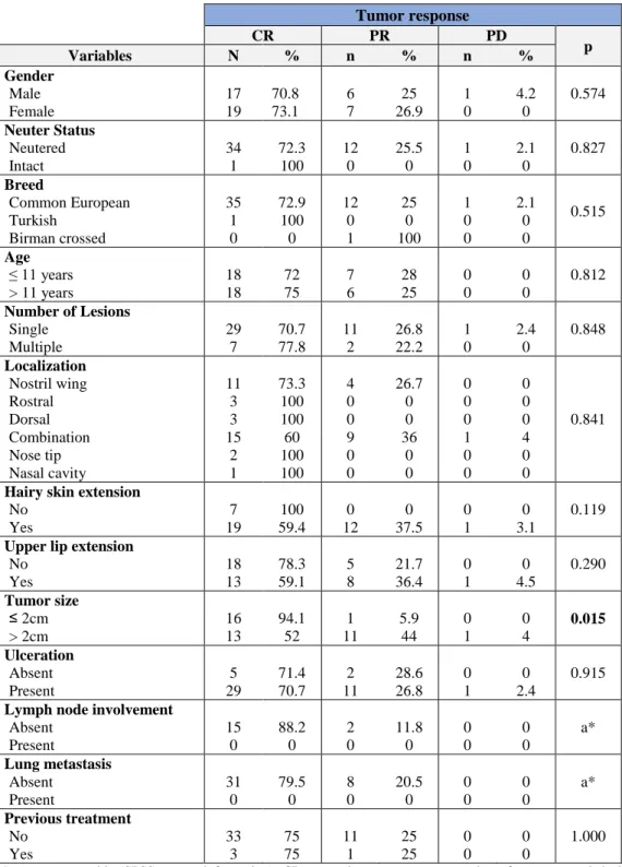
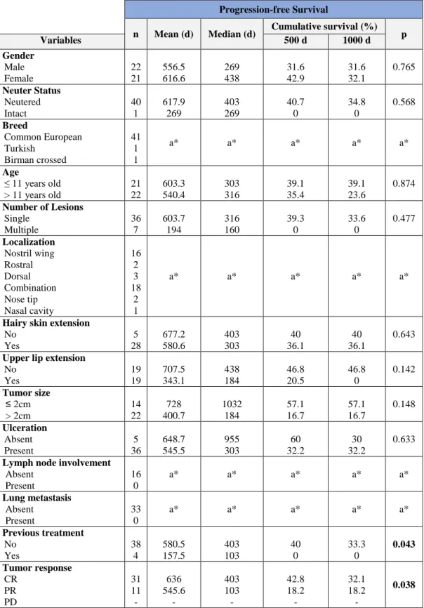
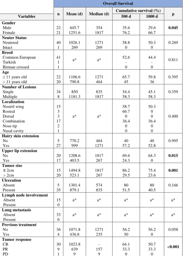
The total radiation dose delivered was 30 Gy, administered in five fractions of 6 Gy for a period of 3 to 4 days. Complete response was achieved in 72% (n=36) of the cats, partial response in 26% (n=13) and 2% (n=1) did not respond. Median PFS and OS times were 316 and 835 days, respectively. The gender of the cats treated with HDR brachytherapy affected OS time (p=0.045), with male cats living less. Tumor size showed a significant effect on tumor response (p=0.015) and on OS (p=0.001), so cats with tumors equal or smaller than 2 cm achieved better treatment responses and lived longer than cats with larger lesions. Results also reported that cats without tumor extension to the upper lip lived longer (p=0.015). Cats that received other treatment before HDR brachytherapy showed shorter PFS times. Finally, tumor response to HDR brachytherapy was found to be statistically associated with PFS (p=0.038)
X
and OS (p<0.001), so cats with better tumor responses showed better local tumor control times and lived longer.
In conclusion, lesions should be treated as soon as possible and can be achieved very good results with HDR brachytherapy in what concerns to PFS, OS and cosmetic outcomes.
XI
RESUMO
O carcinoma de células escamosas (SCC) é um dos tumores malignos mais comuns da pele dos gatos. A incidência é maior em gatos com pêlo de cor clara, falta de pigmento e áreas de pêlo escasso ou sem pêlo, e as orelhas, pálpebras, plano nasal e têmporas são as áreas mais afetadas, sendo mais comum nos gatos mais velhos. A exposição solar crónica é considerada o fator indutor da carcinogénese: induz alterações actínicas na pele que evoluem para lesões pré-neoplásicas, carcinoma in situ e SCC invasivo. O diagnóstico definitivo é feito por biópsia.
Como a taxa de metastização do SCC do plano nasal é muito baixa, e dado o seu caráter localmente invasivo, o principal objetivo do tratamento é o controlo local do tumor. Várias modalidades de tratamento são utilizadas no maneio deste tumor, mas com resultados pouco satisfatórios. A radioterapia externa tem sido usada nos últimos anos com melhores resultados nos tempos de recidiva e sobrevida global. A braquiterapia de alta dosagem (HDR) é um tratamento de radioterapia de contacto, com um curto período de tratamento global e um efeito poupador dos tecidos não neoplásicos.
Devido à falta de literatura sobre braquiterapia HDR no tratamento deste tumor, este estudo retrospetivo foi realizado para descobrir se existiam variáveis individuais e do tumor que pudessem ser fatores de prognóstico para a sobrevida livre de progressão (PFS) e sobrevida global (OS). Os dados recolhidos são referentes a 58 gatos com SCC do plano nasal tratados com braquiterapia HDR, e incluem: género, estado reprodutivo, raça, idade, número de lesões, localização e tamanho do tumor, extensão do tumor para além do plano nasal, ulceração, metástases ganglionares e pulmonares, qualquer tratamento prévio, resposta à braquiterapia HDR, recidiva e sobrevida global.
A dose total de radiação foi 30 Gy, administrada em cinco frações de 6 Gy, durante 3 a 4 dias. Resposta completa foi alcançada em 72% (n=36) dos gatos, resposta parcial em 26% (n=13) e 2% (n=1) não responderam. A média dos tempos de PFS e OS foram 316 e 835 dias, respetivamente. O sexo dos gatos tratados com braquiterapia HDR afetou o tempo de OS (p=0,045): os machos viveram menos tempo. O tamanho do tumor mostrou um efeito significativo na resposta ao tratamento (p=0,015) e na OS (p=0,001): gatos com tumores iguais ou menores que 2 cm responderam melhor ao tratamento e viveram mais tempo. Os gatos sem progressão tumoral para o lábio superior viveram mais tempo (p=0,015). Os gatos que receberam outro tratamento antes da braquiterapia HDR apresentaram tempos de PFS menores. Finalmente, a resposta do tumor à braquiterapia HDR estava estatisticamente associada à PFS
XII
(p=0,038) e OS (p<0,001): gatos que responderam melhor apresentaram melhores tempos de controlo local do tumor e viveram mais tempo.
Concluindo, as lesões devem ser tratadas o mais rápido possível e podem ser obtidos resultados muito bons com a braquiterapia HDR no que diz respeito à PFS, OS e resultados cosméticos.
Palavras-chave: gato, carcinoma de células escamosas, plano nasal, braquiterapia, fatores de prognóstico.
XIII
GENERAL INDEX
AKNOWLEDGEMENTS ... VII ABSTRACT ... IX RESUMO ... XI GENERAL INDEX ... XIII LIST OF FIGURES ... XV LIST OF TABLES ... XV LIST OF GRAPHS... XVII LIST OF ABBREVIATIONS, ACRONYMS AND SYMBOLS ... XIX
INTRODUCTION ... 1
1. Incidence and risk factors ... 1
2. Pathogenesis ... 2 3. Clinical evaluation ... 2 3.1. Diagnosis ... 2 3.2. Clinical Staging ... 4 4. Prevention ... 4 5. Treatment ... 5 5.1. Surgery ... 5 5.2. Cryosurgery ... 6 5.3. Photodynamic therapy ... 6 5.4. Cytotoxic chemotherapy ... 8 5.5. Electrochemotherapy ... 9 5.6. Cyclooxygenase-2 inhibitors ... 10 5.7. Radiotherapy ... 10
5.7.1. External beam Radiotherapy / Teletherapy ... 12
5.7.2. Strontium-90 plesiotherapy ... 13
5.7.3. High-dose rate brachytherapy ... 13
5.7.4. Clinical trials of RT in feline SCC of the nasal planum ... 14
OBJECTIVES ... 21
MATERIALS AND METHODS ... 23
1. Case selection ... 23
2. Data acquisition ... 23
XIV 4. Statistical analysis... 24 RESULTS ... 27 1. Population characteristics ... 27 1.1. Gender ... 27 1.2. Neuter status ... 27 1.3. Breed ... 27 1.4. Age ... 28 2. Tumor characteristics ... 28 2.1. Number of lesions ... 28
2.2. Localization of the tumor ... 29
2.3. Affection of surrounded areas ... 29
2.4. Tumor size ... 30
2.5. Tumor ulceration ... 31
3. Lymph node involvement ... 31
4. Lung metastasis ... 31
5. Tumor response to brachytherapy ... 32
6. Tumor recurrence ... 32
7. Associations between clinical variables and tumor response to brachytherapy ... 33
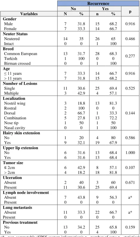
8. Associations between clinical variables and tumor recurrence ... 34
9. Survival analysis ... 35 9.1. Progression-free Survival ... 35 9.2. Overall Survival ... 37 DISCUSSION ... 43 CONCLUSION ... 49 REFERENCES ... 51
XV
LIST OF FIGURES
Figure 1 - WHO TNM staging system for skin tumors. ... 4
Figure 2 - Acute and late effects of radiotherapy to the skin. ... 12
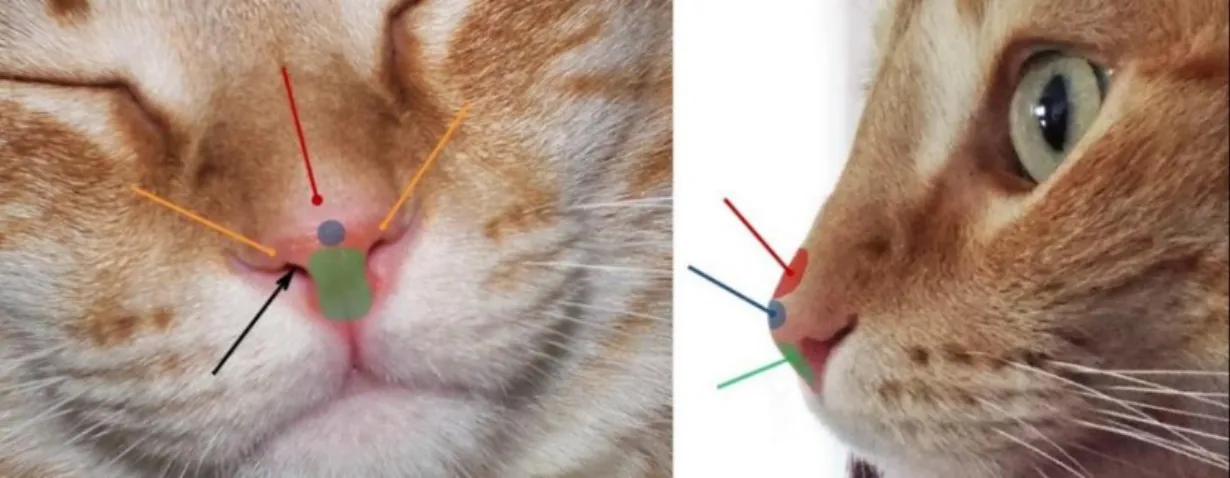
Figure 3 - Considered anatomical localizations of SCC of the nasal planum of the cats of the study ... 24
LIST OF TABLES
Table 1 - Clinical trials of RT in SCC of the nasal planum. ... 15Table 2 – Associations between clinical variables and tumor response to HDR brachytherapy. ... 33
Table 3 - Associations between clinical variables and tumor recurrence. ... 34
Table 4 - Relation between variables and progression-free survival at day 500 and 1000. ... 36
XVII
LIST OF GRAPHS
Graph 1 - Distribution of the population (n=58) by gender. ... 27
Graph 2 - Distribution of the population (n=56) by neuter status. ... 27
Graph 3 - Distribution of the population (n=58) by breed. ... 28
Graph 4 - Box and whisker plot of age. The central line corresponds to the mean age 11 years old. ... 28
Graph 5 - Distribution of the population (n=57) by number of lesions. ... 29
Graph 6 - Distribution of the population (n=56) by localization of the tumor... 29
Graph 7 - Distribution of the population (n=45) by extension of the tumor to hairy skin. ... 30
Graph 8 - Distribution of the population (n=52) by extension of the tumor to the upper lip. .. 30
Graph 9 - Distribution of the population (n=48) by tumor size ... 30
Graph 10 - Distribution of the population (n=55) by tumor ulceration. ... 31
Graph 11 - Distribution of the population (n=50) by the tumor response to brachytherapy.... 32
Graph 12 - Distribution of the population (n=43) by recurrence. ... 32
Graph 13 – Kaplan-Meier curve comparing progression-free survival of cats treated with HDR brachytherapy without (blue) and with (green) a previous treatment; p=0.043. ... 37
Graph 14 - Kaplan-Meier curve comparing progression-free survival of complete (blue) or partial (green) tumor response to HDR brachytherapy; p=0.038. ... 37
Graph 15 - Kaplan-Meier curve comparing overall survival of male (blue) and female (green) cats treated with HDR brachytherapy; p=0.045. ... 39
Graph 16 - Kaplan-Meier curve comparing overall survival of cats without (blue) and with (green) extension of the tumor from the nasal planum to the upper lip, treated with HDR brachytherapy; p=0.015. ... 40
Graph 17 - Kaplan-Meier curve comparing overall survival of cats treated with HDR brachytherapy with a tumor size equal or smaller than 2cm (blue) and larger than 2cm (green); p=0.001. ... 40
Graph 18 – Kaplan-Meier curve comparing overall survival of complete (blue), partial (green) or progressive (golden) tumor response to HDR brachytherapy; p<0.001. ... 41
XIX
LIST OF ABBREVIATIONS, ACRONYMS AND SYMBOLS
5-ALA – 5-aminolaevulinic acid AK – actinic keratosis
BISC –Bowenoid in situ carcinoma BNCT – boron neutron capture therapy
BPA-BNCT – Boronophenylalaninemediated boron neutron capture therapy CI – confidence interval
COX-2 – cyclooxygenase-2 CR – complete response CT – computed tomography DFI – disease free interval DFS – disease free survival DNA – deoxyribonucleic acid ECT – electrochemotherapy
FIV – feline immunodeficiency virus Gy – gray (unit)
HDR – high-dose rate LDR – low-dose rate LED – light-emiting diodes
MRI – magnetic resonance imaging
MSCCIS – multicentric squamous cell carcinoma in situ NR – no response
NSAIDs – nonsteroidal anti-inflammatory drugs OS – overall survival
PD – progressive disease PDT – photodynamic therapy PDR – pulsed-dose rate PF – proliferative fraction PFS – progression free survival PR – partial response
PV – papillomaviruses RT – radiotherapy
XX
SCC – squamous cell carcinoma Sr-90 – Strontium-90
TNM – tumor, nodes, metastasis TTP – time to progression TTR – time to recurrence UV – ultraviolet
VRTOG – veterinary radiation therapy oncology group WHO – World Health Organization
INTRODUCTION
1
INTRODUCTION
About 25% of cats’ tumors are skin and subcutaneous neoplasias, and more than 50% are malignant.1 Squamous cell carcinoma (SCC) is one of the most common malignant tumors of the skin of cats (about 15%). Around 10% of these skin SCC occur in a variant form known by multicentric squamous cell carcinoma in situ (MSCCIS) or Bowenoid in situ carcinoma (BISC) due to its histological similarity to human’s Bowen’s disease.2–4
1. Incidence and risk factors
The development of cutaneous SCC is strongly related to sunlight exposure: ultraviolet (UV) radiation, especially the B portion (UV-B), can cause skin damage if the fur or pigmentation do not grant an adequate protection to the skin by reflecting the light and preventing it to reach the skin surface.1,5–8 Therefore, higher incidence rates are associated to cats with light-colored fur, lack of protective pigment, and sparsely haired / hairless areas like the ears, eyelids, nasal planum and temples, which make these the most affected areas.2,8,9 Other environmental factors, such as the atmospheric conditions and the geographic location of the cats, contribute to the difference of incidence rates: cats inhabiting globe zones near to the equatorial line will be more exposed to the risk when compared to those living farthest, above or below the equator, as seen with humans in what concerns to skin cancer induced by sunlight.8
Squamous cell carcinoma of the nasal planum is relatively common in the cat.9 Cats with white fur have a risk to develop head and neck SCC 13.4 more times than cats with dark fur.10 Papillomavirus (PV) is also thought to be involved in the development of this tumor in cats, especially in BISC lesions.2,4,9,11
Older cats are the most affected by SCC, with a median age of 10 to 12 years. There is no sex or breed predisposition, although it is known that light-colored and short-haired breeds are more susceptible in contrast to long-haired breeds; also Siamese cats are considered naturally protected due to their pigmentation on the most affected areas.2
Cats infected with feline immunodeficiency virus (FIV) were thought to be more prone to the development of SCC, but there are studies reporting that their responses to radiotherapy do not differ from those of the non-infected cats, and times of tumor control and survival were not affected, so the FIV infection status should not be an hindrance when it comes to treatment.7,12
However, these cats are considered of higher risk to more severe radiation side effects, showing more exuberant reactions at the irradiated area.7
INTRODUCTION
2
2. Pathogenesis
Chronic exposure to UV-B radiation is considered the inducer of photocarcinogenesis, and later tumor progression is associated with individual characteristics.7 Solar dermatosis could be the first signal of a skin tumor development. Chronic solar radiation exposure induces actinic changes on the skin, beginning with a sunburn and evolving into pre-neoplastic lesions - actinic keratosis (AK), followed by carcinoma in situ (pre-invasive) with possible evolution to invasive SCC if the integrity of epidermal basal membrane is compromised.8,13 Different stages of evolution of the disease can be observed in the same area at the same time. Hammond et al.12 reported that 33% of the cats with nasal planum SCC under treatment developed new lesions. That is not surprising since all the body is exposed to the same amount of solar radiation and so it is possible the coexistence of lesions at different stages of development.
Bowenoid in situ carcinoma is manifested by multifocal superficial hyperpigmented and crusty plaques (with gradual thickening and posterior ulceration14) in any part of the cat’s body,
without any relation to the amount of fur nor his color, suggesting that it has no relation with UV radiation exposure.2,14,15 This disease is usually benign (although it may progress to invasive SCC) and some cases are linked to PV infection.14 According to Munday et al.11 BISC can be a neoplastic transformation of a viral plaque induced by PV; in their study PV DNA and increased p16 protein were detected within the SCCs protected, in contrast with UV-exposed SCCs since these presumably develop from actinic keratosis lesions.
3. Clinical evaluation
3.1. Diagnosis
In case of solar induced SCC, numerous lesions can be detected coexisting at the same time, although at different stages: solar dermatitis, pre-cancerous lesions (actinic keratosis), carcinoma in situ and invasive SCC. In case of actinic keratosis, changes are consistent with chronic solar exposure: solar elastosis, skin thickening plaque-like, epithelial desquamation and curling in the specific case of the ears.8,16
Cats with SCC of nasal planum usually attend to the consultation because the owner noticed a lesion that is not healing, and less often due to actinic changes. SCC can appear as a single lesion or as multiple lesions, generally in the format of a superficial erosion, then an ulcer and, in more advanced stages, invasive crateriform lesions, preceded by a protracted course. They can have a plaque form, papillary form or fungiform. They can also be nodular or proliferative,
INTRODUCTION
3
and erythema is often observed, as well as scales, scabs and or swelling. 2,5,17–22 Depending on the extension of the mass, serous, mucoid or purulent discharge, epistaxis, sneezing, dyspnea, stertorous breathing and facial deformation can be observed in addition.17,21
Anamnesis should be performed in order to know the complete clinical history. Information about date of first identification of the lesion, current size, rate of growth and presence of adherences and ulceration must be collected.5 Additionally, during physical examination the regional lymph nodes should be examined in order to detect any alteration. Cytology of regional lymph nodes is also recommended in case of enlargement or painful on palpation.
Radiography of the nose, under anesthesia, can be used to identify characteristics already known as been related to malignancy, such as tissue opacification and changes compatible with bone lysis. However, x-ray has low specificity and sensibility, and so computed tomography (CT) is the imaging test of choice to evaluate the extension of the tumor.17 In any case, diagnosis should never be presumptive based on the CT images since there are evidences that nasal neoplasia and rhinitis share CT characteristics that prevent imaging distinction.23
Magnetic resonance imaging (MRI) could help to better analyze a possible brain injury due to extension of the tumor when the mass crosses the cribriform plate.17
Definitive diagnosis of SCC is achieved with biopsy (either multiple punch biopsy or incisional or excisional biopsies) as histopathology is fundamental to evaluate the behavior of the tumor, the degree of differentiation, cellular morphology and vessel invasion.1,2,5 Additionally, with the assessment of regional and distance metastases, it is possible to establish the clinical stage, recommend the most adequate treatment protocol and predict prognosis. Cytology with fine needle aspiration is not the most adequate methodology for diagnosis due to the superficial nature of the majority of nasal planum SCC, and also due to the inflammation and bleeding commonly associated.2 Although it is easy and brief, cytology identification of the cell type is possible but it is not diagnostic1,17. So, to avoid inaccuracy, in cases where only cytology is allowed, the tumor should not be fit in a T stage just by visual measurement of the lesion and assumption of the invasiveness.
Although the TNM classification system proposed by the World Health Organization (WHO)24 (Figure 1) dates back to 1980, it is still used and referenced in recent publications2,5,25.
INTRODUCTION
4
3.2.
Clinical Staging
Primary tumor assessmentLesions should be direct measured at their larger diameter. However this could be hard for larger or more invasive lesions, and so the measurement and extension assessment should be done with the help of a CT scan.5
Regional lymph nodes assessment
Regional extension should be assessed by cytology after a fine needle aspiration of the lymph nodes, even if they do not seem enlarged or reactional on palpation.1 If the results of cytology
are unreliable or shady, an histology of the lymph node by biopsy should be carried out to clear the doubts.5
Distant metastasis assessment
The distant extension of a tumoral process should be done when in presence of a malignant neoplasia to detect metastasis. Chest radiographs in three views are the most used, but a CT scan could also be done in place of the first. 2,5
In the original WHO publication24 a stage grouping was not specified. With all the modifications and adaptations from the original, and as regional and distant metastases are very rarely observed, SCC of the nasal planum is commonly staged based on tumor size and invasiveness degree, considering just the T letter from the assessment of the primary tumor (stage Tis and stage T1 to T4).7,12,15,26–29
4. Prevention
Solar induced SCC can be prevented through owner’s education. Light-colored and short-haired cats should avoid sunlight exposure, at least in the hours of the highest incidence of radiation.1,2,9,10,20,30 If it is an indoor cat that could be easier to control, even with UV light
blocking films for windows.2
Figure 1 - WHO TNM staging system for skin
INTRODUCTION
5
Topical sunscreens rarely help when in the nasal planum, as it is quite impossible to no be licked off.9,10,26
Tattooing to add protective pigment to the skin is not proven to be effective, since the tattoo is applied to the dermis, and the SCC arise from the squamous epithelium of the epidermis.2,9,10,30 Thomson30 suggests that henna tattoos could protect from some of the solar damage as they are applied superficially to the epidermis in despite of the need to reapplications. Vitamin A synthetic derivatives like 13-cis-retinoic acid and etretinate were used to increase epithelial differentiation, and it was concluded that these substances can help managing preneoplastic lesions, controlling their progression.9,10
5. Treatment
For a long time in veterinary medicine, anticancer treatment was practically based on surgery, and only over the last decades, new protocols, techniques and methodologies were extrapolated from the human medicine and started to be applied.31
The main goal of treating nasal planum SCC is to achieve the best local control of the tumor, given its locally invasive character and low metastatic rate.17 In this regard, treatment approach is usually chosen according to the tumor clinical stage.2 After a complete staging of the disease the therapeutic strategy could be established, and decided of what intent would it be: curative, adjuvant or palliative.31
5.1. Surgery
Surgical excision, if feasible should be the first approach to cutaneous SCC.5 However, in tumors of the nasal planum, due to the complex anatomical localization and depending on the level of invasion, performing a complete tumor excision with proper margins (at least 5 mm ideally) could be difficult.17,30 For early stages and superficial lesions of nasal planum SCC surgery alone could be curative if a complete excision is achieved, with better tumor control times than if first treated with external beam radiotherapy.10,20 Nonetheless, for advanced tumor
stages surgery is also the treatment of choice - a complete resection / planectomy in this case, to have a chance of totally remove the tumor.2,10,22
In a study published in 1990 by Withrow et al.32 nine cats with nasal tumors were submitted to a complete excision of the nasal planum, and even so, in three cats with SCC the tumor relapsed; the other remained tumor-free till the end of follow-up. In another study10, in cats that
INTRODUCTION
6
underwent a complete resection of the nasal planum, recurrence was not observed; however, in 29% of the cats with incompletely resected nasal planum, recurrence was observed.
Nasal planum resection has good prognosis and has proved to be functional in cats, with relative good esthetic results, yet sometimes challenging to the owner’s acceptance. Surgery also allows histopathological analysis of the margins, does not need special facilities (like for radiotherapy), and the total treatment time is very short.2,10
Taking all the above into account, and knowing that nasal planum SCC could be successfully treated with radiation therapy and with better cosmetic outcomes, surgery is not always considered.5
5.2. Cryosurgery
Cryosurgery is an alternative treatment for small SCC lesions, and it consists on freezing and thawing the abnormal tissue to destroy it, by the local application of a cryogen (liquid nitrogen or nitrous oxide).2,30 Rapid freezing to -20°C is used to form intracellular and
extracellular small ice crystals that will damage cell membranes, followed by a slow thawing that causes the formation of bigger crystals by the recrystallization of the smaller ones, maximizing the tissue fragmentation.30,31 Three cycles of consecutive freezing-defreezing should be performed and a safety margin of 10mm should be included in the treatment field.30,31 For bigger lesions (greater than 5mm) is difficult to ensure that the entire tumor is destroyed and the margins are clear, and for this reason local recurrence rates are high (73% of the cats in one study recurred, with a median time to recurrence (TTR) of 184 days).30 In another study, SCC on the pinnae and eyelids responded better to cryosurgery than those of the nasal planum, and for the last ones cryosurgery was not enough, with a median TTR of 254 days.2 In conclusion, cryosurgery is cost-effective and readily accessible but has shorter tumor control times when compared to surgery or radiotherapy.20
5.3. Photodynamic therapy
Photodynamic therapy (PDT) consists in the intravenous, topical or oral administration of a photodynamic agent (photosensitizer) for which tumor cells are the target by selective accumulation, and later the lesion undergoes a specific wavelength light to activate the photosensitizer that induces cell death through oxygen and other free radicals production, as well as tumor vasculature damage; it is performed under anesthesia and analgesia.2,33–35 Is has
INTRODUCTION
7
been used in human medicine as a recognized treatment but in veterinary medicine so far it is not well established, but the major applicability it has been for feline SCC.34,35 PDT is considered effective as a single modality treatment for early stages of SCC because results are conditioned by the depth of penetration of both photosensitizer and light (maximum depth of 1.5 cm).2,34,35
Although the good esthetic outcome for cats’ nasal planum and the sparing effect of the normal tissue there could be minor adverse effects, such as acute erythema, and edema, and increased heart rates associated with injection site pain, even with analgesia and anesthesia, already reported in human patients that underwent PDT with meta-(Tetrahydroxyphenyl)Chlorin (m-THPC; Foscan®).2,34,36 Acute effects can be observed during and immediately after the treatment and are usually solved in 3 to 5 days, except in the cases of facial edema outside the treatment area that can cause dyspnea and respiratory stertors, taking longer to heal.36 A scab develops on the treated area and will last for a few weeks; tissue necrosis
following PDT is not very common and rarely ocurrs.34–36 Additional care must be taken to
avoid direct sunlight exposure after treatment with an intravenous photosensitizer (due to generalized light sensitivity), an Elizabethan-collar must be used to avoid post treatment self-trauma, and also provide supportive therapy to avoid or control secondary infections of the treated field.35–37
PDT can be performed with different photosensitizers and light sources.33 In a study by Magne et al.36 PDT was performed with an intravenous photosensitizer (pyropheophorbide-alpha-hexyl-ether) and a laser light source in feline facial SCC: 49% (30/61) achieved a complete response (CR), 12% (7/61) a partial response (PR) and 39% (24/61) did not respond, with a 1-year local control rate of 61.7%; adverse effects were minimal and chronic alopecia was reported in all the cats. In this same study from 1997 was concluded that response and local control rates were significantly associated with tumor stage, proving once again the effectiveness of PDT for superficial SCC.
Later, in 2001, Stell et al.38 performed PDT with a topical (cream) photosensitizer (5-aminolaevulinic acid, 5-ALA) and a light-emitting diodes (LED) light source with CR in 9/10 cats with nasal planum SCC, with a complete response rate of 85% for Tis and T1 lesions with
a single treatment. Hematological and hepatotoxicity associated with 5-ALA were avoided by its administration topically instead of intravenously; only discomfort on the tumor site was observed and managed with a lidocaine/prilocaine cream and opioids.35
INTRODUCTION
8
In 2007 Buchholz et al.34 used a laser light source with an intravenous newly developed liposomal photosensitizer (new liposomal formulation of m-THPC) that improves the tropism to the tumor cells sparing the normal ones, and decreases the generalized light sensitivity time. This study revealed to be well tolerated and very effective with 100% of CR (n=20), recurrence of 20% and a 1-year local control rate of 75%, greater than the one reported by Magne et al.36.
With the same protocol used by Stell et al., in 2008 Bexfield et al.33 performed PDT in a larger group of cats with nasal planum SCC. A clinical benefit was observed in 96% (53/55) of the cats, with 85% (47/55) achieving a CR; 51% recurred with median disease free survival (DFS) of 157 days.
One year after, Ferreira et al.37 administered an intravenous photosensitizer (hematoporphyrin) and used a LED light source to treat highly invasive as well as non-infiltrative nose and nasal planum SCC in cats. The first ones did not respond to two applications of PDT, and clinical response was only achieved after surgical excision of the tumor; the second ones responded with PR to one application of PDT and CR to two applications. The use of this photosensitizer allows local tumor control to early stages of the disease and can also be used with surgery for more advanced stages of SCC to reduce recurrence times.
In conclusion, PDT can be an alternative to other modalities on the management of nasal planum SCC in cats, despite the still relative short local tumor control after a good response to a single treatment, which can be improved with the development of more selective photosensitizers.2,33,34 While awaiting more efficient drugs PDT can be considered in a multimodal therapeutic approach.37 However, PDT in pets turns out very expensive and is only performed in a few institutions, and this is a reason why this treatment approach is still unknown to many veterinarians.35
5.4. Cytotoxic chemotherapy
The information available about the use of cytotoxic drugs in nasal planum SCC of the cats is limited, and relies mostly on intralesional chemotherapy and electrochemotherapy. Performing a chemotherapy is a more invasive treatment approach than the conservative options such as radiation or photodynamic therapy, but the cost is smaller, it does not require specific facilities nor expensive equipment, and it is more easily accessible.
Intratumoral chemotherapy consists in the deeper infiltration of a cytotoxic drug right in the neoplastic tissue and in the surrounding healthy tissues, allowing an increased drug
INTRODUCTION
9
concentration in the tumor.39,40 Carboplatin started to be used for intratumoral treatments in cats after therapeutic gains were observed with cisplatin in other species; knowing the pulmonary and nephrotoxicity of this drug in cats, carboplatin is the less toxic alternative drug from the same group that also presents activity against carcinomas.40,41 In 1996, Théon et al.40 diluted carboplatin in purified sesame seed oil to act as a slow-release formulation by limiting carboplatin release from the injection point and therefore limiting the systemic absorption of the drug, avoiding general adverse effects. In this study administrations were performed weekly, and this treatment showed to be effective since CR was achieved in 73.3% (11/15) of the cats, with a progression-free survival (PFS) of 16 months and 1-year survival rate of 55.1%. The study published by de Vos et al.39 in 2004 (discussed in section 5.7.4.) combined superficial x-rays RT with intralesional administration of carboplatin to treat feline SCC of the nasal planum; here carboplatin was used as a radiopotentiating agent for radio-chemotherapy which has more power administered intratumoral than intravenously. Treatment was well tolerated and all the cats of the study achieved CR, a promising result considering the advanced stage of the tumors treated.
Individual protection equipment should be used when performing injection of intratumoral carboplatin due to the hazard of aerosol formation if it sprays back from the needle to the air or surrounding surfaces.2,39 A leakage of the cytotoxic from the tumor is also possible to happen and it should be cleaned with absorbent paper.39,40
Intravenous mitoxantrone is also mentioned in the literature for malignant tumors of the cats but with a low response rate (4/32 cats).2
5.5. Electrochemotherapy
Electrochemotherapy (ECT) consists in the administration of a cytotoxic drug (like bleomycin or cisplatin) in association with high-voltage electric pulses that stimulates the drugs to enter into the tumor cells changing their permeability.25,42
Bleomycin is an antibiotic with cytotoxic properties that is indicated in the treatment of SCC.41 ECT with intratumoral bleomycin is reported in 2009 by Spugnini et al.42 as a safe and effective treatment modality for feline SCC. After the tumor and margins injection with bleomycin, biphasic electric pulses were delivered by electrodes. After two sessions of ECT with 1 week interval, of the 7 cats with nasal planum tumor, 6 achieved CR and 1 PR, and 4 were still in remission at the end of follow-up.
INTRODUCTION
10
In 2014, Tozon et al.25 published a study of electrochemotherapy with intravenous bleomycin with 87.5% of CR, with recurrence in one of 5 cats with nasal planum SCC. The cosmetic effect is also very good and much more well accepted when compared to surgical resection of the nasal planum, and there are almost none adverse effects, reasons why the ECT could also be considered to manage these lesions.25
5.6. Cyclooxygenase-2 inhibitors
It is still unknown the role of Cyclooxygenase-2 (COX-2) inhibitors on the prevention and management of SCC of the nasal planum, and further investigation is needed.
COX enzymes has been associated with neoplastic formation and tumor growth, and while isoenzyme COX-1 is involved in physiologic processes, the isoenzyme COX-2 can be induced to produce prostaglandins which affect pathologic processes (inflammation, wound healing and tumor development).2,43,44 COX-2 was the target of the studies since it is overexpressed in
humans with SCC UV-light induced, AK in human is treated with COX-2 inhibitors, the risk of developing AK lesions or even an epithelial tumor is lower in humans treated with non-steroidal anti-inflammatory drugs (NSAIDs) COX-2 specific, and COX-2 was detected in canine SCC.2,43,44 Beam et al.43 observed that COX-2 was not detected for none of the cutaneous SCC in cats, suggesting a species difference of COX-2 expression in canine and feline neoplasia or a feline COX-2 concentration lower than the immunohistochemistry detection level. Based on this, their conclusion was that COX-2 inhibitors would not have an anticancer activity for cats.
In contrast, in a study performed by Bardagí et al.44 in 2012, it is demonstrated a strong COX-2 expression in feline AK lesions, SCC and inflammatory dermatosis, raising again the issue of the use of COX-2 inhibitors in these cases.
5.7. Radiotherapy
Ionizing radiation is used in anticancer therapy through two major types: photon radiation (x-rays and gamma rays) and particle radiation (electrons and protons).31,45 Radiation acts on
the surface or in depth, inducing direct damage to cell DNA or to biological molecules, consequently affecting cellular division, growth and lesion repair capacity; this way, radiation can be used to kill neoplastic cells and reduce tumors’ volume.28,31,45 Radiotherapy (RT) is a
INTRODUCTION
11
local treatment without systemic toxicity and the possible side effects, both acute or late, only affect the irradiated field.46
The intent of RT could be definitive / curative if there is a high possibility of a long-term tumor control, adjuvant when used in association with other treatment modalities (as surgery or chemotherapy), or palliative in case of advanced disease, when survival maintenance is hopeless but an improvement in cat’s quality of life can be achieved.31,45 Nasal carcinomas in
cats are expected to have a good response to hypofractionated radiotherapy for both curative and palliative intents.47
Radiotherapy is considered nowadays as the first choice option for radiosensitive tumors, for those difficult to surgically resect and to use in cases of recurrence.31,48 However, due to the need of special facilities, trained technicians and elevated costs, radiotherapy is not widely used. The ionizing radiation can be administered by distinct methods classified in: external beam RT or teletherapy when the radiation source is distanced from the treating field (generally using X rays, Cobalt-60 or electrons); brachytherapy or curietherapy when the radiation is delivered through a sealed source placed directly in the tumor - on the surface or interstitially (using Cobalt-60, Cesium-137, Strontium-90 or Iridium-192); plesiotherapy, a kind of brachytherapy, that uses a Strontium-90 (Sr-90) applicator to deliver low-energy radiation; and systemic or intracavitary radiotherapy when radioisotopes, or radioisotope molecules, are administered through a non-sealed source (orally or injected), being delivered to the target tissue by own physiological processes (Iodine-131and Samarium-153).28,31,45–47,49
Radiotoxicity is related to the radiosensitivity of the irradiated tissues (that is proportional to the mitotic index), and so, depending on the tissue response to radiation, acute or late effects can be observed.31 The skin is considered a radiosensitive tissue with fast proliferating cells and therefore acute reactions are more likely to occur.31,46 Acute effects occur during the treatment course or within a few days and they are usually self-limiting or completely solved with supportive treatment in two to four weeks; late effects could be observed several weeks or months, or even years, after the treatment, and are usually irreversible.45,50 Acute and late effects of RT on the skin are mentioned in Figure 2, adapted from the toxicity criteria of the Veterinary Radiation Therapy Oncology Group (VRTOG)51.
INTRODUCTION
12
5.7.1. External beam Radiotherapy / Teletherapy
External beam RT is the most used method of RT in veterinary medicine, adequate to treat superficial and invasive SCC lesions on the nasal planum, using different amounts of energy: the higher the energy, the deeper the radiation penetrates the tissues. Therefore, low penetrating beams (gamma rays from cobalt units) are preferred for superficial tumors, as well as electron beams (from linear accelerators) because of their short penetrating distance despite their high-energy, and high penetrating beams (x-rays from linear accelerators) are preferred for deeper located tumors, or for those with a big volume. This is important when thinking of sparing the non-neoplastic underlying tissues, reducing the occurrence of side effects.46
Full-course protocols of external beam RT are usually performed over three to four weeks, during which multiple fractions of radiation are given, for both superficial and deep lesions. The literature reports that tumors with small volume respond better to external RT; in a study is also concluded that there is no difference between tumor stages T2, T3 and T4 in what concerns to response to radiotherapy.7 Other survival data will be developed in section 5.7.4.,
where clinical trials of RT in feline SCC of the nasal planum are discussed.
Although the reports of numerous protocols, the outcomes are similar among them: the cosmetic results are good when compared to surgery, recurrence rate is high and too many anesthesias are needed.
INTRODUCTION
13
5.7.2. Strontium-90 plesiotherapy
Plesiotherapy consists in the direct application of Sr-90 on the surface of the lesion with an ophthalmic probe. This method showed efficacy in the management of ocular SCC in humans.26 This treatment modality is adequate for superficial lesions because the dose decreases with depth (only less than 10% of the maximal dose delivered at the surface will penetrate to a depth of 3mm) and therefore, lesions with more than 2mm should not be considered for this modality.2,10,12,26,52 Van Vechten et al.52 (abstract from 1993), Goodfellow et al.26 (2006) and Hammond et al.12 (2007) reported the successful use of Sr-90 plesiotherapy in cats with nasal planum SCC with, in the last two publications, 13/15 cats and 43/49 cats achieving CR; more results on this topic are also presented in clinical trials’ section (5.7.4.).
5.7.3. High-dose rate brachytherapy
Brachytherapy is a method of delivering radiation at short distance, through a sealed source (radioactive seeds or wires) placed directly in the tumor - interstitially, temporarily or permanently, delivering radiation continuously as the source deteriorates.46,47,49,53
Small size and well delimited tumors, tumors non-amenable for surgical excision and tumors with high recurrence rates can benefit from brachytherapy.54
Iridium-192 was the most used radioisotope in low-dose rate (LDR) brachytherapy (dose rate 0.4 - 2 Gy/h), producing gamma rays by its radioactive decay and with a half-life of 74.2 days which allowed its reuse, making treatment more economical.53–56 In this treatment modality, Iridium-192 seeds sealed with a thick stainless steel capsule and placed in a flexible polyethylene were implanted into the tumoral tissue; more than one seed could be sealed in one tube forming wires that were implanted through the tumor. 54,55 The total dose administered was calculated by the product of dose rate and time the seed was in place (the longer the time, the higher the total dose).55 Therefore, the radioisotope was implanted for the time needed to achieve the prescribed dose.54 This treatment was advantageous in comparison to external RT because higher doses of radiation could be achieved within the tumor without minimal effects to the non-neoplastic surrounding tissues, as the dose rate exponentially decreased with distance from the source.49,55,57 Secondary effects within the treated area with LDR brachytherapy were minimal (dermatitis sunburn-like and loss of pigmentation, mainly) and occurred during the first weeks after treatment.53 Due to the radioactive hazard when performing Iridium-192 LDR brachytherapy, radioprotection should not be neglected. The therapy room should be duly equipped and constructed according to legislation; to minimize direct contact with the
INTRODUCTION
14
radioactive source the manipulator should use adequate clamps and all the personal involved should use a radiation monitoring dosimeter; the animals should ideally stay in a separate room with leaded walls during the treatment time.53
Since 2014, in France, it is impossible to use LDR brachytherapy with Iridium-192 wires due to the end of its commercialization, and so after-loading machines (curietherapy systems) are the alternative, offering two treatment possibilities based on the dose rate: pulsed-dose rate (PDR) and high-dose rate (HDR).58–61 To keep performing interstitial brachytherapy, the PDR was the only option, but in the case of skin and even more the nasal planum, the thickness is not enough to implantation, and so HDR brachytherapy was developed to irradiate superficial lesions in a fast treatment modality (dose rate over 12 Gy/h).56,58
In HDR brachytherapy vectors are responsible for delivering a radioisotope, from a remote radioisotope delivery unit computer-controlled, directly to the neoplastic tissue, which allows a sparing effect on the surrounding normal tissues and none dissipation of radiation to the room and, mostly, to the technicians, although a proper shielded room is not dismissed.61 To perform
this superficial irradiation, molds or applicators could be specifically made, in wax for example, to help positioning the vectors.58
In contrast to LDR, HDR brachytherapy have more severe side effects which is not surprising due to the elevated doses of radiation: local irritation, ulcers, osteonecrosis, mucositis radiation related, and possible irreversible depigmentation, sneezing and swelling.
HDR brachytherapy is a fast therapeutic modality and can be completed in a period less than a week with an overall treatment time very short.
The literature on this topic is very poor, even more when it comes to the use of HDR brachytherapy in feline SCC of the nasal planum, without any published studies.
5.7.4. Clinical trials of RT in feline SCC of the nasal planum
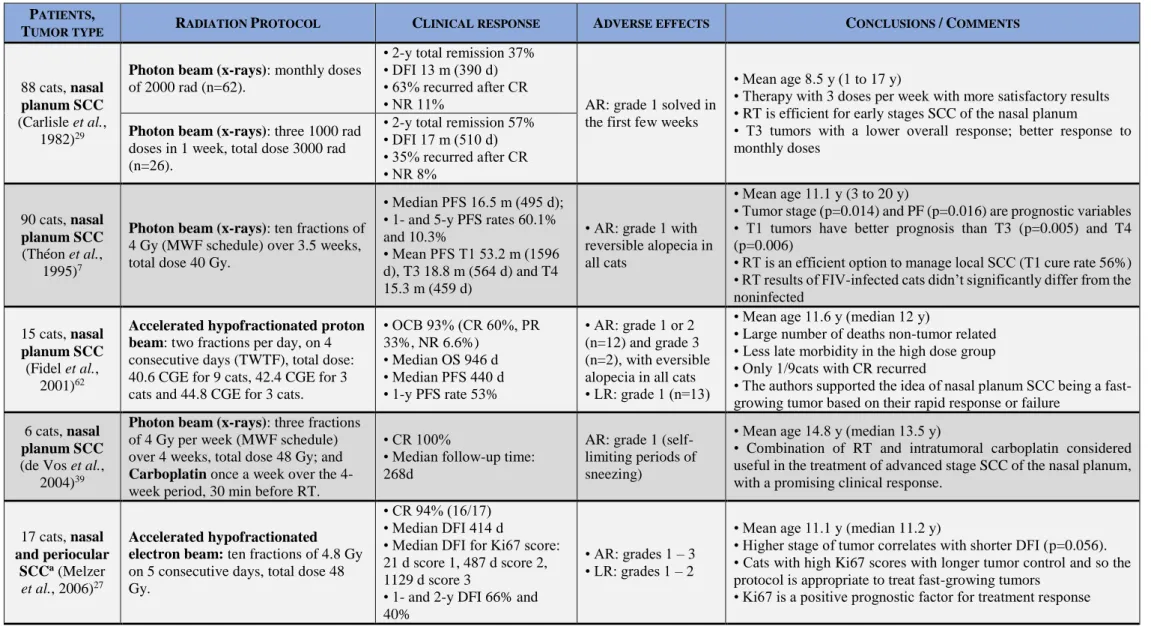
Radiation therapy has been used over the past years in cats with the purpose of treating SCC of the nasal planum. So far, to the author's knowledge, there are few publications of clinical trials studying the clinical outcome and possible prognostic factors associated with the use of RT in the treatment of this tumor. Table 1 summarizes the publications reviewed.
15
1
5
Table 1 - Clinical trials of RT in SCC of the nasal planum. PATIENTS,
TUMOR TYPE RADIATION PROTOCOL CLINICAL RESPONSE ADVERSE EFFECTS CONCLUSIONS /COMMENTS
88 cats, nasal
planum SCC
(Carlisle et al., 1982)29
Photon beam (x-rays): monthly doses
of 2000 rad (n=62).
• 2-y total remission 37% • DFI 13 m (390 d) • 63% recurred after CR
• NR 11% AR: grade 1 solved in the first few weeks
• Mean age 8.5 y (1 to 17 y)
• Therapy with 3 doses per week with more satisfactory results • RT is efficient for early stages SCC of the nasal planum
• T3 tumors with a lower overall response; better response to monthly doses
Photon beam (x-rays): three 1000 rad
doses in 1 week, total dose 3000 rad (n=26).
• 2-y total remission 57% • DFI 17 m (510 d) • 35% recurred after CR • NR 8% 90 cats, nasal planum SCC (Théon et al., 1995)7
Photon beam (x-rays): ten fractions of
4 Gy (MWF schedule) over 3.5 weeks, total dose 40 Gy.
• Median PFS 16.5 m (495 d); • 1- and 5-y PFS rates 60.1% and 10.3%
• Mean PFS T1 53.2 m (1596 d), T3 18.8 m (564 d) and T4 15.3 m (459 d)
• AR: grade 1 with reversible alopecia in all cats
• Mean age 11.1 y (3 to 20 y)
• Tumor stage (p=0.014) and PF (p=0.016) are prognostic variables • T1 tumors have better prognosis than T3 (p=0.005) and T4 (p=0.006)
• RT is an efficient option to manage local SCC(T1 cure rate 56%) • RT results of FIV-infected cats didn’t significantly differ from the noninfected
15 cats, nasal
planum SCC
(Fidel et al., 2001)62
Accelerated hypofractionated proton beam: two fractions per day, on 4
consecutive days (TWTF), total dose: 40.6 CGE for 9 cats, 42.4 CGE for 3 cats and 44.8 CGE for 3 cats.
• OCB 93% (CR 60%, PR 33%, NR 6.6%) • Median OS 946 d • Median PFS 440 d • 1-y PFS rate 53% • AR: grade 1 or 2 (n=12) and grade 3 (n=2), with eversible alopecia in all cats • LR: grade 1 (n=13)
• Mean age 11.6 y (median 12 y)
• Large number of deaths non-tumor related • Less late morbidity in the high dose group • Only 1/9cats with CR recurred
• The authors supported the idea of nasal planum SCC being a fast-growing tumor based on their rapid response or failure
6 cats, nasal
planum SCC
(de Vos et al., 2004)39
Photon beam (x-rays): three fractions
of 4 Gy per week (MWF schedule) over 4 weeks, total dose 48 Gy; and
Carboplatin once a week over the
4-week period, 30 min before RT.
• CR 100%
• Median follow-up time: 268d
AR: grade 1 (self-limiting periods of sneezing)
• Mean age 14.8 y (median 13.5 y)
• Combination of RT and intratumoral carboplatin considered useful in the treatment of advanced stage SCC of the nasal planum, with a promising clinical response.
17 cats, nasal
and periocular SCCa (Melzer
et al., 2006)27
Accelerated hypofractionated electron beam: ten fractions of 4.8 Gy
on 5 consecutive days, total dose 48 Gy.
• CR 94% (16/17) • Median DFI 414 d
• Median DFI for Ki67 score: 21 d score 1, 487 d score 2, 1129 d score 3
• 1- and 2-y DFI 66% and 40%
• AR: grades 1 – 3 • LR: grades 1 – 2
• Mean age 11.1 y (median 11.2 y)
• Higher stage of tumor correlates with shorter DFI (p=0.056). • Cats with high Ki67 scores with longer tumor control and so the protocol is appropriate to treat fast-growing tumors
16 16 Table 1 – continuation 15 cats, nasal planum SCC (Goodfellow et al., 2006)26
Strontium-90 plesiotherapy: five
fractions of 10 Gy delivered at a depth of 2 mm over a 10-day period (MWF schedule), total dose 50 Gy.
• OCB 100% (CR 87%, PR 13%)
• Median OS 780 d
• 1-, 2- and 5-y OS rates 77%, 51% and 15%
• Median DFI 652 d
AR: grade 1 and long term alopecia
• Mean and median age 12 y
• All cats with CR remained disease-free for the duration of the study
• In the authors opinion Sr-90 therapy is simple, safe, with good outcome and cosmetic results, well tolerated.
49 cats, nasal
planum SCC
(Hammond et
al., 2007)12
Strontium-90 plesiotherapy: single
high-dose fraction of one or multiple overlapping site applications of a Sr-90 ophthalmic applicator (median dose 128 Gy, 1 to 6 applications).
• OCB 98% (CR 88%, PR 10%)
• Median OS >8 y (>2920 d) • 1-, 2- and 5-y OS rates 98%, 92% and 55%
• Median PFS 4.7 y (1715 d)
AR: 1 solved in the first few weeks
• Mean and median age 12 y (4 to 16 y) • All SCC in situ had CR without recurrence
• Cats with CR had a longer OS (p<0.001) than cats with PR • Advantageous when compared to surgery or external beam RT
3 cats, nasal
planum SCC
(Trivillin et al., 2008)63
BNCT: administration of 300mg kg-1
of 10BPA IV and irradiation with
thermalized epithermal neutron beam 3h after (average flux 3.4 ±0.3x10⁸
neutrons cm-2 s-1 over 40 min, fluence
oh thermal neutrons 8.2x10¹¹ neutrons
cm-2).
PR in all cats
AR: grade 1 for skin and grade 2 for mucous membrane
• Treatment improved cats’ clinical condition
• BPA-BNCT can induce partial tumor control, reduction in tumor volume and radiation-induced tumor cell damage non-affecting normal tissue.
• BPA-BNCT may not be adequate for smaller and early stage SCC
15 cats, facial
SCC (Cunha et
al., 2010)15
Hypofractionated electron beam:
four fractions of 7.6 or 10 Gy each, with 1 week interval, total dose 30.4 or 40 Gy.
SCC nasal planum: 8/15 cats, with CR 50% (n=4) and NR 50% (n=4)b
For all 15 cats: mean OS 224 d; mean DFI 271 d AR: grade 1 (n=1) and grade 3 (n=1) resolved with supportive treatment. • Mean age 11 y (7 to 17 y)
• Response rates were lower than the previous published, possibly because of the advanced stage lesions (60% were T3 and T4) • The authors concluded that modifications to the protocol are needed to improve treatment responses
Abbreviations: AR acute reactions; BNCT boron neutron capture therapy; 10BPA 10B-boronophenylalanine; CGE cobalt gray equivalents; CR complete response; d days; DFI disease free
interval; FIV feline immunodeficiency virus h hours; IV intravenous; LR late reactions; m months; min minutes; MWF monday/wednesday/friday; n number of cats; NR no response; OCB overall clinical benefit; OS overall survival; p statistical significance; PF proliferative fraction; PFS progression-free survival; PR partial response; RT radiotherapy; SCC squamous cell carcinoma; Sr-90 Strontium-90; TTR time to recurrence; TWTF tuesday, wednesday, thursday, Friday; y years. Notes: a - Of the 17 cats, 12 had nasal planum SCC, and the clinical response data is for all the 17 cats, no differentiating them. b - Only 8 cats had nasal planum SCC and the survival analysis was not available to this localization alone to easy comparison with the other studies.
INTRODUCTION
17
In 1982, Carlisle et al.29 studied the response of a population of 88 cats with SCC of the nose to RT with x-rays in two regimens: a monthly irradiation of 2000 rad dose each time with the total dose varying with the healing state, and a one week treatment of 3 irradiations of 1000 rad, with a total dose of 3000 rad. Total remission was achieved with both protocols in more than 85% of the cats despite the later recurrence of some of them. RT with 3 doses in one week had the best results with 57% of the cats in total remission for 2 years, compared with the 37% achieved with the other regimen, supporting the advantages of dose fractionation. Tumors staged T1 showed better responses than T2 and T3, as they are smaller in size and superficial; T3 lesions showed a lower overall response (marked initially but not maintained, remaining static or growing despite further irradiation), but the response was better with monthly doses and so it was suggested that this regimen should be considered.
A study using Strontium-90 plesiotherapy for early stages of SCC of the nasal planum was presented as an abstract at the annual conference of the Veterinary Cancer Society in 1993 by Vechten et al.52. Treatment was done with using an applicator with an active diameter of 8.7
mm. The dose used was 200 Gy using one, two or three fields. Mean age of the 25 cats was 10.6 years (range 4 - 19 years). Mean PFS was 34 months (1020 days), 1 and 3-year PFS rates were 89% and 82%, respectively, and recurrence was only observed in three cats. Acute toxicity was minimal and there were no late effects.
In 1995 Théon et al.7 wanted to analyze which prognostic factors could have effect on the response to RT with x-rays fractionated protocol, and the effectiveness of this protocol. PFS was analyzed considering the color of the fur, the presence of multiple facial lesions, the histologic grade, the tumor proliferative fraction (PF) and the stage according to the TNM classification. Tumor stage (p=0.014) was found to be a prognostic variable. T1 tumors have better prognosis than T3 (p=0.005) and T4 (p=0.006), with a mean PFS for T1, T3 and T4 tumors of 53.2 months (1596 days), 18.8 months (564 days) and 15.3 months (459 days), respectively. T1 tumors had a 5-year PFS rate of 56%, compared to the 10.6% of T2 tumors. Tumor PF (p=0.016) was also found to be a prognostic variable for tumor response. Whereas stage related to the total dose of radiation, PF related to total treatment time, and both could be managed with the appropriate fractionation regimen, to be possible to increase the irradiated dose avoiding radiation effects. In this study 11/90 cats were FIV positive but RT results of the infected cats didn’t significantly differ from the non-infected, so the authors encourage that FIV infection status shouldn’t be a decision variable regarding the treatment. The only concern
INTRODUCTION
18
following RT in these cats was related with the observed higher risk of chronic ulceration of upper lip (p=0.001).
In 2001, Fidel et al.62 treated 15 cats with an accelerated protocol of proton radiation which allowed irradiation of a confined area with high doses radiation. Total doses were increased when the adverse effects were considered minimal, and they concluded that the used doses could still be escalated as there was no acute nor late reactions for none of the cats. In this study, cats that poorly responded to RT had a fast tumor progression (30, 38 and 56 days), and that is why the authors supported the idea of nasal planum SCC being a fast-growing tumor as they quickly respond or fail.
Three years later, another study was publish by de Vos et al.39, combining superficial x-rays RT with intralesional administration of carboplatin, with the intent of potentiating the effect of RT in a population of 6 cats. All the cases achieved CR. Only one cat was euthanized following the evolution of his rhinitis into a therapy-resistant disease and the others remained in complete remission at the end of follow-up. Median follow-up time was 268 days and at the time, time to progression (TTP), TTR and overall survival (OS) were not met. Despite this, the combined protocol showed promising clinical responses considering the advanced stages of the cats’ tumors (n=1 T2, n=2 T3 and n=3 T4).
First in 2006 Melzer et al.27 tested the response to electron beam RT of nasal and periocular SCC, according to their proliferative activity, using Ki67 immunohistochemistry as proliferation index. They hypothesized that tumors with higher proliferative activity would have better responses and tumor control. Although the data is relative to cats with nasal and periocular SCC, the majority was nasal (12/17) and the results confirm the hypothesis: median DFI for Ki67 score 1 was 21 days, for score 2 it was 487 days and for score 3 it was 1129 days. So, this protocol was considered appropriate to treat fast-growing tumors, with 94% CR. Tumor control was similar to the previous published studies: median disease free interval (DFI) of 440 days, compared to 390 and 510 days (13 and 17 months)29, 495 days (16.5 months)7 and 440 days62. It was also reported that higher stage of tumor correlates with shorter DFI (P=0.056).
In the same year (2006) was published a retrospective study with 15 cats treated with Sr-90 plesiotherapy by Goodfellow et al.26. All cats had an overall clinical benefit with 87% (n=13) achieving CR and 13% (n=2) achieving PR, and only two cats needed a second RT cycle following a PR, achieving after that a CR. All cats with CR remained disease-free for the duration of the study, and the median DFI was 652 days (range 134 – 2043). Median OS was 780 days. In the authors opinion Sr-90 plesiotherapy is an excellent alternative to orthovoltage
INTRODUCTION
19
radiation in the management of superficial SCC since it allows a much precise irradiation of the target area, with high radiation doses at the surface of the skin but sparing the underlying non-neoplastic tissues.
Hammond et al.12 published in 2007 a retrospective study in order to analyze the outcome of 49 cats following RT with a single fraction of Strontium-90. All the 49 cats were diagnosed with superficial SCC: 35 presented a SCC and 14 has a SCC in situ. CR was achieved by 88% (n=43) and PR by 10% (n=5) while 2% (n=1) did not respond to treatment. CR was achieved by 100% of the cats with SCC in situ. It was observed that cats with CR had a longer OS (3076 days) than cats with PR (581 days) (p<0.001). Median follow-up time was 1018 days (166 to 4168 days) and recurrence occurred in 10/35 cats. OS and times of tumor control were larger than those previously published. Median OS was 3076 days and 1- and 5-years OS rates were 98% and 55%, compared to 77% and 15%, respectively, reported by Goodfellow et al.26. The same is applied to times of tumor control: median PFS was 1710 days, compared with the median DFI of 652 days reported by Goodfellow et al.26. Based on these results this treatment
protocol was considered advantageous to manage small and superficial SCC when compared to surgery or external beam RT, also due to short treatment times, submission to a single anesthesia event and lower costs.
The first boron neutron capture therapy (BNCT) study in spontaneous feline tumors was published in 2004 by Rao et al.64. It was assessed the possibility of treating SCC in cats considered to be terminal due to their tumor with a low dose BPA-BNCT (Boronophenylalanine mediated BNCT) - flux of thermal neutrons was 3x10⁸ neutrons/cm2s over 10 min, resulting in a fluence of thermal neutrons of 1.8x10¹¹ neutrons/cm2. Of the 3 cats included in the study, 2 had nasal SCC, and 12 and 13 years old. Both cats had their tumors partially controlled after treatment, with a clinical condition improvement. After the treatment, one was euthanized at 2.5 months and the other at 9.5 months (but this one was submitted to a new BPA-BNCT 7.5 months after the first treatment). Despite the low dose used, therapeutic effect was achieved and no radiotoxic effects were registered. In the sequence of this publication, Trivillin et al.63, in 2008, published another study with BNCT after the promising results of their first. This one included 3 cats with nasal planum SCC and the irradiations lasted 40 min, resulting in a larger fluence of thermal neutrons. In all cats was observed a PR to treatment and tumor restarted growing 1 month (30 days), 5 months (150 days) and 4.5 months (135 days) after the treatment. And although the cats were euthanized 2 months (65 days), 5 months (150 days) and 7.5 months (225 days) after BPA-BNCT, they showed an improvement clinical condition after an important
INTRODUCTION
20
reduction in tumor volume. According to the 10BPA biodistribution studies made in this publication, boron concentration was greater the larger the tumor was, and so BPA-BNCT may not be the ideal approach for smaller and early stage SCC.
In 2010, a study published by Cunha et al.15 used an hypofractionated protocol in the treatment of cutaneous SCC. Of the total 15 cats, 8 (32%) had nasal planum SCC. The pinna was the most affected site (33%) and nasal planum was next; eyelid and tempora were also affected (16% and 12%). Survival analysis was made to all the cats hampering comparisons with the previous publications. Mean OS was 224 days. The tumors that achieved a CR showed signs of recurrence with a mean time of 271 days (64 - 720), with a median follow-up of 5 months. Of the 8 cats, 50% (n=4) achieved CR (2/4 were T3 lesions and 2/4 were T4 lesions) and 50% (n=4) did not respond to the treatment (1/4 was a T2 lesion and 3/4 were T4 lesions). To the author’s knowledge none of the published studies on radiotherapy treatment for feline SCC of the nasal planum are on high-dose rate brachytherapy. Therefore, the next part of this work is a preliminary retrospective study and the first clinical investigation to assess the outcome of cats with SCC of the nasal planum treated with HDR brachytherapy.