i
INSTITUTO POLITÉCNICO DE LISBOA
Instituto Superior de Engenharia de Lisboa
Escola Superior de Tecnologia da Saúde de Lisboa
Development of cloning-free protocols for generation of gene knockouts using
CRISPR-Cas9 technology in the model organisms Danio rerio,
Drosophila melanogaster and Mus musculus
Catarina Filipe da Costa Craveiro
Trabalho Final de Mestrado para obtenção do grau de Mestre em Engenharia Biomédica
Orientadores
Ana Catarina Certal (Fundação Champalimaud) Isabel Campos (Fundação Champalimaud)
Cecília R.C. Calado (ISEL- Instituto Superior de Engenharia de Lisboa)
Júri
Presidente: Manuel Matos (ISEL- Instituto Superior de Engenharia de Lisboa) Vogais: Rita Aires ( Instituto Gulbenkian Ciência)
Ana Catarina Certal (Fundação Champalimaud)
ii
Agradecimentos
Gostaria de agradecer às minhas orientadoras principais, Ana Catarina Certal e Isabel Campos pela oportunidade que me derem para realizar este projeto assim como a confiança que depositaram em mim para obter resultados. Foram o grande alicerce de todo o meu projeto e estiveram sempre disponíveis para colmatar todas as minhas dúvidas.
À minha coorientadora Cecília Calado pelas informações institucionais e também pelo apoio total na elaboração desta tese, mostrando-se também sempre disponível para qualquer questão.
À minha colega Joana Monteiro que se disponibilizou a acompanhar-me na fase inicial deste projeto, nomeadamente em questões relativas à Biologia Molecular, ensinando-me muitas das técnicas aqui aplicadas.
À minha colega Sofia Silva por toda a ajuda relativa aos cruzamentos de moscas, assim como toda a equipa da Fly Platform que também sempre me deu o seu apoio e tempo para qualquer questão. À Plataforma de Biologia Molecular da FC, nomeadamente Ana Cunha e Raquel Tomás, por toda a ajuda e esclarecimento de dúvidas.
Às minhas colegas Andreia Madalena e Natacha Leonardo por serem as colegas mais empenhadas em quererem o resultado positivo deste projeto e também por toda a ajuda delas em questões como recolhas de amostras, superovulação de fêmeas, e muito mais.
Às minhas colegas de faculdade, que apesar de não estarem dentro da engenharia genética se disponibilizaram na ajuda à elaboração desta dissertação.
iii
Development of cloning-free protocols for generation of gene knockouts using
CRISPR-Cas9 technology in the model organisms Danio rerio,
Drosophila melanogaster and Mus musculus.
Catarina Filipe da Costa Craveiro
2017
This thesis was fully performed at Fish Facility, Fly Facility and Vivarium at
Champalimaud Foundation under the direct supervision of Ana Catarina Certal and Isabel
Campos, in the scope of the Master Thesis in Biomedical Engineering of the ISEL-Instituto
Superior de Engenharia de Lisboa and ESTeSL-Escola Superior de Tecnologia da Saúde de
Lisboa.
iv
Index
Agradecimentos ...ii List of Abbreviations ... v Resumo ... 1 Abstract ... 4 1. Introduction... 51.1. Model organisms and their manipulation to produce knockouts ... 5
1.2. Bacteria CRISPR adaptive immunity system ... 14
1.3. The tyrosinase and yellow gene ... 18
2. Aims ... 19
3. Material and Methods... 20
3.1 Production of guideRNA ... 20
3.2 Cas9 Protein ... 22
4. Danio rerio (zebrafish) 4.1 Methods ... 23
4.2. Results ... 27
5. Drosophila melanogaster (fruit fly) 5.1 Methods ... 34
5.2 Results ... 35
6. Mus musculus (mouse) 6.1 Methods ... 40 6.2. Results ... 43 7. General discussion 8. General conclusions ... 50 9. Bibliography ... 51 Appendix ... 59
v
List of Abbreviations
ADN – Ácido desoxiribonucléico ARN – Ácido ribonucléico Cas - CRISPR-associated
CF – Champalimaud Foundation
CRISPR - Clustered Regularly Interspaced Palindromic Repeats CrRNA – CRISPR RNA
DNA – Deoxyribonucleic acid DSB – Double Strand Break
EDTA - Ethylenediaminetetraacetic acid ES cells - Embryonic stem cells EtOH - Etanol
gRNA - guideRNA
hCG – Human chorionic gonadotropin Hpf – hours post fertilization
Hpi – hours post injection
HR – Homologous Recombination KCl - Potassium chloride
Mgcl2 - Magnesium chloride
MiMIC - Minos Mediated Integration Cassette mRNA – messenger Ribonucleic acid
NaOH – Sodium hydroxide
NHEJ – Non-Homologous End Joining PAM - Proto-spacer adjacent motif PCR – Polymerase Chain Reaction
vi PMSG – Pregnant’s mare´s serum gonadotropin
RNA – Ribonucleic acid
SRSRs - Short Regularly Spaced Repeats TracrRNA - Trans-activating RNA Tris - tris(hydroxymethyl)aminomethane Tris-HCl - Tris hydrochloride
Tyr - Tyrosinase
1
Resumo
Charles Darwin (1809-1882) apresentou a sua teoria da evolução em 1859 quando publicou “Origem das Espécies por Meios de Seleção Natural ou a Preservação das Raças Favorecidas na Luta pela Vida” que indica que todos os seres vivos têm um ancestral comum. Esta teoria leva à conclusão de que a maioria das funções biológicas moleculares e celulares do organismo humano podem ser estudadas de uma forma mais eficiente e simples em organismos não-humanos. A utilização de modelos animais não humanos para determinados estudos de investigação em vez do ser humano traz vantagens a níveis experimentais e, principalmente, a nível ético. A experimentação animal traz benefícios não só ao ser humano mas também aos próprios animais. Organismos Modelo são assim espécies não humanas que são biologicamente estudadas na expectativa de descobrir funções de genes, curas para doenças ou melhorias na qualidade de saúde que podem ser aplicadas a outros organismos. Espécies como Danio rerio (peixe-zebra), Drosophila melanogaster (mosca-da-fruta) e Mus musculus (murganhos), são exemplos de animais usados como organismos modelo pela comunidade científica. Os murganhos por exemplo, constituem o organismo modelo geneticamente mais semelhante ao ser humano, sendo cerca de 85% das regiões codificadoras dos murganhos idênticas à do ser humano, chegando para alguns dos genes mesmo a 99% de semelhança.
Apesar do genoma humano estar completamente sequenciado, para muitos genes ainda é desconhecida a sua função. Para estudar a função dos genes, um organismo knockout é essencial porque ao tornar o gene inativo permite quantificar/qualificar a consequência dessa inatividade, e daí inferir a função génica. Um knockout pode ser conseguido através de uma mutação no gene. A tecnologia de CRISPR/Cas9 é um mecanismo encontrado na resposta imunitária das bactérias, que tornou possível provocar mutações dirigidas a genes específicos. Para este sistema funcionar é necessário a proteína CRISPR associated 9 (Cas9) (para cortar o ADN), uma região proto-spacer
adjacent motif (PAM) (região no ADN reconhecida pela proteína Cas9) e um guideRNA (que guia a
Cas9 à região alvo). A proteína Cas9 provoca um corte na dupla cadeia de ADN e a célula tenta reparar esse corte através do mecanismo Non Homologous End Joining (NHEJ), mas durante este processo podem ocorrer várias mutações, como deleções ou inserções, provocando uma frameshift que, ou produz uma proteína deficiente ou impossibilita a produção da proteína - qualquer das opções é um knockout do gene. Não existe um protocolo de produção de guideRNA e consequente produção de knockouts que seja facilmente intermutável entre os 3 organismos modelo abordados neste projeto, sendo esse o nosso maior objectivo na elaboração deste trabalho.
Para alcançar o objectivo da tese foi usado um protocolo já estabelecido para produção de
guideRNA e consequente produção de animais mutantes em peixe-zebra: primeiramente como prova
2 esse protocolo em peixe-zebra e de termos obtido animais mutantes estáveis, tentámos optimizar o mesmo protocolo para mosca-da-fruta e para murganho de acordo com as diferenças de desenvolvimento embrionário inerentes a cada organismo.
Para a realização deste projeto, foram escolhidos genes que provocariam um efeito fenotipicamente visível aquando mutados de modo a facilitar o processo de rastreamento de mutantes. No caso do peixe-zebra e do murganho, o gene escolhido foi tyrosinase, envolvido na produção do pigmento preto no corpo e nos olhos dos animais. Para a mosca-da-fruta, o gene escolhido foi o
yellow, também envolvido na produção do pigmento acastanhado da cutícula deste insecto. Em
peixe-zebra, o gene tyrosinase foi mutado com sucesso, ficando assim inoperativo. Esta mutação causou mosaicismo fenotípico e genético: algumas células destes animais não tinham pigmento e confirmou-se a preconfirmou-sença de diversos alelos mutantes diferentes no genoma.
Exemplo de algumas limitações que existiram na elaboração deste projeto foi, no protocolo de produção de guideRNA e produção de animais mutantes e a extração de ARN a partir do ADN transcrito. Para extração de ARN o protocolo utiliza o Qiagen micro-RNA extraction kit. No entanto, a quantidade extraída de ARN com recurso a este reagente foi diminuta. Face a estes resultados, fizemos uma comparação direta entre a extração de ARN com esse mesmo kit e extração com fenol/clorofórmio a partir do mesmo produto de transcrição. Com o fenol/clorofórmio foi possível extrair quase 10 vezes mais ARN do que com o kit. Após estes resultados, todos os outros guideRNAs foram extraídos com o método de fenol/clorofórmio.
Outra limitação existente no seguimento do protocolo usado neste projecto, foi a amplificação a partir de ADN genómico extraído de embriões com 24h de peixe-zebra. Para concluir que essa região do gene poderia não estar acessível no estadio de desenvolvimento de embrião de 24h, testámos dois factores: o protocolo de extração de ADN em embriões de 24h e os estadios de desenvolvimento até aos 5 dias de idade. Para testar a extração de ADN em embriões de 24h, comparámos a amplificação a partir de ADN genómico extraído de embriões de 24h para dois genes: tyrosinase e
DIA1R (amplificação deste gene em embriões de 24h já tinha sido anteriormente observada) como
controlo. Foi possível observar que para o o gene DIA1R continuava a existir amplificação do gene, ao contrário do gene da tyrosinase. De seguida, para testar em que estadio de desenvolvimento a amplificação da região pretendida do gene da tyrosinase começava a ser observada, extraímos ADN de embriões de 24h, larvas de 72h, larvas com 3 dias e larvas com 5 dias de idade, seguidas de reações de PCR para amplificação dessa mesma região. Amplificação da região pretendida do gene tyrosinase a partir de ADN genómico extraído de larvas de 5 dias foi observada, no entanto é uma amplificação muito diminuta.
A microinjeção em mosca-da-fruta de guideRNA in vitro ao contrário de em plasmídeo, apesar de ter sido mostrado por outros investigadores, ser mais eficiente, leva a um processo de produção de
3
guideRNA mais dispendioso e demorado. Ao optimizar este protocolo em mosca-da-fruta estaríamos a
ultrapassar essas dificuldades. No entanto, não foi possível terminar a experiência sendo por isso necessária a continuação deste projecto. Pudemos apenas concluir que a co-microinjeção de guideRNA com proteína Cas9 não é eficiente, uma vez que a concentração necessária de proteína Cas9 é muito maior do que a que foi possível utilizar neste projeto.
Por último, o protocolo foi utilizado em murganhos e neste caso, obtivemos 41 animais provenientes de microinjeção de guideRNA e proteína Cas9, mas nenhum apresentava fenótipo facilmente observável ao nível da pigmentação da pelagem. No entanto, estudos em tyrosinase em murganhos mostram resultados de animais sem fenótipo de pigmentação mas que apresentavam mutações quando genotipados, passo essencial para uma conclusão definitiva quanto à aplicabilidade deste método na geração de mutantes em murganho, mas que, infelizmente e por constrangimentos temporais não conseguimos efetuar em tempo útil.
Concluimos que conseguimos reproduzir com sucesso o protocolo em peixe-zebra. Em mosca-da-fruta, o mesmo protocolo de produção e injeção de guideRNA poderá funcionar mas será preciso adpatar a entrega da proteína Cas9. Por útlimo, em murganhos parece que o protocolo a usar poderá ser muito semelhante ao do peixe-zebra, no entanto fica por confirmar o sucesso na produção de mutantes.
4
Abstract
Model organisms are non-human species, that due to similarities with the human organism, are studied in the expectation of discovering gene functions, cure for diseases, improvements in healthcare and welfare. Danio rerio, Drosophila melanogaster and Mus musculus are examples of model organisms widely used in all biomedical research fields. To study gene function, production of knockout animals is an important approach. The CRISPR/Cas9 targeted mutagenesis technology offers the possibility of targeting any gene of interest as long as there is a proto-spacer adjacent motif (PAM) in that region, a gRNA and a Cas9 protein. Cas9 protein makes a DSB in the DNA that the cell tries to fix through the NHEJ mechanism. This mechanism is not always efficient and small base deletions or insertions may arise, causing a frameshift that leads to the production of a deficient protein or null protein, causing a knockout of the gene. A common protocol for gRNA production and knockout generation that fits all three model organisms above referred, is not yet available. In this project, we first did a proof of principle with a pre-existing protocol for gRNA production and knockout zebrafish production. When establishing the zebrafish protocol, the main objective was to use the same protocol structure to produce knockout animals in both fruit fly and mouse, making the necessary optimizations regarding differences in embryonic development. To do this, genes that would cause a phenotypic readout were chosen: tyrosinase in zebrafish and mouse, and yellow in fruit fly. The tyrosinase gene in zebrafish was successfully mutated and mosaic phenotypic and genotypic disruption was observed. Co-microinjection of gRNA for the yellow gene in fruit fly with Cas9 protein didn’t produce a positive result, since Cas9 protein is required in a much higher concentration in the cell. For this animal model, we concluded it was best to micro-inject the gRNA in embryos already producing the Cas9 protein. In mouse, injection of Cas9 protein and gRNA targeting the tyrosinase gene resulted in the successful generation of 41 animals, but we fail to observe a clear tyrosinase mutant.
5
1.
Introduction
1.1 Model organisms and their manipulation to produce knockouts
Naturalist Charles Darwin (1809-1882) was on a voyage around the world on board of “Beagle” for 5 years. His observations and studies of specimens during those 5 years, led him to present his evolutionary theory and in 1859, he published “On the Origin of Species by Means of Natural Selection, or the Preservation of Favoured Races in the Struggle for Life”. Charles Darwin’s
evolutionary theory (Darwin, 1869) says that all living beings have a common ancestor. Based on that theory, all organisms share a common ancestor and biology. This argument follows naturally to the conclusion that non-human organisms can be studied to pursue the ultimate goal of medicine development. Using animals in such studies instead of humans brings obvious advantages both at the experimental and ethical planes. Experiments in non-human organisms can bring benefits not just to humans, but also to the animals themselves. Work of Mendel in pea plants and Morgan in fruit flies, that identified Mendel’s determinants as the chromosomes, are a clear example. Model organisms are all non-human species that are biologically studied in the expectation of discovering gene functions, cure for diseases, improvements in healthcare and welfare that can then be applied in other organisms. Models are chosen according to the experimental manipulation that is planned. Characteristics such as life cycle, genetic manipulation tools, housing requirements (cage, vials, type of feed, etc.…) and genetic similarity are important when choosing a model organism. According to Nature Glossary the definition for model organism is “An organism suitable for studying a specific trait, disease, or phenomenon, due to its short generation time, characterized genome, or similarity to humans; examples are a fly, fish, rodent or pig, whose biology is well known and accessible for laboratory studies” (Nature Education, 2017).
Drosophila melanogaster as model organism and transgenesis techniques used
Drosophila melanogaster was probably the first animal being used for genetic studies by
Thomas Morgan, who received the Nobel Prize in 1933 of Physiology or Medicine for discovering the role of chromosomes in heredity (Nobel Prize in Physiology or Medicine, 1933). Drosophila is a model organism widely used by researchers for several reasons: flies are easy and inexpensive to maintain in laboratory; there are almost non-ethical issues in using Drosophila, allowing almost any genetic modification; females can lay about 100 eggs per day (Shapiro, 1932); at 25 ºC it takes 10 days to have adult flies so many flies can be generated very fast; it has only 4 pairs of chromosomes, being the 4th so small that is usually discarded; females show meiotic recombination but males don’t
6 (Morgan, 1912); balancer chromosomes (modified chromosomes used to prevent crossing over between homologous chromosomes during meiosis) exist carrying genetic markers that are easily visible. These chromosomes allow for an homozygous lethal mutation to be maintained heterozygous in the population (Bloomington Drosophila Stock Center, 2013); and the full genome is already sequenced and fully annotated. The National Human Genome Research Institute made a comparison between the fruit fly and human genomes estimating that 60% of genes are conserved in both species and about 75% of human disease genes have a recognizable match in the fruit fly genome.
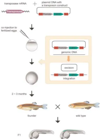
Transgenesis in the fruit fly was detected in 1982 through the works of Rubin and Spradling with transposable elements, a technique now called P-element Transgenesis, that is based on a transposon called P element, a highly mobile element present in the DNA (Rubin & Spradling, 1982). P-elements encode a functional transposase that enables them to “jump” inside a genome (Hummel, 2008). For production of transgenic flies according to this technique, two constructs need to be microinjected into the embryo, one that contains the gene of interest and a marker gene (mini-white) and another one, called Helper plasmid, that contains the transposase that will catalyse the “jump” of the DNA of interest into the fly genome (Fig.1). Mini-white rescues the red colour of the fly eye, since microinjected flies have a white background. In P-element Transgenesis, the transgene is randomly incorporated in the fly genome making it a useful tool to produce transgenic animals, because the transgene of interest is expressed either way. It is possible to produce mutants using P element Transgenesis since an incomplete excision can occur. Fly embryos are microinjected in the syncytial stage, when the embryo is a multinucleated cell with no cytoplasmic membranes involving the nuclei. Microinjection in this stage increases the possibility of targeting every nucleus. Also, microinjection is made in the posterior end of the embryo where the pole cells will appear (pole cells give origin of the germline cells), increasing the chances of germ line transgene incorporation and subsequent transmission to progeny.
In P-element Transgenesis, integration of DNA is random but transgenesis in fly evolved and new techniques arise where DNA is integrated in known sites of the fly genome. One of those techniques is based on Integrase φC31, where an integrase isolated from a phage induces recombination between two non-identical sequences, one called attP (from phage) and the other called attB (from bacteria). This mechanism was translated into the production of transgenic flies, microinjecting a plasmid that contains an attB region into the fly embryo that already possesses an attP site similar to the attP site of the phage (Fig 2). The microinjected plasmid also contains a mini-white as marker gene (Groth et al., 2004). Integrase φC31 can be used in another approach using stocks originated from integration of a specific cassette randomly into the genome, that cassette stands for Minos Mediated Integration Cassette (MiMIC). MiMIC makes use of the transposon Minos, containing a DNA cassette flanked by 2 inverted attP regions. Replacement of this DNA cassette by a functionally relevant DNA element (enhancer, gene trap, etc) is achieved by φC31-mediated
7
Figure 1 – Drosophila Random P Transgenesis (Abdul Razzaq, n.d.)
integration. In this case, original flies have yellow background and the cassette has a yellow+ marker, so the flies are phenotypically wildtype, but when microinjection is successful and the cassette is replaced, flies loose the yellow+ marker and become yellow (Venken et al., 2011). The latter 2 techniques are not random like P-element Transgenesis but attP/MiMiC regions are in known regions of the genome and are useful to produce transgenic animals, not to study gene function (unless the gene of interest has an attP/MiMiC region and in that case, some strategies can be employed to produce a knockout, like the Gal4-UAS: system) (Ou & Lei, 2013).
8 Mice were first used by Mendel in 1860 but he was forbidden to breed mice within the monastery so he started his work in sweet peas (The Jackson Laboratory). Lucien Cuénot, in 1902, was then the first person to use mice and he demonstrated the Mendelian inheritance in mammals, using the coat colours in mice (Cuenot, 1905). Mice are biologically very similar to humans and suffer from the same diseases for the same genetic reasons making them one of the most used model organism. On average, 85% of mouse coding regions are identical to human. Some genes are 99% similar but others are just 60% (National Human Genome Research Institute). Besides genetic similarity, there are other reasons that make mouse a good model organism, such as: one year in the mouse equals to 30 human years (Dutta & Sengupta, 2016), this accelerated lifespan allows the study of an entire life cycle; their maintenance is cost-effective, they are small to handle, reproduce fast; and can be genetically manipulated to mimic any human disease or condition.
Jon Gordon, in 1980, was able to produce the first transgenic mouse by microinjecting purified DNA directly into the pronuclei of fertilized mouse oocytes (Gordon et al., 1980). This became a widely used technique for mouse transgenic production. But the integration of this DNA seems to be random (Lacy et al., 1983) making it impossible to replace, for example, a gene that causes a certain disease. Other technique used for transgenic and mutant mice production consists in the manipulation of mouse embryonic stem cells (ES cells). Using this approach it is possible to manipulate a desired locus by introducing a loss or gain of function in vitro (Bradley et al., 1984; Thomas & Capecchi, 1987). ES cells are present in 3.5 day blastocysts and are pluripotent, meaning that are able to contribute to different cell lineages (Martin, 1981). When in a petri dish, these ES cells may be transfected with the desired DNA that is introduced into the cell’s genome by homologous recombination between the donor DNA and the target genomic locus of ES cell’s DNA. Transformed ES cells that contain the desired alteration are then injected into blastocysts that are in turn transferred to a surrogate mother. Typically, surrogate mothers and ES cell donor animals have different colour coats. This way, the born pups that will have incorporated the altered ES cells will display a quimeric colour coat (Bradley et al., 1984; Koller & Smithies, 1992). The ES cells technique made it possible to knockout a gene through the homologous recombination mechanism but it is still a long and expensive process (Hall et al., 2009).
9
Figure 3 – Zebrafish Tol2 transgenesis system (Kawakami, 2007)
Danio rerio as model organism and transgenesis techniques used
In 1981, George Streisinger was the first to clone a vertebrate and it was the zebrafish Danio rerio. George was the father of zebrafish as a research model and has turned it into a very useful scientific model organism to study development and gene function (Streisinger et al., 1981). There’s an online resource, the Zebrafish Information Network (ZFIN) where genetic, genomic and developmental information can be found. The reasons why zebrafish is such a good model organism are: its genome is already sequenced; has a rapid embryonic development attaining sexual maturity in 60-90 days; adults are small and are housed in large groups, requiring few space and lowering the maintenance costs; adult zebrafish breed very fast and can produce until 300 embryos at a time;
fertilization in zebrafish is external, allowing the easy manipulation; embryos also have the advantage of being large and transparent (Burke, 2016). Zebrafish has similar behaviour as compared to mammalian models concerning toxicity testing and diurnal sleep cycle (Jones, 2007). Even existing 70% of gene similarity between human and zebrafish (Howe et al., 2013), limitations in using zebrafish as an organism model exists, for example as a human disease model. Some human diseases are caused by genes that do not exist in zebrafish, making impossible to use this organism as a human disease model for a variety of human diseases. Zebrafish is also not a good organism model for human diseases that take place in a body part that zebrafish don’t have, like mammary glands or prostate (Burke, 2016).
10
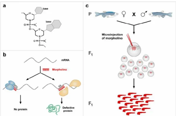
Figure 4 – Zebrafish Morpholinos (Codarin et al., 2009)
Stuart and colleagues, in 1988, after publication of the first transgenic mice production (Gordon et al., 1980), applied successfully the same technique in zebrafish, with their group being the first to produce a transgenic zebrafish (Stuart et al., 1988). However, those results had a very low efficiency rate , and although this was being increased over time, a new technique using transposons has been developed in zebrafish, called the Tol2 transposon system (Kawakami & Shima, 1999; Kawakami et al., 2000). Evidence of this active transposon was first reported in Medaka fish in 1996 (Koga et al., 1996). A couple of years later, Tol2 was isolated from a mutational insertion in the Medaka tyrosinase locus and showed to have autonomous mobility (Kawakami et al., 1998). The Tol2 system consists on a construct containing 2 cis-regulatory sequences (CREs) from the Tol2 element positioned 5´and 3´ of a promoter sequence followed by a fluorescent protein. This construct was named Tol2 vector. The Tol2 vector is co-injected with mRNA encoding for the Tol2 transposase into a one-cell stage embryo. Once translated, the Tol2 protein will catalyse the excision of the region of the Tol2 vector between the CREs and its integration in the genomic DNA (Fig. 3) (Kawakami & Shima, 1999; K Kawakami et al., 2000; Kawakami, 2007). There’s another approach capable of blocking a gene in initial stages of embryo development, allowing the study of its function, the morpholinos (Nasevicius & Ekker, 2000; Summerton, 1999). Morpholinos are synthetic molecules and exist in two types: the ATG morpholinos, that block the initiation of translation of proteins, and the Splice morpholinos that bind and interfere with the RNA splicing machinery resulting on a truncated protein (Fig. 4). However, this mechanism is transient because morpholinos are degraded through time (Bill et al., 2009; Morcos, 2007).
11 Neither the Tol2 system nor the Morpholinos are able to induce targeted mutagenesis in zebrafish and, in 2008, zinc finger nucleases (ZNFs) were adapted to create targeted double strand breaks in the zebrafish genome (Doyon et al., 2008; Meng et al., 2008). ZNFs were produced to cleave DNA (Kim et al., 1996) and are a fusion between a restriction enzyme, FokI, and a DNA recognition domain containing 3 (or more) zinc finger motifs. ZNF heterodimerization in a position of the DNA leads to a double-strand break (DSB).
12
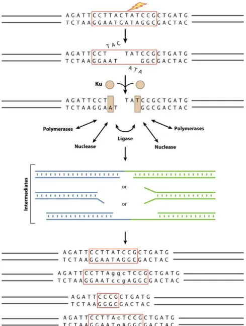
Figure 5 – DSB repair: flexibility of enzyme functions lead to different repairs in a DSB
break (Lieber, 2010).
DNA double strand break repairs
Cells fix double strand breaks in the DNA by two different mechanisms, homologous recombination (HR) or Non-Homologous End Joining (NHEJ). Homologous recombination only occurs if a donor sequence with homology arms is present (Filippo et al., 2008), a technique used to produce transgenic animals. Otherwise, NHEJ will occur and for that mechanism a nuclease to reconstruct the damaged DNA, a polymerase to fill in the gaps and a ligase to restore the strand integrity are required (Ma et al., 2004). It seems obvious to think that these enzymes work by this order to reconstruct the DNA cut but these enzymes have a functional flexibility big enough to allow the NHEJ mechanism to occur in many ways (Fig. 5).
13 This flexibility can result in loss of nucleotides or junctions with nucleotide addition (Lieber, 2007, 2010), causing a frameshift that can result on a different protein translation or the complete gene knockout (Puchta et al., 2015).
An animal that has a gene knockout is an organism in which a particular gene or genes have been made inoperative. And knocking out genes is important for research purposes. If we remove a piece from a machine it’s possible to know how it works and what’s the importance of that piece and its function, for genes is the same logic, by knockin out a gene it’s possible to understand what is its function. Nowadays, despite several animal genomes being sequenced, many genes still have an unknown function, and by knocking out a gene it is possible to study its function. Knockins (insertion of a gene) and knockouts are also widely used to produce and create disease models (Hall et al., 2009).
14
Figure 6 - Direct repeated sequences of iap gene of E. coli. There are 29 highly conserved nucleotides, 14 of which
(underlined in the bottom) contain a dyad symmetry. In brackets are the nucleotide numbers in the gene (Ishino et al.,
1987).
1.2 Bacteria CRISPR adaptive immunity system
Viruses are the biggest predators of bacteria, infecting prokaryotic cells with its DNA or RNA and making the bacteria machinery transcribe and translate its genetic material. Bacteria have both an innate immune system, that recognizes certain infection characteristics, and an adaptive immune system that can recognize specific pathogen characteristics (Rath et al., 2015). In 1987, in
Escherichia coli, five homologous sequences of 29 nucleotides arranged in direct repeats with 32
nucleotides interspacing were found (Fig. 6). Those sequences were called REP (from repeats) sequences and were thought to act as mRNA stabilizers (Ishino et al, 1987).
Later in 2000, Mojica’s group identified that those short-repeated elements, generally in clusters, had one peculiarity: sequences were always regularly spaced by a unique sequence of constant length (Mojica et al., 2000). They called those clusters SRSRs (Short Regularly Spaced Repeats). Another feature present in those clusters is the presence of a conserved sequence, called leader, that is located upstream of every cluster locus. This leader directs transcription (Rath et al., 2015). Searching these SRSRs in all available microbial genomes, resulted in hits in 20 microbial species widespread among physiological and phylogenetic groups (Mojica et al., 2000). In 2002, those sequences were named CRISPR (Clustered Regularly Interspaced Palindromic Repeats) (Jansen et al., 2002), name that is used nowadays to refer to this molecular system. Alongside with CRISPR, three Cas (CRISPR-associated) genes were also identified. Cas genes are present in prokaryotes that contain CRISPR, absent in non-CRISPR-containing prokaryotes and are found to be located invariably adjacent to the CRISPR locus, suggesting that Cas genes and CRISPR have a functional relationship. Cas genes showed characteristic motifs of helicases and exonucleases (Jansen et al., 2002).
In 2005, work in S. pyogenes showed that CRISPRs could acquire phage DNA by discovering that seven out of the nine spacers included in S. pyogenes CRISPRs corresponded to a phage sequence (Pourcel et al., 2005). Another work, in this case in S. thermophilus, showed that about 75% of CRISPR spacers from this bacterium corresponded to S. thermophilus phages and 20% corresponded to S. thermophilus and Lactococcus lactis plasmids (Bolotin et al., 2005). Both these works pointed that CRISPR spacers have phage DNA and extra chromosomal origin, but it was Mojica’s group that proposed a role for CRISPRs in microbial immunity showing that those extra chromosomal elements,
15
Figure 7 – Bacteria CRISPR/Cas immunity system (Doudna lab http://rna.berkeley.edu/crispr.html)
included in the spacers, fail to infect the cells (Mojica et al., 2005). In 2007 this hypothesis was further reinforced by experimental work in S. thermophilus by Barrangou and colleagues. They showed that resistance against a bacteriophage could be acquired by integrating a genome fragment of that phage into the CRISPR locus (Barrangou et al., 2007). Each spacer integration promotes a duplication of a new repeat, creating a new spacer-repeat unit. S. thermophilus also allowed the discovery of plasmid cleavage in this system. Cleavage of DNA was performed 3 nucleotides upstream of a proto-spacer adjacent motif (PAM) by an endonuclease. When an invading DNA appears, selection of which spacer precursors (proto-spacers) will integrate the CRISPR locus, is determined by the recognition of PAM. PAMs are usually 3 nucleotides long and differ between CRISPR types (Barrangou et al., 2007; Deveau et al., 2008; Horvath et al., 2008). In 2008, Brouns and colleagues demonstrated how those acquired spacers are used. CRISPRs are transcribed and a complex of Cas proteins cleaves the CRISPR RNA (crRNA) in each repeat, and retains the cleavage product that corresponds to a certain phage (Fig. 7). CrRNAs serve as guide RNAs that allow the Cas protein complex to interfere with the (Brouns et al., 2008).
Three types of CRISPR systems were identified, but type II is the system currently used to manipulate eukaryotic cells. In Type II CRISPR system, phage or plasmid DNA that tries to infect a cell is cut into small fragments halting the infection. In addition, those small fragments are
incorporated into the CRISPR locus in short repeats (about 20 bp each). When new infection occurs, those loci are transcribed and those transcripts are processed into small RNAs (called CRISPR RNA – crRNA) that will guide Cas proteins to the target invading DNA based on sequence complementarity of crRNA and invading DNA (Fig. 8). In this system, only one protein, Cas9, is required to inactivate a gene (Jinek et al., 2012). The Cas9 protein, discovered in Streptococcus species, has a key role in Type II CRISPR system, participating in processing crRNA and destroying target DNA. Cas9 contains
16
Figure 8 – CRISPR/Cas9 mechanism after 1st phage infection and 2nd infection by the same phage (Charpentier &
Barrangou, 2017).
two nuclease domains, a RuvC-like nuclease domain and a HNH-like nuclease domain, that cut the upstream strand and the downstream strand, respectively (Sapranauskas et al., 2011).
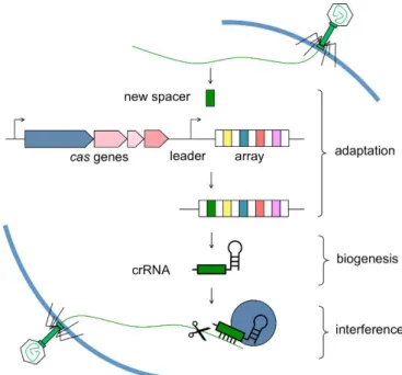
The CRISPR immunity system is divided into three stages: adaptation/acquisition, biogenesis/expression and interference. In the adaptation stage, a unique sequence from invading DNA, the protospacer, is incorporated into the CRISPR locus becoming a new spacer. This stage gives bacteria a genetic memory of invading DNA. Cas1 and Cas2 are two nucleases that are the key factors for the spacer integration into the CRISPR locus, but the mechanism through which these nucleases effect that integration is not fully understood. In Type II CRISPR, Cas9 is essential to identify the sequence that will be the protospacer by recognizing PAM sequences, and it is assumed that after that recognition, Cas9 recruits Cas1 and Cas2 to deliver the new protospacer into the CRISPR locus (Hille & Charpentier, 2016; Rath et al., 2015). The second stage, expression/biogenesis, refers to the transcription of the CRISPR locus to produce a CRISPR ribonucleoprotein complex. Primarily, the CRISPR locus is transcribed into pre-crRNA that is later processed into guide crRNAs, each containing memorized sequences of previous invaders. In type II system, it is known that a separate trans-activating RNA (tracrRNA) is required for the maturation of crRNA, but its mechanism is still unknown (Hille & Charpentier, 2016). In the third and final stage, interference, crRNAs binds to Cas9 protein and the complex locates the corresponding targets to be degraded. For interference to occur, the presence of PAM and complementarity between crRNA and invader DNA are necessary (Hille & Charpentier, 2016; Rath et al., 2015).
To target mutagenesis in vitro, Cas9 is complexed with crRNA and tracrRNA (Deltcheva et
17
Figure 9 – Stages in CRISPR/Cas immunity system: adaptation, biogenesis and
interference (Marraffini Laboratory -
http://marraffini.rockefeller.edu/research.html)
upstream of the PAM sequence, which in the case of Cas9 protein is NGG. Doudna and Charpentier showed that Cas9 protein required a base-paired structure between crRNA and tracrRNA to cleave DNA, so they developed a simpler system which combined crRNA and tracrRNA into a single guide RNA (sgRNA). Cas9 is effective with separate tracrRNA and crRNA as it is with sgRNA (Jinek et al., 2012).
To produce mutant animals using the CRISPR/Cas9 technology, only microinjection of Cas9 (in protein or in mRNA) and a sgRNA complementary to the chosen gene target is required. This technology offers many advantages over all techniques referred above: it is easier and cheaper to design and produce, since only Cas9 protein (or Cas9 mRNA) and sgRNA are necessary; sgRNA and Cas9 protein (or mRNA) can be directly injected into embryos; it is possible to make more than one mutation at once by co-injecting 2 or more gRNAs; the possibilities to target the mutation are bigger than ever since you can almost target any gene as long as a PAM sequence exists; it is also possible to make knock-ins with this technique by co-injecting oligonucleotides that will be incorporated into the genome by homologous recombination. However, limitations also exist: one of the major limitations of the CRISPR/Cas9 technology is off-targets: the mutation can occur in a non-specific region with similar homology to the real target site (even tough, off targets of CRISPR technology are fewer than other techniques); even with microinjection in 1-cell stage embryos, it does not mean that the mutation will occur in all cells nor that it happens in both alleles, creating mosaic animals; and the generations of multiple different mutated alleles. When DSB happens, the repair process of NEHJ is different in every animal producing different mutations from the same cut (The Jackson Laboratory b). CRISPR/Cas9 has been successful in many animals, invertebrates and vertebrates. Indels have been
18
Figure 10 – Different body color between a Drosophila
wildtype female (1) and a Drosophila yellow female (2)
(Rampasso & Vilela, 2017)
introduced at about 90% efficiency in C. elegans, Drosophila, rabbit, chicken, mouse, zebrafish and human cells (Bortesi et al., 2016).
1.3 The tyrosinase and the yellow gene
Tyrosinase is an enzyme responsible for the conversion of tyrosine into melanin in
melanocytes. Melanin gives colour to skin, hair and eyes and is also found in the retina were has it a role in vision (Genetics Home Reference - U.S. National Library of Medicine, 2017). Mutations in the
tyrosinase gene cause oculocutaneos (OCA1) in humans and identical phenotypes are found in mice
(King et al., 2003). In mice, a single nucleotide exchange in the coding region of tyrosinase causes the classical albino mutation (Jackson & Bennett, 1990). In zebrafish, the tyrosinase gene is expressed first in the retinal pigment epithelium and then in the neural crest (Camp & Lardelli, 2001). The
tyrosinase gene was chosen in this project exactly because it is expected that after knocking out the tyrosinase gene a lack of pigmentation phenotype would be easily observed.
The yellow gene (y) is located in the drosophila X chromosome and controls the pigmentation pattern of the adult fly cuticle and larval mouth parts. When the yellow gene is mutated adult flies have a phenotypically distinct yellow pigmentation on its cuticle (Biessmann & Alberts, 1985). Just as for the tyrosinase gene, the yellow gene was chosen for convenient phenotype scoring:
yellow knockout mutants will have yellow cuticles
and can be easily differentiated from wildtype flies with normal brownish cuticle (Fig. 10).
19
2.
Aims
Despite the fact that the CRISPR/Cas9 technique is already well established in these three organisms, there is a lack of a general common protocol suitable for all 3 animals. In this project, our focus was to establish a single protocol for guide RNA production and CRISPR mediated knockout generation that would fit all 3 model organisms: Danio rerio, Drosophila melanogaster and Mus
musculus.
The main objective of this MSc project was to optimize a general protocol of guideRNA production that would fit three model organisms, Danio rerio, Drosophila melanogaster and Mus
musculus, commonly used at CF and produce mutant animals using CRISPR technology. To achieve
this goal, we:
• Chose a gene that would be responsible for an observable phenotypic characteristic. The knockout of that gene was expected to generate a different phenotype;
• Used a guideRNA production protocol that had been already shown to be successful in Danio rerio;
• Started with the validation of such a protocol in Danio rerio using embryo microinjection of guideRNA and Cas9 protein;
• Moved to Drosophila melanogaster and adjusted microinjection concentrations of guideRNA and Cas9 protein to produce mutant individuals;
• Performed embryo microinjection in Mus musculus also adjusting the microinjection concentrations to produce mutant individuals.
A second objective of this project, after producing mutant animals, was to establish a mutant stable line. To achieve this goal, we:
• Crossed Danio rerio mutants between themselves and screened the progeny for non-pigmented individuals
• To establish a line tyrosinase knockout, injected animals need to be crossed with wildtype individuals
• Crossed Drosophila melanogaster mutants with yellow flies for two generations and screened the progeny for yellow cuticle colour to establish a mutant stock for yellow knockout;
• Crossed Mus musculus mutants with albino animals and screened the progeny for non-pigmented animals, then crossed between themselves to establish a stock for
20 Due to the organism diversity, every step after the production of the guideRNA is different so, the next chapters, Material and Methods and Results, will be divided by organism.
3.
Material and Methods
3.1 Production of guideRNA
To produce guideRNA, a modified protocol from Gagnon et al (2014) was used (Gagnon et
al., 2014).
Template for guideRNA production
To produce guideRNA, a first template is generated, consisting on two oligos annealed. The first is a variable gene-specific oligo, comprising a suitable promoter, the target site (without the PAM region) and an overlap region. This overlap region will anneal with the second oligo that is constant and contains tracrRNA that will bind to the Cas9 protein.
A suitable promoter can be T7 or SP6. If the guideRNA sequence starts with GG, a suitable promoter is T7, if it starts with GA then is SP6. If there is no GG or GA, a GGG upstream to the guideRNA and the T7 promoter are added. The sequence for the T7 promoter is TAATACGACTCACTATA and for the SP6 promoter is ATTTAGGTGACACTATA. The overlap region is GTTTTAGAGCTAGAAATAGCAAG. In the end, there are two template possibilities: T7: TAATACGACTCACTATA -N20-GTTTTAGAGCTAGAAATAGCAAG
SP6: ATTTAGGTGACACTATA-N20-GTTTTAGAGCTAGAAATAGCAAG where N20 is the target site sequence chosen to the target gene.
The constant oligonucleotide, regardless of the choice of the promoter or target gene, is: 5’AAAAGCACCGACTCGGTGCCACTTTTTCAAGTTGATAACGGACTAGCCTTATTTT AACTTGCTATTTCTAGCTCTAAAAC 3’
These oligonucleotides were synthesized by Sigma.
The first step of the guideRNA production protocol was the annealing of both oligonucleotides:
21 Table 3 – Fill in with T4 polymerase
Table 2 – Thermocycler conditions for annealing
oligonucleotides
Table 1 – Solution for annealing of oligonucleotides
Reagent μl
dNTPs (10nM) 2.5
10x NEB buffer 2.2
100x NEB BSA 0.2
T4 NEB DNA Polymerase 0.5
Water 4.8
10 μl total
95ºC 5 minutes
95ºC ramp. Rate to 85ºC ´-2ºC/second 85ºC ramp. Rate to 25ºC ´-0.1ºC/second
4ºC Hold Oligonucleotide μl Gene-specific (100 μM) 1 Constant (100 μM) 1 Water 8 10 μl total
Followed by a temperature cycle using a thermocycler:
The next step was the Fill in with T4 polymerase (NEB) to produce a double strand oligonucleotide:
Samples were incubated for 20 minutes at 12ºC. After incubation 80 μl of water was added to the template followed by purification using a PCR cleanup column, eluting in 30 μl of water. Expected DNA yield should be between 100-200 ng/µl. After measuring the DNA, a 1% agarose gel or QIAxcel ScreenGel®, was performed to verify that the product had the correct size of ~120bp.
Transcription of template to produce guideRNA
After purification, the template was in vitro transcribed with Ambion Megashortscript T7 or Megascript SP6 kit, depending on the promoter, to produce guideRNA. From this point onward all procedures were made in RNase-free conditions. To maximize the transcription, the incubation step was prolonged to an overnight incubation when using T7 kit and 4 hours when using SP6 kit.
22
Extraction and Purification of guideRNA
Recovery of guideRNA was performed with Qiagen micro-RNA purification Kit or with a Phenol/chloroform extraction: to 20 µl of transcription product, 115 µl of nuclease-free water and 15 µl of Sodium Acetate Stop (Ambion kit) were added. Next, 150 µl of phenol/chloroform pH 4.5 (or pH 8) was added, mixed well and centrifuged for 5 minutes at 4ºC. Upper layer (lower layer if using phenol/chloroform pH 8) was transferred to another tube with 350 µl 100% EtOH, incubated for 15 min at -80ºC or with dry ice to precipitate RNA. RNA was centrifuged 20 minutes at 4ºC to form a RNA pellet. The supernatant was discarded and the pellet washed with 500 µl 70% EtOH spinning 15 minutes at 4ºC. The supernatant was discarded again and the pellet was left to dry and resuspended in 20 µl of nuclease-free water. RNA was aliquoted according to the concentration needed for microinjection.
3.2 Cas9 Protein
Cas9 Protein was batch-produced at 1 mg/ml in 20mM Tris Ph 8, 10mM MgCl2 and 0.2M KCl buffer, at the Weizmann Institute of Science, Israel.
23
4.
Danio rerio (zebrafish)
4.1
Methods
Targeting strategy
For zebrafish, the tyrosinase gene was chosen for mutation. Tyrosinase is responsible for the black pigmentation of the body and eyes of the animal. So, a knockout of this gene should produce a visible phenotype with lack of pigmentation.
Choosing guideRNA
In zebrafish, the CRISPRz database was used to look for guides already validated for the zebrafish tyrosinase gene. We chose the one used by Jao et al. (2013): GGACTGGAGGACTTCTGGGGAGG (PAM site underlined). Since tyrosinase guideRNA (without the PAM site) started with GG, a suitable promoter was T7, thus being the tyrosinase gene-specific oligonucleotide (ordered from Sigma):
TAATACGACTCACTATAGGACTGGAGGACTTCTGGGGTTTTAGAGCTAGAAATAGCAA G
The guideRNA production protocol was performed and guideRNA was ready for microinjection.
Danio rerio breeding
Zebrafish were housed at the CF Fish Facility. Wildtype TU adults (around 6 males and 12 females) were crossed for each microinjection trial, setting 6 crosses for each trial (1 male to 2 females). Those crosses were made between 4 and 6 p.m. with fish housed in spawning tanks. Spawning tanks contain an insert reservoir, that have holes in the bottom, and a spacer that fits the tank, separating males and females from physical contact, but sharing the water. Animals stay overnight in these tanks being close to each other but not being able to breed, so when the spacer is removed early in the morning (when the lights turn on), fish spawn and eggs are fertilized. Spacers are taken one at a time, meaning that each cross produced embryos for a single microinjection. When the first laying of the first cross was injected, then the spacer from the second cross was removed, and so on. Eggs fall through the holes in the insert reservoir, preventing the cannibalization of the embryos
24
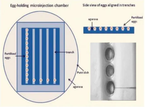
Figure 11 – Agarose plates and zebrafish embryo alignment. Image modified from
Wang et al. and Lu Zhe (Wang et al., 2013; Zhe, n.d.)
by the parents. Fish were used to breed once a week. Eggs were collected into petri dishes with the help of a tea strainer and blue water (Methylene blue) and were ready to be aligned for microinjection. After laying, animals were housed back into the housing tanks (Martins et al., 2016). All animal procedures were made under rigorous standards of animal welfare and complied with the 2010/63/EU (European Parliament and the Councli pf the European Union, 2010).
Microinjection needles and microinjection set-up
Agarose petri dishes with trenches (Fig. 11) were used to align and microinject embryos. Alignment and microinjection of embryos were performed under a Zeiss Discovery V8 scope and microinjection with a PV820 Pneumatic Picopump (WPI). Microinjection needles were bought from Biomedical Instruments and were loaded with Eppendorf Microloader™ tips.
Embryo Microinjection
Collected embryos were aligned with the cell positioned to the right side so it can be directly injected (Fig. 11) Only one-cell stage embryos were microinjected, embryos in other stages were discarded. Uninjected embryos were also kept as controls for each laying/cross.
Different concentrations of guideRNA and Cas9 protein were tested (Table 4). Phenol red was added to the mix to serve as a visible marker for the injection into the embryo. After injection,
25
Table 4 – Microinjection concentrations of gRNA and Cas9
protein into zebrafish embryos
Cas9 Protein (ng/µl) sgRNA (ng/µl)
210
526
260
174
260
438
315
526
500
438
600
200
embryos were incubated and bleached (to disinfect embryo surface) at 24hpf. Survival rates were recorded 24 hours after injection (See Appendix 1).
Screening for mutations
Forty-eight hours after injection it was possible to evaluate the result from targeting the
tyrosinase gene because tyrosinase expression already started (Camp & Lardelli, 2001). Individuals
with visible phenotypes were incubated until 5 days old, at which age larvae entered the nursery. For non-phenotype targeting there is a possibility to screen for the mutation in early stages by DNA extraction from 24hpf embryos, following HotSHOT protocol (Meeker et al., 2007).
The HotSHOT protocol consists in collecting pools of two embryos into a PCR strip (without blue water; if not possible, blue water can be removed with a micropipette) with 50 μl of 50mM NaOH to cause cell lysis during a 15 minutes incubation at 95ºC, followed by a cooling step at 4ºC. If using a thermomixer for incubation, ice can be used to cool down the samples; if using a thermocycler, an additional step of 4ºC can be added. For buffering, 5 μl of 1mM Tris-HCL pH 7.5 was added to the samples. After embryo DNA extraction, a 25 μl PCR reaction mix was prepared using 5 μl of extracted embryo DNA (See Appendix 5 for primers used). Uninjected embryos were always used as controls. After PCR reaction (Table 5), samples were loaded in a 3% agarose gel for 1 hour at 80V. Uninjected eggs should have a single band while injected positive eggs should have a smear or more than one single band. This method can and should be used to test guide efficiency, according to which a respective number of fish are raise to adulthood. The PCR product was purified and sent for sequencing. If the CRISPR process is successful and there is integration or deletion of nucleotides in the target region, this can be easily seen in the sequence chromatogram that will be a mix of different alleles present in the sample.
26
Initial Denaturation 95°C 30 seconds
95°C 30 seconds
60°C 30 seconds
68°C 2 minute
Final Extension 72°C 5 minutes
Hold 12ºC
Temperature
34 Cycles
Time
Primers
For PCR from embryo DNA extraction, amplicons should be around 100bp, 50bp from the cut site to each side, the smaller the better to search for indels, but for the tyrosinase gene the primers used were the same as Joa et al. (2013) and amplicons were around 315bp (Appendix 5).
Genotyping the adults
By two months old, fish are big enough to be fin clipped to sample tissue for genotyping. DNA extraction was performed with proteinase K: Tissue was sampled into 200 μl of lysis Buffer (50 Nm Tris-HCL pH 8.5, 1 mM EDTA and 0.5% Tween-20) and proteinase K was added to a final concentration of 200 μg/μl immediately before DNA extraction (samples can be frozen before extraction), samples were incubated for 2h at 55ºC in a thermomixer followed by a denaturation step of 10 minutes at 95ºC. After incubation, samples were centrifuged 10 minutes at 13.200rpm at 4ºC. The supernatant was collected to a new tube and stored at 4ºC for up to 3 months or -20ºC for longer periods.
After DNA extraction, a 25 μl PCR reaction (Table 5) was performed and samples loaded on QIAxcel ScreenGel®. Different band sizes (or a smear – QIAxcel ScreenGel® has an “smear analysis” option”) should appear.
Table 5 PCR cycle for tyrosinase gene
Sequencing
For sequencing the mutation, positive fish should be outcrossed and the F1 genotyped. The PCR product sent for sequencing and screened for jammed chromatogram near the PAM site. For the
tyrosinase gene in particular, incrosses between F0 mutant individuals were made and non-pigmented
27
Figure 12 – QIAxcel ScreenGel®, for tyrosinase gRNA
template
4.2 Results
GuideRNA production
After annealing the oligos and fill-in with T4 polymerase, the template was purified using a PCR column (QIAquick PCR Purification Kit) and eluted in 30 μl of water. DNA concentration was expected to be 100-200ng/μl, tyrosinase template was 145 ng/μl. Template was then loaded in QIAxcel ScreenGel®, and a band of ~120bp was observed (smaller fragments are primer dimer) (Fig. 12) (as described in protocol).
RNA extraction with Qiagen micro-RNA purification kit showed that very few RNA was extracted, 54 ng/μl in our first attempt and 10 ng/μl in our second attempt, which was not sufficient to microinject. Based on these results, we did a comparison between RNA extraction with the Qiagen kit and Phenol/chloroform extraction. After in vitro transcription, the sample was divided into two tubes and we tested the 2 protocols. With phenol/chloroform we could extract 3278 ng/μl while with Qiagen kit we were only able to extract 20,8 ng/μl. According to the ratios of the absorbance parameters and by applying the Beer-Lambert Law, guideRNA extracted with Phenol/chloroform was contaminated with protein (A260nm/A280nm = 1.66) but free from organic contaminants (A260nm/A230nm = 2.26), so we therefore chose to follow the phenol/chloroform protocol for RNA extraction.
28 2
1 3 4
Figure 13 – PCR amplification of
314bp fragment of the tyrosinase zebrafish gene run in 1% Agarose gel: Amplification from fin sample genomic DNA preparation (1) Amplification from a 24hpi embryos genomic DNA preparations (2 and 3), geneRuler 200bp (Thermo Scientific) (4).
2
Screening for mutations
DNA embryo extraction
At 24hpi embryo DNA was extracted with 10 pools of 2 injected embryos each and 2 pools of two non-injected embryos as control but nothing was amplified in the PCR reaction. A new pair of primers and different enzymes were tested but still nothing was amplified. Next, extraction from DNA embryos and fin samples were compared. Only by using DNA extracted from fin samples was it possible to amplify the correctly sized band (Fig. 13).
In an effort to understand and troubleshoot lack of amplification of the tyrosinase fragment from 24hpi genomic DNA template, we tested different amplification protocols:
Protocol 1 – protocol from HotSHOT using Thermocycler for incubation and 5 μl of template DNA for PCR reaction
Protocol 2 – protocol from HotSHOT but using Thermomixer for incubation and an additional final step of 5 minutes centrifugation at 13550 rpm and 1,5 μl template DNA for PCR reaction
Different genes regions were amplified, in the tyrosinase gene and DIA1R gene, as a control. The primers used for amplification of tyrosinase gene were the same ones that worked for the fin sample and that were used by Jao et al. (2013). For amplification of the DIA1R gene another pair of primers that had also worked for the fin sample, were used. Results from the above PCR reactions are in Figure 14.
29
Figure 14 - Left panel: QIAxcel ScreenGel® analysis (1) protocol 1 for tyrosinase gene; (2) protocol 1 for DIA1R gene; (3)
protocol 2 for tyrosinase gene and (4) protocol 2 for DIA1R gene. On the right, there’s an overall result table with DNA concentration measured in the samples
Figure 15 – PCR amplification of 314bp fragment of the tyrosinase
zebrafish run in 1% Agarose gel: Amplification from 72h larvae genomic DNA preparation (1); Amplification from 5-day-old larvae genomic DNA preparation (2); GeneRuler 200bp (Thermo Scientific) (3)
As we can see from figure 14, we suceeded in amplifyng DIA1R gene with template DNA extracted from 24h embryos but fail to amplify tyrosinase gene., in both embryo DNA extraction protocols. With these results and since extraction using the HotSHOT protocol clearly worked, we decided to extract genomic DNA from different ages and use genomic DNA extracted as DNA template for PCR reaction to see when was the tyrosinase gene amplified. For genomic DNA extracted from 72hpf larvae, no amplification was detected but with genomic DNA extracted from 5-day-old larvae a very faint band starts to appear (Fig. 15).
1
1 3
2 3 4
30
Figure 16 – PCR amplification of 341bp fragment of the tyrosinase zebrafish
run in 1% Agarose gel: GeneRuler 50 bp (Thermo Scientific) (1); Amplification from 24 h embryos genomic DNA preparation (in pools of two embryos) (2, 3, 4, 5, 6 and 7).
Figure 17 – Injected 72h larvae for tyrosinase knockout: Lack of pigmentation in some cells of the
eye (A) and wild type phenotype (B).
In another approach, we tested the addition of DMSO to the PCR reaction mix of 24hpf and 72hpf embryo DNA extraction (Fig.16). These results were not pursued since animals were already growing and for
tyrosinase in specific (that causes a phenotypic result when mutated), DNA
embryo extraction was not essential.
Phenotype screening
At 48hpf, injected embryos were screened for lack of pigmentation and the number of individuals with mosaicism was scored. Although in our first microinjection trial we did not see a lack of pigmentation in larvae, we still decided to grow some fish and some adult individuals did grow with non-pigmented cells. So, despite the fact that this mutation causes lack of pigmentation, it is possible that sometimes it could not have a larval phenotype. See Appendix 2 for mosaic individuals that were identified and respective microinjection mix concentrations. Eighteen larvae were identified with mosaicism, but in total we had 40 fish that showed pigmented mosaicism as adults (see Figure 17 for mosaicism example in larvae). This phenotype was achieved with different concentration mixes (Appendix 2). Mosaic animals that were raised but that didn’t show a lack of pigmentation as larvae, were obtained from our first injection with 260 ng/μl of Cas9 protein and 174 ng/μl of sgRNA.
1
2 3 4 5 6 7
31 Graphic 1: Embryo survival rate: Control embryo survival rate vs injected embryo survival rate in different microinjection concentrations of Cas9 protein and guideRNA (A, B, C, D and E). A – 260 ng/μl Cas9 protein + 174 ng/μl guideRNA, B – 260 ng/μl Cas9 protein + 438 ng/μl guideRNA, C – 500 ng/μl Cas9 protein + 438 ng/μl guideRNA, D – 315 ng/μl Cas9 protein + 526 ng/μl guide RNA and E – 600 ng/μl Cas9 protein + 200 ng/μl guideRNA
Table 6 Mosaic animals with lack of pigmentation phenotype found according to concentrations mixes
Embryo survival rates were different from uninjected embryos and injected embryos, being lower in the injected ones. Despite results from Condition A (see Graphic 1) that are probably due to the lack of experience, that was optimized trough trials, injected embryo survival is close to control embryo survival rate in all other conditions (B, C, D and E). Also, it seems that toxicity does not affect embryo survival, since that the lowest concentration mix condition (B) showed very similar survival rate to the most concentration mix conditions (D and E).
Genotyping
Genotyping F0 injected animals
Fourteen adult fish (F0 injected animals) were genotyped for screening indels. TU wildtype DNA was used as a control. All samples amplified the wildtype band, but also amplified smaller or bigger fragments that indicate indel occurrence (Fig. 18). Overall table of results of measured band sizes can be found in Appendix 3.
32
Figure 18 – Screening for indels: Wildtype sample (F04 TU) amplified DNA with ~315 bp, all
other samples were from mosaic individuals that in addition to wildtype band, also showed smaller or bigger fragments proving that guideRNA cut the DNA and NEHJ events have occurred.
Sequencing
PCR products from these samples were sent for sequencing, but it was a very jammed chromatogram making it impossible to draw any conclusion. To really get conclusions of which indels have really occurred it may be best to run a high concentration agarose gel and extract each amplicon and send it for sequencing, which was not done due to time constrains.
Genotyping F1 fish
Non-pigmented larvae (Fig. 19) from incrosses of mosaic animals, were grown and fins sampled at 2-month-old fish. From figure 19, it’s possible to see that all non-pigmented individuals (D01 – D08) lack the wildtype amplicon (D09), and instead, are composed of different F0 mutations.
33
Figure 19 – Genotyping of Non-pigmented incrossed animals (left side); Non-pigmented
34
5.
Drosophila melanogaster (fruit fly)
5.1. Methods
Targeting strategy
For fruit fly, yellow was the targeted gene to be knockout. Yellow is a spontaneous recessive mutation that gives a yellow colour to the body of the fly (Biessmann & Alberts, 1985). Yellow mutation already exists and it’s been part of fly crossings strategies. In this case we tried to mimic the existing spontaneous mutation of yellow body colour in wildtype flies and vasa_Cas9 flies. Vas_cas9 (Bl #51324) are flies that express Cas9 protein under the germ-line promotor vasa and were used for microinjection of only guideRNA instead of Cas9 protein and guideRNA co-injection in non-expressing cas9 flies.
Choosing guideRNA
In Drosophila, guideRNA was found in BreakingCas site (Oliveros et al., 2016) using the
yellow gene sequence (NM_143655.4) as template for search fit guides. The guide chosen had a 99.9
score: GGTTTTGGACACTGGAACCGTGG (PAM site underlined). This guide was also used in Basset et al. (2013) experiments. Since yellow guideRNA (without the PAM site) started with GG the suitable promotor was T7, being the yellow gene-specific oligonucleotide (ordered from Sigma): TAATACGACTCACTATAGGGGGTTTTGGACACTGGAACCGGTTTTAGAGCTAGAAATA GCAAG
Production of guideRNA was performed according to the protocol described in “Production guideRNA” methods section, page 16 and it was ready to microinject.
Microinjection needles and microinjection set-up
Embryos were aligned under a Leica MZ6 scope and microinjected under a Zeiss Primovert microscope adapted to microinjection, with a Narishige micromanipulator connected to a PV820 Pneumatic Picopump. Capillaries from WPI (Thin wall single- barrel Standard Borosilicate 1mm with filament) were pulled on a Sutter P-2000 needle puller to produce microinjection needles. Needles were loaded with Eppendorf Microloader™ tips.