Faculdade de Medicina de Lisboa
The interplay between alpha-synuclein and ATP13A2:
towards the understanding of the molecular basis of
Parkinson's disease
Tomás Ribeiro da Silva Lopes da Fonseca
Tese orientada por:
Professor Doutor Tiago Fleming de Oliveira Outeiro
Tese especialmente elaborada para obtenção do grau de Doutor em Ciências Biomédicas, Especialidade em Neurociências
Presidente: Doutor José Luís Bliebernicht Ducla Soares, Professor Catedrático em regime de tenure e Vice-Presidente do Conselho Científico da Faculdade de Medicina da
Universidade de Lisboa. Vogais:
- Doutora Ana Cristina Carvalho Rego, Professora Auxiliar com Agregação da Faculdade de Medicina da Universidade de Coimbra;
- Doutor Duarte Custal Ferreiral Barral, Professor Auxiliar Convidado da Faculdade de Ciências Médicas da Universidade Nova de Lisboa; - Doutora Cecília Maria Pereira Rodrigues, Professora Catedrática da Faculdade de Farmácia da Universidade de Lisboa;
- Doutora Luísa Vaqueiro Lopes, Investigadora do Instituto de Medicina Molecular da Faculdade de Medicina da Universidade de Lisboa;
- Doutor Tiago Fleming de Oliveira Outeiro, Professor Associado Convidado da Faculdade de Medicina da Universidade de Lisboa;
- Doutor Joaquim José Coutinho Ferreira, Professor Associado Convidado da Faculdade de Medicina da Universidade de Lisboa.
Financiamento – Fundação para a Ciência e Tecnologia
Medicina de Lisboa, Universidade de Lisboa and at the Department of Neurodegeneration and Restorative Research, University Medical Center Göttingen. The financial support to TLF was provided by the Fundação para a Ciência e Tecnologia under the fellowship SRFH/BD/74881/2010.
O trabalho experimental documentado na presente tese foi realizado na Unidade de Neurociência Celular e Molecular, Instituto de Medicina Molecular, Faculdade de Medicina de Lisboa, Universidade de Lisboa e no Departamento de Neurodegeneration and Restorative Research,, University Medical Center Göttingen. O apoio financeiro foi garantido pela Fundação para a Ciência e Tecnologia pela atribuição de bolsa de Doutoramento com a referência SRFH/BD/74881/2010.
As opiniões expressas nesta publicação são da exclusiva responsabilidade do seu autor.
A impressão desta dissertação foi aprovada pelo Concelho Científico da Faculdade de Medicina de Lisboa em reunião de 17 de Novembro de 2015.
Table of Content
List of Figures ... ix Acknowledgements ... xi Abstract ... xiii Resumo ... xv Abbreviations ... xviii 1. Introduction ... 1 1.1. Parkinson’s Disease ... 1 1.1.1. PD Genetics ... 4 1.2. Alpha-Synuclein ... 6 1.2.1. Structure ... 6 1.2.2. Function ... 81.2.3. Toxicity and Disease ... 9
1.3. ATP13A2 ... 13
1.3.1 Structure ... 13
1.3.2. Function ... 13
1.3.3 Toxicity and Disease ... 15
1.4. Protein Degradation ... 17
1.4.1. The lysosome ... 17
1.4.1.1. Macroautophagy ... 18
1.4.1.2. The Endolysosomal Pathway ... 19
1.4.2. The interplay between α-Syn, ATP13A2, and protein degradation pathways ... 22
1.5. Endoplasmic Reticulum Homeostasis ... 26
1.5.1. α-Syn, ATP13A2 and ER homeostasis ... 30
2. Aims of the study ... 32
3. Materials and Methods ... 33
3.1. Plasmid constructs ... 33
3.2. Bacterial transformation and plasmid DNA purification ... 33
3.5. Live-cell imaging microscopy ... 35
3.6. Immunoblotting analysis ... 35
3.7. Proteinase K Resistance ... 37
3.8. Size Exclusion Chromatography and Dot blot ... 37
3.9. RNA extraction and cDNA synthesis ... 39
3.10. Real-time PCR analysis ... 39
3.11. Mitosox assay ... 40
3.12. Cytotoxicity assay ... 41
4. Results ... 42
4.1. The ATP13A2 Dup22 mutant exhibits altered intracellular localization ... 43
4.2. ATP13A2 Dup22 increases the propensity of α-Syn to aggregate in a cellular model ... 44
4.3. ATP13A2 Dup22 promotes the aggregation of SynT ... 51
4.4. ATP13A2 Dup22 promotes the formation of higher molecular weight species in a cell model of α-Syn oligomerization ... 55
4.5. ATP13A2 Dup22 promotes the formation high molecular weight species of untagged α-Syn ... 58
4.6. The formation of higher molecular weight α-Syn species is independent of CatD maturation ... 61
4.7. ATP13A2 Dup22 and α-Syn co-localize in abnormal ER structures ... 64
4.9. ER Stress is accompanied by mitochondrial impairment ... 71
4.10. Coexpression of ATP13A2 Dup22 and α-Syn increases cytotoxicity ... 75
5. Discussion ... 78
6. Conclusions and Outlook ... 85
Figure 1. An overview on PD: from patient to pathology ... 3
Figure 2. α-Syn: structure, function and toxicity ... 12
Figure 3. ATP13A2 structure and hypothetic role in physiological and pathological conditions ... 16
Figure 4. Mechanisms of protein degradation: autophagy and endolysosomal pathways ... 21
Figure 5. The interplay between α-Syn and macroautophagy ... 23
Figure 6. The hypothetical interplay between ATP13A2, Zn2+ and α-Syn ... 26
Figure 7. The two stages of ER stress ... 29
Figure 8. The effect of α-Syn and ATP13A2 on the ER ... 31
Figure 9. SEC-HPLC Gradient and Dotblot ... 39
Figure 10. Dup22 mutation alters ATP13A2 cellular localization ... 43
Figure 11. Effect of ATP13A2 on α-Syn aggregation ... 47
Figure 12. Rab8a modulates α-Syn aggregation in human cells. ... 50
Figure 13. Effects of ATP13A2 on SynT aggregation ... 54
Figure 14. ATP13A2 Dup22 promotes the formation of high molecular weight α-Syn species ... 57
Figure 15. ATP13A2 Dup22 enhances the formation of higher molecular weight α-Syn species, when co-expressed with untagged α-α-Syn ... 60
Figure 16. ATP13A2 Dup22 effect on α-Syn is independent of CatD maturation . 63 Figure 17. ATP13A2 Dup22 and α-Syn co-localize with reticular, altered ER ... 67
Figure 18. The interplay between ATP13A2 Dup22 and α-Syn induces ER stress ... 70
Figure 19. Co-expression of ATP13A2 Dup22 and α-Syn alter mitochondrial homeostasis ... 74
Figure 20. The interaction between ATP13A2 Dup22 and α-Syn increases cellular toxicity... 77
I would like to start by expressing my genuine thanks to Prof. Dr. Tiago Outeiro. Thanks to him I had the opportunity to develop my PhD in Germany and to get in contact with a broad-range of techniques and cutting edge science. I want to thank him for giving me the opportunity to pursue this project and to established several collaborations with renowned scientists. Lastly, I also thank him for giving me the space to develop a critical and creative thinking, and to enable me to grow as a scientist.
In the last years I was fortunate to get help and guidance from truly amazing scientists. Our post-docs Dr. Ellen Gerhardt, Dr. Éva Szegő, and Dr. Anna Villar-Piqué were always available to help with my lab problems and share the frustrations (and joys) of life in the lab. I want to thank Sibylle Eisbach for the fun times inside and outside the lab, translations and for forgetting to mention my name in her thesis defence (and I am still waiting for the monkey bread!). I want to thank also Mariana Dias for reasons yet to be discovered. It appears I should be grateful for providing her with my (almost free) Internet.
An enormous Danke to Christianne Fahlbusch. Her “ninja”-like work (silent but efficient) was essential to keep the lab operational. She is, without a doubt, the person that brings the lab together. To Omar Diaz I would like to thank for all the refreshing non-scientific conversations, football games and the technology help throughout these 4 years. For helping me to get a place to live in Göttingen, and for all the support in several occasions I want to thank Sonja Reisenauer. I also want to thank Brigitte Salzmann-Aue for our “kind-of” german conversations, and to Kristine Hutalle for her help in the lab in the last months. Many people passed through the lab in the last four years, and either for good or not so good reasons, they all influenced the person I am now. For this I can only thank them all.
When you have a good family you have everything. In the last three years I was lucky to have my sister with me, in Göttingen. I shared the good and bad moments with her, we had a lot of fun and celebrated several landmarks and victories of the last years. No matter what, I know that wherever I will go she will be there the next year!
Throughout my entire life I never walked alone. I always had the unconditional support from my parents. I want to thank them for everything and I would not be
want to thank them for the financial support that was essential for me to have the chance to present my work in international conferences. They are my inspiration and mentors, and I would be happy if one day I could be 50% of what they are. My PhD is the end of a long scientific journey that I started at a young age. Since then, I was unfortunate to lose people that cared about me and changed my life. To my grandparents José Ribeiro da Silva, Luís Lopes da Fonseca and Maria Lopes da Fonseca, I can only thank and remember them with love.
Despite being in Germany for the last years I never forgot the people in Lisbon. In particular Dr. Hugo Miranda and Dr. Rita Oliveira were always available and helpful. Also in Lisbon I met true kindness and compassion when I did volunteer work at “Focinhos e Bigodes”. Their team taught me that you can always overcome adversity and that even in the worse moments you need to find time to help and love others.
I keep the most important acknowledgement for last. Even if I were Shakespeare, no words would ever be enough to express the gratitude I have to Raquel Pinho. She was my strength, motivation and biggest help during my entire PhD. Without her the completion of this thesis would have been much harder. I also want to thank her for the enormous patience and for supporting all the crazy ideas I had in the last years. By far, she is the best that happened to me during my PhD and my stay in Göttingen.
Parkinson’s disease (PD) is the most common neurodegenerative disorder with motor impairment. While PD is clinically well characterized, the molecular and cellular basis underlying both the onset and progression of the disorder are still unknown. Gaining deeper knowledge on PD pathophysiology has been hindered by the fact that only a minority of PD patients have a defined genetic cause, with the remaining 90% of the cases being classified as sporadic. Thus far, mutations in more than 20 genes are considered risk factors for developing PD. These PD-related genes are linked to several distinct intracellular pathways, hardening the quest to pinpoint the exact molecular imbalance responsible for PD onset. One of these pathways, the Endolysosomal, has recently gained notoriety due to its importance in alpha-Synuclein homeostasis (α-Syn). α-Syn is a small protein with unknown function and, perhaps, the most extensively studied in PD context. Several point mutations and gene multiplications in α-Syn gene, SNCA, have been linked to PD and the protein is found in Lewy Bodies (LB), the pathological hallmark of the disease. In the Endolysosomal machinery another PD-associated protein has been under the spotlight: ATP13A2. This protein is a transmembrane ATPase located at the late endosomes and lysosomes, with yet unknown function, that is also present in LB. In the last years important steps have been taken towards understanding the interplay between ATP13A2 and α-Syn, with contradictory results reported. Inarguable though is that ATP13A2 can, at least partially, affect the intracellular fate of α-Syn.
Our work confirms that the Endolysosomal pathway plays an important role in α-Syn homeostasis. We start by showing that two proteins members of this pathway, Raba8a and ATP13A2, could alter α-Syn aggregation in a well-established cellular model. Nevertheless, while we found that Raba8a exerts its effect by direct binding to α-Syn, the PD-associated protein ATP13A2 may affect specific cellular mechanisms. We describe that, in human cells, a mutation in ATP13A2, a duplication of 22 base pairs (Dup22), enhances α-Syn aggregation and increases its resistance to proteinase K digestion. The mutated protein could also promote the formation of oligomers and higher α-Syn molecular weight species. In addition, the dynamics between α-Syn and ATP13A2 Dup22 can severely impact cellular homeostasis. Here we report that α-Syn and ATP13A2 Dup22 can be found in a
reticulum (ER). This alteration in the ER morphology was correlated with an unmitigated increase in ER stress that culminates with the activation of apoptotic pathways and cell death. Besides ER changes we also identified significant alterations in mitochondria morphology, along with increased susceptibility to oxidative stress.
Altogether our work provides novel insights into the effect of Endolysosomal proteins on α-Syn. Additionally we describe that the interaction of two PD-associated proteins, ATP13A2 and α-Syn, may trigger a cascade of deleterious intracellular events that involve ER stress and mitochondria alterations, and ultimately lead to cell death. Most importantly, our findings shed new light on the cellular dyshomeostasis that may underlie the development of PD.
A doença de Parkinson (PD) é a condição neurodegenerativa com sintomas motores mais comum. Embora o quadro clínico da doença esteja bem caracterizado, os mecanismos moleculares e celulares responsáveis pelo seu aparecimento e progressão são ainda desconhecidos. O estudo aprofundado sobre a patofisiologia de PD tem sido dificultado pelo escasso número de pacientes que apresentam uma causa genética definida, sendo que aproximadamente 90% dos casos são classificados como esporádicos. Atualmente, um total de 20 genes são considerados factores de risco para o aparecimento de PD. O envolvimento desses genes em diferentes mecanismos intracelulares confere complexidade adicional à investigação da causa da doença. Um desses mecanismos é a via endo-lisossomal, a qual tem recentemente ganho relevo devido à sua importância na homeostasia da proteína alpha-sinucleina (α-Syn). A α-Syn é uma proteína com função desconhecida e, provavelmente, a mais estudada no contexto de PD. Várias mutações e multiplicações no gene da α-Syn, SNCA, foram descritas em doentes com PD e a proteína está presente nos Corpos de Lewy (LBs), considerados um dos principais marcos histopatológicos da doença. A via endo-lisossomal é regulada por várias proteínas, sendo que a ATP13A2 tem merecido destaque por estar geneticamente associada a PD. Esta proteína é uma ATPase transmembranar com localização nos endossomas e lisossomas. À semelhança da α-Syn, a ATP13A2 é também encontrada nos LBs e a sua função é ainda desconhecida.
Nos últimos anos têm sido dados passos importantes para melhor compreender a interação entre estas duas proteínas, embora resultados contraditórios tenham sido descritos. Apesar de algumas discrepâncias, é indiscutível que a ATP13A2 afecta, pelo menos parcialmente, o destino intracelular da α-Syn.
O nosso estudo confirma que a via endo-lisossomal desempenha um papel crucial na homeostasia da α-Syn. Inicialmente, mostramos que duas proteínas que intervêm nesta via, ATP13A2 e Raba8a, têm a capacidade de alterar a formação de inclusões intracelulares de α-Syn. No entanto, embora a Raba8a exerça o seu efeito através de uma interação direta com a α-Syn os nossos resultados indicam que a ATP13A2 afecta mecanismos intracelulares específicos. Em células humanas, uma mutação familiar na ATP13A2, que consiste na duplicação de 22
sua resistência à digestão com proteinase K. Esta proteína mutada é também capaz de promover a formação de oligómeros e espécies de peso molecular superior de α-Syn. Paralelamente, a dinâmica de interação entre α-Syn e ATP13A2 Dup22 tem um efeito severo na homeostasia celular. Neste trabalho reportamos que a α-Syn e a ATP13A2 Dup22 co-localizam em estruturas reticulares membranosas, as quais descobrirmos serem compostas por reticulo endoplasmático (ER). Esta alteração da morfologia do ER está associada a um aumento do stress do ER, que culmina na ativação de mecanismos apoptóticos e morte celular. Para além do ER, também observámos alterações significativas na morfologia da mitocôndria acompanhada por um aumento de susceptibilidade a stress oxidativo.
Em resumo, estes resultados oferecem novos conhecimentos sobre o efeito de proteínas associadas à via endo-lisossomal na homeostasis da α-Syn. Adicionalmente descrevemos que a interação de duas proteínas associadas a PD, ATP13A2 e α-Syn, ativa uma cascata perniciosa de eventos intracelulares que envolvem stress do ER e alterações na mitocôndria, e culminam em morte celular. Mais importante, estas descobertas fornecem uma melhor compreensão dos mecanismos celulares que poderão influenciar o aparecimento da doença de PD.
6-OHDA - 6-hydroxydopamine α-Syn - Alpha-Synuclein
ATF - Activating Transcription Factor Atg - Autophagy Related genes AD - Autosomal Dominant AR - Autosomal Recessive
BiFC - Bimolecular Fluorescence Complementation CHOP - C/EBP homologous Protein
CatD - Cathepsin D Cd2+ - Cadmium
CMA - Chaperone Mediataed Autophagy CSPα - Cysteine-string protein-alpha DLB - Dementia with Lewy Bodies Dup22 - Duplication of 22 base pairs EE - Early Endosome
eIF2a - eukaryotic translation initiation factor 2a ER- Endoplasmic Reticulum
ERAD - ER-associated protein degradation
ESCRT - Endosomal Sorting Complexes Required for Transport ILV - Intraluminal vesicles
IRE-1a - inositol-requiring enzyme 1 alpha KD - Knockdown KO - Knockout KRS - Kufor-Rakeb syndrome LDH - Lactate dehydrogenase LB - Lewy Body LE - Late Endosome LN - Lewy Neurite Mn+ - Manganese
MSA - Multiple System Atrophy
mTor - mammalian target of Rapamycin MVB - MultiVesicular Body
NAC - non-Aβ component of plaque NCL - Neuro Ceroid Lipofuscinosis Ni2+ - Niquel
NMR - Nuclear Magnetic Resonance PAF - Pure Autonomic Failure
PBS – Phosphate buffered saline PD - Parkinson’s Disease
PE - Phosphatidylethanolamine
PERK - protein kinase RNA-like ER Kinase PK - Proteinase K
PTM - Post-translational modification ROS - Reactive Oxygen Species
SDS-PAGE - sodium dodecyl sulfate polyacrylamide Se2+ - Selenium
SEC-HPLC - Size Exclusion Chromatography by High-Performance Liquid Chromatography
SN - Substantia nigra pars compacta
SNARE – Soluble NSF Attachment Protein Recpetor TGN - TransGolgi Network
UPR - Unfolded Protein Response VAMP2 - Synaptobrevin-2
XBP1 – X-box binding protein Zn2+ - Zinc
1. Introduction
The continuous improvement of life conditions and medical assistance worldwide, but particularly in developed countries, is bringing new challenges to the clinical community in particular, and to modern societies in general. One of these matters is that population aging is drastically increasing the occurrence of neurodegenerative diseases, which many now consider an epidemic. In this picture, it is important to take into consideration the deriving high medical costs that, just in Europe, reached more than 798 billion euros in 2010 (1).
Understanding the micro- and macro-environmental alterations occurring in a neurodegenerative brain has gained enormous interest in the research field over the last century. The ultimate goal is to gather a deep understanding of the many faces of the neurodegeneration process and, ultimately, bring new treatments and therapeutics from bench to clinics.
1.1. Parkinson’s Disease
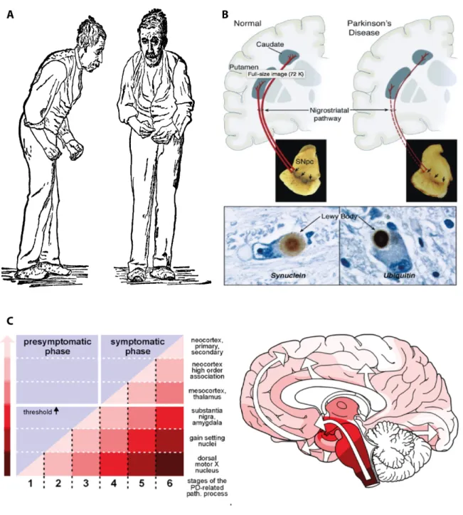
Parkinson’s disease (PD) was first described in 1817 by James Parkinson, and is nowadays the second most common neurodegenerative disorder after Alzheimer’s disease (2). At the clinical level, PD is classically characterized by motor symptoms that include bradykinesia, resting tremor, postural instability, and muscular rigidity (3) (Fig. 1A). Pathologically the degeneration of dopaminergic neurons in the substantia nigra (SN) pars compacta, and the presence of intracellular proteinaceous inclusions in the surviving neurons, known as Lewy Bodies (LBs) and Lewy Neurites (LNs) (3), are the main hallmarks of PD (Fig. 1B). Although initially classified as a movement disorder, it is now well accepted that non-motor symptoms can precede and succeed motor disabilities, and PD is currently regarded as a whole-brain disease (4). In fact, non-motor symptoms are gaining momentum in PD research for possible clinical biomarkers or predictors of the disease onset (5). Furthermore as the understanding on PD symptomatology evolves, the mechanisms underlying the pathological progression of the disease
come under spotlight. Despite lacking full validation, Braak’s staging theory is appraised as the most comprehensive data on PD progression. Braak hypothesises that PD pathology might start in either the olfactory bulb or in the lower brainstem. Later on, with the disease progressing, the pathology ascends, affecting other brain regions and promoting the appearance of other symptoms including the motor ones (6-9) (Fig. 1C). Supporting Braak’s concept, recent data suggests a “spreading”-like phenomena in PD. In particular it has been reported a time-dependent pathology appearance in normal mesencephalic grafts after being grafted in PD patients. The pathology included LBs and reduced dopamine transporter (10-14).
Phenotypically several other diseases have fallen under the PD umbrella and are commonly denominated as PD-related disorders. Kufor-Rakeb syndrome (KRS) is one of these disorders. KRS patients display a similar clinical chart to severe PD cases with an early disease onset. Besides the common symptomatology, KRS patients also exhibit pallido-pyramidal degeneration and supranuclear upgaze paresis and are levodopa responsive (15).
Figure 1. An overview on PD: from patient to pathology. A) The typical posture of a
PD patient was first illustrated by James Parkinson in his 1918 essay. B) PD has two main pathological hallmarks: the degeneration of dopaminergic neurons at the SN and the presence of intracellular proteinaceous inclusions, known as LBs. C) Braak’s hypothesis for PD progression suggests that the symptomatic phase appears when the pathology is no longer confined to the brainstem and it ascends to other brain areas. Adapted from (2, 16, 17).
1.1.1. PD Genetics
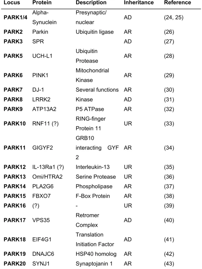
The cellular triggers underlying PD remain as unknown as the disease progression itself. Only a minority of PD cases, less than 10%, have a defined genetic cause, with the majority of cases being reported as sporadic due to their unknown origin. Thus far alterations in approximately 20 genes have been associated with PD onset (18) (Table 1). The fact that these genes are involved in a broad spectrum of cellular processes, ranging from mitochondrial homeostasis to protein degradation machinery, reflects the complexity of PD pathophysiology. This fact is also a notorious obstacle to understand which alterations in proteins, pathways or organelles are the tipping point that promotes the disease onset and progression. Furthermore, several environmental factors such as metals, pesticides and insecticides are known to cause, or at least increase the risk of developing PD (19-21).
Despite the wide genetic variability, one protein stands out from the crowd and has been extensively studied in the last decades: alpha-Synuclein (α-Syn). Mutations in SNCA, encoding for α-Syn, were the first being associated with the disease, but also duplications and triplications of the gene were later found in PD patients. Besides its involvement in the familial forms of PD, the protein is commonly found in LBs (22) and LNs (23) in sporadic cases.
Table 1. PD associated genes
AD – Autosomal dominant; AR – Autosomal Recessive; UR – Unknown Relevance.
Locus Protein Description Inheritance Reference
PARK1/4
Alpha-Synuclein
Presynaptic/
nuclear AD (24, 25)
PARK2 Parkin Ubiquitin ligase AR (26)
PARK3 SPR AD (27)
PARK5 UCH-L1 Ubiquitin
Protease AR (28)
PARK6 PINK1 Mitochondrial
Kinase AR (29)
PARK7 DJ-1 Several functions AR (30)
PARK8 LRRK2 Kinase AD (31)
PARK9 ATP13A2 P5 ATPase AR (32)
PARK10 RNF11 (?) RING-finger Protein 11 UR (33) PARK11 GIGYF2 GRB10 interacting GYF 2 AR (34)
PARK12 IL-13Ra1 (?) Interleukin-13 UR (35)
PARK13 Omi/HTRA2 Serine Protease UR (36)
PARK14 PLA2G6 Phospholipase AR (37)
PARK15 FBXO7 F-Box Protein AR (38)
PARK16 (?) - UR (39)
PARK17 VPS35 Retromer
Complex AD (40)
PARK18 EIF4G1 Translation
Initiation Factor AD (41)
PARK19 DNAJC6 HSP40 homolog AR (42)
1.2. Alpha-Synuclein
1.2.1. Structure
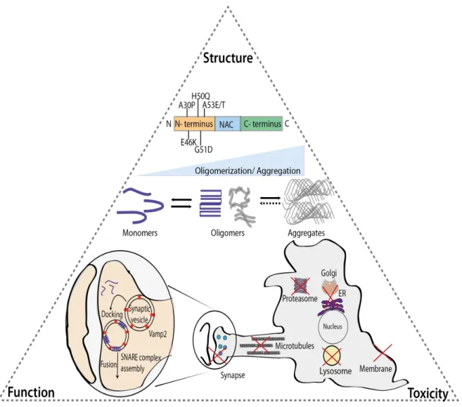
Structurally speaking the 140 amino acids of α-Syn can be separated in three distinct domains: a N-terminus prone to mutations, a central domain necessary for aggregation and a C-terminus negatively charged (Fig. 2). Besides all the 6 familial mutations (A30P, E46K, H50Q, G51D, A53E and A53T) that are found in the boundaries of the N-terminal domain (1 to 60), this region is also known for its several imperfect repeats of the consensus motif (KTKEGV) (24, 44-48). These repeats are responsible for the membrane binding and helix folding abilities to α-Syn (49, 50) and they are also found in other proteins that display identical conformational alterations (51). Artificial mutations in this domain block the interaction with membranes and enhance the protein’s intracellular toxicity (52). The next 34 amino acids (61 to 95) form the central region, also known as non-Aβ component of plaque (NAC) domain, and represent the main structural difference between the three members of the synuclein family. Highly hydrophobic, this region contains 12 amino acids (71 to 82) essential for α-Syn filament formation (53). The C-terminal part of α-Syn that includes 44 amino acids is rich in glutamates and aspartates imparting a negative charge to the protein. These negative residues can modulate α-Syn aggregation propensity (54) and, according to some reports, this domain may be responsible for the protein’s chaperone-like activity (55).
Classically, α-Syn has been considered an unfolded monomeric protein. In 2011, two reports brought controversy to the field by suggesting a native α-Syn tetrameric conformation enriched in alpha-helical structure (56, 57). These findings where rapidly questioned by other groups who found evidence for a predominance of the protein in monomeric and disordered state (58, 59). Nevertheless, this year the tetramer hypothesis was again highlighted with two novel papers reaffirming the presence of α-Syn tetrameric in physiological conditions (60, 61). These two reports put the KTKEGV imperfect repeats as the key mediator of α-Syn tetramerization and describe how some of the familial mutations can shift the equilibrium from tetramer to monomer leading to an increase in cellular toxicity.
α-Syn is highly susceptible to several post-translational modifications (PTMs), which seem to modulate the protein's propensity to aggregate, as well as its cellular behaviour and fate. The protein can be phosphorylated at two serines (S129 and S87) and three tyrosines (Y125, Y133 and Y135). Phosphorylation at S129 is considered a biochemical marker of LBs since approximately 90% of α-Syn present in LBs is phosphorylated in this residue (62, 63). Several kinases including Casein Kinases, Polo-Like Kinases and G protein-clouples receptor Kinases can phosphorylate α-Syn at position 129 (62, 64-68) with some of those enzymes being found up-regulated in PD brains (67), or present in LBs (65, 69). The S129 phosphorylation is described to affect α-Syn fibrillation although it is still unclear if this specific PTM enhances or inhibits this cellular process (63, 70-72). As for the phosphorylation at position 87 an inhibitory effect on aggregation has been observed (73). Less is known regarding phosphorylation of the tyrosines residues although, in flies, it has been described that Y125 phosphorylation leads to a decrease of α-Syn oligomeric species. Unfortunately, the full functional relevance of α-Syn phosphorylation both in a physiological and disease context is still unknown (74, 75).
In addition to phosphorylation, tyrosine residues (Y39, Y125, Y133, Y136) can also be nitrated, a PTM known to increase α-Syn toxicity. Nitrated α-Syn was found in LBs (76) and nitrated oligomers promoted mitochondrial impairment and cell death in mammalian cells (77). Substantial loss of dopaminergic neurons at the SN, accompanied by down-regulation of striatal dopamine and dopamine receptor D2, was observed upon injection of nitrated α-Syn into SN of rats (78). Interestingly nitration of position Y39 can inhibit α-Syn fibril assembly and reduce monomer degradation via the ubiquitin proteasome system (79).
Truncated α-Syn has also been found in LBs and animal models, with findings suggesting that familial mutations can increase its occurrence (80-82). In vitro truncation of α-Syn at the C-Terminus can enhance fibril formation and promote the fibrilization of full-length α-Syn (83, 84). Nevertheless the opposite effect was reported upon calpain 1- or neurosin-mediated α-Syn truncation (85-88). These two proteases cleave α-Syn near the NAC domain highlighting the importance of this region in α-Syn aggregation.
α-Syn can also undergo other PTMs including ubiquitination (82, 89-91), sumoylation (92-94) and acetylation (95-97). Ubiquitinaton occurs mainly at the
N-terminus of α-Syn protein sequence (98) although it has been described that important Ubiquitin ligases require α-Syn C-terminus to proper modify the protein (99). Ubiquitinated α-Syn can be found in LBs and this PTM can affect the protein’s degradation via de endolysosomal and autophagy pathways (89, 99, 100). Identical to ubiquitination, sumoylation also targets lysines and, in regards to α-Syn, the lysines 96 and 102 are the main amino acids affected by this PTM (92). The presence of sumoylated α-Syn has been descibred in aggregates of α-Syn although it is believe that this PTM can inhibit the aggregation formation (92, 93). A recent report presented a more functional role for α-Syn sumoylation describing an important role of this PTM on the protein’s secretion by exosomes (101).
Lastly, N-terminus acetylation of α-Syn has been has been repetitively associated to the protein’s capability for membrane binding and this PTM was also described to increase α-Syn resistance to aggregation (95-97).
1.2.2. Function
α-Syn was initially described in 1988 and its name derives from its intracellular localization: Syn- from synapse and -nuclein from nucleus (102). More than a quarter of a century later, the scientific community has yet to reach a consensus regarding the intracellular function of α-Syn. Several reports have so far have suggested a wide spectrum of hypothetical roles ranging from neurotransmitter release, to DNA binding, and mitochondrial homeostasis (103-105). The strongest evidence points towards a pre-synaptic function. Supporting this line of evidence is the protein’s abundance in the brain (106) as well as the described pre-synaptic localization (102, 107) and its co-localization with the reserve pool of synaptic vesicles (108, 109). Initial reports suggested that α-Syn could play several roles in the cycling of synaptic vesicles, modulating the vesicle pool size, mobilization and endocytosis (110, 111). Accordingly, altered synaptic vesicles dynamics, along with decreased striatal dopamine, are the minor alterations described in α-Syn knockout (KO) mice (112, 113). Notorious concerns regarding potential compensatory mechanisms, generated by other members of the synuclein family (b and g), led to the generation of double and triple KO animals. In KO mice for
α-Syn and b-α-Syn no alterations in synaptic vesicle dynamics were observed, though the animals exhibited alterations in pre-synaptic protein levels together with a distinct decrease of dopamine levels throughout the brain (114). Likewise the triple-synuclein KO mice also showed changes in the levels of pre-synaptic proteins and a more severe phenotype, including decrease of synaptic terminal size and higher lethality (115).
Important work developed by Südhof and colleagues suggests that α-Syn interacts with synaptobrevin-2 (VAMP2) and with phospholipids via its C- and N-termini, respectively (116). This interaction promotes Soluble NSF Attachment Protein Receptor (SNARE) complex assembly and recent data indicates that α-Syn multimerization ability is required upon binding to membranes (116-118). The role of α-Syn might be extremely specific since this interplay was only reported in docked membranes at the plasma membrane (117) (Fig. 2). Nevertheless VAMP2 has been linked to other intracellular pathways besides neurotransmitter release so it is possible that α-Syn has a broader role in the intracellular trafficking mechanisms (119, 120). Supporting this idea is the fact that both α-Syn and VAMP2 and strongly present in erythrocytes, blood cells that do not perform synaptic vesicles transmission (56, 121).
Nevertheless, reinforcing a putative function of α-Syn on synaptic homeostasis, the protein was also shown to compensate the loss of the pre-synaptic cysteine-string protein-alpha (CSPα) in mice. CSPα KO animals display impairment of SNARE assembly, followed by pronounced neurodegeneration and early lethality. Overexpression of α-Syn in these mice was able to partially rescue the phenotype and increase their life expectancy with the authors suggesting that α-Syn could enhance SNARE-complex assembly acting downstream of CSPα. Additionally KO animals for both α-Syn and CSPα present the most severe phenotype, confirming the involvement of α-Syn in the compensatory mechanism (122, 123).
1.2.3. Toxicity and Disease
Despite many open questions regarding the physiological function of α-Syn, this 14.5 kDa protein has taken a central stage in research on neurodegeneration due to its involvement in several brain-related diseases, commonly described as synucleinopathies. PD is one of these disorders and, as mentioned above, α-Syn
is considered an important component of the LBs and LNs although the positive or negative cellular effect of these inclusions is still elusive. A total of 6 mutations in its 140 amino acids have been found in PD patients and duplication or triplication of the SNCA gene lead to the same clinical phenotype (24, 25, 44-48, 124). Recent studies also revealed that polymorphisms in the SNCA gene lead to an increase of PD risk (39, 125-127).
In addition to PD, the group of synucleinopathies is composed of three other neurological disorders: Multiple Systems Atrophy (MSA), Dementia with Lewy Bodies (DLB) and Pure Autonomic Failure (PAF). Of the three, MSA is the disorder with a strongest connection to α-Syn, although in terms of pathology is the more distinct one. MSA patients present proteinaceous inclusions in oligodendrocytes (128-130). Two recently described mutations in α-Syn (A53E and H50Q) were found in patients displaying MSA pathology, and polymorphisms in the α-Syn gene can also enhance the risk for disease onset (48, 131-133). Patients with DLB exhibit dementia and other Alzheimer-like symptoms due to a stronger pathological effect in the cortex area (134). The clinical discrimination between PD and DLB is challenging since almost half of all PD patients also develop dementia as the disease progresses (135). More distinct from the other disorders is PAF. This disease affects the peripheral nervous system and also presents α-Syn-positive LBs and LNs, although some researchers postulate that PAF in no more than an intermediate disease state that later progresses into other neurological disorders (136-138).
The origin of α-Syn toxicity is still a debatable subject but its deleterious effects have been associated to the majority of the intracellular organelles and pathways. In fact it has been described that α-Syn can impair vesicle and protein trafficking, protein degradation systems, mitochondrial respiration, microtubule polymerization and neurite network (139-145). As mentioned above, the structure of α-Syn confers its aggregation propensity. Thus, understanding the mechanisms underlying this process has become the central quest in the field of synucleinopathies. Three main α-Syn species are commonly represented in the “aggregation pathway” that start with monomers, followed by the formation of intermediate oligomers, and finalized with aggregates/inclusions (Fig. 2). Once again a clear lack of consensus exists regarding the toxic elements of this pathway with some evidence pointing towards the oligomers (146-148), while other suggest
the aggregates (149, 150) (Fig. 2). Knowing that LBs are found in surviving dopaminergic neurons, it has been speculated that they may represent a pro-survival response to the cellular toxicity. Nevertheless a recent work concluded that different α-Syn assemblies could lead to distinct but yet complementary molecular and behavioural patterns (151). These data suggest that more than one culprit species could be underlying the disease onset.
Additionally, since the function of α-Syn might require a multimerization process upon membrane binding, it is likely that the protein is constantly shifting between cytosolic monomer and membrane-bound multimer. It has now become essential to chemically, structurally and biologically distinguish between “functional oligomer” and “toxic oligomer”. Altogether this data points to an extremely sensitive cellular balance between of function and dysfunction that weight towards one of them according to the protein propensity to bind membranes.
Figure 2. α-Syn: structure, function and toxicity. α-Syn is an intrinsically disordered
protein with 6 PD-related point mutations described so far. The protein can undergo oligomerization/multimerization that, under pathological conditions, can lead to the formation of beta-sheet structured aggregates. The outcome of the oligomerization can determine the existence of a functional (blue) or dysfunctional (grey) α-Syn with recent data suggesting a crucial role for the protein's membrane binding domains in this process. In terms of function, α-Syn has been strongly associated with vesicular trafficking, and particularly with synaptic vesicles, interacting with important players in this pathway. Although it is still not fully understood which α-Syn species are toxic, putative deleterious effects on several intracellular organelles and pathways, including proteasomes, lysosomes, synapses, microtubules, ER-to-Golgi trafficking and membranes, have been described (152).
1.3. ATP13A2
1.3.1 Structure
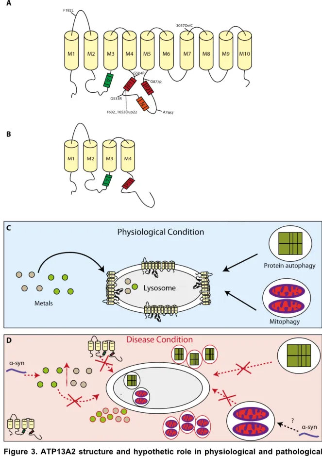
ATP13A2, also known as Park9, is a transmembrane protein of 1180 amino acids that mainly localizes in lysosomes and late endosomes (32). The protein is a member of the P5 type pump ATPases family, together with 4 other proteins, ATP13A1 and ATP13A3-5. Bioinformatic analyses suggest that ATP13A2 has 11 transmembrane domains and four functional domains: an actuator domain (A), a catalytic phosphorylation site (P1), and two nucleotide-binding domains (P2 and N) (153) (Fig. 3A). The protein is highly expressed in the brain, particularly in the SN and is upregulated in the dopaminergic neurons of this region in the brains of PD patients (32, 154). A recent work provided new insights about the intracellular behaviour and homeostasis of ATP13A2. Both C- and N-terminus of the protein are faced towards the cytosol with the latter terminal having a unique conformation that is provided by the eleventh transmembrane domain (155). Additionally it was observed that ATP13A2 could be autophosphorylated in its inactive state with several phosphates being able to induce this process also via the N-terminus (155).
1.3.2. Function
The cellular function of ATP13A2 is still elusive, as in the case of α-Syn. Fibroblasts obtained from patients with ATP13A2 mutations, revealed profound alterations in mitochondrial homeostasis. This impairment was associated with reduced ATP production and increased maximum respiration capacity, due to an impairment of mitochondrial degradation and subsequent accumulation of defective organelles. These phenotypes could be partially rescued upon ATP13A2 overexpression (156). The process of autophagic mitochondria degradation, known as mitophagy, is a crucial quality control mechanism to ensure the proper function of the organelle (157). An imbalance in this process has been associated with PD (158).
ATP13A2 has been directly linked to mitophagy in several studies (156, 159, 160) but little is known about which mechanisms are involved (Fig. 3C).
Apart from mitophagy ATP13A2 has been connected with normal protein autophagy (161-163) and metal/cation homeostasis (154, 160, 163-172) (Fig. 3C). Regarding the latter, ATP13A2 was shown to have a protective effect in manganese (Mn+)-mediated α-Syn toxicity, in both yeast and SH-SY5Y cells (166) (Fig. 3C and D). This role in Mn+ homeostasis was further explored in yeast and allowed the identification of several genes involved in the process (168). Furthermore some ATP13A2 mutants were unable to rescue Mn+ induced toxicity in mammalian cells (170) and two ATP13A2 polymorphisms enhance Mn+ neurotoxic effect in patients (169). Considering that this metal has been linked to Parkinsonism (173), and specifically to α-Syn oligomerization and aggregation (174, 175), one can speculate that Mn+ could be an intermediate between α-Syn and ATP13A2. This hypothesis is not fully accepted since a recent report concluded that in mammalian cells, ATP13A2 levels had no effect on Mn+ sensitivity (164).
Besides Mn+, ATP13A2 was shown to exert a protective effect against niquel- (Ni2+), cadmium- (Cd2+) and selenium- (Se2+) induced toxicity, in yeast and in
mammalian cell culture (171, 172), yet little is known about the role of these metals in the context of α-Syn toxicity and PD.
More recently, ATP13A2 was also linked to zinc (Zn2+) homeostasis in a study
showing that mitochondrial impairment, due to increased amounts of Zn2+, could be rescued upon overexpression of ATP13A2 (160). The interplay between α-Syn, ATP13A2 and Zn2+ has been strongly associated with autophagy and extracellular release and it will be further developed ahead.
The first results with ATP13A2 KO mice showed that the animals exhibit both PD and NCL pathology, including the formation of intracellular aggregates positive for α-Syn and ubiquitin, sensorimotor deficits and lipofuscinosis (161), suggesting that the phenotypes may not be solely gene dependent. A recent report further indicated that, in these mice, α-Syn accumulation was a side effect from a general impairment of the endolysosomal pathway (176).
1.3.3 Toxicity and Disease
Mutations in ATP13A2 have been associated with different diseases including PD, KRS (32, 177-185), and also Neuroid Ceroid Lipofuscinosis (NCL) (186-188). Of the several disease-associated mutations identified in ATP13A2, thus far only a few were investigated in detail. In cells ATP13A2 mutants exhibited loss of protein function, subcellular mislocalization in the endoplasmic reticulum (ER), increased cellular toxicity, and shorter protein half-life (Fig. 3D) (189). In a detailed study of the effects of ATP13A2 missense mutations associated with early-onset parkinsonism several novel phenotypes were identified, including disruption of the protein vesicular localization, impairment of ATPase activity and of neurite outgrowth (190). One of these mutations, commonly known as Dup22, was initially described in a Jordanian family. The patients (four in total) exhibited an early age of onset (12-15 year old) with rapid disease progression. Cardinal PD symptoms like rigidity, bradykinesia and postural instability were present, accompanied by non-motor symptoms such as hallucinations. Genetically the Dup22 mutation consists in a duplication of 22 base pairs (1632-1653) that promotes the formation of 236 erroneous amino acids followed by a stop codon. At a structural level, this mutated protein lacks 6 of the 10 transmembrane domains of ATP13A2 (Fig. 3B) (32, 191, 192). Despite the believe that the lost of transmembrane domains could lead an intrinsic incapability of ATP13A2 to be targeted to its final destination, a recent study revealed that the presence of N-terminus is enough for the protein’s colocalization at the late endosomes and lysosomes (155).
Figure 3. ATP13A2 structure and hypothetic role in physiological and pathological conditions. A) ATP13A2 is an 1180 amino acids protein with 10 transmembrane domains
and four functional domains: catalytic phosphorylation (P1), nucleotide binding (P2 and N) and actuator domain. B) The familial mutation Dup22 removes 6 of the transmembrane
domains that constitute the ATP13A2 WT C) ATP13A2 is thought to play a role on metal homeostasis and autophagy process, including protein and mitochondrial degradation via the lysosome. D) A failure in metal processing, caused by mutations or reduced activity of ATP13A2, would lead to the toxic accumulation of metals in the cytoplasm. In disease conditions, α-Syn may increase the intracellular levels of metals, exacerbating cytotoxic effects. Regarding protein and mitochondria degradation it has been reported that deficient ATP13A2 activity can lead to the accumulation of defective mitochondria or proteins (such as α-Syn) that ultimately contribute to increased cytotoxicity. Adapter from (193).
1.4. Protein Degradation
The correct degradation of proteins is an important process to assure the proper intracellular homeostasis and it is ensured by two independent, but complementary, systems. These two mechanisms, the autopaghy-lysosomal pathway and the Ubiquitin Proteasome System are named upon their final destination organelle, the lysosome and the proteasome, respectively. Monomeric α-Syn can be actively degraded by both organelles (194, 195) that can compensate each other upon one’s failure (196). When it comes to eliminating higher molecular species the lysosome is the only organelle able to deal with them and promote their correct degradation (197). Considering that both ATP13A2 and α-Syn have been extensively linked to the lysosome, I will further develop the pathways associated with this organelle.
1.4.1. The lysosome
Cell survival requires the constant turnover of its functional machinery, such as proteins and organelles, and the lysosome constitutes the main cellular component responsible for this task. This turnover is crucial for the removal of deleterious intracellular components, and for the recycling of macromolecules to guarantee proteome renewal (198). Autophagy (meaning “self-eating”, in Greek) consists in the process of decomposition and degradation of cellular components and organelles via the lysosomal compartment. Autophagy itself serves two main
purposes: the clearance of deleterious intracellular components, and the recycling of macromolecules from functional pre-existing organelles and proteins to guarantee proteome renewal (198).
When it comes to protein and organelle degradation, the lysosome can be the end point of several pathways that can be categorized in two different groups: the autophagy pathway and the Endolysosomal pathway. The first group can be divided in three pathways based on the cargo delivery method: chaperone-mediated autophagy (CMA), macroautophagy and microautophagy. The Endolysosomal pathways, despite also culminating in the lysosome, display distinct molecular players and can have two distinct origins: the ER or endocytic mechanisms.
For the purpose of this thesis, I will focus on macroautophagy and the endolysosomal pathways.
1.4.1.1. Macroautophagy
Macroautophagy is a bulk, content-blind, cellular degradation mechanism that requires the formation of de novo double membrane-bound vesicles to sequester intracellular components, including whole organelles, towards the lysosome (199, 200). The entire mechanism deeply relies on the so-called autophagy-related proteins (Atg) that were first described in yeast (201-203). In fact, the formation of the de novo membranes mainly depends on the autophagy related protein (Atg) 9, both in yeast and humans (204-207). Macroautophagy is found constitutively active but it can be enhance either via the mTOR pathway, the mammalian target of rapamycin (208), or via the PI3kinase/beclin/vsp34 pathway (209). The autophagosome formation requires two ubiquitination steps highly regulated by Atg proteins (201-203). Initially Atg12 is conjugated with Atg5, a process involving Atg7 and Atg10 (210, 211). The Atg12-Atg5 complex is later targeted to the autophagosome together with Atg16 (212, 213). The localization of this complex at the membrane is required for the second ubiquination step to occur, via Atg8 (also known by LC3) (214, 215). LC3 is C-terminally cleaved by Atg4 to form LC3-I (216, 217), which is then conjugated to the lipid phosphatidylethanolamine (PE) by Atg7 and Atg3 to generate LC3-II (211, 218). LC3-II is the most common marker for
autophagy since it is specific to this pathway and degraded only after the fusion of the autophagosome to the lysosome (217). After formation, the autophagosomes travel along the microtubules in a dynein-dependent manner towards the microtubule organizing centers where the lysosomes are located (219) (Fig. 4A). The release of the autophagosome content to the lysosome can be performed either by fusion of both compartments or just by a transfer of the autophagosome content to the lysosome without loss of the first structure (219). The mechanisms underlying the fusion of both compartments are largely unknown in mammalian cells, except for the fact that Rab7 plays a role on this process (220).
1.4.1.2. The Endolysosomal Pathway
The endosomal pathway is a complex intracellular trafficking mechanism that has been extensively associated to PD (221). In this pathway the early endosomes (EE) are the main sorting pit stops that receive content originating in the plasma membrane via endocytosis, and from the Trans-Golgi network (TGN). Once inside the EE the content may have two destinations: either it is targeted to the lysosome to be degraded or it is recycled towards the plasma membrane. Depending on their faith, the proteins are usually found in different sections of the EE. Specifically, proteins that will be recycled are commonly found in the tubular sorting endosome while the ones targeted for the lysosomes are present in the vacuolar sorting endosome. In the latter the content is stored in intraluminal vesicles (ILV) that originate by inward budding of the EE membrane. The content of the ILVs and their formation is strongly dependent on the endosomal sorting complexes required for transport (ESCRT) (222-224). The cargo identification by the ESCRT typically demands a previous ubiquitination step, although an ESCRT-independent mechanism has also been described (222, 225). The vacuolar endosome suffers a process of continuous maturation that culminates in the formation of late endosome (LE), via multivesicular Bodies (MVBs). At this stage the LE contains few proteins intended for recycling after endocytosis, and the majority of the content being targeted for the lysosome. Importantly, differences are also observed in the protein composition at the membrane of the LE with the most well described alteration being a Rab conversion, consisting in a switch between Rab5, present in the EE, and Rab7 found in LE (226). A second sorting
event takes place at the LE/MVBs complex, enabling the content can either be forwarded towards the lysosome for degradation or targeted to the extracellular environment via exosomes (Fig. 4B). The biological function of exosomes is still elusive, but some speculate that these vesicles may constitute a cellular clearance route to proteins that cannot be degraded by the lysosome (227).
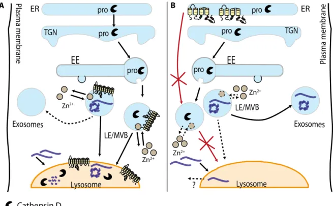
Besides the notorious role in protein degradation, the endolysosomal pathway itself is of an extreme importance for the lysosome biogenesis and function. It is through this pathway that lysosome membrane proteins and intralysosomal hydrolases are delivered to the lysosome. The most studied pathway is the M6PR-dependent transport, used by proteins such as Cathepsin D (CatD), and relies on the addition of an M6P-tag to the hydrolases, though some proteins can undergo different processing (228, 229). Regarding M6P-independent pathways, recent findings point that hydrolases can migrate to the lysosomes bounded to lysosomal membranes, like β-Gluocerebrosidase, a protein which gene, GBA, has been extensively linked to PD (230, 231).
The extensive network of the endosomal pathway requires the constant movement of the vesicles inside the cell. Rab proteins are major regulators of the dynamics underlying these movements, and the overall intracellular trafficking (232, 233). More than 60 members compose the highly conserved Rab family (234) with Rab5, Rab7 and Rab22B being the ones more linked to this endosomal mechanism (235) (Fig. 4B). Nevertheless Rab8a, which has been previously linked to α-Syn (236), has recently emerged as an important protein in the normal function of this pathway (237). In detail, it was shown that normal endolysosomal requires the presence of the complex Rab8 and optineurin at the Golgi membrane (237). A mislocalization of the complex leads to decrease in CatD maturation and impaired lysosomal protein degradation.
Figure 4. Mechanisms of protein degradation: autophagy and endolysosomal pathways. A) Three main cellular pathways can enhance macroautophagy: mTor
dependent, mTor independent and JNK/Blc2. All terminate in the complex Beclin-1/VPS34 that will promote the formation of the autophagosome that surrounds the content targeted to degradation. This autophagosome formation starts with an ubiquitination reaction where Atg12 is conjugated to Atg5, a process mediated by Atg7 and Atg10. After the binding of Atg16L, the Atg12/Atg5/Atg16L complex is targeted to the autophagosome. This complex localization at the membrane is necessary for the second ubiquination and requires LC3. Initially, LC3 is cleaved at the C-terminus by Atg4 to form LC3-I, which is then conjugated to a PE by Atg7 and Atg3 to generate LC3-II. The membrane of the autophagosome is originated from other organelles and transported by Atg9. After closing the autophagosomes is moved towards the lysosomes where it fuses, to deliver the cargo for
degradation. B) The endolysosomal machinery involves several intracellular players. The content being delivered into the lysosome can arise from the TGN, where Rab8a is a master player, or through the extracellular environment via endosomes that are positive for Rab11. In either case, the content is targeted to the Vacuolar Sorting component of the EE. This structure will then give origin to the LE/MVBs a process that requires a Rab conversion from Rab5 to Rab7 at the membrane level. These LE/MVBs that can undergo two distinct pathways: 1) form Rab27a positive exosomes and release the content to the extracellular media or 2) fuse with the lysosomes to allow the degradation of proteins or the delivery of lysosomal membrane proteins and hydrolases. Adapted from (238).
1.4.2. The interplay between α-Syn, ATP13A2, and protein degradation pathways
Monomeric α-Syn degradation is divided into two complementary mechanisms, the ubiquitin proteasome system and the CMA (194, 195) that compensate each other upon one’s failure (196). When it comes to eliminating higher molecular species the burden shifts entirely to the lysosome (197). Nevertheless, the CMA cannot handle species larger than dimers (239, 240) so another mechanism, macroautophagy, is activated.
The degradation of α-Syn via macroautophagy has been mostly studied by using specific inhibitors and enhancers of this pathway. Macroautophagy can degrade both WT and mutant α-Syn in a Beclin-1 dependent process (99, 241, 242), though some literature points to a mutant-specific degradation (243, 244). Formation of high molecular weight species has been reported upon blockage of this pathway, although the results are controversial (245, 246). No overall consensus has been reached on the effect of macroautophagy in α-Syn homeostasis, although in yeast, a model organism that lacks CMA, it was demonstrated that phosphorylation and sumoylation of α-Syn are able to modulate the protein’s degradation via this pathway and, ultimately, inclusion formation (71, 94).
On the other hand, α-Syn also impacts macroautophagy. The overexpression of the protein can inhibit macroautophagy via an interaction with Rab1a that culminates in a mislocalization of Atg9 (141). In DLB patients and αSyn
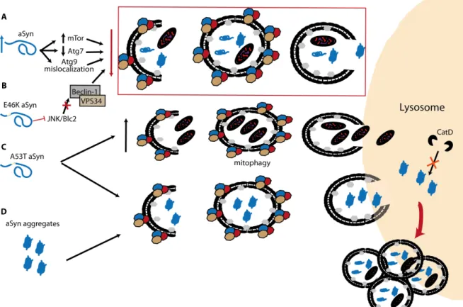
overexpressing transgenic mice an increase in mTor and decrease in Atg7 levels has been observed (140) (Fig. 5A). The same study also reported the presence of enlarged autophagosomes and lysosomes, as it was first observed in cells overexpressing α-Syn (241). A recent work reported a drastic effect in the presence of α-Syn aggregates, as these species appear to be resistant to macroautophagy and promote a general failure of the pathway with subsequent accumulation of autophagosomes (150) (Fig. 5D). Besides the WT α-Syn, also the familial PD-associated mutations can have a deleterious effect on macroautophagy. The familial mutation E46K was reported to impair macroautophagy via inactivation of the JNK1/Blc2 pathway (247) (Fig. 5B). As for the A53T mutation opposite effects were described, with one group suggesting an enhancement of macroautophagy, especially the mitophagy pathway (248), while other suggested an impairment of degradation leading to the accumulation of autophagosomes (249) (Fig. 5C).
Figure 5. The interplay between Syn and macroautophagy. A) Accumulation of
α-Syn can increase mTor, decrease Atg7 levels and promote mislocalization of Atg9 that, individually or combined, can alter the delicate homeostasis of macroautophagy. B) α-Syn familial mutation E46K was described to inhibit macroautophagy via JNK/Blc2, an
mTor independent pathway. C) On the other hand, two opposite effects have been associated with the A53T mutation: increase in mitophagy, and accumulation of autophagosomes due to impaired degradation. D) As for α-Syn aggregates, these species cannot be degraded by macroautophagy, leading to the impairment of the pathway. Adapted from (238).
Once inside the lysosome, α-Syn degradation is a task usually performed by CatD (250). Synthetized in the ER, this protease is initially cleaved generating an inactive proCatD (251). The proCatD is then targeted to the lysosome, a process that was found to be endosome-mediated (252, 253). Upon reaching an acidic environment, cysteine proteases are responsible for the cleavage of proCatD at the N-terminus to originate the active form of CatD (254, 255). Confirming the relevance of CatD in α-Syn homeostasis, overexpression of CatD was shown to protect against α-Syn-induced toxicity (256). CatD KO mice exhibit the formation of insoluble α-Syn species while in cell models the expression of an inactive mutant of CatD generated identical effects (257, 258). Increased levels of mutated CatD enhanced the expression of Cathepsin B, a protease that can promote α-Syn aggregate formation (256, 259). Recent findings indicate that treating cells with α-Syn aggregates leads to lysosomal rupture and cathepsin B dependent ROS production (260). At the pathological level, decrease of CatD has been described in neurons of the SN with α-Syn-positive aggregates, in PD patients (261). Nevertheless other groups reported no difference in CatD between PD patients and controls (262, 263).
As mentioned above, ATP13A2 has been strongly associated with autophagy, mainly due to its localization at the LE and lysosomes membranes. ATP13A2 knockdown (KD) and KO models exhibit an impairment of α-Syn degradation (161, 162). Further results pointed towards an overall autophagy impairment due to alterations of lysosomal pH and the levels of hydrolases leading to a failure in autophagosomes clearance, and a decreased proteolytic processing (162, 163, 165). Recently, it was suggested that aggregate formation in ATP13A2 KO mice was α-Syn-independent, and a clear impairment of the endolysosomal pathway was reported. This impairment resulted in a reduction in CatD maturation, and localization at the lysosome (176). These findings are in agreement with a previous report showing a decrease in CatD activity upon ATP13A2 mutations in
Medaka fish (264), and puts both CatD and ATP13A under the spotlight. Curiously mutations in both CatD and ATP13A have been associated to NCL and, alike ATP13A2, CatD KO mice exhibit a NCL phenotype, which may suggest that similar cellular mechanisms are taking place (161, 186, 265, 266) (Fig. 6A and B). Another strong line of research combines protein clearance with metal homeostasis in the paradigm correlating ATP13A2 and α-Syn. Two recent studies suggest unbalanced Zn2+ homeostasis as the starting point of a chain of events responsible for α-Syn clearance. The first study showed that alterations in Zn2+ intracellular levels and cellular sub-localization could promote lysosomal dysfunction and α-Syn accumulation. This phenotype was enhanced upon ATP13A2 KD, in cells and in fibroblasts from patients carrying ATP13A2 mutations, and could be rescued after ATP13A2 overexpression (165). On the other hand, an independent study focused upstream of the endolysosomal pathway, in particularly the LE/MVBs. The authors found that MVBs are targeted to exocytosis, instead of autophagy, and this mechanism is the main pathway underlying the decrease of intracellular α-Syn levels (164). In this perspective, ATP13A2 was shown to modulate Zn2+ levels, which, in turn, could influence the biogenesis of exosomes (Fig. 6A and B).
Altogether, it becomes obvious that the endolysosomal pathway, either due to metal homeostasis or protein degradation imbalance, plays an important role in neurodegeneration and, more specifically, in PD.
Figure 6. The hypothetical interplay between ATP13A2, Zn2+ and α-Syn. A) In normal
conditions CatD is processed via the TGN and delivered to the lysosome via the endosomal pathway. Importantly ATP13A2 appears to play an important role in the maturation of CatD. ATP13A2, together with Zn2+, appear to regulate the intracellular fate
of α-Syn towards either the lysosome or the extracellular medium via exosomes. B) Mutations in ATP13A2 or the absence of the protein can inhibit the maturation of CatD and its localization at the lysosome. In ATP13A2 KO mice the formation of aggregates positive for α-Syn has been described, an identical situation reported in CatD KO mice. In cell models mutations or KD of ATP13A2 promoted the release of α-Syn to the extracellular medium in a process that involved Zn2+ homeostasis.
1.5. Endoplasmic Reticulum Homeostasis
To ensure proper protein function, macromolecules need to undergo a meticulous process of synthesis and folding. The ER is one of the main organelles involved in this process, and is also an important fall-back player upon proteostasis impairment.
After production in the cytosol, two main protein types go through ER processing: water-soluble and transmembrane proteins, like ATP13A2. The ER also has a role
in the synthesis of lipids, that will be integrated in the majority of cell organelles such as mitochondria, lysosomes, peroxisomes, Golgi and even the plasma membrane (267). As previously mentioned, the ER also provides de-novo membranes to the autophagosome, via Atg 9, and is essential in macrosecretion (268, 269).
The accumulation of misfolded, mutated or aggregated proteins at the ER lumen triggers an organelle response, commonly referred to as ER stress, and the activation of a cascade of cellular pathways, known as Unfolded Protein Response (UPR). Depending on the amount of accumulated misprocessed proteins, and the duration of the stress, two opposite pathways can be activated. More specifically, under transitory stress, pro-survival pathways are initiated but upon prolonged stress, apoptotic cell death mechanisms assume control. Both cellular faiths rely in common initiators that lay on the ER membrane: the inositol-requiring enzyme 1 alpha (IRE1alpha), the protein kinase RNA-like ER kinase (PERK) and the activating transcription factor 6 (ATF6) (270-273). Under resolvable ER stress, the three cascades work in symbiosis by activating or inhibiting several pathways to promote cell survival. Briefly upon phosphorylation, PERK activates its downstream target eiF2a (eukaryotic translation initiation factor 2a), leading to the inhibition of new protein translation mechanisms and activation of an antioxidant, autophagy response (273, 274).
The IRE1alpha pathway is based on an unconventional splicing of the X-Box binding protein 1 (XBP1) that will actively promote the transcription of genes involved in protein folding, autophagy and ER-associated protein degradation (ERAD) (275, 276). IRE1alpha can also upregulate the Regulated IRE1-alpha-Depdendent Decay (RIDD) pathway that is responsible for mRNA degradation. Recent data suggests that RIDD action is crucial to maintaining the Death Receptor 5 (DR5) mRNA levels reduced and avoid the activation of caspase pathways. As for ATF6 the cytosolic fragment generated upon its cleavage will translocate to the nucleus and enhance the transcription of chaperones and ERAD associated genes (277). This latter cascade is the only of the three that is not active in the apoptotic and cell death signalling.
Prolonged (chronic) ER stress leads to an alternative, pro-apoptotic cascade. The majority of ER-mediated death signalling originates from the PERK pathway that, via ATF4, promotes the stable upregulation of the C/EBP-homologous protein