2017
UNIVERSIDADE DE LISBOA
FACULDADE DE CIÊNCIAS
DEPARTAMENTO DE QUÍMICA E BIOQUÍMICA
Prodrugs of weak acids with activity against M.tuberculosis
Ana Rita Narciso Pratas
Mestrado em Bioquímica
Especialização em Bioquímica Médica
Dissertação orientada por:
Luís Filipe Vicente Constantino
Maria Teresa Troina Pamplona Berry
i
Acknowledgements
Ao meu orientador Doutor Luís Filipe Vicente Constantino pela sua orientação e oportunidade de realizar este trabalho.
À minha co-orientadora Doutora Maria Teresa Troina Pamplona Berry por me ter aceite como sua mestranda.
À professora Doutora Emília Alice dos Reis Torroaes Valente por todo o apoio, acompanhamento e dedicação prestada ao longo de todo o trabalho.
Ao Doutor Nuno Carmo por me ter realizado os estudos de actividade e por toda a disponibilidade demonstrada em tirar qualquer dúvida que me surgisse.
A todos os membros da unidade de Química Medicinal do iMed-ULisboa, que directa ou indirectamente tornaram possível a realização deste trabalho. Todo o vosso apoio e amizade foi bastante importante para mim e não poderia desejar melhores colegas. Em particular, quero agradecer aos amigos que fiz nesta etapa e que melhoraram todos os meus dias – Alessandro, Dário, Elizabeth, Gustavo, Jorge, Lara, Luís, Margarida, Ricardo e Rita. Sem vocês não teria sido o mesmo e foi mesmo um prazer enorme conhecer-vos e ter passado os meus dias convosco. Quero também agradecer à Patrícia por me ter acompanhado ao longo de todo o meu percurso académico e em particular durante esta etapa em que passamos todos os dias juntas, por todo o apoio, conversas, desabafos, sorrisos e pela boa amiga que se revelou.
Aos meus amigos da licenciatura – Beatriz, Daniel, Filipa, Margarida, Raquel, Ricardo e Tomás por toda a amizade, carinho e apoio desde que entrei na faculdade. Foram os melhores amigos que a vida académica me podia ter dado.
As minhas amigas Bárbara, Inês e Patrícia por simplesmente serem minhas amigas e estarem presentes todos os dias na minha vida.
À minha família por pelo apoio e carinho que me transmitem.
Aos meus pais e ao meu irmão por sempre me apoiarem, por tudo o que me proporcionaram e continuam a proporcionar, por serem um porto de abrigo e de conforto que mais ninguém consegue ser. Por serem fantásticos todos os dias e pelo amor incondicional permanente.
ii
Resumo
A tuberculose é uma doença infecciosa que afecta milhoões de pessoas em todo o mundo. Em 2015 foram relatados 9,6 milhões de novos casos. O agente infeccioso causador da doença,
Mycobacterium tuberculosis, desenvolveu várias resistências aos tratamentos existentes. Esta
bactéria possui um envelope celular bastante complexo, constituído por ácidos micólicos interferindo na sensibilidade da bactéria aos antiobióticos hidrofóbicos. Os tratamentos actualmente existentes consistem em combinações de fármacos de primeira linha (isoniazida, rifampicina, pirazinamida e ethambutol). No entanto, surgiram estirpes resistentes a alguns dos fármacos administrados no tratamento dando origem a outros dois tipos da doença: tuberculose multi-resistente e tuberculose extensivamente multi-resistente. Nestes casos o tratamento envolve outro tipo de fármacos: fármacos de 2ª e 3ª linha. Estes tipos de fármacos são mais dispendiosos e apresentam diversos efeitos secundários.
Nos fármacos de 2ª linha, existe um grupo de fármacos que são as fluroquinolonas. Estes fármacos derivam do ácido nalidíxico e contêm fluor na sua estrutura, sendo activos contra um grande número de bactérias. Este grupo de compostos tem como alvo enzimas envolvidos no processo de transcrição do DNA, processo crucial para a vitalidade da bactéria. O ácido nalidíxico é uma quinolona usada normalmente no tratamento de infeções urinárias causadas por bactérias. Este antibiótico actua através da inibição do ADN girase, um enzima fulcral no processo de transcrição do ADN bacteriano.
Estudos anteriores demonstraram que alguns ácidos fracos apresentam atividade antimicobacteriana contra Mycobacterium tuberculosis em virtude da sua acumulação no interior da célula, devido a um mecanismo de efluxo deficiente. Sendo o ácido nalidíxico um ácido fraco, alterações na sua estrutura poderão melhorar as suas propriedades farmacocinéticas bem como a sua activida antimicobacteriana.
Os pró-fármacos são compostos que não são biologicamente activos, ou seja, necessitam de ser activados através de reações químicas ou enzimáticas. Este tipo de compostos é sintetizado quando a substância activa possui propriedades indesejadas, como baixa absorção, degradação rápida por parte de enzimas, deficiente entrada nas células das micobactérias, entre outras. Na preparação de um fármaco contra a tuberculose, é necessário que este apresente boas propriedades de absorção, distribuição, metabolismo e excrecção, sendo ativo no local desejado e não tóxico contra o organismo humano. Neste sentido, é necessária a síntese de compostos estáveis à hidrólise por parte de enzimas, plasmáticos e hepáticos do hospedeiro, que penetrem a membrana celular da bacteria, portanto lipofílicos e que sejam activos contra a bactéria
Mycobacterium tuberculosis. Para serem ativos, é necessário que os compostos sejam
hidrolisados pelos enzimas da micobácteria, de forma a converter os pró-fármacos na sua forma activa. Por último, os derivados sintetizados devem apresentar níveis de toxicidade reduzidos para o hospedeiro.
Uma das estratégias mais comuns na síntese de pró-fármacos é a formação de grupos éster pois estes grupos levam a um aumento da lipofilia do composto e consequentemente facilitam a sua passagem através das membranas biológicas. Estudos anteriores demonstraram que o
iii Neste trabalho foram sintetizados derivados do ácido nalidíxico (1). Os derivados sintetizados são ésteres pois estes são mais lipofílicos, e por isso penetrarão mais facilmente nas micobactérias que o ácido correspondente. Para que os compostos sejam ativos, é necessário que cheguem ao local de acção sem sofrer alterações na sua estrutura. Assim, é necessário que estes ésteres sejam resistentes à hidrólise por enzimas humanos e, por outro lado, susceptíveis à hidrólise por parte de enzimas micobacterianos.
Os ésteres sintetizados apresentam diferentes cadeias alcoxílicas (entre 2 e 14 carbonos), de forma a estudar o efeito da lipofilia na estabilidade e actividade destes. Na porção acilo, foi feita uma substituição em relação ao ácido nalidíxico de forma a verificar se aquela porção da molécula tem influência na actividade do composto. Pretendeu-se avaliar se a substituição do grupo C7-CH3 pelo grupo C7-CCl3 levava a melhores resultados a nível de inibição do crescimento micobacteriano bem como se os compostos eram susceptíveis ou resistentes à hidrólise enzimática. De facto, estudos anteriores demonstram que aquela porção da molécula apresenta um papel crucial na actividade deste tipo de compostos. Para além de ésteres, foi sintetizado também um derivado do ácido carboxílico que continha apenas a substituição do grupo C7-CH3 pelo grupo C7-CCl3, de forma a testar a sua actividade bem como para controlo de ensaios de estabilidade.
Na síntese de todos os derivados, dois métodos distintos foram usados e observou-se que os rendimentos eram bastante diferentes entre os dois grupos de ésteres: ésteres com o grupo C7-CH3 tiveram um rendimento de síntese acima dos 51% enquanto que ésteres com o grupo C7-CCl3 tiveram um rendimento de síntese inferior a 35%.
Os estudos de actividade foram realizados na estirpe H37Rv de Mycobacterium tuberculosis. Nestes estudos, foram analisadas as concentrações mínimas necessárias para inibição de 90% e de pelo menos 50% do crescimento bacteriano. Também foi analisada a percentagem de inibição do crescimento bacteriano para uma concentração administrada de composto de 100 µM. Observou-se que praticamente todos os derivados do ácido nalidíxico inibem o crescimento micobacteriano. Para além disso, verificou-se que os derivados com o substituinte C7–CCl3 são mais activos, de um modo geral, que os derivados com o substituinte C7-CH3.
Foram realizados estudos de estabilidade em plasma humano e em tampão fosfato pH 7,4. Os estudos em plasma humano permitiram observar se os compostos são susceptíveis à hidrólise enzimática por partes dos enzimas presentes neste compartimento corporal. Os ensaios em tampão fosfato pH 7,4 foram realizados de forma a avaliar a estabilidade química dos compostos. Verificou-se que os derivados sintetizados são muito resistentes à hidrólise química, bem como à hidrólise enzimática. De facto, os compostos apresentam tempos de semi-vida bastante elevados revelando-se muito estáveis. Os resultados obtidos demonstram que os compostos sintetizados apresentam tempos de semi-vida superiores a 2,8 dias, no caso da hidrólise enzimática, e 7,8 dias no caso da hidrólise química. Para além disso, no estudo da susceptibilidade à hidrólise
R1: CH3, CCl3
R2: different alkoxyl chains 1
iv enzimática, houve compostos que não demonstraram degradação durante as 72 horas do ensaio em plasma humano.
Fez-se também uma comparação entre as constantes cinéticas da hidrólise enzimática e da hidrólise química para avaliar se a hidrólise química tem impacto nos estudos de estabilidade em plasma humano. Verificou-se que para alguns compostos este tipo de hidrólise não é significativa, porém, em alguns deles, esta apresenta um impacto semelhante ao da hidrólise enzimática. Com base neste estudo, pretende-se o desenvolvimento de novos pró-fármacos com actividade contra a tuberculose, de modo a combater as estirpes resistentes aos tratamentos atuais.
Neste trabalho, foi possível concluir que os derivados ésteres desenvolvidos do ácido nalidíxico são muito estáveis à hidrólise enzimática bem como à hidrólise química. Verificou-se que os derivados que apresentam o substituinte C7–CCl3 são mais activos contra a estirpe estudada do que os compostos que apresentam o substituinte C7-CH3. Apesar de praticamente todos os derivados sintetizados apresentaram actividade contra a bacteria, alterações na sua estrutura são necessárias de modo a aumentar a inibição do crescimento bacteriano, a menores concentrações administradas, mas mantendo a estabilidade à hidrólise enzimática no hospedeiro.
No futuro mais estudos com este tipo de derivados serão necessários, tais como, ensaios de toxicidade, estudos de modelação, estudos em homogenato de fígado, entre outros. Para além disso, é necessário compreender qual o mecanismo que leva à inibição do crescimento bacteriano por parte dos derivados do ácido nalidíxico sintetizados.
v
Abstract
Tuberculosis is one of the most prevelent diseases in the world with high rates of incindence and mortality. Mycobacterium tuberculosis, the causative agent of tuberculosis, is one of the leading bacterial cause of infection. There are several treatment regiments available to treat
Mycobacterium tuberculosis but the development of new strains and co-infection with HIV led
to the need of new antibiotics to treat this pathogen. One of the antibiotics to treat multidrug resistant and extensively drug resistant tuberculosis are fluoroquinolones. These drugs are fluorinated nalidixic acid derivatives and are active against a broad spectrum of bacteria. Nalidixic acid is a quinolone used in the treatment of urinary tract infections caused by bacteria. This drug inhibitis DNA gyrase, an enzyme with an important role in bacterial transcription. Many organic acid drugs have small half lives, high protein binding and difficulties in celular penetration. In order to test if we could obtain derivatives with better activity/improved pharmacokinetics several ester derivatives (1) were synthesized.These derivatives will be converted inside the bacterial cell to their correspondent acids. In order to be effective, these compounds need to be resistant to hydrolysis by human enzymes, but must be hydrolyzed by the mycobacterial enzymes.
Almost all of the synthesized derivatives have shown antibacterial activity against M. tuberculosis H37Rv strain. Besides that, it was shown that derivatives containing the C7-CCl3 group instead of a C7-CH3 group inhibit more the bacterial growth.
Stability assays were performed in human plasma and phosphate buffer pH 7.4 in order to evaluate enzymatic and chemical hydrolysis, respectively. With these assays, it was possible to observe that all the studied compounds have a good stability, being resistant to the action of the enzymes in human plasma. Besides that, it is possible to conclude that the synthesized compounds are also very resistant to chemical hydrolysis in phosphate buffer pH 7.4.
In the present work it was possible to conclude that nalidixic acid derivatives are very stable to enzymatic and chemical hydrolysis. It was also observed that almost all the synthesized compounds are active against Mycobacterium tuberculosis and changes in the acyl structure lead to a higher inhibition of bacterial growth. Although, further changes in the structure can probably improve further the activity of the compounds while keeping the high stability to host enzymatic hydrolysis.
Keywords: Tuberculosis, Nalidixic acid, Prodrugs, Stability, Activity
R1: CH3, CCl3
R2: different alkoxyl chains
vii
Index
1. Introduction ... 2 Tuberculosis ... 2 1.1.1 Epidemology ... 2 1.1.2 Disease Characterization ... 2 1.1.3 Mycobacterium tuberculosis ... 3 1.1.4 Treatment ... 4 1.1.5 Fluoroquinolones ... 5 1.1.6 Nalidixic Acid ... 6 Prodrugs ... 6 1.2.1 Esters as Prodrugs ... 7 1.2.2 Chemical Hydrolysis ... 8 1.2.3 Enzymatic Hydrolysis ... 8 Esterases ... 91.3.1 Esterases of Human Plasma ... 10
1.3.2 Mycobacterial Esterases ... 11
The Aim of the Thesis ... 12
2. Synthesis and Structural Identification ... 15
2.1 Ester Synthesis ... 15
2.1.1 Esters from Acyl Chlorides ... 15
2.2 Synthesis of Nalidixic Acid Esters ... 16
2.2.1 First library of Esters ... 18
2.2.2 Seconds Library of Esters ... 19
2.3 Structural Identification of Synthetized Esters ... 20
2.3.1 Nuclear Molecular Ressonance ... 20
2.3.2 Mass Spectrometry ... 24
3. Stability and Activity Assays ... 27
3.1 Stability Assays in Phosphate Buffer ... 27
3.2 Stability Assays in Human Plasma ... 29
3.3 Ratio between Human Plasma Stability/Phosphate Buffer pH 7.4 Stability ... 32
3.4 Activity Studies in vitro ... 33
4. Discussion ... 37
4.1 Synthesis and Structure Analysis ... 37
4.2 Stability Assays ... 39
4.3 Activity Assays ... 40
viii
5. Conclusion ... 49
6. Future perspectives ... 51
7. Experimental Procedure ... 53
7.1 Equipment and material ... 53
7.2 Reagents and Solvents ... 53
7.3 Synthesis ... 54
7.4 Ester stability assays... 64
7.4.1 Stability assays in phosphate buffer pH 7.4 ... 64
7.4.2 Stability assays in human plasma ... 64
8. Bibliography ... 67
Anex I………..…………xvi
Appendix I……….xviii
ix
Figures Index
Figure 1. 1 - Estimated TB incidence rates in 20142. ... 2
Figure 1. 2 - Four generations of quinolones. Ciprofloxacin, levofloxacin and moxifloxacin are FQ. Scaffolds are colored black and peripheral chemical modifications are colored red32. ... 5
Figure 1. 3 - NAL structure. ... 6
Figure 1. 4 - Hydroxynalidixic acid structure. ... 6
Figure 1. 5 - Scheme of chemical hydrolysis of carboxylic derivatives (esters and amides). (a) acid catalysis (b) base catalysis54. ... 8
Figure 1. 6 - Catalytic groups of hydrolases involved in ester and amide bond hydrolysis54. ... 9
Figure 1. 7 – Scheme of the α/β-hydrolase fold. β-Sheets (1-8) are shown as blue arrows, α-helices (A-F) as red columns and the relative positions of the amino acids residues of the catalytic triad are indicated as orange circles. ... 10
Figure 1. 8 - NAL derivatives representation. R1 = -CH3, -CCl3; R2= different alkoxyl chain. 13 Figure 2. 1 - General reaction of ester synthesis. ... 15
Figure 2. 2 - Mechanism of acid-catalyzed esterification53. ... 15
Figure 2. 3 - General reaction of acyl chlorides synthesis with thionyl chloride53. ... 16
Figure 2. 4 - Mechanism of acyl chlorides synthesis using thionyl chloride53. ... 16
Figure 2. 5 – Libraries of NAL esters. (a) library 1; (b) library 2. ... 17
Figure 2. 6 – Scheme of ester synthesis by method A. ... 17
Figure 2. 7 - Scheme of ester synthesis by method B. ... 18
Figure 2. 8 – Structure of compound 10. ... 19
Figure 2. 9 - Structure of compound 10a. ... 20
Figure 2. 10 - Mass spectrometry result of compound 9a. ... 25
Figure 3. 1 – Variation of ester and acid concentration during phosphate buffer (pH=7.4, T=37ºC) assay of ester 4. ... 28
Figure 3. 2 - Variation of ester and acid concentration during phosphate buffer (pH=7.4, T=37ºC) assay of ester 5a. ... 29
Figure 3. 3 - Variation of ester and acid concentration during human plasma assay of ester 2. . 31
Figure 3. 4 - Variation of ester and acid concentration during human plasma assay of ester 5a. 31 Figure 3. 5 - Estimation of 50% of inhibition of M. tuberculosis H37Rv strain growth by compounds of library 1. ... 34
Figure 3. 6 - Estimation of 50% of inhibition of M. tuberculosis H37Rv strain growth by compounds of library 1. ... 35
Figure 4. 1 - Starting material of the obtained compounds. In compound 1, R1 is a CH3 group. In compound 1a, R1 is a CCl3 group. ... 37
Figure 4. 2 - General structure of compounds from libraries 1 and 2 with the main carbons numbered. ... 37
Figure 4. 3 – Ester hydrolysis in human plasma and phosphate buffer assays. ... 39
Figure 4. 4 - Model of quinolone binding to GyrA-GyrA dimer of DNA gyrase. Substitute on carbon C7 is near Gly81. Carboxyl and keto oxygens are near Asp87 and Ser83, respectively95. ... 42
Figure 4. 5 – Relationship between alkoxyl chain length and kobs value (plasma). ... 44
Figure 4. 6 - Relationship between alkoxyl chain length and inhibition percentage of M. tuberculosis H37Rv growth for a concentration of 100µM of compounds of library 1. ... 44
x Figure 4. 7 - Relationship between alkoxyl chain length and inhibition percentage of M.
tuberculosis H37Rv growth for a concentration of 100µM of compounds of library 2. ... 45 Figure 4. 8 - Relationship between alkoxyl logP and percentage of inhibition of M. tuberculosis H37Rv strain growth for a concentration of 100µM for compounds of library 1. ... 46 Figure 4. 9 - Relationship between logP and percentage of inhibition of M. tuberculosis H37Rv strain growth for a concentration of 100µM for compounds of library 2. ... 46 Figure 4. 10 – Relationship between lipophilicity and plasma stability. ... 47
xi
Table Index
Table 1. 1 - Drugs used in TB treatment. ... 4
Table 2. 1 - First library of NAL esters. ... 18 Table 2. 2 - Second library of NAL derivatives. ... 19 Table 2. 3 - 1H-NMR signal (ppm) of aromatic protons and protons attached to C
1’, C2’ and C1’’ in libraries 1 and 2. ... 20 Table 2. 4 - 13C-NMR signal (ppm) of carbons in libraries 1 and 2. ... 23
Table 3. 1 – Hydrolysis of compounds of libraries 1 and 2 in phosphate buffer pH=7.4. ... 27 Table 3. 2 - Hydrolysis of compounds of libraries 1 and 2 in 80% of human plasma suspension in phosphate buffer pH=7.4. ... 30 Table 3. 3 – Ration between (103x)k
obs/(h-1) of plasma hydrolysis and (103x)kobs/(h-1) of phosphate buffer pH 7.4 hydrolysis... 32 Table 3. 4 – Results of activity assays in M. tuberculosis H37Rv performed by PhD Nuno Carmo from Host-Pathogen Interactions group of iMed ULisboa, lead by Professor Elsa Anes. ... 33
Table 4. 1 – Percentage of inhibition of the bacterial growth M. tuberculosis H37Rv strain to a concentration of 100µM. ... 41 Table 4. 2 – Structural characteristics, plasma stability and activity against H37Rv strain of M.
tuberculosis results for each compound. The logP values were calculated with AlogPs software
xii
List of Abbreviations
TB – Tuberculosis
M. tuberculosis – Mycobacterium tuberculosis
NTM - Non-tuberculous mycobacteria
MTC – Mycobacterium tuberculosis complex
MDR-TB – Multidrug resistance tuberculosis
XDR-TB – Extensively drug-resistance tuberculosis
FQ – Fluoroquinolones
CIP – Ciprofloxacin
LEV – Levofloxacin
MOX – Moxifloxacin
NAL –Nalidixic acid
QRDR - Quinolone resistance-determining region
ADMET – Absorption, distribution, metabolism, excretation and toxicity
Ser – Serine His – Histidine Gly – Glycine Leu – Leucine BChE – Butyrylcholinesterase PON 1 – Paraoxonase 1 PON 2 – Paraoxonase 2 PON 3 – Paraoxonase 3
HAS – Human serum albumin
Tyr – Tyrosine
LipF - Rv3487c
LipH – Rv1399
Eq – Equivalent
h – hour
1H-NMR – Proton nuclear magnetic ressonance
13C-NMR – Carbon nuclear magnetic ressonance
xiii
HMQC – Heteronuclear multiple-quantum correlation
HMBC – Heteronuclear multiple-bond correlation
APT – Attached proton test
J – Coupling constant
HPLC – High performance liquid chromatography
ACN – Acetonitrile
kobs – pseudo-first order constant
t1/2 – half time live
MIC – Minimum inhibitiory concentration
I – inductive effect
Asn – Asparagine
Lys – Lysine
1
Introduction
2
1. Introduction
Tuberculosis
1.1.1
Epidemology
Tuberculosis (TB) is a major global health problem that infects nearly one-third of the worlds population1. In 2014, there were an estimated 9.6 million new TB cases and 1.5 million TB deaths (1.1 million among HIV-negative persons and 0.4 million among HIV-positive persons)2. In figure 1 is shown the estimated TB incidence rates in 2014 and since this number and the number of TB deaths are unacceptable there is an emergence to find new alternatives for treatment2.
Figure 1. 1 - Estimated TB incidence rates in 20142.
1.1.2
Disease Characterization
TB in an infectious disease caused by Mycobacterium tuberculosis (M. tuberculosis). The infection starts usually by the inhalation of droplet nuclei (1-5 µm in diameter particles) containing M.
tuberculosis, expectorated when patients with active TB cough. The inhaled droplet nuclei avoid
defenses of the bronchi due to their small size and penetrate into terminal alveoli of the lungs where they are engulfed by non-specifically activated alveolar macrophages, lung macrophages and dendritic cells. If the macrophages are unable to destroy mycobacteria, they continue to multiply until the macrophages rupture. The released organisms are subsequently ingested by inactivated blood macrophages that are attracted to the lung by chemotactic factors3,4. In the lungs, M. tuberculosis can also infect endothelial cells, M cells and type 1 and 2 epithelial cells, which means that M. tuberculosis can infect non-phagocytic cells also3.
3 In the initial phase of the infection, M. tuberculosis internalized by macrophages and dendritic cells replicates intracellularly and immune cells, infected with mycobacteria, can cross the alveolar barrier and cause systemic dissemination5.
The typical pattern of TB occurs in four stages. The first stage occurs in 3-8 weeks and corresponds to the implementation of inhaled mycobacteria in alveoli. This is followed by the dissemination through the lymphatic circulation to the regional lymph nodes in the lungs which results in the formation of the primary complex. In the second stage, the mycobacteria circulate through the bloodstream to other organs. This stage occurs in the ensuing approximate 3 months and some patients suffer fatal disease during this time. The third stage can occur at any time up to two years of infection and is marked by severe chest pain and inflammation of the pleural surface. The final stage consists in the resolution of the primary complex and can take several years and, in some cases, extrapulmonary disease can become manifest during this time6.
1.1.3
Mycobacterium tuberculosis
Mycobacterium is a bacteria genus responsible for several diseases, such as TB and leprae. They are
acid-fast bacilli, aerobic obligate, immobile and with a cell wall rich in lipid content7. The scientific classification is:
Kingdom: Bacteria Phylum: Actinobacteria Class: Actinobacteria Order: Corynebacterineae Family: Mycobacteriaceae Genus: Mycobacterium8
These bacteria are divided in 2 groups: Non-tuberculous mycobacteria (NTM) and Mycobacterium
tuberculosis complex (MTC). The NTM species are ubiquitous in nature, they are considered
opportunistic pathogens and several species are associated with human diseases9. Species like M. avium,
M. smegmatis and M. ulcerans belong to this group10,11. In contrast, MTC is always considered pathogen and this group includes: M. tuberculosis, M. bovis and M. caprae10,12.
The most pathogenic bacteria of this genus are M. tuberculosis, that causes tuberculosis, M. leprae that is the causative agent of leprosy and M. ulcerans, that causes ulcer, a serious superficial infection4. M.
tuberculosis grows very slowly and has a complex cell envelop that is constituted with mycolic acids13,14. Mycolic acids are very long-chain branched fatty acids attached to the cell wall. They are an important determinant of fluidity of the cell wall and they are also related to the sensitivity of the mycobacteria species to hydrophobic antibiotics13,15. The generation time of M. tuberculosis in infected animals is typically 24h. The state of dormancy in which the bacillus remains inactive within infected tissue results from the action of the cell-mediated immune response that can’t eradicate the infection16. In order to prevent the growth of M. tuberculosis during persistent infections, the strategy used by the host consists in forming granulomas (aggregates of immune cells around infected tissues)17. The mycobacteria can stay in this state of dormancy for decades and later a failure of the immune system may permit their revival and the activation of the disease18.
4 There is no simple answer to what makes M. tuberculosis virulent because this bacteria does not have classical virulence factors like other bacterial pathogens e.g. produced toxins, so it is still necessary to find factors that are important for the progression of TB and therefore to a better understanding of this disease19,20. There is still limited knowledge of how M. tuberculosis causes disease but so far the standard terms “mortality” and “morbidity” have been used for a description of M. tuberculosis virulence and then it is important to understand the pathogenesis associated with TB20.
1.1.4
Treatment
The current recommended treatment of TB consists in a drug combination. Treatment requires a minimum of 6 months duration in 2 phases: 2 months of 4 first-line drugs (isoniazid, rifampicin, pyrazinamide and ethambutol) in the intensive phase followed by 4 months of isoniazid plus rifampicin in the continuous stage21,22.
Improper prescriptions and patient noncompliance has led to the emergence of new strains of M.
tuberculosis resistant to some or all current antitubercular drugs23. Multidrug resistance tuberculosis (MDR-TB) is defined as tuberculosis resistant to isoniazid and rifampicin and is associated with high death rates of 50% to 80%23,24. Treatment regiments of MDR-TB include at least 4 second-line drugs and total duration therapy can go up to 28 months21. Extensively drug-resistance tuberculosis (XDR-TB) is another type of resistant TB, that is resistant to isoniazid, rifampicin, one fluroquinolone and to at least one of the three injectable second-line drugs that is associated with high mortality rates, takes substantially longer to treat than MDR-TB and requires the use of third-line anti-TB drugs, which are expensive and often with more side effects than first-line and second-line drugs21.
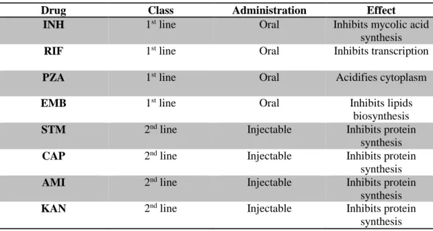
In table 1.1 is presented some of the drugs used nowadays in the treatment of TB.
Table 1. 1 - Drugs used in TB treatment21,25–28.
Drug Class Administration Effect
INH 1st line Oral Inhibits mycolic acid
synthesis
RIF 1st line Oral Inhibits transcription
PZA 1st line Oral Acidifies cytoplasm
EMB 1st line Oral Inhibits lipids
biosynthesis
STM 2nd line Injectable Inhibits protein
synthesis
CAP 2nd line Injectable Inhibits protein
synthesis
AMI 2nd line Injectable Inhibits protein
synthesis
KAN 2nd line Injectable Inhibits protein
5
Drug Class Administration Effect
OFX 2nd line Oral Inhibits DNA
supercoiling
CYC 2nd line Oral
Inhibits peptidoglycan
synthesis
CFZ 3rd line Oral Interact with
bacterial DNA
AMX/CLV 3rd line Oral Inhibition of β-lactamase
IPM/CLN 3rd line Injectable
IPM – Inhibition of β-lactamase CLN – Inhibition of enzyme responsible for degradation IPM
INH – isoniazid; RIF – rifampicin; PZA – pyrazinamide; EMB – ethambutol; STM –
streptomycin; CAP – capreomycin; AMI – amikacin; KAN – kanamycin; OFX – ofloxacin;
CYC – cycloserine; CFZ – clofazimine; AMX/CLV – amoxicillin plus clavulanate; IPM/CLN – imipenem plus cilastatin.
1.1.5
Fluoroquinolones
Fluoroquinolones (FQ), fluorine-containing nalidixic acid derivatives (Figure 1.2), are a group of compounds that target DNA gyrase and DNA topoisomerase II and have a broad-spectrum of antimicrobial activity21,29. These compounds are recommended and widely used in the treatment of bacterial infections of the respiratory, gastrointestinal, and urinary tracts, as well as sexually transmitted diseases and chronic osteomyelitis. It has been shown that FQ can also penetrate into macrophages and have bactericidal activity there29. Besides that, FQ have shown potent activity against M. tuberculosis and are used as second-line drugs in TB therapy30,31.
Figure 1. 2 - Four generations of quinolones. Ciprofloxacin, levofloxacin and moxifloxacin are FQ. Scaffolds are colored black and peripheral chemical modifications are colored red32.
Ciprofloxacin (CIP), an inhibitor of DNA gyrase, is used in the most potent fluoroquinolone and is active against a broad range of bacteria and the most susceptible are aerobic gram-negative bacilli33,34.CIP was the first fluoroquinolone indicated for infections outside the urinary tract and was considered a major advance in therapy for aerobic gram-negative infections. However, CIP was noted to have poor activity against anaerobes35. This drug can also interact with a number of drugs, some herbal and natural supplements, and certain thyroid medications34.
Levofloxacin (LEV) is an isomer of ofloxacin (used in treatment of MDR-TB) and has increased in vitro antibacterial activity against a variety of bacteria including anaerobes36. LEV is also used in the treatment of MDR-TB if the strain is susceptible or if the agent is thought to have efficacy31. The mode of action of this drug is the inhibition of DNA gyrase and DNA topoisomerase II37.
6 Moxifloxacin (MOX) is synthetic fluoroquinolone antibiotic agent used in the treatment of sinus and lung infections. The bactericidal action of these drugs results from inhibition of DNA gyrase and DNA topoisomerase II38. MOX is also a second-line drug and is the most potent fluoroquinolone used in TB treatment although is less active than isoniazid (first-line drug)31,39.
1.1.6
Nalidixic Acid
Nalidixic acid (NAL) (figure 1.3) was the first member of the quinolone family of antibacterial agents synthesized and it’s used in the treatment of urinary tract infections caused my susceptible gram-negative microorganisms40,41. NAL is administrated orally, is rapidly absorbed from the gastrointestinal tract and has a bioavailability of approximately 96%. NAL metabolism is hepatic and 30% of the administrated dose is converted to the active metabolite, hydroxynalidixic acid (Figure 1.4)42.
NAL is an antibiotic that inhibits DNA gyrase, an enzyme that catalyses the negative supercoiling of DNA, that is essential for DNA replication, transcription and recombination43,44. This enzyme is constituted by two A subunits and two B subunits, encoded by gryA and gryB, respectively44.Quinolone sensitivity is controlled by subunits A, which contain the breakage-reunion active site while subunits B promote ATP hydrolysis, needed for energy transduction23,44.
It was shown that fluoroquinolones had the potential to reduce the duration of therapy in TB murine models21
. NAL and other quinolones were previously tested in M. tuberculosis with a mutation in gryA and have shown poor DNA cleavage stimulation44. These mutations occur in a conserved region that is called quinolone resistance-determining region (QRDR) of the gyrA and gyrB genes. Mutations within the QRDR are associated with quinolones resistance29.
Prodrugs
Prodrugs are compounds that are not biologically active and have to be transformed into active products by enzymatic or chemical reactions45. The active compounds have undesirable properties that may become pharmacological, pharmaceutical or pharmacokinetic barriers in clinical drug application. Using drug derivatization with retention of the desirable therapeutic activity has an important role on improving drug efficacy46. Optimizing drug activity is a two-dimensional problem because the activity at the target is essential but is just as important as the efficient delivery of the agent in that same target47.
Figure 1. 3 - NAL structure. Figure 1. 4 - Hydroxynalidixic acid structure.
7 Prodrugs can be applied to achieve increased chemical or metabolic stability, higher water solubility or higher solubility in lipid membranes, reduced toxicity, improved oral or local absorption45. The main objectives of prodrugs are:
Modification of drug pharmacokinetics; Longer action;
Decrease in toxicity and side-effects; Increase in selectivity;
Resolution of formulation problems, such as stability, solubility and organoleptic properties48. Prodrugs can be classified in two main classes: carrier-linked prodrugs and bioprecursor prodrugs. In carrier-linked prodrugs, the active compound is temporary linked to a carrier through a bioreversible covalent linkage. Once in the body, the carrier-linked prodrug undergoes biotransformation, releasing the drug and the carrier. The carrier should be nonimmunogenic, easy to synthesize at a low cost, stable under the conditions of prodrug administration and undergo biodegradation to nonactive metabolites. This class includes a group of prodrugs called co-drugs that are prodrugs with two pharmacologically active agents coupled together into a single molecule. Bioprecursors are prodrugs that result from a molecular modification of the active compound itself. The bioprecursor prodrug is transformed metabolically or chemically by hydration, oxidation or reduction into the active agent49.
Prodrugs can also be classified in two another types, depending on the site of conversion to the active metabolite: Type I prodrugs are metabolized intracellularly (Type IA, at the cellular targets of their therapeutic action and Type IB, by metabolic tissues), whereas Type II prodrugs are metabolized extracellularly (Type IIA, in the milieu of the gastrointestinal fluid; Type IIB, in the circulatory system and/or other extracellular fluid compartments and Type IIC, near or inside therapeutic target/cells)49,50. Some of the first- and second-line agents used in the treatment of TB are prodrugs (for example, isoniazid and pyrazinamide). These antimycobacterial prodrugs can be divided in two main groups depending on their mechanism of action: mycobacteria bioactivated and host bioactivated. The mycobacteria bioactivated prodrugs have increased bioavailability and solubility, and decreased toxicity. The main benefits of host-bioactivated drugs are: increased bioavailability and therapeutic effectiveness; improved solubility, chemical stability; increased organ/tissue-selective delivery of the active agent and decreased toxicity. However, the main problem of antimycobacterial prodrugs, already used in TB treatment, is the emergence of strains resistant to prodrugs due to mutations in the gene encoding the activator50.
1.2.1
Esters as Prodrugs
The prodrug strategy is one of the most promising approaches to enhance the therapeutic efficacy of pharmacologically active agents by optimizing the absorption, distribution, metabolism, excretion and toxicity (ADMET) properties of the parent drugs. This is a faster strategy than searching for entirely new therapeutic agents with suitable ADMET characteristics50.
Prodrug design is used to improve the active drugs and some of the most common functional groups, such as carboxylic, hydroxyl, amine, phosphate and carbonyl are transformed in esters, carbonates, carbamates, amides and phosphates.51
8 The features of an ideal prodrug include: hydrolysis resistance during absorption; weak or no activity; aqueous solubility; good permeability through cells; chemical stability at different pH and kinetics that allow the release of the parental drug. Among the chemical bonds used to link the parental drug and carrier, esters have proven to be promising due to their amenability to hydrolysis both in vivo and in
vitro52.
Esters of active agents with carboxyl, hydroxyl or thiol functionalities are the most commonly used prodrugs because these prodrugs enhance the lipophilicity, thus the passive membrane permeability, and decrease the water solubility by masking charged groups49,51. An ideal ester prodrug should have chemical stability across pH range, high aqueous solubility and exhibit good transcellular absorption phase49.
1.2.2
Chemical Hydrolysis
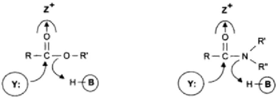
Carboxylic acid derivatives hydrolysis can be catalysed by acids or bases. The hydrolysis mechanisms, in both acid- and base-promoted, involve a nucleophilic addition-elimination at the acyl carbon53.
Figure 1. 3 - Scheme of chemical hydrolysis of carboxylic derivatives (esters and amides). (a) acid catalysis (b) base catalysis54.
In acid catalysis (Figure 1.5a), the first step is the protonation of the ester carbonyl to make it more electrophilic. Then, the water oxygen functions as the nucleophile attacking the electrophilic carbon in the C=O, creating the tetrahedral intermediate. The oxygen from the water is deprotonated to neutralise the charge and the XH group is protonated to convert it into a good leaving group: the carboxylic acid product is obtained and the acid catalyst is regenerated53–55.
In base hydrolysis (Figure 1.5b), the hydroxy nucleophile attacks at the electrophilic carbon of the ester
C=O, creating the tetrahedral intermediate. Then, the intermediate expels an alkoxyl ion, leading to the
carboxylic acid. After, transfer of a proton leads to the products of the reaction53,55.
1.2.3
Enzymatic Hydrolysis
The enzymatic hydrolysis is a common hydrolysis reaction that is catalyzed by an enzyme. Enzymatic hydrolysis is more efficient than chemical hydrolysis due to the decrease in the Gibbs energy of the transition state54. Esters and amides, endogenous or exogenous, are one of the major substrates for
9 hydrolases, enzymes of class 3 that catalyze the attack to functional groups by water molecules. Hydrolases have three catalytic features at the active site that accelerate the rate of hydrolysis (Figure
1.6)54,56. These catalytic sites are: an electrophilic component, which increases the polarization of the carbonyl group in the substrate (Z+); a nucleophile to attack the carbonyl C-atom, leading to the
formation of a tetrahedral intermediate (Y:) and a proton donor to transform -OR’ and –NR’R’’ into better leaving groups (H-B)54.
Figure 1. 4 - Catalytic groups of hydrolases involved in ester and amide bond hydrolysis54.
Based on the structures of their catalytic sites, hydrolases can be divided in five classes: serine hydrolases, threonine hydrolases, cysteine hydrolases, aspartic hydrolases and metallohydrolases54. Hydrolases have different substrates such as: peptides, ethers, esters, glycosylases, acid anhydrides and halide bonds57.
Esterases
Esterases are enzymes that catalyze the cleavage and formation of ester bonds. These enzymes belong to hydrolases class of enzyme, EC 3 and can act as hydrolyses when catalyzing the cleavage of ester bonds57,58. Within the ester formation bonds esterases can catalyze three types of reactions: esterifications, interesterification and transesterification reactions with good chemo-, region- and/or enantioselectivity58. Esterases are subdivided in the type of reaction they catalyze:
Carboxylic ester hydrolases (EC 3.1.1); Thioester hydrolases (EC 3.1.2); Phosphatases (EC 3.1.3);
Phosphodiester hydrolases (EC 3.1.4);
Triphosporic monoester hydrolases (EC 3.1.5); Sulfatases (EC 3.1.6);
Diphosphoric monoesterases (EC 3.1.7); Phosphoric triester hydrolases (EC 3.1.8); Exonucleases (EC 3.1.11-16);
Endonucleases (EC 3.1.21-31)57.
The major carboxylic ester hydrolases classes are carboxylesterases (EC 3.1.1.1, carboxylic-ester hydrolase) and carboxylesterases (EC 3.1.1.3, triacylglycerol hydrolases)59. Carboxylesterases catalyze the hydrolysis of a wide variety of substrates including esters, amides and carbamates (endogenous or exogenous)60. These enzymes catalyze hydrolysis of esters, by the water, resulting in alcohol and carboxylate61. The three-dimensional structure of carboxylesterases show the characteristic
α/β-10 hydrolase fold59 (Figure 1.7). In this structure, each parallel β-segment is attached to its neighbour by an α-helical segment56. The catalytic triad is composed of serine-aspartic acid-histidine (Ser-Asp-His) (Figure 1.7) and usually a consensus sequence glycine-x-x-leucine (Gly-x-x-Leu motif, where x can be any amino acid residue) is around the active site. The mechanism for ester hydrolysis is based in of four steps: first, the substrate is bound to the active serine, yielding a tetrahedral intermediate stabilized by the catalytic His and Asp residue. Then, the alcohol is released and an acyl-enzyme complex is formed. Next, attack of water forms again a new tetrahedral intermediate, which after resolution yields the acid and free enzyme59.
Figure 1. 5 – Scheme of the α/β-hydrolase fold. β-Sheets (1-8) are shown as blue arrows, α-helices (A-F) as red columns and the relative positions of the amino acids residues of the catalytic triad are indicated as orange circles59.
Carboxylesterases are widely present throughout the body (e.g., intestine, brain, skin, etc.) with their highest activity being in the liver. These esterases act also as an effective biological barrier to limit the distribution of substrates that might be toxic and induce their elimination by turning them into polar molecules. Ester prodrugs are intentionally designed to be activated by these esterases since most compounds will be detoxified by them62.
1.3.1
Esterases of Human Plasma
Human plasma esterases have an important role in drug metabolism. These enzymes participate in activation of ester prodrugs, inactivation of drugs and detoxify natural and synthetic ester-containing poisons63.
The human plasma has three esterases groups: Butyrylcholinesterase (EC 3.1.1.8); Paraoxonase (EC 3.1.8.1);
Acetylcholinesterase (EC 3.1.1.7)63.
Besides this three groups, there is a very important and abundant protein in the human plasma with ester hydrolytic capacity: albumin63.
Paraoxonases are called “A-esterases” whereas butyrylcholinesterase and acetylcholinesterase are called “B-esterases”64.
11 Butyrylcholinesterase (BChE) is also known as pseudocholinesterase, non-specific cholinesterase or simply cholinesterase. This enzyme acts on butyrylcholine and acetylcholine. BChE has higher activity in liver, intestine, lungs, heart and kidney. Serum BChE is synthesized in liver and secreted into plasma, where it has higher activity65,66. BChE has also an important role as detoxification enzyme because it can hydrolyse compounds with carboxylic or phosphoric acid ester groups. Therefore, this enzyme can hydrolyse compounds like: succinyldicholine (neuromuscular blocking drug); cocaine; aspirin; heroin and others65.
Paraoxonase is an enzyme family with three members: paraoxonase 1(PON1), paraoxonase 2 (PON2) and paraoxonase 3 (PON3). These esterases are capable of hydrolysing organophosphates as well as aromatic esters64. PON1 and PON3 can be found in the cholesterol-carrying particles HDL and can inhibit lipid oxidation in LDL whereas PON2 is found in many tissues67. Only PON1 is present in human plasma. PON1 is a proefficient esterase toward several synthetic substrates whereas PON2 and PON3 exhibit high lactonase activity64.
Acetylcholinesterase (AChE) or “true cholinesterase” is known to be abundant in brain, muscle and erythrocyte membrane being present in negligible amounts in human plasma63,65. Like BChE, AChE belongs to the α/β hydrolase-fold family66. The main function of AChE is hydrolysis of the neurotransmitter acetylcholine at cholinergic synapses and it is one of the fastest enzymes known65. Albumin, most known as human serum albumin (HSA), is the most soluble protein in the body (about 4% in serum) and the most prominent protein in plasma. This protein is responsible for a lot of multiple functions in the body, including the maintenance of blood osmolarity, acting as an antioxidant and serving as a solubilising agent and carrier for many endogenous and exogenous compounds68. HSA has also the capacity of transporting different substances including fatty acids, hormones, enzymes, dyes, trace metals and drugs. Substances that are toxic in the unbound or free state are generally not toxic when bound to HSA. These properties give to HSA the capability to regulate the extracellular concentration of numerous endogenous as well exogenous administrated substances69. HSA does not have an enzyme commission number which means that this protein is considered to be inert, without any catalytic activity. However, it was proven that HSA has esterase activity. The active site of this protein is one tyrosine residue (Tyr411). Although the enzymatic activity of a single molecule of HSA is low, the concentration of this protein is very high, which means that HSA has a significant contribution to drug metabolism63.
In the human plasma, these four proteins types of esterases are the only ones that have contribution on ester hydrolysis.
1.3.2
Mycobacterial Esterases
As described before, M. tuberculosis has a complex cell envelope with mycolic acids, suggesting that there must be a large number of enzymes involved in lipid metabolism70. Besides, it was discovered the existence of lipid inclusions in mycobacteria cytoplasm which indicates the presence of lipolytic enzymes71. In fact, after the whole genome sequence of this bacteria being studied by the Sanger Center and the Institut Pasteur, in 1998, it showed at least 250 enzymes related to lipid metabolism including extracellular secreted enzymes, integrated cell wall enzymes and intracellular lipases/esterases70. From the genome annotation of M. tuberculosis, it has been concluded that 24 genes encode lipolytic enzymes. These proteins have been classified “Lip family” (LipC to LipZ)72,73. However, this classification is only based on the presence of the consensus sequence Gly-x-Ser-x-Gly, which is characteristic of the
12 members of the α/β hydrolase family. This classification does not allow the distinction between lipases and esterases but Blast analysis has shown that the “Lip family” is composed of both lipases and esterases17. Esterases preferentially break ester bonds of shorter chain fatty acids and lipases catalyze lipid hydrolysis, which means that lipases display a much broader substrate range than esterases72. A fundamental difference between esterases and lipases is their ability to act on solubilised substrates. Esterases act on water-soluble molecules and lipases are highly efficient at hydrolysing molecules aggregated in water, such as an emulsion or a micellar solution74.
Analysis of the M. tuberculosis genome revealed the existence of some carboxylesterases that are responsible for ester hydrolysis. The carboxylesterases identified are members of the “Lip family”: Rv3487c (LipF) and Rv1399 (LipH)75,76. LipF belongs to the α/β hydrolase-fold family and hydrolyze efficiently short chain esters. The active site of many α/β hydrolases is constituted by a catalytic triad with serine as the nucleophile, histidine as a proton donor and aspartate or glutamate forming the charge-relay network. In LipF, the potential catalytic triad is: Ser90-Glu189-His219, which means that it contains a glutamate residue (Glu) instead of aspartate residue. The aspartate or glutamate residue is strongly bonded to the nitrogen into histidine residue, stabilizing the protonated positively charged protonated, the correct tautomer and the correct conformation of His76. Like LipF, LipH belongs to the α/β hydrolase-fold family. This enzyme hydrolyses efficiently short-chain triacylglycerols and vinyl esters and has no detectable activity against emulsified substrates. The catalytic triad of LipH: Ser162-Asp260-His29075.
In 2010, Guo et. al demonstrated that Rv0045c is an esterase of M. tuberculosis, involved in ester/lipid metabolism70. Compared with LipF and LipH, Rv0045c has shown no sequence identity in a multiple sequence alignment. Although, this alignment showed that the active site (Gly-x-Ser-x-Gly sequence motif) characteristic of hydrolases is highly conserved and the main catalytic residues are Ser89, Asp113, Ser206 and His234, usually present on the catalytic triad of esterases. Like LipF and LipH, Rv0045c can efficiently catalyse short chain ester substrates70.
The Aim of the Thesis
In the present work, the main objective was to synthesize nalidixic acid (NAL) derivatives with activity against M. tuberculosis. The pretended derivates are long chain esters because they can have an adequate plasma stability and can be activated by mycobacterial esterases77. Besides that, NAL is a weak acid and it was previously discovered that M. tuberculosis is susceptible to weak acids, suggesting a relation between antimycobacterial activity and pKa of acids78.
So, different nalidixic acid derivatives were synthesized with modifications on carbon C7 (R1 = -CH3 or –CCl3) and different alkoxyl chains (R2), as it is shown in Figure 1.8:
13 Stability studies in human plasma and phosphate buffer were also performed in order to evaluate enzymatic and chemical hydrolysis, respectively. The phosphate buffer assays are useful to evaluate if chemical hydrolysis has or not contribution to the velocity of human plasma hydrolysis because it is used a suspension of human plasma 80% in phosphate buffer. Human plasma assays are also useful to evaluate if the synthesized prodrugs are stable and will not degradate until they achieve the target. Activity studies against M. tuberculosis H37Rv strain were done in order to evaluate if compounds inhibit bacterial growth.
With this work, it is pretended to have an ester of nalidixic acid or of a nalidixic acid derivative with slow plasma hydrolysis and activity against M. tuberculosis.
15
Synthesis and Structural Identification
2. Synthesis and Structural Identification
2.1 Ester Synthesis
The esterification reaction is one of the most important and fundamental reactions in organic synthesis79. Carboxylic acids react with alcohols to form esters through a condensation reaction (Figure 2.1).
Ester synthesis can be achieved in acid-catalysed reactions, that are called Fischer esterifications, or from alkanoyl chlorides (acyl chlorides). In Fischer esterifications (Figure 2.2), the carboxylic acid accepts a proton from the strong catalyst. Then the alcohol attacks the protonated carbonyl group to give a tetrahedral intermediate. After that, a proton is lost from one oxygen atom and gained by another one. The loss of a water molecule gives a protonated ester and finally the transfer of that proton to a base leads to the ester53.
Figure 2. 2 - Mechanism of acid-catalyzed esterification53.
2.1.1
Esters from Acyl Chlorides
One of the best methods to synthesize esters is the reaction of acyl chlorides with alcohols (Figure 2.3). This reaction occurs rapidly and does not require an acid catalyst53.
16 Figure 2. 3 - General reaction of acyl chlorides synthesis with thionyl chloride53.
Acyl halides are the most reactive of the acyl derivatives; the best synthetic route to an ester is synthesis of an acyl halide, chloride as an example, from the carboxylic acid first and then the conversion of the acyl chloride to the ester, through the reaction with an alcohol.
The reagent used to form acyl chlorides was thionyl chloride because it reacts with carboxylic acids to give acyl chlorides in good yield (Figure 2.4):
Figure 2. 4 - Mechanism of acyl chlorides synthesis using thionyl chloride53.
These reactions involve nucleophilic addition-elimination by a chloride ion on a highly reactive intermediate, a protonated acyl chlorosulfite. This intermediate contains even better acyl leaving group than the acyl chloride product53
2.2 Synthesis of Nalidixic Acid Esters
17 Figure 2. 5 – Libraries of NAL esters. (a) library 1; (b) library 2.
To synthesize the esters of these different libraries, two methods were performed. The first method (method A), was used to synthesize the first library of compounds (Figure 2.5a), esters with different alkoxyl chains with the C7 methyl group on the heteroaromatic acyl group. To synthesize the second esters library, with different alkoxyl chains with the trichloromethyl group on the C7 heteroaromatic acyl portion (Figure 2.5b), method B was used. Method A was based on the description done by Sachdeva
et. al. in 201580 and Method B was based on the description done by Cynamom et. al. in 199581.
Method A
To 1 equivalent (eq.) of NAL in a round bottom flask, 19.2 eq. of thionyl chloride were added and the flask was kept in fuming chamber aside for 15 min. Later 34.4 eq. of the alcohol were added to the solution drop by drop and mixed thoroughly after each addition. Then the reaction was kept under reflux during 1 hour (h) (Figure 2.6)80.
Figure 2. 6 – Scheme of ester synthesis by method A.
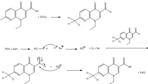
Method B
To 1.2 eq. of NAL in a round bottom flask, 16.5 eq. of thionyl chloride were added and this reaction was kept under reflux for 2h. The acyl chloride was isolated as solid residue by evaporation of the thionyl chloride in excess under vacuum on a rotary evaporator. Meanwhile, 1.5 eq. of NaH were added to 1 eq. of alcohol to prepare its alkoxide. Then, the alkoxide was added to the acyl chloride solid residue previously dissolved in dried dichloromethane and this reaction was kept under reflux to synthesize the ester (Figure 2.7). The reaction was followed by thin layer chromatography.
18 Figure 2. 7 - Scheme of ester synthesis by method B.
2.2.1 First library of Esters
The first esters library was synthesized by method A. These esters have different alkoxyl chain, ranging from 2 until 14 carbons length.
Table 2. 1 - First library of NAL esters.
R Reflux (h) Yield (%) Description
2 C2H5 1 65 White solid 3 C4H9 1 68 White solid 4 C6H13 1 59 White solid 5 C9H19 1 51 White solid 6 C10H21 1 60 White solid 7 C11H23 1 65 White solid 8 C12H25 1 66 Yellow solid 9 C14H29 1 63 Yellow solid 10 CH(CH3)C7H15 1 79 Yellow solid
In the first library of compounds, 9 different esters were synthesized with different alkoxyl chains from 2 to 14 carbons length and they were obtained with a range of yields from 51% to 79%. The esters 2-9
19 have linear alkoxyl chains and the ester 10 has a linear alkoxyl chain of 8 carbons with one ramification (methyl group) in the first carbon of the chain (figure 2.8).
Figure 2. 8 – Structure of compound 10.
2.2.2 Seconds Library of Esters
The second esters library was synthesized by method B. These esters have different alkoxyl chains, from 4 until 14 carbons length.
Table 2. 2 - Second library of NAL derivatives.
R Reflux (h) Yield (%) Description
1a H overnight 100 Brown solid
3a C4H9 18 21 White solid 5a C9H19 19 5 White solid 6a C10H21 11 5 Yellow solid 7a C11H23 11 15 Yellow solid 8a C12H25 40 15 Yellow solid 9a C14H29 16 35 Yellow solid
10a CH(CH3)C7H15 33 20 Yellow oil
In the second ester library group, 7 different esters were synthesized and the yields obtained were very low, ranging from 5% to 35%. This may be due to the excess of acid in this method instead the alkoxide (like in method A, but with the alchohol).
20 This method starts with 2h of reflux of the acid with thionyl chloride. This step led to the synthesis of the acyl chloride and also the chlorination of the methyl group in carbon C7 of the aromatic acyl group. This substitution occurs because chlorination occurs rapidly in methyl groups attached ortho or para to a nitrogen atom in a multi-heteroatom ring82.
Compound 1a is not an ester, is a carboxylic acid derivative of nalidixic acid. This compound was synthesized as a control for the stability assays. Like the synthesis of the esters of this library, nalidixic acid was kept under reflux with thionyl chloride for 2 hours. Then, the excess of thionyl chloride was evaporated and water was added until the carboxylic acid was formed.
The esters obtained through this method have linear alkoxyl chains except the compound 10a which has a linear alkoxyl chain of 8 carbons with one ramification on the first carbon of the chain (Figure 2.9).
Figure 2. 9 - Structure of compound 10a.
2.3 Structural Identification of Synthetized Esters
2.3.1 Nuclear Molecular Ressonance
The proton (1H) and carbon (13C) NMR spectra were obtained in deuterated chloroform (CDCl 3). All the NMR spectra of the synthesized compounds are characterized in chapter 7.
All the signals identification have been made with the help of Heteronuclear Multiple-Quantum Correlation (HMQC) and Heteronuclear Multiple Bond Correlation (HMBC) and Attached Proton Test (APT) spectra for carbons besides the normal proton (1H) and carbon (13C) spectra, as can be seen an example in Appendix 1.
Table 2. 3 - 1H-NMR signal (ppm) of aromatic protons and protons attached to C1’, C2’ and C1’’ in libraries 1 and 2.
Compound Library 1
Aromatic
protons C1’-H C2’-H C1’’-H
21 1 8.91 (C2-H); 8.67 (C5-H); 7.41 (C6 -H) 4.63 1.55 2.75 2 8.65 (C5-H); 8.63 (C2-H); 7.24 (C6 -H) 4.49 1.50 2.66 3 8.63 (C5-H); 8.60 (C2-H); 7.23 (C6 -H) 4.48 1.48 2.65 4 8.57 (C5-H); 8.54 (C2-H); 7.16 (C6 -H) 4.41 1.42 2.59 5 8.57 (C5-H); 8.53 (C2-H); 7.16 (C6 -H) 4.41 1.42 2.58 6 8.65 (C5-H); 8.61 (C2-H); 7.24 (C6 -H) 4.33 1.49 2.66 7 8.65 (C5-H); 8.61 (C2-H); 7.24 (C6 -H) 4.48 1.49 2.66 8 8.65 (C5-H); 8.61 (C2-H); 7.24 (C6 -H) 4.48 1.49 2.66 9 8.57 (C5-H); 8.54 (C2-H); 7.17 (C6 -H) 4.41 1.42 2.59 Compound Library 1 Aromatic protons C1’-H C2’-H C1’’-H 10 8.64 (C5-H); 8.57 (C2-H); 7.23 (C6 -H) 4.48 1.49 2.65 Compound Library 2 Aromatic protons C1’-H C2’-H C1’’-H 1a 9.03 (C2-H), 8.99 (C5-H); 8.22 (C6 -H) 4.67 1.63 -3a 8.92 (C5-H); 8.70 (C2-H); 8.06 (C6 -H) 4.52 1.57 -5a 8.93 (C5-H); 8.71 (C2-H); 8.06 (C6 -H) 4.53 1.58 -6a 8.93 (C5-H); 8.71 (C2-H); 8.07 (C6 -H) 4.36 1.58 -7a 8.92 (C5-H); 8.69 (C2-H); 8.06 (C6 -H) 4.52 1.57 -8a 8.93 (C5-H); 8.71 (C2-H); 8.07 (C6 -H) 4.53 1.58 - 9a 8.84 (C5-H); 8.62 (C2-H); 7.98 (C6 -H) 4.44 1.49 - 10a 8.91 (C5-H); 8.66 (C2-H); 8.05 (C6 -H) 4.52 1.58 -
22 After 1H-NMR spectra analysis, it is observed that chemical shifts of aromatic protons are different between the two libraries of compounds staying in general at higher positions in the second library (table
2.3). In the first library, C5-H protons have a chemical shift between 8.57-8.65 ppm, C2-H between 8.53-8.63 ppm and C6-H between 7.16-7.24 ppm and in the second library, C5-H protons have a chemical shift between 8.93-8.84 ppm, C2-H between 8.71-8.62 ppm and C6-H between 8.07-7.98 ppm. These differences occur due to the substitution of the methyl group hydrogens for three chlorines. The same fact is also observed in proton spectra of the two acids: 1 with C7-methyl and 1a with C7-chloromethyl groups.
Also, when comparing each group of esters with their acid (table 2.3) it is possible to conclude that after ester formation, all the 1H-NMR signals of the spectra for both groups of esters have a lower chemical shift more evident in the signal of C2-H and an exchange between the positions of C2-H and C5-H signals is observed from the acid to the esters.
Other difference between the 1H-NMR spectra of the two libraries is the existence of a singlet at 2.58-2.66 ppm on library 1 which corresponds to the methyl group attached to carbon C7. This singlet signal does not exist in compounds of library 2 because the protons of the methyl group attached to carbon C7 were substituted by three chloro atoms on the trichloromethyl group.
Compounds 10 and 10a with a methyl group connected to carbon C1’’’ of the alkoxyl chain transforms this carbon in a chiral carbon, that is an asymmetric atom or stereocenter, and this fact implies that the two protonsof the next carbon on the alkoxyl chain C2’’’ are nonequivalent and are called diastereotopic, as such two different signals appear in the proton 1H-NMR spectra. Therefore instead of the signal C
2’’’
-H of the two equivalent protons, like on the other esters, due to the splitting with C1’’’ and C3’’’ protons resulting in a tt which appears as a multiplet with two protons integration, these signals will result in a
ddt splitting appearing as two multiplet signals with one proton integration each.
The aromatic protons C5-H and C6-H appear as duplets and the coupling constants (J) were:
J5-6 = 8.1 Hz (library 1),
J5-6 = 8.1-8.4 Hz (library 2),
With the ethyl group N1-C1’H2-C2’H3 the C1’-H appears as a quadruplet and C2’-H as a triplet with a coupling constant:
JC1’H-C2’H = 7.2 Hz (both groups of compounds).
On the other hand with the alkoxyl protons the chains last carbon appears as a triplet with coupling constants:
J(last carbon-H)≈ 6.9-7.8 Hz.
And for both groups of compounds, except 10 and 10a, C1’’’-H appears as a triplet with coupling constants:
J C1’’’-H’ ≈ 6.6-7.2 Hz
Whereas in compounds 10 and 10a the protons CH3-C1’’’appear as a duplet with a coupling constant: