Journal of Biomaterials Science: Polymer Edition
About the effect of eye blinking on drug release from pHEMA based hydrogels: an in
vitro study
--Manuscript
Draft--Manuscript Number:Full Title: About the effect of eye blinking on drug release from pHEMA based hydrogels: an in vitro study
Short Title: Blinking effect on drug release from pHEMA
Article Type: Original Research Paper
Keywords: controlled drug release; levofloxacin; pHEMA hydrogels; contact lenses; friction. Corresponding Author: Ana Paula Serro
Instituto Superior Técnico Lisbon, PORTUGAL Corresponding Author Secondary
Information:
Corresponding Author's Institution: Instituto Superior Técnico Corresponding Author's Secondary
Institution:
First Author: Raquel Galante
First Author Secondary Information:
Order of Authors: Raquel Galante
Patrizia Paradiso
Maria Guilhermina Moutinho Ana Isabel Fernandes José Mata
António Matos Rogério Colaço Benilde Saramago Ana Paula Serro Order of Authors Secondary Information:
Manuscript Region of Origin: PORTUGAL
Abstract: The development of new ophthalmic drug delivery systems capable of increasing the residence time of drugs in the eye and improve its bioavailability relatively to eyedrops has been object of intense research in recent years. Several studies have shown that drug loaded therapeutic soft contact lenses (SCLs) constitute a promising approach, with several potential advantages as compared with collyria. The main objective of this work is to study the effect of repetitive load and friction cycles caused by the eye blinking, on the drug release from hydrogels used in SCLs which, as far as we know, was never investigated before.
Two poly-2-hydroxyethylmethacrylate based hydrogels, pHEMA-T and pHEMA-UV, were used as model materials. Levofloxaxin was chosen as model drug. The hydrogels were fully characterized in what concerns structural and physicochemical properties. PHEMA-UV revealed some superficial porosity and a lower short range order than PHEMA-T.
We observe that the load and friction cycles enhanced the drug release from pHEMA-UV hydrogels. The application of a simple mathematical model, which takes into account the drug dilution caused by the tear flow, showed that the enhancement of the drug release caused by blinking on this hydrogel may be relevant in in vivo conditions. Powered by Editorial Manager® and ProduXion Manager® from Aries Systems Corporation
Conversely, the more sustained drug release from pHEMA-T is not affected by load and friction cycles. The conclusion is that, depending on the physicochemical and microstructural characteristics of the hydrogels, blinking is a factor that may affect the amount of drug delivered to the eye by SCLs and should thus be considered.
About the effect of eye blinking on drug release from pHEMA based hydrogels: an in vitro study
R. Galantea, P. Paradisoa, M.G. Moutinhob, A.I. Fernandesb, J.L.G. Mataa, A.P.A. Matosb, R. Colaçoa,c, B. Saramagoa, A.P. Serroa,b*
a
Centro de Química Estrutural, Instituto Superior Técnico, Universidade de Lisboa, Av. Rovisco Pais, 1049-001 Lisboa, Portugal
b
CiiEM, Instituto Superior de Ciências da Saúde Egas Moniz, Campus Universitário, Quinta da Granja, Monte de Caparica, 2829-511 Caparica, Portugal
c
Bioengineering Department, Instituto Superior Técnico, TU Lisboa, Av. Rovisco Pais, 1049-001 Lisboa, Portugal
Acknowledgements
To BASF for the kind offer of PVP Kollidon® 30. To Dr. Miguel Ângelo Rodrigues from CQE-IST for access to the HPLC equipment. To Fundação para a Ciência e a Tecnologia for P. Paradiso PhD Grant SFRH/BD/71990/2010 and for funding through the projects PEst-OE/QUI/UI0100/2013 and
M-ERA.NET/0005/2012
. To CRUP for funding the bilateral action with Germany (NMI - Natural and Medical Sciences Institute at the University of Tübingen, A29/12).*Corresponding author Ana Paula Serro
Centro de Química Estrutural - Instituto Superior Técnico Av. Rovisco Pais, 1049-001 Lisboa, Portugal
[email protected] Tel: +351 21 8419240
Fax: +351 21 8464455 Abstract
Manuscript
The development of new ophthalmic drug delivery systems capable of increasing the residence time of drugs in the eye and improve its bioavailability relatively to eyedrops has been object of intense research in recent years. Several studies have shown that drug loaded therapeutic soft contact lenses (SCLs) constitute a promising approach, with several potential advantages as compared with collyria. The main objective of this work is to study the effect of repetitive load and friction cycles caused by the eye blinking, on the drug release from hydrogels used in SCLs which, as far as we know, was never investigated before.
Two poly-2-hydroxyethylmethacrylate based hydrogels, pHEMA-T and pHEMA-UV, were used as model materials. Levofloxaxin was chosen as model drug. The hydrogels were fully characterized in what concerns structural and physicochemical properties. PHEMA-UV revealed some superficial porosity and a lower short range order than PHEMA-T.
We observe that the load and friction cycles enhanced the drug release from pHEMA-UV hydrogels. The application of a simple mathematical model, which takes into account the drug dilution caused by the tear flow, showed that the enhancement of the drug release caused by blinking on this hydrogel may be relevant in in vivo conditions. Conversely, the more sustained drug release from pHEMA-T is not affected by load and friction cycles. The conclusion is that, depending on the physicochemical and microstructural characteristics of the hydrogels, blinking is a factor that may affect the amount of drug delivered to the eye by SCLs and should thus be considered.
Keywords: controlled drug release; levofloxacin; pHEMA hydrogels; contact lenses; friction.
3
Topical application of medicated eyedrops is still the most common treatment for a wide range of ocular diseases caused by pathogenic agents. After instillation, about 90-95% of the drug contained in the collyrium is either absorbed through the conjunctiva or lost due to tear drainage [1, 2]. A significant amount of the drug enters rapidly into the bloodstream and may reach important organs, causing serious side effects. Besides the low penetration rates in the cornea, the short drug residence time leads to high variability of the drug concentration in the intraocular tissues and to the need of frequent administration, resulting in poor patient compliance and limiting the therapeutic efficacy.
The development of new drug delivery systems capable of enhancing the ocular bioavailability of drugs has been subject to special attention in the last few years and is regarded as a major advance in ophthalmological therapy. To achieve this goal, scientists of different areas have investigated several possibilities: for example, the use of colloidal systems, such as microemulsions, nanosuspensions or nanostructures (e.g. liposomes, nanoparticles or niosomes) and the in-situ application of gels or solid polymeric inserts [3-5]. Although, in general, these approaches increase drug bioavailability, they entail several problems [5]. Colloidal systems are subject to clearance mechanisms and tend to be washed away by reflex tearing [5]. Production of nanostructures at a large scale may be expensive and technically complex. Moreover, the potential of these structures as ophthalmic drug delivery systems is restricted by their lack of stability, limited drug-loading capacity and affinity of the matrix components (e.g. lipids or polymers) towards the drug. Aqueous gels and ointments exhibit relatively poor patient compliance due to some discomfort, blurring of vision and occasional irritation after application [6]. Moreover, the use of solid devices in the pre-corneal region, such as the inserts, also faces significant resistance from the patients [7]. Therefore, the development of new drug delivery systems, which are inexpensive and patient friendly, exhibit suitable drug release profiles and resist to the required final sterilization, still remains a challenge.
Recent works [8, 9], have shown that the use of soft contact lenses (SCLs) as vehicles for storage and ocular delivery of drugs seems to be a promising approach, due to its high degree of comfort, biocompatibility and prolonged contact with the eye surface. The application of SCLs for therapeutic purposes is a well established technique in current clinical ophthalmology [10, 11]. However, they are typically used with the purpose of protecting the eye, relieving pain, maintaining
4
corneal epithelial hydration and, eventually, to correct refractive errors [8-13]. In the post-surgical period, when the cornea is more vulnerable, their role is particularly relevant, since they protect the eye during the processes of cell adhesion and growth, which lead to healing. They may also be very useful in the case of keratic ulcers or after refractive surgery. Sometimes, contact lenses are prescribed for use in combination with eye drops, in order to prolong the residence time of the drugs in the cornea. Drug pre-loaded SCLs, which to our knowledge are not commercially available yet, may lead to a more effective treatment, because they avoid repeated applications. In fact, once placed on the eye, the drug released from the lenses will diffuse into a thin fluid layer trapped between the lens and the cornea (the post-lens tear film) whose mixture with the outside tear fluid is limited [14]. A sustained release of adequate amounts of drug will allow a prolonged therapeutic action.
The development of such type of devices has raised great interest among the clinical and academic community and also at the industrial level, as shown by the large number of studies on the subject published in recent years [2, 15, 16]. Different strategies to load the hydrogels with drugs were attempted, e.g. the direct incorporation of the drugs into the monomer mixture before the polymerization step [17, 18], the incorporation or surface immobilization of drug-loaded nanostructures (like nanoparticles and liposomes) in the hydrogels’ matrix [1, 19-22] or the use of supercritical fluids to enhance drug loading efficiency [23, 24]. However, soaking the lenses in drug solutions still remains the most simple and inexpensive method of loading, besides presenting fewer risks to the integrity of the drug molecules and lenses materials. Moreover, it offers the possibility to load commercial contact lenses, whose properties and production are already optimized. Nevertheless, many authors claim that drug release from hydrogels loaded by soaking only lasts for a few hours [21]. The release kinetics depends on numerous factors, such as the drug properties, the hydrogel composition and crosslinking degree, the temperature and, obviously, the volume of the dissolution media and hydrodynamic conditions used in the release experiments. Typically, these experiments are carried out in static conditions, with large volumes of solution, which are very different from those present in the eye. Therefore, a careful interpretation of the results is required and attempts to extrapolate for the behavior of the hydrogels in the eye have to be cautious.
5
The main objective of the present work was to study the effect of repetitive load and friction cycles associated to the eyelid movement, present in in vivo conditions, on the drug release from ophthalmic hydrogels, which, to our knowledge, has never been investigated before. The systems chosen were poly(2-hydroxyethyl methacrylate) (pHEMA) based hydrogels, containing levofloxacin, as model drug. The reason for this choice was the fact that pHEMA is commonly used as base material for the production of conventional soft contact lenses [25], while the antibiotic, levofloxacin, presents a broad activity spectrum, being widely used both in the prophylaxis and treatment of ocular infections [26].
Two types of pHEMA based hydrogels containing a small amount of poly(vinylpyrrolidone (PVP) were prepared. PVP increases the hydrophilicity of the material and has self-lubricant properties essential to the comfort and good performance of the contact lenses [27, 28]. One hydrogel was prepared using an anhydrous mixture. In the other case, acrylic acid and water were added to the mixture to enhance the water absorption capacity of the material, as well as the oxygen permeability [29]. The hydrogels were characterized with respect to the properties that are important in lens materials’ performance, namely, swelling kinetics, ion permeability, transmittance, friction coefficient, elasticity, wettability, morphology/topography and microstructure. After characterization, the samples were loaded with the antibiotic by immersion in the drug solution. The effect of dynamic cyclic compression (thereafter designated friction) on the drug release was studied using an apparatus especially conceived to simulate the movement of the eyelids. A mathematical model which mimics the hydrodynamic conditions in the eye was applied to the results to infer about the importance of blinking on the in vivo drug release.
2. Materials and Methods 2.1 Hydrogels preparation
Two types of pHEMA based hydrogels were prepared. In the first case (pHEMA-T), appropriate amounts of the crosslinker ethylene glycol dimethacrylate (EGDMA, Sigma-Aldrich, 98%) and of the initiator 2,2′-azobis(methylpropionitrile) (AIBN, Sigma-Aldrich, 98%), were dissolved in 2-hydroxyethyl methacrylate (HEMA, Sigma-Aldrich, HPLC grade 98%), to obtain concentrations of 80 mM and 10 mM, respectively. Poly(vinylpyrrolidone) (PVP, Kollidon® 30, kindly provided by BASF) was added to the mixture to a final concentration of 0.02 g/mL. After complete dissolution,
6
the solution was degassed by ultra-sonification (5 minutes), bubbled with a gentle stream of nitrogen (15 min), and injected into a mould consisting of two glass plates separated by a spacer of polyurethane. In order to facilitate the hydrogel removal from the mould, the glass plates were previously silanized by immersion in 2% solution of dimethyldichlorosilane (Fluka, 99.5%) in carbon tetrachloride (Riedel-de Haën, 99.8%) for one hour, followed by rinsing with dichloromethane (Sigma-Aldrich, 99.5%) and drying with nitrogen [30]. The thermopolimerization of the mixture was achieved in an oven in two stages: 50°C for 12 h and 70°C for 24 h. The hydrogel sheet obtained was boiled in water for 15 min to remove unreacted monomers and to facilitate the cutting of the samples used in the study (5x1cm2). The hydrated samples (thickness 0.35 mm) were dried overnight (16 h) inside an oven at 50°C.
A second type of hydrogels, pHEMA-UV, was prepared using a similar procedure. In this case, acrylic acid (Sigma-Aldrich, 99%) was added to the previous mixture to get a concentration of 100 mM, and the obtained mixture was diluted in water at a volume ratio of 1:1. Since thermopolymerization resulted in an opaque hydrogel, an alternative method of polymerization by UV radiation was used, which led to a faster polymerization and prevented the occurrence of phase separation. The polymerization was done using 4 UV lamps of 15 W at 350 nm (distance from light source 10 cm) during 90 min, at room temperature. In this case, the hydrogel sheet was boiled for a longer period (30 min), replacing the water after 15 min, since a larger amount of unreacted monomers was released.
2.2 Materials characterization 2.2.1 Swelling kinetics
The dry hydrogel samples were weighted (W0) and immersed in water at 25°C. At defined time
intervals, the samples were removed from the incubating medium and their surface was carefully dried with absorbent paper. They were weighted (Wt) and re-immersed in water. The procedure
was repeated several times during 10 h in order to determine the kinetics of swelling of the material. All tests were performed in triplicate.
The swelling capacity (SC) was calculated by:
0 0
100
tW
W
SC
W
(1)7
In the equilibrium (when the weight achieves a constant value, W∞) the water absorption capacity (WAC) is defined by:
WAC
W
W
0100
W
(2). 2.2.2 Ionic permeabilityThe ionic permeability of the hydrogels was measured using an in-house built diffusion cell comprising two separated compartments: a donor and a receiving chamber. The hydrated hydrogel samples were placed in between the two compartments. The receiving chamber was filled with deionized water and the donor chamber with 130 mM sodium chloride solution (NaCl, Merck, 99.5%) to simulate the lachrymal fluid. The ionic permeability of the materials was determined by measuring the rate of ion transport across the hydrogel. The electric conductivity of the solution in the receiving chamber was recorded as a function of time for a minimum of 10 hours, using a conductivity meter with temperature sensor (Cond 340i/SET, WTW). The conductivity data (in S/cm) were converted into NaCl concentration (in mg/mL) through a calibration curve previously obtained. The rate of ion transport (F) corresponds to the slope of the linear regression applied to the concentration data versus time. Solving the diffusion equation under the pseudo-steady state conditions allows the calculation of the ion permeability (also referred as the ionoflux diffusion coefficient, Dion)[31]:
F V
D
iondC
A
dx
(3)where V is the volume of the receiver solution, A is the area of the hydrogel sample and dC/dx corresponds to the NaCl concentration gradient in the hydrogel.
The results are the average of three independent measurements.
2.2.3 Transmittance
The visible light transmittance (T) of the hydrogel samples was determined in the wavelength range of 450-700 nm using a UV-Vis Beckmam DU-70 spectrophotometer. The lenses were
8
hydrated by soaking in 130 mM NaCl solution overnight, and then mounted on the outer surface of a quartz cuvette. Measurements were done in triplicate.
2.2.4 Friction coefficients
A CSM nanotribometer was used to determine the friction coefficient of the hydrogels against a smooth polymeric surface. The hydrated hydrogel samples were placed in a cell containing NaCl solution (130 mM). Polymethylmethacrylate (PMMA) semi-spheres, with a curvature radius of 2 mm, were used as counterbody. The normal load applied was 20 mN and the sliding velocity 0.7 cm/s. The results are the average of three measurements.
2.2.5 Elasticity
The Young’s modulus of each hydrogel was estimated from the slope of the stress-strain curves obtained during tensile tests conducted with hydrated samples of the materials (at least five independent measurements with each load). For that, samples were carefully suspended in a vertical support and submitted to increasing tension by placing hanged weights (0.1 - 1.2 N) on their lower extremity. The elongation produced was monitored with a cathetometer, assuring that the hydrogels were kept well hydrated at all times and, then, the Young’s modulus (E) was determined.
2.2.6 Wettability
The hydrophilicity of the hydrogels was determined by measuring the water contact angle on dry samples (dried 3 h in a vacuum oven), through the sessile drop method. Drops (2-3 μL) were deposited onto the dried hydrogels surfaces using a micrometer syringe, in a chamber previously saturated with water vapor. Drops images were acquired during 10 min using a video camera (JAI CV-A50) attached to a microscope (Wild M3Z) connected to a frame grabber (Data Translation DT3155). The acquisition and analysis of the images were performed using the ADSA-P software (Axisymmetric Drop Shape Analysis Profile). Eight to ten drops were done for each hydrogel.
9
The hydrogels’ surface was observed using a scanning electron microscope (SEM) Hitachi S2400 (15 KeV). The hydrated samples were cracked in liquid nitrogen, kept at -80°C for 4 hours and then freeze-dried overnight. Prior to the SEM analysis the hydrogels were covered by a thin gold coating (thickness 30 nm).
An atomic force microscope (AFM) NanoSurf Easyscan 2 (tip-scanning type) was used to get topographic images at a lower scale. The scan of the hydrated hydrogels surface was done in water, at room temperature, in contact mode atomic force microscopy on areas of 20×20 μm2
, at a scan rate of 1 Hz. Gold coated PPP-CONTSCAuD Nanosensor cantilevers (force constant 0.06 N/m) were used. The average roughness (Ra) was determined from the images obtained using the WSxM 5.0 develop 6.4 software.
2.2.8 Microstructure
Transmission electron microscopy (TEM) of both hydrogels was carried out with a Jeol 1200EX TEM equipment operated at 80 kV. The dry hydrogel samples were
embedded
in an Epon-Araldite mixture and thin sections were cut with a diamond knife in a Reichert Ultracut E ultramicrotome. Some sections were contrasted with 2% aqueous uranyl acetate for 30 minutes followed by lead citrate for 4 minutes, others were observed unstained. Besides bright field TEM observations, selected area electron diffraction (SAED) patterns were obtained from the samples.2.3 Drug release tests
The hydrogel samples were loaded with levofloxacin (Sigma-Aldrich, 98%) by immersion in a drug solution (13 mL, concentration 5 mg/mL) for 16 h. After loading, the sample surface was dried with absorbent paper and weighted.
In order to determine the total amount of drug loaded in the hydrogels, the loaded samples were immersed in a large volume of water (200 mL) during 4 days. After that period, the whole volume of water was renewed daily, till no more antibiotic was detected (the drug quantification method is described in the end of this section). The experiment was prolonged typically till the 7th day. The total amount of drug released was assumed to be equal to the drug loaded.
In the release tests without friction, the loaded samples were immediately immersed in 13mL of NaCl solution (130 mM) in closed containers. Tests were carried out both without stirring and
10
under stirring (150 rpm). The tests with friction were done using the same containers (liquid cells) and the same volume of liquid in the equipment Simublink (Fig. 1), designed and conceived in our laboratory to simulate the blinking movement over the contact lenses in the eye. Basically, the equipment transforms the rotation motion of a stepping motor into alternate linear motion of adjustable elongation. All the movement parameters are managed by an Arduino interface (Uno +EasyDriver), controlled by a computer. The counterbody used in the tests was a PMMA cylinder with 3.5 g, which slides over the hydrogels at a velocity of 14 cm/s, with 2 s of pause between “blinkings”, and exerts a pressure of 16 kPa, to mimic the eyelid effect [32]. All drug release experiments were done at least in triplicate, and at room temperature.
At set time intervals, aliquots of 1 mL of the supernatants were collected and the same volume of fresh NaCl solution was replaced. The levofloxacin concentration in the release medium was determined by high performance liquid chromatography (HPLC). A chromatograph with a Jasco UV-VIS detector and a C-18 column Nova-Pak Watters was used. The wavelength of the detector was set at 290 nm. The mobile phase, consisting of water, acetonitrile (Fisher Scientific, HPLC grade, 99.9%) phosphoric acid (Sigma-Aldrich, 85%), and triethylamine (Sigma-Aldrich, 99%) (86/14/0.65/0.3 in volume), was run into the column at a flow rate of 1 mL/min and a pressure of 14 MPa, according to the method described by Wong et al. [33].
2.4 Determination of the antimicrobial drug activity
The minimum inhibitory concentrations (MICs) of levofloxacin for two bacteria that typically colonize the eye surface (Staphylococcus aureus ATCC 6538 and Pseudomonas aeruginosa ATCC 27853) were estimated by agar diffusion tests. Cultures of both microorganisms were inoculated and a solution with final optical bacterial density of 1 McFarland was prepared by dilution with distilled sterilized water. A volume of 350 μL of this suspension was added to 50 mL of Muller Hinton broth solution (Becton, Dickinson and Company). The inoculated medium was poured into square plates and let to solidify; paper discs impregnated with 15 μL of a levofloxacin solution of defined concentration (between 7.8 and 500 g/mL) were carefully placed on the plates. Sterile water-loaded discs were used as negative controls. Growth inhibition diameters were measured with an electronic caliper, after 18 h incubation at 37 °C. The assays were
11
repeated 3 times in duplicate. MICs correspond to the minimum concentration tested, which led to the observation of growth inhibition zones.
To assess the activity of the drug after release, the resulting drug solutions, from tests with and without friction, were divided in two sets: one for the microbiological tests, the other for HPLC determinations. The concentrations of active drug determined from the microbiological calibration curve (growth inhibition diameters vs concentration) were compared to the concentrations measured by HPLC.
3. Results and Discussion
3.1 Characterization of the hydrogels
Two types of pHEMA based hydrogels were prepared: pHEMA-T, by thermopolymerization of an anhydrous mixture, and pHEMA-UV, by photopolymerization of a mixture containing water. The addition of water was intended to promote the formation of a more opened structure with increased water absorption capacity. According to Yañez et al., when the water fraction in the mixture exceeds the saturation water content of the gel being synthesized, the excess water will phase separate and be dispersed in micro/nano sized regions inside the gel, giving rise to pore formation [34]. However, these pores can act as light scattering centers and origin loss of transparency [34, 35]. For this reason, UV polymerization was chosen for the water-containing hydrogels, as explained in Materials and Methods section. Therefore, microstructural as well as physicochemical differences between the two types of hydrogels are expected.
The good performance of contact lenses depends on a set of properties which were experimentally assessed and are described below in this section.
The swelling kinetics of the hydrogels in water can be inferred from Fig. 2. In both cases the equilibrium was reached after approximately three hours. As predicted, the photopolymerized hydrogels absorbed a higher amount of water than the thermopolymerized: 63.5% against 56.1% (mass %). These swelling capacities correspond to water contents (WAC) of 39.7% and 35.9%, respectively, which are similar to the water content of pure pHEMA (around 38% [36]).
In pHEMA based hydrogels which do not contain silicon, water is the main vehicle for the passage of oxygen [37]. Although undetermined in the present study, the oxygen permeability of these materials is known to be adequate for the normal oxygenation of the cornea [38].
12
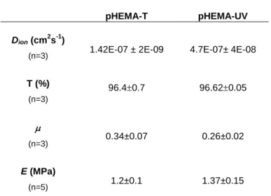
The values of the ionic permeability, Dion, determined for the hydrogels (Table 1) are clearly above
the minimum ion permeability of 1.067 x 10⁻⁹ cm2 s-1, claimed to ensure sufficient contact lens movement [39]. PHEMA-UV samples present a higher permeability which can be explained by the higher water content and lower crosslinking density of this hydrogel.
As shown in Table 1, the hydrogels, in the hydrated state, present a high transmittance to visible light. The transmittance values obtained fall within the range reported in literature for soft contact lenses materials [40, 41]. These values remain practically constant after loading the hydrogels with the antibiotic (96.570.03% for PHEMA-T and 95.970.04% for PHEMA-UV).
In lubricated conditions (NaCl solution) both hydrogels led to friction coefficients of approximately 0.3 (Table 1). Although the experimental conditions (e.g. counterbody geometry, normal load, sliding velocity) vary from study to study, the values obtained are of the same order of magnitude of those previously published for similar hydrogels: Yanez et al. [34] reported analogous values for pHEMA hydrogels containing PVP, and Roba et al. [28] also found comparable values for various commercial contact lenses.
The values of the Young’s modulus (E) for the hydrogels produced in this work (see Table 1) are of the same order of magnitude of those reported in the literature for various pHEMA based hydrogels (between 0.4 and 0.9 MPa [42]) and silicon hydrogel lenses (between 0.3 and 1.9 MPa [43] ).
Figure 3 presents the water contact angle variation during the first 10 minutes of measurement for pHEMA-T and pHEMA-UV samples. Although there is some overlapping of the error bars, pHEMA-T seems to be slightly more hydrophilic. The results are in agreement with data available in the literature relative to similar hydrogels in the dry state [23]. Lower values are expected for the hydrated hydrogels [44].
SEM images of pHEMA-T and pHEMA-UV after drug release tests in static conditions (Fig. 4a and b) show a similar morphology for both hydrogels. Samples that were submitted to load cycles in
Simublink (images not shown) did not present significant changes relatively to the former ones.
Figure 5 shows the AFM images of the surface of both type of hydrogels. While pHEMA-T reveals a featureless surface (Fig. 5a), pHEMA-UV presents a small amount of surface nanopores with sizes is in the range of 100-300 nm (Fig. 5b). The average roughness of the surfaces, determined
13
from the AFM images (20X20 µm2), varies between 4.5 and 6.2 nm. These values lie in the range of the roughness values reported for commercial contact lenses [45].
The TEM observation of the hydrogels reveals a homogeneous structure, without any noticeable bulk pores in either of the hydrogels. However, select area electron diffraction (SAED) patterns (Fig. 6) are much more interesting. As expected for any amorphous material, both hydrogels present a scattered diffraction halo with decreasing intensity from the centre to the periphery. However, in what concerns pHEMA-T (Fig. 6a) the halo is much broader than in pHEMA-UV (Fig. 6b), as can be clearly seen from the corresponding intensity distribution histograms (Fig. 6c and 6d). On the other hand, pHEMA-T SAED presents a ring (marked with an arrow in Fig. 6a and 6c) which does not occur in pHEMA-UV. This result shows that pHEMA-T shows a higher short range order than pHEMA-UV, which may result from the higher cross link density/low water content of pHEMA-T [46].
3.2 Drug release tests
Levofloxacin release from pHEMA-UV and pHEMA-T was followed for 10 hours in static conditions, under stirring and with friction (in Simublink). The release profiles for the three situations are shown in Fig. 7, for both hydrogels. PHEMA-UV consistently releases a higher amount of antibiotic than pHEMA-T, which may be explained by the higher drug uptake that occurs in the former type of hydrogel, not only due to its higher water absorption capacity (see section 3.1), but also due to the presence of acrylic acid. In fact, extended drug release tests carried out in water showed that the amount of drug loaded in PHEMA-T was 8.8 0.5 µg/mg dry gel, while for PHEMA-UV it was 19.9 0.7 µg/mg dry gel. This is in agreement with the results of Alvarez-Lorenzo et al. [29], who found that addition of acrylic acid to pHEMA hydrogels remarkably increased their affinity to norfloxacin, a fluoroquinolone such as levofloxacin.
In respect to release conditions, stirring did not lead to significant differences in the drug release profiles, in comparison to the static conditions, for both hydrogels. However, the effect of friction depends on the hydrogel: while for pHEMA-T, friction almost did not affect the drug release profile, for pHEMA-UV, the mechanical action gave rise to a higher release rate and the process of drug release was complete within 5 hours. This behavior may be explained by the simultaneous presence of nanosized superficial pores in the pHEMA-UV matrix (absent from the pHEMA-T) and
14
lower short range order and smaller cross linking degree of polymer chains, when compared with pHEMA-T. The fluid distribution within the polymeric network should be affected by the mechanical stimulation which implies that the effect of friction becomes more accentuated in less cross linked polymers and, eventually, is even potentiated if some superficial porosity exists, as in the case of pHEMA-UV. To our knowledge, this is the first report on the effect of friction on drug release from hydrogels for contact lenses, although other authors have addressed the effect of mechanical stimulation in model release systems. For example, Lee et al. [47] found that the release of a growth factor from alginate hydrogels increased under mechanical loading, in agreement with our findings.
3.3 Microbiological tests
The minimum inhibitory concentrations (MIC) for Staphylococcus aureus ATCC 6538 and
Pseudomonas aeruginosa ATCC 27853 were estimated to be 16 µg/mL and 62 µg/mL,
respectively.
The concentrations of levofloxacin in the solutions resulting from drug release experiments, carried out with and without friction, were determined by microbiological tests and HPLC. The obtained values were identical, indicating that the antibiotic activity is maintained.
3.4 Estimation of the in vivo efficacy of the studied systems
In order to predict the importance of friction in in vivo conditions, a mathematical model, which takes into account the physiological parameters of the eye, described in detail elsewhere [48], was applied. For the sake of simplicity, the model considers only the drug release in the post-lens tear film and neglects any heterogeneity in the tear fluid composition. The application of more complex drug release models, that take into consideration the transport of the drug into the cornea [49, 50], is outside the scope of the present work.
The model assumes that the amount of drug delivered by a drug-loaded, commercial sized lens to the lachrymal fluid (
M
t) during a given time interval (
t
)
can be estimated by:15
where
m
lis the dry mass of the lens andq
is the drug release rate per unit mass of dry gel which may be obtained by fitting the drug release data to the Korsmeyer-Peppas equation:
q
n K t
n1 (5)where n is the diffusional exponent which can be related to the drug transport mechanism, K is a proportionality constant, and t is the time. This equation is known to adequately describe the cumulative drug release from hydrogels[51, 52].
The drug concentration in the lachrymal fluid at a given time t (in min),
t
Levo
, following the lens application, is given by:
t(1
)
1 r t t tM
Levo
R
Levo
V
(6)where
V
tis the total tear volume in the eye andR
ris the fraction of renovation of the lachrymal fluid. According to the literature, the renewal rate of the lachrymal fluid is 1.2 µL/min [53]. Assuming that the total tear volume in the eye is in averageV
t = 7 μL at each instant [53, 54], the volume renovated in each minute will correspond to 17% ofV
t.The model was applied to the levofloxacin release from pHEMA-UV, the friction-sensitive hydrogel as reported before. For lenses with a dry mass of 66 mg (approximate mass of a lens of the hydrogels under study with 14 mm in diameter), the curves obtained under friction (in Simublink) and in static conditions are shown in Fig. 8. The difference between the curves confirms that friction significantly affects the drug release from pHEMA-UV, even when the tear turnover is taken into account.
For comparative purposes, Fig. 8 shows the estimated antibiotic concentration in the eye over time, as a result of the application of commercial levofloxacin eyedrops (e.g. QUIXIN®, 5 mg/mL). This estimation was based on the assumption that a volume of 7 μL of collyrium remained in the eye, after the application of the recommended dosage (1-2 drops every hour). A saw shape like curve was obtained. The MICs for Staphylococcus aureus and Pseudomonas aeruginosa are also shown in Fig. 8.
16
According to our model, pHEMA-UV hydrogels lead to an initial burst effect in the drug release, after application of the lens in the eye, which is more pronounced under friction conditions. The model also predicts that, in static conditions, the hydrogel keeps the levofloxacin concentration in the tear fluid above the MIC for both microorganisms for at least 10 h (the time of the experiment). Furthermore, after the initial three hours, a sustained release is achieved, in contrast with the saw-type concentration profile resultant from the application of eyedrops.In the presence of friction, the drug level in the lachrymal fluid falls abruptly after 5 h, since no further drug release could be measured afterwards. Results show that blinking may be a relevant factor in the drug release from hydrogels for contact lenses when their cross-link degree is low and/or they present a nanoporous structure.
Conclusion
In this work, the effect of blinking on the drug release from hydrogels for contact lenses was investigated, for the first time, through in vitro studies. Two types of pHEMA based hydrogels with adequate properties for that application (swelling capacity, ionic permeability, transmittance, friction coefficient, elasticity, wettability) and different structural and physicochemical properties were used: pHEMA-T and pHEMA-UV. In all drug release tests, pHEMA-UV showed higher release rates than pHEMA-T. Friction was found to enhance the drug release kinetics only in the case of pHEMA-UV. The lower short range order of this hydrogel (which indicates a lower cross linking degree) and the small nanoporosity of its surface are probably responsible for this behavior. In order to predict the importance of friction in in vivo conditions, the data were further analyzed using a mathematical model based on known physiological parameters of the eye. The drug concentrations predicted for in vivo conditions show that the effect of blinking in the increase of drug release kinetics from pHEMA-UV is maintained, even when the drug dilution caused by tear flow is considered. These results highlight the importance of taking friction into consideration when testing the drug release from materials with some nanoporosity, since materials that seem adequate when tested in static conditions may lead to undesired faster release under friction.
17
References
1. Gulsen D, Chi-Chung L, Chauhan A. Dispersion of DMPC Liposomes in Contact Lenses for Ophthalmic Drug Delivery. Curr Eye Res. 2005;30:1071-80.
2. Xinming L, Yingde C, Lloyd AW, Mikhalovsky SV, Sandeman SR, Howel CA, et al. Polymeric hydrogels for novel contact lens-based ophthalmic drug delivery systems: a review. Cont Lens Anterior Eye. 2008;31:57-64.
3. Meisner D, Mezei M. Liposome ocular delivery systems. Adv Drug Delivery Rev. 1995;16:75-93.
4. Gaudana R, Jwala J, Boddu SS, Mitra A. Recent Perspectives in Ocular Drug Delivery. Pharm Res. 2009;26:1197-216.
5. Rathore K, Nema R. An insight into ophthalmic drug delivery system. Int J Pharm Scs Drug Delivery. 2009;1:1-5.
6. Sasaki H, Yamamura K, Mukai T, Nishida K, Nakamura J, Nakashima M, et al. Enhancement of ocular drug penetration. Crit Rev Ther Drug Carrier Syst. 1999;16:85-146. 7. Kumari A, Sharma PK, Garg VK, Garg G. Ocular inserts - Advancement in therapy of eye diseases. J Adv Pharm Technol Res. 2010;1:291-6.
8. Peng C-C, Kim J, Chauhan A. Extended delivery of hydrophilic drugs from silicone-hydrogel contact lenses containing Vitamin E diffusion barriers. Biomaterials. 2010;31:4032-47. 9. Ciolino JB, Hoare TR, Iwata NG, Behlau I, Dohlman CH, Langer R, et al. A drug-eluting contact lens. Invest Ophthalmol Vis Sci. 2009;50:3346-52.
10. Rubinstein MP. Disposable contact lenses as therapeutic devices. J Brit Cont Lens A. 1995;18:95-7.
11. Shah C, Raj CV, Foulks GN. The evolution in therapeutic contact lenses. Ophthalmol Clin North Am. 2003;16:95-101.
12. Millis EAW. Chapter 13 - Therapeutic contact lenses. Medical Contact Lens Practice. Edinburgh: Butterworth-Heinemann; 2005. p. 129-35.
13. McDermott ML, Chandler JW. Therapeutic uses of contact lenses. Surv Ophthalmol. 1989;33:381-94.
14. Creech JL, Chauhan A, Radke CJ. Dispersive Mixing in the Posterior Tear Film Under a Soft Contact Lens. Industrial & Engineering Chemistry Research. 2001;40:3015-26.
15. Ciolino JB, Dohlman CH, Kohane DS. Contact lenses for drug delivery. Semin Ophthalmol. 2009;24:156-60.
16. White C, Tieppo A, Byrne M. Controlled drug release from contact lenses: a comprehensive review from 1965-present. Journal of drug delivery science and technology. 2011;21:369-84.
17. Wu L, Brazel CS. Surface crosslinking for delayed release of proxyphylline from PHEMA hydrogels. Int J Pharm. 2008;349:1-10.
18. Kapoor Y, Thomas JC, Tan G, John VT, Chauhan A. Surfactant-laden soft contact lenses for extended delivery of ophthalmic drugs. Biomaterials. 2009;30:867-78.
18
19. Danion A, Brochu H, Martin Y, Vermette P. Fabrication and characterization of contact lenses bearing surface-immobilized layers of intact liposomes. Journal of Biomedical Materials Research Part A. 2007;82A:41-51.
20. Alvarez-Lorenzo C, Hiratani H, Concheiro A. Contact Lenses for Drug Delivery: Achieving Sustained Release with Novel Systems. American Journal of Drug Delivery. 2006;4:131-51. 21. Gulsen D, Chauhan A. Ophthalmic Drug Delivery through Contact Lenses. Invest Ophthalmol Vis Sci. 2004;45:2342-7.
22. Jung HJ, Abou-Jaoude M, Carbia BE, Plummer C, Chauhan A. Glaucoma therapy by extended release of timolol from nanoparticle loaded silicone-hydrogel contact lenses. J Control Release. 2013;165:82-9.
23. Yanez F, Martikainen L, Braga ME, Alvarez-Lorenzo C, Concheiro A, Duarte CM, et al. Supercritical fluid-assisted preparation of imprinted contact lenses for drug delivery. Acta Biomater. 2011;7:1019-30.
24. Costa VP, Braga MEM, Guerra JP, Duarte ARC, Duarte CMM, Leite EOB, et al. Development of therapeutic contact lenses using a supercritical solvent impregnation method. The Journal of Supercritical Fluids. 2010;52:306-16.
25. Karlgard CCS, Wong NS, Jones LW, Moresoli C. In vitro uptake and release studies of ocular pharmaceutical agents by silicon-containing and p-HEMA hydrogel contact lens materials. Int J Pharm. 2003;257:141-51.
26. Dajcs JJ, Thibodeaux BA, Marquart ME, Girgis DO, Traidej M, O'Callaghan RJ. Effectiveness of ciprofloxacin, levofloxacin, or moxifloxacin for treatment of experimental Staphylococcus aureus keratitis. Antimicrob Agents Chemother. 2004;48:1948-52.
27. Lee SG, Brunello GF, Jang SS, Bucknall DG. Molecular dynamics simulation study of P (VP-co-HEMA) hydrogels: effect of water content on equilibrium structures and mechanical properties. Biomaterials. 2009;30:6130-41.
28. Roba M, Duncan EG, Hill GA, Spencer ND, Tosatti SGP. Friction Measurements on Contact Lenses in Their Operating Environment. Tribol Lett. 2011;44:387-97.
29. Alvarez-Lorenzo C, Yañez F, Barreiro-Iglesias R, Concheiro A. Imprinted soft contact lenses as norfloxacin delivery systems. J Control Release. 2006;113:236-44.
30. Vazquez R, Nogueira R, Orfao M, Mata JL, Saramago B. Stability of triglyceride liquid films on hydrophilic and hydrophobic glasses. J Colloid Interface Sci. 2006;299:274-82.
31. Peng C-C, Chauhan A. Ion transport in silicone hydrogel contact lenses. Journal of Membrane Science. 2012;399–400:95-105.
32. Rennie AC, Dickrell PL, Sawyer WG. Friction coefficient of soft contact lenses: measurements and modeling. Tribol Lett. 2005;18:499-504.
33. Wong FA, Juzwin SJ, Flor SC. Rapid stereospecific high-performance liquid chromatographic determination of levofloxacin in human plasma and urine. J Pharm Biomed Anal. 1997;15:765-71.
19
34. Yañez F, Concheiro A, Alvarez-Lorenzo C. Macromolecule release and smoothness of semi-interpenetrating PVP–pHEMA networks for comfortable soft contact lenses. Eur J Pharm Biopharm. 2008;69:1094-103.
35. Tighe BJ, Jones L, Evans K, Franklin V. Patient-dependent and material-dependent factors in contact lens deposition processes. Adv Exp Med Biol. 1998;438:745-51.
36. McArthur SL, McLean KM, St. John HAW, Griesser HJ. XPS and surface-MALDI-MS characterisation of worn HEMA-based contact lenses. Biomaterials. 2001;22:3295-304.
37. Efron N, Morgan PB, Cameron ID, Brennan NA, Goodwin M. Oxygen permeability and water content of silicone hydrogel contact lens materials. Optom Vis Sci. 2007;84:328-37.
38. Lakes RS, Parker J. Biomaterials: An Introduction. 3rd ed: Springer; 2007.
39. Baron RC, Chabrecek P, Court J, Domschke A, Griesser HJ, Ho A, et al., Extended wear ophthalmic lens. United States of America patent US 5776999 A. 1998.
40. Efron N, Maldonado-Codina C. Development of Contact Lenses from a Biomaterial Point of View – Materials, Manufacture, and Clinical Application. In: Ducheyne P, editor. Comprehensive Biomaterials. Oxford: Elsevier; 2011. p. 517-41.
41. Gulsen D, Chauhan A. Effect of water content on transparency, swelling, lidocaine diffusion in p-HEMA gels. Journal of Membrane Science. 2006;269:35-48.
42. Tranoudis I, Efron N. Tensile properties of soft contact lens materials. Cont Lens Anterior Eye. 2004;27:177-91.
43. Horst CR, Brodland B, Jones LW, Brodland GW. Measuring the Modulus of Silicone Hydrogel Contact Lenses. Optometry & Vision Science. 2012;89:1468-76.
44. Li L, Xin Z. Surface-hydrophilic and protein-resistant tris(trimethylsiloxy)-3-methacryloxypropylsilane-containing polymer by the introduction of phosphorylcholine groups. Colloids and Surfaces A: Physicochemical and Engineering Aspects. 2011;384:713-9.
45. Giraldez MJ, Serra C, Lira M, Real Oliveira ME, Yebra-Pimentel E. Soft contact lens surface profile by atomic force microscopy. Optom Vis Sci. 2010;87:E475-81.
46. Czigány Z, Hultman L. Interpretation of electron diffraction patterns from amorphous and fullerene-like carbon allotropes. Ultramicroscopy. 2010;110:815-9.
47. Lee KY, Peters MC, Anderson KW, Mooney DJ. Controlled growth factor release from synthetic extracellular matrices. Nature. 2000;408:998-1000.
48. Paradiso P, Galante R, Santos L, Matos AP, Colaço R, Serro AP, et al. Comparison of two hydrogel formulations for drug release in ophthalmic lenses. . J Biomed Mater Res B. 2013 49. Ferreira JA, Oliveira P, M.Silva P, A.Carreira, Gil H, Murta JN. Sustained drug release from contact lens. Comp Modeling in Engineering and Science. 2010;60:152-79.
50. Ferreira JA, Oliveira P, M.Silva P, A.Carreira, Gil H, Murta JN. Drug delivery: from a ophthalmic lens to the anterior chamber. Comp Modeling in Engineering and Science. 2011;79:164-74.
51. Rekhi GS, Nellore RV, Hussain AS, Tillman LG, Malinowski HJ, Augsburger LL. Identification of critical formulation and processing variables for metoprolol tartrate extended-release (ER) matrix tablets. J Control Release. 1999;59:327-42.
20
52. Siepmann J, Peppas NA. Modeling of drug release from delivery systems based on hydroxypropyl methylcellulose (HPMC). Adv Drug Delivery Rev. 2001;48:139-57.
53. Hoang-Xuan T, Baudo C. Inflammatory Diseases of the Conjunctiva: Thieme Medical Publishers, Incorporated; 2001.
54. Yokoi N, Bron AJ, Tiffany JM, Maruyama K, Komuro A, Kinoshita S. Relationship between tear volume and tear meniscus curvature. Arch Ophthalmol. 2004;122:1265-9.
Figure captions
Figure 1. Simublink - equipment used to simulate the eyelid movement over the hydrogels. 1 - Liquid cell, 2 - Power supply, 3 - Motor controller, 4 - Step-by-step motor and mechanical structure.
Figure 2. Swelling kinetics of the hydrogels in water (n=3).
Figure 3. Water contact angle variation with time for pHEMA-T and pHEMA-UV (n=8-10).
Figure 4. SEM images of pHEMA-T (a) and pHEMA-UV (b) after drug release tests in static conditions (Images magnification 3000x).
Figure 5. AFM topographic images of (a) pHEMA-T and (b) pHEMA-UV unloaded samples. Dimensions of a pore found in pHEMA-UV are visible in the insert of (b).
Figure 6. TEM SAED of (a) pHEMA-T and (b) pHEMA-UV, with the corresponding intensity histograms (c and d).
Figure 7. Levofloxacin release profiles of pHEMA-T and pHEMA-UV in static conditions, under stirring and with friction.
Figure 8. Levofloxacin concentration in the lachrymal fluid, as estimated from the model, following the application of drug loaded pHEMA-UV lenses, under friction and in static conditions. The antibiotic concentration resultant from the application of a commercial collyrium and the minimum inhibitory concentrations for S. aureus and P. aeruginosa are included for comparative purposes.
Figure 1
Figure 2
Figure 3
Figure 4
Figure 5
Figure 6
Figure 7
Figure 8
Table 1: Estimated ion permeability (Dion), transmittance (T), friction coefficient () and Young’s modulus (E) of the studied pHEMA based hydrogels in the hydrated state. Results are mean±standard deviation of n determinations.
pHEMA-T pHEMA-UV
Dion (cm2s-1)
(n=3) 1.42E-07 ± 2E-09 4.7E-07± 4E-08
T (%) (n=3) 96.40.7 96.620.05 (n=3) 0.34±0.07 0.26±0.02 E (MPa) (n=5) 1.2±0.1 1.37±0.15 Table 1