FACULDADE DE MEDICINA
NATURALLY OCCURRING MUTATIONS IN REGULATORY PROTEINS AMONG STREPTOCOCCUS PYOGENES ISOLATES FROM DISTINCT
HUMAN INFECTIONS
CATARINA TERESA CONDINHO PATO
Orientador: Professor Doutor Mário Nuno Ramos de Almeida Ramirez
Tese especialmente elaborada para a obtenção do grau de Doutor em Ciências e Tecnologias da Saúde, especialidade em Microbiologia
FACULDADE DE MEDICINA
NATURALLY OCCURRING MUTATIONS IN REGULATORY PROTEINS AMONG STREPTOCOCCUS PYOGENES ISOLATES FROM DISTINCT
HUMAN INFECTIONS
CATARINA TERESA CONDINHO PATO
Orientador: Professor Doutor Mário Nuno Ramos de Almeida Ramirez Tese especialmente elaborada para a obtenção do grau de Doutor em Ciências e
Tecnologias da Saúde, especialidade em Microbiologia Júri:
Presidente:
Doutor José Augusto Gamito Melo Cristino, Professor Catedrático e Presidente do Conselho Científico da Faculdade de Medicina da Universidade de Lisboa
Vogais:
Doctor Shiranee Sriskandan, Professor of Faculty of Medicine, Imperial College
London;
Doutor Paulo Jorge Pereira Cruz Paixão, Professor Auxiliar da Faculdade de Ciências Médicas - Universidade NOVA de Lisboa;
Doutora Isabel Maria dos Santos Leitão Couto, Professora Auxiliar do Instituto de Higiene e Medicina Tropical - Universidade NOVA de Lisboa;
Doutora Maria Constança Matias Ferreira Pomba, Professora Associada da Faculdade de Veterinária da Universidade de Lisboa;
Doutor Mário Nuno Ramos de Almeida Ramirez, Professor Associado com Agregação da Faculdade de Medicina da Universidade de Lisboa (Orientador);
Doutora Ana Isabel de Aquino Friães, Professora Auxiliar da Faculdade de Medicina da Universidade de Lisboa.
Fundação para a Ciência e Tecnologia (SFRH/BD/81329/2011) 2018
In first place I would like to express my sincere gratitude to my PhD supervisor, Professor Mário Ramirez. First, for having accepted me as PhD student but mostly for his will and availability to share his immense knowledge and offering suggestions during the innumerous work discussions and revisions, that were definitely essential for the development of my critical way of thinking and were the basis of all my work decisions.
My sincere thanks to Professor José Melo Cristino, whom I deeply respect and admire, for having provided me with the opportunity to join his team and whose professional life is an inspiration.
I am also deeply grateful to Ana Friães, with whom I worked more closely, for having introduced me to the majority of the methods used in this thesis, for all the work revisions and of course for all the guidance and support.
A special thank to my PhD partners Andreia Horácio, Jorge Miranda, (MRamirez Lab), and Sofia Cerqueira (PSimas Lab), who shared this journey with me. I want to thank them for their friendship, support and for all the incentive.
I also wish to thank all my other colleagues at MRamirez Lab, for all the technical support and mostly for the friendly environment and team spirit. These include Sandra Aguiar, Maria João Frias, Elizabete Martins, João Carriço, Margarida Carrolo, Catarina Costa, Marcos Pinho, Filomena Costa, Alice Faria, Joana Lopes, Raquel Garcia, Mickael Silva and Miguel Machado.
For the financial support, I want to thank Fundação para a Ciência e Tecnologia for having granted me with a Ph.D. scholarship (SFRH/BD/81329/2011).
My final thanks are to my friends and family, especially to my parents, for always believing in me, for keeping me motivated and mostly for all their love. I could not have done it without them.
Keywords: Streptococcus pyogenes, CovRS, SpeB, skin and soft tissue infections
Streptococcus pyogenes (Group A Streptococcus, GAS) is among the most prevalent bacterial pathogens of humans and it is responsible for a wide range of infections, from pharyngitis and impetigo to life-threatening conditions such as necrotizing fasciitis and streptococcal toxic shock syndrome. However, until today, despite many years of research, there is still no consensus regarding which are the genotypic or phenotypic characteristics that confer to an isolate a certain tissue preference or a more invasive potential. The nasopharyngeal mucosa and skin are considered the primary sources of isolates responsible for invasive infections. This suggests a further ability of these isolates to invade and survive in deeper tissues. In the last years, spontaneous mutations occurring in the covRS two-component regulatory system have been considered a possible explanation for the transition from localized to systemic infection. It is estimated that this system controls directly or indirectly the expression of 10-15% of the GAS genome. As a consequence of these mutations, among the distinct patterns of expression of several virulence factors, the downregulation of the extracellular cysteine protease SpeB, has been considered crucial for the switch to a hypervirulent phenotype. The downregulation of SpeB has also been described as a consequence of mutations in a stand-alone transcriptional regulator named RopB. However, contrasting results were reported whether covRS mutations were more prone to occur in certain lineages, namely those more frequently associated with invasive infections or if they occur in a similar proportion among invasive and non-invasive isolates. To address these questions, in the present thesis, we determined the sequence of the covRS and ropB of 191 isolates from invasive infection and pharyngitis and evaluated the production of SpeB, as well of NAD glycohydrolase (NADase) and streptolysin S (SLS), which are two virulence factors supposed to be under the influence of CovRS. Moreover, skin and soft tissue infections (SSTI) are frequently considered the focal points for the development of invasive disease. However, most of the knowledge about GAS skin isolates is from studies that intended to find differences between isolates recovered from invasive and non-invasive infections generically. The majority of these studies use isolates recovered from SSTI but mostly from pharyngitis and compared those together against the isolates recovered from
invasive infections, resulting in few data regarding isolates recovered from SSTI. Therefore, we characterized by multiple typing methods a total of 320 isolates from SSTI recovered in Portugal and performed the comparison with invasive isolates recovered during the same period which were previously characterized. All SSTI isolates were also tested for SpeB activity and for those without detectable SpeB activity we determined the sequence of covRS and ropB genes.
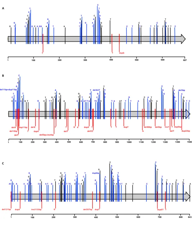
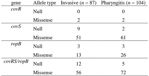
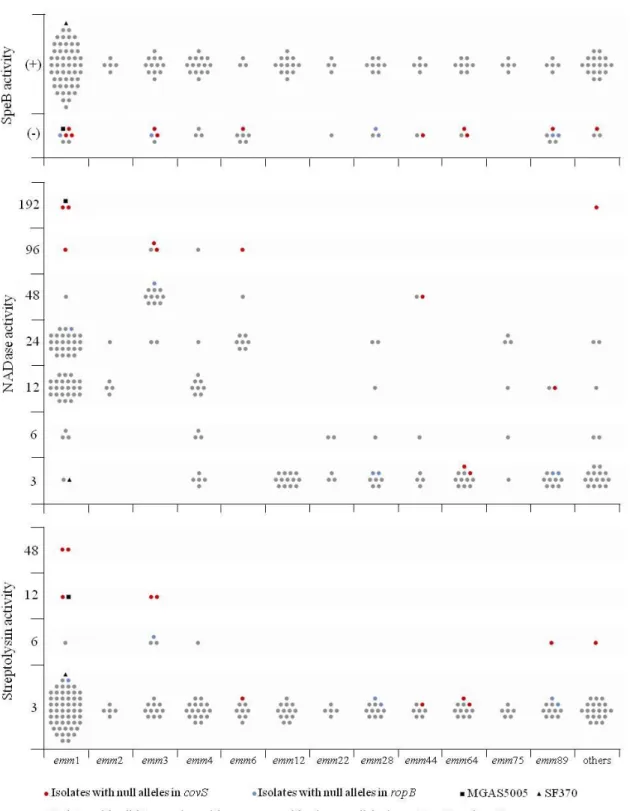
Overall, we found that isolates with null covS alleles, which are predicted to eliminate the protein function have a significant association with invasive infections comparative with isolates from pharyngitis and SSTI. Additionally, none of these isolates, as expected, had SpeB activity, and, with few exceptions, they showed an increased activity of both NADase and SLS that could explain their potentially higher invasiveness. Even so, this mechanism was found to be uncommon, corresponding to only 10% of invasive isolates, which could be due to an overall fitness cost of these mutations. Moreover, null covS alleles were not more prevalent among isolates from clones frequently associated with invasive infection such as emm1 and emm64 and instead they were distributed throughout diverse genetic backgrounds. The few exceptions regarded the levels of NADase and SLS points to the complexity of the regulatory networks among distinct GAS lineages. Additionally, no null covR alleles were detected in our isolates and ropB null alleles were found in a low fraction of GAS isolates and were not associated with any infection type. Regarding SpeB activity, it was detected in a similar proportion in isolates recovered from the different sources, and therefore its absence was not associated to any type of infection, suggesting that its abrogation cannot by itself explain the higher ability of certain clones to cause invasive disease. Among SSTI isolates, we found that emm89 type isolates were the most prevalent and were significantly associated with these infections when compared with invasive isolates. In contrast, emm1, emm3, and emm64 isolates were associated with invasive infections. Within emm89 isolates, SSTI were only associated to those that lack the hasABC locus, corresponding to a recently emerged acapsular clade (clade 3) that also carries a variant of the ngs-ifs-slo locus. These results suggest that for some unknown reason these isolates may have an increased potential to cause SSTI. As a consequence of known differences in the emm-type between isolates causing these two types of infections, we also found significant associations between the ability to bind to different host proteins. This ability was presumed by inferring the emm-cluster through the emm-type results. The emm-cluster is a recent classification based on the entire
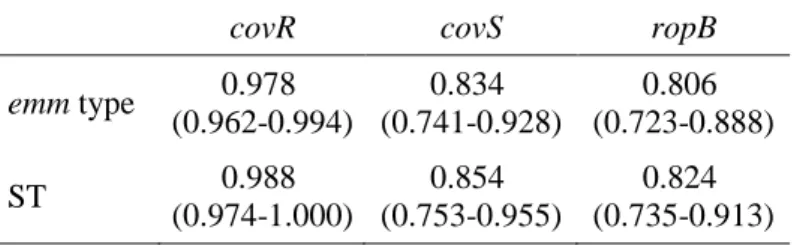
proteins and other structural properties. Therefore, the ability to bind fibrinogen and albumin were significantly associated with invasive isolates, whereas the ability to bind to C4BP and IgG were associated with SSTI isolates. Differences in the presence of superantigen (SAg) genes, SAg profiles and in the distribution of sequence types (ST) determined by Multilocus Sequence Typing (MLST) were also noted. The possible impact of these differences in the ability of the isolates to cause these distinct infections remains to be clarified. Moreover, within each emm type the same MLST defined lineages and SAg profiles could be found in both types of infection, questioning the possibility that these characteristics dictate the tissue tropism of each isolate.
In summary, the results described in this thesis indicate that isolates responsible for SSTI are genetically distinct from those recovered from normally sterile sites and while some GAS clones have more capacity to invade deeper tissues, others are more prone to cause SSTI. Moreover, the significant presence of null covS mutations among invasive isolates and the fact that no association was observed regarding the absence of SpeB activity in isolates from different types of infections, suggests that the role of spontaneous mutations impairing the CovRS activity is probably related with the regulation of others virulence factors under its control in addition to SpeB.
Palavras-Chave: Streptococcus pyogenes, CovRS, SpeB, infeções da pele e tecidos moles
As bactérias da espécie Streptococcus pyogenes estão entre os principais agentes bacterianos responsáveis por infeções no Homem. São também frequentemente denominadas por estreptococos pertencendo ao Grupo A de Lancefield, por aglutinarem com o soro A da classificação de Lancefield, uma investigadora cujos trabalhos foram cruciais para o atual conhecimento sobre esta espécie. Uma das características mais particulares desde agente é a capacidade de causar um grande espectro de infeções variando relativamente ao local e à gravidade. Pode causar desde doenças autolimitadas da faringe e pele, como faringo-amigdalites e lesões de impetigo, a infeções invasivas frequentemente associadas a elevada morbilidade e mortalidade, como são o caso da fasceíte necrosante e a síndrome do choque tóxico. Também de referir que o seu principal impacto mundial advém das complicações autoimunes, hoje em dia ainda com elevada prevalência nos países em desenvolvimento, onde estão incluídas a febre reumática e a glomerulonefrite. Assim sendo, para causar esta variedade de infeções, este microrganismo tem que se adaptar a diferentes ambientes. A orofaringe e a pele são consideradas como os principais focos de estirpes1 invasivas, o que implica que estas estirpes têm que invadir e sobreviver nos tecidos celulares subcutâneos. Porém, mesmo depois de décadas de investigação, ainda não há consenso relativamente a quais são as características moleculares ou fenotípicas que conferem a uma estirpe um maior potencial invasivo ou uma determinada preferência por um local anatómico.
Em 2006, um estudo com um modelo murganho de infeção da pele e tecidos moles sugeriu a ocorrência de mutações espontâneas num sistema regulador durante o processo de infeção como uma explicação para a transição da infeção localizada para sistémica. Vários estudos foram publicados de seguida a apoiar esta hipótese, incluindo alguns que descreveram a presença destas mutações em estirpes recolhidas de infeções no Homem. O sistema regulador em causa é constituído por dois componentes e denomina-se de CovRS. Estima-se que este sistema controle direta ou indiretamente cerca de 10 a 15% de todo o genoma de S. pyogenes. Entre as alterações verificadas na
1
Neste resumo, a palavra estirpe é utilizada para referir um microrganismo isolado de um determinado produto biológico
expressão de vários fatores de virulência, a abolição da atividade de uma protease extracelular denominada SpeB tem sido considerada como um evento crucial para uma maior virulência. Teoricamente, na ausência de SpeB, são preservados vários fatores de virulência presentes na superfície bacteriana que contribuem para o processo invasivo. Adicionalmente, a diminuição da atividade de SpeB tem também sido descrita como resultado de mutações num outro regulador, designado RopB. Porém, enquanto alguns estudos defendem que as mutações nestes reguladores ocorrem mais frequentemente em estirpes recolhidas de infeções invasivas, outros defendem que estas ocorrem na mesma proporção em estipes de infeções não invasivas. Alguns defendem ainda que estas mutações estão limitadas às linhagens frequentemente associadas a infeções invasivas. De modo a abordar estas questões, determinámos as sequências dos genes covRS e ropB de 191 estirpes recolhidas de infeções invasivas e de amigdalites. Determinámos também, a atividade de SpeB de cada uma destas estirpes e os respetivos valores de NAD glicohidrolase e de estreptolisina S que são dois fatores de virulência aparentemente também sob o controlo do regulador CovRS. Por sua vez, as infeções da pele e tecidos moles são frequentemente consideradas como um foco para o desenvolvimento de infeções invasivas. Porém, a informação disponível sobre estirpes da pele é escassa porque a maioria do conhecimento atual advém de estudos cujo objetivo é a identificação de diferenças entre estirpes recolhidas de infeções invasivas e não invasivas. Os estudos em questão tendem a analisar as características das estirpes isoladas da pele em conjunto com estirpes recolhidas de exsudados faríngeos, o que resulta numa difícil avaliação das características específicas desta população. Desta forma, caracterizámos 320 estirpes recolhidas de infeções da pele e tecidos moles em Portugal através de vários métodos de tipagem e procedemos à sua comparação com estirpes invasivas recolhidas durante o mesmo período previamente trabalhadas. Avaliámos ainda, para as 320 estipes, a presença de atividade de SpeB e naquelas que não apresentavam atividade, determinámos as respetivas sequências dos genes covRS e do ropB.
Verificámos existir uma associação significativa entre estirpes cujas mutações se preveem eliminar a função proteica do CovS e as infeções invasivas, comparativamente com estirpes recolhidas de faringo-amigdalites e de infeções da pele e tecidos moles. Adicionalmente, assim como esperado, nenhuma destas estirpes apresentava atividade SpeB e, salvo algumas exceções, apresentavam um aumento dos níveis de atividade de NAD glicohidrolase e de estreptolisina S. Embora estas diferenças sejam concordantes
estirpes invasivas analisadas. Pensa-se que estas mutações possam estar associadas a um elevado “fitness cost”, dificultando por exemplo a capacidade de colonização e de transmissão, o que pode explicar a sua reduzida prevalência. Verificou-se ainda que estas mutações se encontram dispersas por estirpes de diversas linhagens e não estão apenas restritas às mais frequentemente associadas a infeções invasivas, como estirpes do tipo emm1 e emm64. As exceções detetadas nos valores de atividade de NAD glicohidrolase e estreptolisina S realçam a complexidade já conhecida dos mecanismos de regulação de S. pyogenes e as potenciais diferenças entre estirpes de linhagens distintas. Relativamente ao covR, não foi detetada nenhuma mutação que se preveja eliminar a função proteica. Por sua vez, para o gene ropB, este tipo de mutações foram detetadas num reduzido número de estirpes e não foram associadas a nenhum tipo de infeção. No que se refere à atividade de SpeB, esta foi detetada na maioria das estirpes e numa proporção semelhante em estirpes de diferentes origens, pelo que a sua ausência não foi associada a nenhum tipo de infeção. Estes resultados sugerem que a ausência de SpeB por si só não explica a maior capacidade de certas estirpes para causar infeção invasiva.
Estirpes do tipo emm89 eram as mais prevalentes e estavam significativamente associadas às infeções da pele e tecidos moles, quando comparadas com as estirpes invasivas. Por outro lado, estirpes do tipo emm1, emm3 e emm64 foram significativamente associadas a infeções invasivas. Porém, de entre as estirpes do tipo emm89, as infeções da pele e tecidos moles apenas foram associadas às estirpes que não têm o locus hasABC, responsável pela síntese da cápsula de ácido hialurónico. Estas estipes correspondem a uma linhagem recente sem cápsula que possui uma variante do locus ngs-ifs-slo. Estes resultados sugerem que, por alguma razão ainda desconhecida, estas estirpes podem apresentar uma maior apetência para causar infeções da pele e tecidos moles. Refletindo os tipos de emm associados a cada tipo de infeção, outras associações significativas foram detetadas, nomeadamente a presumida capacidade de ligação a diferentes proteínas do Homem durante o processo de infeção. Esta capacidade foi presumida deduzindo os “clusters” de emm através dos tipos de emm. Os “clusters” correspondem a uma classificação mais recente que se baseia na totalidade da sequência do gene emm, ao contrário dos tipos de emm que se baseiam apenas numa porção deste. Segundos os autores, as estirpes de cada “cluster” partilham zonas de ligação a proteínas do hospedeiro. Assim sendo, enquanto a capacidade de ligação ao
fibrinogénio e à albumina foram significativamente associadas às estirpes invasivas, a capacidade de ligação ao C4BP e IgG foram associadas às estirpes da pele e tecidos moles. Também foram notadas diferenças na presença de genes codificantes de superantigénios, nos perfis de superantigénios e nos “sequence types” determinados através de “Multilocus Sequence Typing”. Embora se tenham verificado diversas diferenças entre estirpes com diferentes origens, permanece por esclarecer se estas influenciam a apresentação da doença e qual o mecanismo subjacente para que isto ocorra. Por sua vez, ao considerar estirpes do mesmo tipo de emm, verificaram-se os mesmos perfis de superantigénios e os mesmos “sequence types” em estirpes de ambos os tipos de infeção, o que sugere que é pouco provável que estes indicadores estejam relacionados com a preferência de cada estipe para um determinado local anatómico ou a um maior potencial invasivo.
Resumidamente, os resultados descritos nesta tese indicam que as estirpes responsáveis por infeções da pele e tecidos moles são uma população geneticamente distinta das estirpes recolhidas de locais normalmente estéreis e enquanto algumas linhagens têm uma maior capacidade invasiva outras são mais propensas a causar infeções da pele e tecidos moles. Por fim, o facto de se ter verificado uma presença significativa de mutações no covS que se preveem que eliminem a função proteica entre as estirpes invasivas associado ao facto de não se identificarem diferenças na atividade SpeB entre estirpes de infeções distintas, sugere que o mecanismo subjacente às mutações espontâneas no CovRS como causa de maior virulência, pode estar relacionado com outros fatores de virulência sob o controlo deste sistema para além do SpeB.
The work described in the present thesis intended to evaluate the importance of CovRS and RopB regulators among Streptococcus pyogenes isolates recovered from distinct types of infections including in isolates from skin and soft tissue infections that were fully characterized through several typing methods and compared with contemporary invasive isolates.
The thesis comprises 4 chapters, organized as follows:
Chapter 1 corresponds to the general introduction that highlights the importance of Streptococcus pyogenes including a historical overview, the infections for which it is responsible and their burden, a description of the best known virulence factors, their mechanisms of action, their contribution to virulence and some of their regulators. It also includes a brief description of typing methods, therapeutic management of infections and mechanisms of antimicrobial resistance.
Chapter 2 is dedicated to the study of CovRS and RopB regulators and the consequences of their variability in several virulence factors, for which we specifically screened the strains. In this study, we used a collection of isolates from pharyngitis and from invasive infections to evaluate potential differences.
Chapter 3 consists in a detailed characterization of a collection of isolates recovered from skin and soft tissues infections using several typing methods and their comparison with a collection of invasive isolates recovered during the same period that were previously characterized. We also screened skin and soft tissue isolates for the presence of protease activity and sequenced covRS and ropB in a subset of isolates in order to understand their importance in this type of infections and to compare it with the results described in chapter 2.
Chapter 4 corresponds to the general discussion. In this chapter is provided a summary of the main results obtained in this thesis and its integrated discussion. It also includes perspectives for future work.
ADP Adenosine diphosphate
APSGN Acute poststreptococcal glomerulonephritis ARF Acute Rheumatic Fever
ASO Anti-Streptolysin O cADPR cyclic ADP-ribose C4BP C4-binding protein
CDC Centers for disease control and prevention CI Confidence interval
CLSI Clinical and Laboratory Standards Institute DNA Deoxyribonucleic acid
DNase Deoxyribonuclease ECM Extracellular matrix
FCT Fibronectin-binding, collagen-binding, T antigen FDR False discovery rate
FHL-1 Factor H-like protein 1 GAS Group A streptococci HBP Heparin binding protein HLA Human leukocyte antigen IFN Interferon
Ig Immunoglobulin
iGAS Invasive GAS infections IL Interleukin
ITP Invasive transcriptome profile LTA Lipoteichoic acid
M Macrolides (resistance phenotype) MGE Mobile genetic element
MHC Major histocompatibility complex MIC Minimum inhibitory concentration
MLSB Macrolides, lincosamides, streptogramins B (resistance phenotype)
cMLSB: constitutive MLSB
MLST Multilocus Sequence Typing mRNA Messenger ribonucleic acid
MRSA Methicillin resistant Staphylococcus aureus NAD Nicotinamide adenine dinucleotide
NADase NAD glycohydrolase
NET Neutrophil extracellular trap PBPs Penicillin Binding Proteins PCR Polymerase chain reaction PMN Polymorphonuclear leukocyte PTP Pharyngeal transcriptome profile PYR Pyrrolidonylarylamidase
QRDR Quinolone resistance-determining region RADT Rapid antigen detection test
RALP RofA-like protein RHD Rheumatic Heart Disease rRNA ribosomal ribonucleic acid SAg Superantigen
Sic Streptococcal inhibitor of complement SID Simpson’s index of diversity
SLO Streptolysin O SLS Streptolysin S
SMEZ Streptococcal mitogenic exotoxin Z SOF Serum opacity factor
Spe Streptococcal pyrogenic exotoxin SSA Streptococcal superantigen SSTI Skin and Soft tissue infection ST Sequence type
STSS Streptococcal toxic shock syndrome TCR T cell receptor
TCS Two-component system regulator TLR Toll-like receptor
TNF Tumor necrosis factor WB Western Blot
WHO World Health Organization
SUMMARY ... ix RESUMO ... xiii THESIS OUTLINE ... xvii ABBREVIATIONS ... xix TABLE OF CONTENTS ... xxiii
CHAPTER I
GENERAL INTRODUCTION ... 1 1. HISTORICAL BACKGROUND ... 3 2. GENERAL FEATURES ... 3 3. INFECTIONS AND THEIR CONSEQUENCES ... 5 3.1. Pharyngitis and Scarlet fever ... 6 3.2. Impetigo ... 7 3.3. Invasive infections ... 8 3.4. Acute rheumatic fever and rheumatic heart disease ... 10 3.5. Acute poststreptococcal glomerulonephritis ... 11 3.6. Pediatric autoimmune neuropsychiatric disorders ... 11 3.7. Poststreptococcal reactive arthritis ... 11 4. CARRIAGE ... 12 5. VIRULENCE FACTORS ... 13 5.1. M protein ... 13 5.2. Superantigens ... 21 5.3. Hyaluronic acid capsule ... 24 5.4. Pili ... 25 5.5. Streptococcal pyrogenic exotoxin B ... 26 5.6. Streptolysin O ... 28 5.7. Nicotinamide glycohydrolase ... 29 5.8. Streptolysin S ... 30 5.9. Streptokinase ... 30 5.10. Extracellular streptodornase D ... 31 5.11. Streptococcal inhibitor of complement ... 31
5.12. C5a peptidase ... 31 5.13. SpyCEP ... 32 5.14. GRAB ... 32 6. HOST FACTORS AND SUSCEPTIBIBLITY TO INFECTION ... 32 7. VIRULENCE REGULATION MECHANISMS ... 33 7.1. CovRS two component regulatory system ... 33 7.2. Regulator of proteinase B ... 36 8. TYPING METHODS ... 37 8.1. Serotyping ... 37 8.2. emm typing ... 38 8.3. emm-clusters ... 38 8.4. SAG profiling ... 40 8.5. Multilocus Sequence Typing ... 40 9. ANTIMICROBIAL THERAPY AND RESISTANCE MECANISMS ... 41 9.1. Treatment of pharyngitis ... 41 9.2. Treatment of impetigo ... 42 9.3. Treatment of necrotizing fasciitis ... 42 9.4. Mechanisms of action and antimicrobial resistance ... 42 10. VACCINE CANDIDATES ... 44 REFERENCES ... 47 AIMS OF THE THESIS ... 71
CHAPTER II
CONSEQUENCES OF THE VARIABILITYOF THE OF COVRS AND ROPB
REGULATORS AMONG STREPTOCOCCUS PYOGENES CAUSING HUMAN
INFECTIONS ... 73 CHAPTER III
STREPTOCOCCUS PYOGENES CAUSING SKIN AND SOFT TISSUE INFECTIONS HAVE INCREASED PREVALENCE OF THE RECENTLY EMERGED EMM89 CLADE 3 AND ARE NOT ASSOCIATED WITH ABROGATION OF COVRS ... 105
GENERAL DISCUSSION ... 143
CHAPTER I
1. HISTORICAL BACKGROUND
According to a review about history of streptococcal research (Ferretti and Kohler, 2016), the first potential description of streptococcal infections dates to the 4th century BC, in the original writings of Hippocrates where are described the symptoms of childbed fever and erysipelas. In 1874, an Austrian surgeon named Theodor Billroth, described “small organisms as found in either isolated or arranged in pairs, sometimes in chains of four to twenty or more links (Streptococcus; Gr. Strepto, a chain and coccus, a berry)” in cases of erysipelas and wound infections. In 1879, Louis Pasteur established the real importance of streptococci when he isolated the microorganism from the uteruses and blood of women with puerperal fever and demonstrated that Streptococcus was the etiological agent responsible for the disease. At that time, puerperal fever was the cause of high mortality among women and newborns. The name “Streptococcus pyogenes” (Gr., pyo, pus, and genes, forming) came from Julius Rosenbach in 1884, due to his observations of bacteria recovered from suppurative lesions. Meanwhile, other names also emerged such as eryespaltis, scarlatinae, and puerperalis, mostly according to the disease associated. However, in 1932, due to the lack of unique characteristics of the microorganisms isolated from specific diseases, Andrews & Christie suggested that all previous species names be included in the single name Streptococcus pyogenes. Since 1903, with the introduction of blood agar plates by Hugo Schottmüller that is possible to differentiate streptococci based on their type of hemolysis. Those with a clear zone surrounding a colony were termed Streptococcus haemolyticus. Then, in 1933, Rebecca Lancefield developed the classification system, that is currently used, which allowed the differentiation of Streptococcus haemolyticus isolates in distinct groups (Lancefield, 1933). Streptococcus pyogenes corresponds to Lancefield group A antigen and therefore the name Group A Streptococcus (GAS) is commonly used as an alternative.
2. GENERAL FEATURES
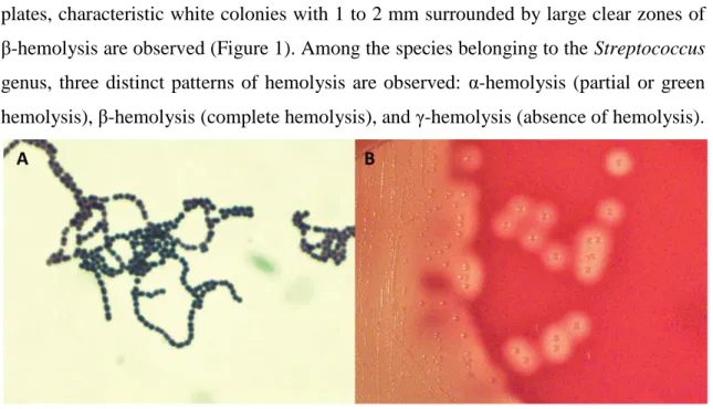
Streptococcus pyogenes is a Gram positive bacterium. Individual cells are presented as spherical cocci with 1 to 2µm in diameter arranged in short chains in clinical specimens. When grown in liquid media longer chains are observed (Figure 1). It is a facultative anaerobe, catalase negative, with optimal growth on blood-enriched
agar media (Murray et al., 2013). After overnight incubation at 35-37°C on blood agar plates, characteristic white colonies with 1 to 2 mm surrounded by large clear zones of β-hemolysis are observed (Figure 1). Among the species belonging to the Streptococcus genus, three distinct patterns of hemolysis are observed: α-hemolysis (partial or green hemolysis), β-hemolysis (complete hemolysis), and γ-hemolysis (absence of hemolysis).
Figure 1. Streptococcus pyogenes. A- Chains with Gram stain. B- Colonies in blood agar with a surrounding area of β-hemolysis. Both images were reproduced from (Murray et al., 2013).
A minority of Streptococcus pyogenes strains exhibiting α-hemolysis and γ-hemolysis on blood agar were also reported, which were associated with the lack of ability to produce streptolysin S (SLS) (discussed later) (Yoshino et al., 2010). Some strains are covered by a hyaluronic acid capsule that is antigenically indistinguishable from the hyaluronic acid in mammalian connective tissues (discussed later) (Murray et al., 2013). Additionally, within the cell wall of Streptococcus pyogenes is the group-specific carbohydrate, which is a dimmer of N-acetylglucosamine and rhamnose. This carbohydrate comprises about 10% of the dry weight of the cell and is the base of the serological classification developed by Rebecca Lancefield in 1933, which identifies Streptococcus pyogenes as Lancefield’s group A streptococci (Lancefield, 1933). After the enzymatic extraction of the carbohydrate, a latex agglutination test with each specific group serum allows the identification of Streptococcus pyogenes (group A), Streptococcus agalactiae (group B), and other species (groups C, F and G) (Murray et al., 2013). There are some strains of Streptococcus dysgalactiae subsp. equisimilis and Streptococcus anginosus group that share the same group-specific carbohydrate with Streptococcus pyogenes reacting positively with group A serum, however these are uncommonly recovered from human infections (Facklam et al., 2002).
Susceptibility to bacitracin was another test used for the identification of Streptococcus pyogenes. However, since the report of bacitracin resistant Streptococcus pyogenes isolates, including in Portugal, this test is no longer reliable (Malhotra-Kumar et al., 2003; Pires et al., 2009; Silva-Costa et al., 2008). Another biochemical feature associated with Streptococcus pyogenes is its pyrrolidonylarylamidase (PYR) activity that can be tested by the PYR test. Among β-hemolytic streptococci, there are strains from other species susceptible to bacitracin or positive for the PYR test. However, Streptococcus pyogenes is the only species reported of having these both characteristics (Facklam, 2002).
3. INFECTIONS AND THEIR CONSEQUENCES
GAS is responsible for a variety of pyogenic infections ranging from mild superficial infections of the respiratory tract and skin, such as pharyngitis and impetigo, to extremely severe invasive infections, such as necrotizing fasciitis and streptococcal toxic shock syndrome (STSS) (Cunningham, 2000; Walker et al., 2014). GAS infections have also been related to the development of postinfection autoimmune sequelae, which are responsible for high morbidity and mortality worldwide. These conditions include, mainly, acute rheumatic fever (ARF) and rheumatic heart disease (RHD), as well as acute poststreptococcal glomerulonephritis (APSGN) (Walker et al., 2014; Cunningham, 2000). Before the advent of antibiotics, streptococcal diseases such as scarlet fever, erysipelas, and puerperal fever were considered major health problems for centuries, however their incidence and mortality rates started to fall just before penicillin’s widespread use after the Second World War, suggesting that other factors, such as the host, the pathogen and the environment contributed to the decrease of the impact of these diseases (Efstratiou and Lamagni, 2016). Since the 1980s, it was observed a resurgence of severe invasive GAS infections with several reports of both suppurative and non-suppurative S. pyogenes sequelae. This re-emergence in the incidence of invasive GAS infections was associated mainly with the rise of the M1 clone, which is dominant among invasive S. pyogenes isolates in most developed countries (Efstratiou and Lamagni, 2016; Cunningham, 2000).
In addition to the wide range of suppurative infections and non-suppurative complications, GAS can also asymptomatically colonize the nasopharyngeal mucosa
and skin. Each of these sites represents the primary reservoirs of GAS (Cunningham, 2000). The ability of GAS to survive in human saliva is thought to allow the transmission from infected persons or asymptomatic carriers via respiratory droplets (Shelburne et al., 2005). Additionally, the ability of GAS to colonize and persist in the skin allows transmission from person-to-person through skin contact. Furthermore, there are reports of GAS disease by food-borne outbreaks (Walker et al., 2014).
According to the World Health Organization (WHO), GAS was considered the ninth leading infectious cause of human mortality, mainly attributed to invasive infections and RHD in developing countries where transmission is promoted by poor living conditions and where there is no access to a prompt and adequate antimicrobial treatment (Carapetis et al., 2005).
3.1. Pharyngitis and Scarlet fever
GAS is the most common bacterial cause of pharyngitis, accounting for over 600 million cases annually among people aged over 4 years (Bisno, 2001; Carapetis et al., 2005). In temperate climates, the incidence of streptococcal pharyngitis is highest during winter and early spring occurring most commonly among school-age children between 5 and 15 years of age, although all ages groups are susceptible (Wessels, 2011). The clinical symptoms of GAS pharyngitis include a sudden-onset of a sore throat, pain on swallowing, fever, chills, malaise, and headache (Bisno, 2001). Symptoms such as abdominal pain, nausea and vomiting can also be present, particularly in younger children (Bisno, 2001; Wessels, 2011). The signs that are frequently observed are tonsillopharyngeal erythema, patchy exudates, soft palate petechiae, beefy red and swollen uvula, and anterior cervical lymphadenitis (Bisno, 2001; Choby, 2009). Occasionally, GAS pharyngitis is also accompanied by scarlet fever, also known as scarlatina. This syndrome is thought that occur after a pharyngeal infection with a strain that secretes bacteriophage-encoded streptococcal pyrogenic exotoxins (SPE), mainly SpeA, SpeC and SSA (Silva-Costa et al., 2014). Although uncommon, scarlet fever may occur due to GAS infections at other sites (Cunningham, 2000). Scarlet fever signs occur within 1 or 2 days after the manifestations of clinical symptoms of pharyngitis and include a characteristic diffuse erythematous rash that appears first on the upper chest and then spreads to the extremities sparing the area around the mouth, palms and soles, and a yellowish-white coating covering the tongue
that when is shed reveals a red and raw surface, which is commonly named “strawberry tongue”. The rash disappears in the next 5 to 7 days followed by the desquamation of the superficial skin layer (Murray et al., 2013).
Usually, uncomplicated GAS pharyngitis is self-limiting even without specific treatment. Fever resolves within 3 to 5 days and throat pain resolves within 1 week (Wessels, 2011). However, occasionally untreated GAS pharyngitis can result in suppurative and nonsuppurative complications and therefore an adequate antibiotic therapy is recommended (Shulman et al., 2012). Suppurative complications include bacteremia, cervical lymphadenitis, endocarditis, mastoiditis, meningitis, otitis media, peritonsillar/retropharyngeal abscess and pneumonia (Choby, 2009).
Transmission occurs by person-to-person contact, probably through nasal secretions or saliva droplets from carriers or infected individuals. Therefore, the highest incidence occurs among people attending crowded places such as schools and military training facilities (Cunningham, 2000). It is estimated that in developed countries, approximately 15% of school-age children and 4-10% of adults may suffer from a symptomatic episode of GAS pharyngitis annually (Carapetis et al., 2005). In developing countries, these values are presumably 5 to 10 times higher (Carapetis et al., 2005).
3.2. Impetigo
Impetigo (pyoderma) is a contagious, purulent skin infection of the superficial epidermis that manifests itself as numerous vesicles that rapidly progress into pustules, which then enlarge and rupture to form the characteristic thick, honey-colored crusts (Bisno and Stevens, 1996). Generally, pyoderma lesions occur on exposed areas such as the face and extremities (Bisno and Stevens, 1996). They are commonly accompanied by regional lymphadenopathy (Bisno and Stevens, 1996).
Impetigo usually heals spontaneously within two weeks without scarring. However, treatment relieves the discomfort, improves cosmetic appearance and prevents the spread of the organism (Cole and Gazewood, 2007). It is the third most common skin disease in children and, additionally to GAS, it can also be caused by Staphylococcus aureus (Sladden and Johnston, 2004). Transmission occurs through direct skin contact and most often affects children between 2 to 6 years old, particularly those who live in tropical and subtropical climates in areas with poor hygiene and
crowded living conditions (Sladden and Johnston, 2004). Accordingly, the highest prevalence is reported among aboriginal Australians and Pacific Islands nations (Carapetis et al., 2005). Overall, in less developed countries, its estimated burden is over 111 million cases of pyoderma annually among children under 15 years (Carapetis et al., 2005).
3.3. Invasive infections
Additionally to superficial infections, GAS has the ability to penetrate epithelial surfaces and produce several invasive disease manifestations ranging from less severe forms, such cellulitis, to diseases associated with high morbidity and mortality rates, such as necrotizing fasciitis or STSS (Cole et al., 2011; Walker et al., 2014). Others infections include bacteremia, puerperal sepsis, septic arthritis, pneumonia, meningitis, abscesses, osteomyelitis, endocarditis and peritonitis (Walker et al., 2014). It is estimated that there are over 660 000 cases of GAS invasive infections each year, of which 97% occur in less developed countries, and over 160 000 deaths (Carapetis et al., 2005).
Cellulitis and erysipelas are two conditions, both manifested by local signs of inflammation such as warmth, erythema, and pain, usually accompanied by fever and leukocytosis (Bisno and Stevens, 1996). Portals of entry may include local trauma or abrasions and psoriatic, eczematous, or tinea lesions (Bisno and Stevens, 1996). Erysipelas involves the superficial layers of the skin, while cellulitis affects the subcutaneous tissues. In contrast to cellulitis, in erysipelas, there is commonly a distinct demarcation of the area of inflammation, which rises above the surrounding normal skin. Although the two conditions are readily distinguishable when they occur in their typical form, there is a range of possible tissue involvement which can make this distinction difficult (Bisno and Stevens, 1996). Erysipelas on the legs and feet account for up to 85% of cases and in only about 5% of cases are the blood cultures positive (Chartier and Grosshans, 1990; Bisno and Stevens, 1996). Facial erysipelas may occur after GAS pharyngitis (Bisno and Stevens, 1996).
Necrotizing fasciitis, also popularly known as “flesh-eating disease”, is a severe GAS infection involving the skin, subcutaneous and deep soft tissue, and muscle. Within hours to a few days, the infection can progress for a small skin lesion, usually mistaken as an insect bite, to a highly lethal condition. This occurs as a result of rapid
bacterial growth and spread along the fascial sheaths that separate adjacent muscle groups, which are then breached, with the progression of infection, resulting in severe necrosis of adjacent tissues (Olsen and Musser, 2010). Rapid surgical debridement of infected tissue in the initial stages (12-24h) of the disease has been demonstrated to be essential for patient survival (Olsen and Musser, 2010). However, initially, patients most often present nonspecific signs such as fever, vomiting, and diarrhea which delays the correct diagnosis and contributes to the high morbidity and mortality associated to necrotizing fasciitis (Olsen and Musser, 2010). Mortality rates are reported to be 24%, 32% and 16% among necrotizing fasciitis cases in United States, Europe, and Japan, respectively (Lamagni et al., 2008; O'Loughlin et al., 2007; Lin et al., 2013). The most frequently affected anatomical sites are the lower and upper extremities. However, cases in the head, neck, upper torso, and abdominal wall were also reported (Olsen and Musser, 2010). The progression of the disease can lead to a widespread muscle cell death named necrotizing myositis which is associated with a poor outcome (Olsen and Musser, 2010).
Several GAS infections may be further complicated by the development of STSS. However, there is a closer association with necrotizing fasciitis, which is present in 50% of the patients with STSS (Bisno and Stevens, 1996). The diagnostic criteria for STSS requires the isolation of GAS from a normally sterile site and hypotension that is refractory to adequate volume resuscitation, plus at least two of the following conditions: renal dysfunction, respiratory distress, hepatic dysfunction, coagulopathy, erythroderma, soft tissue necrosis with pain, tissue destruction and skin discoloration (Reglinski and Sriskandan, 2014). The development of STSS has been associated with the host response to superantigen (SAg) production by GAS (discussed later). STSS is responsible for high rates of mortality with 44% of patients dying within a week of developing the disease (Lamagni et al., 2008). Blood cultures are positive in about 60% to 100% of patients with STSS (McCormick et al., 2001).
Major risk factors for invasive GAS infections and STSS include disease or injury compromising the mucosal surface or skin barrier such as chicken pox, decubitus ulcers, penetrating injuries, minor cuts, burns, splinters and surgical procedures, childbirth, animal bites, and intravenous drug abuse (Bisno and Stevens, 1996; Olsen and Musser, 2010). However, in about one-half of STSS cases, there is no obvious point of entry (McCormick et al., 2001). It is hypothesized that these cases are often preceded by blunt trauma, muscle strain, hematoma or joint effusion at the infection site what could
promote the seeding of bacteria at the site of injury in a patient with a transient bacteremia as a consequence of a superficial infection or colonization of the throat or skin (McCormick et al., 2001; Johansson et al., 2010; Stevens, 2000). According to this hypothesis, symptoms of a sore throat may occasionally precede STSS (Bisno and Stevens, 1996).
3.4. Acute rheumatic fever and rheumatic heart disease
ARF is a delayed systemic disorder that can occur within 1 to 5 weeks following an untreated GAS pharyngitis (Cunningham, 2000). Major clinical manifestations based on the updated Jones criteria include inflammation of the joints (migratory polyarthritis), of the heart (carditis), of the central nervous system (Sydenham´s chorea), and of the skin (erythema marginatum and/or subcutaneous nodules) (Cunningham, 2008). This disease is an autoimmune complication and results partially from the molecular mimicry between the M protein on GAS surface (discussed later) and components of human tissues, such as host cardiac myosin, which leads to the production of cross-reactive antibodies and T cells responsible for tissue destruction (Cunningham, 2008). Although arthritis is the most frequent manifestation, present in 60 to 80% of the cases, carditis, which is present in 30% to 45%, is the most serious because it affects the myocardium and the heart valves, and frequently results in mitral and/or aortic regurgitation, manifested in a heart murmur upon auscultation, that ultimately leads to valve replacement or death (Cunningham, 2000; Lee et al., 2009). Sydenham’s chorea is a neurological disorder, present in 10% of ARF cases manifested as involuntary movements, muscle weakness, and emotional disturbances. Erythema marginatum and subcutaneous nodules are less frequently observed. Erythema marginatum has been described in 2% of the cases and is characterized as a distinct red circinate rash (Cunningham, 2000; Lee et al., 2009). The high rates of morbidity and mortality associated with ARF are mostly due to the long-term damage of the heart, resulting in RHD. It is estimated that RHD affects over 2.4 million children aged 5 to 14 and a total of at least 15.6 million people worldwide, with 282000 new cases and 233000 deaths annually (Carapetis et al., 2005). The highest prevalence is found in indigenous populations and developing countries (Carapetis et al., 2005). Streptococcal strains which commonly cause pyoderma do not lead to rheumatic fever (Bisno and Stevens, 1996).
3.5. Acute poststreptococcal glomerulonephritis
APSGN is an immune-mediated disorder mainly affecting the kidneys. Clinical presentation includes edema, hypertension, hematuria, urinary sediment abnormalities, and decreased serum complement levels, with little fever (Cunningham, 2000). Although some GAS antigens have been proposed to be related with APSGN, the exact nephritogenic components underlining the disease remain to be elucidated (Walker et al., 2014). In fact, the nephritogenicity of GAS appears to be partially related with specific M protein serotypes, however not all strains the same M serotype are nephritogenic. APSGN is particularly associated with pyoderma and skin infections which occur in the summer of southern and temperate climates, however, both pharyngeal and skin infections can trigger the disease. The latency periods differ according to the type of preceding infection. After a skin infection, it may be 3 to 6 weeks, while after a throat infection it may be 1 to 2 weeks (Cunningham, 2000). Recurrences are rare. If supportive care is provided, long term renal damage due to APSGN is uncommon, with a mortality of 1% of the total cases. Even so, it is estimated that there are over 470 000 cases of APSGN each year, affecting mostly children and young adults, with approximately 5000 deaths, 97% of which in less developed countries (Carapetis et al., 2005; Cunningham, 2000).
3.6. Pediatric autoimmune neuropsychiatric disorders
Pediatric autoimmune neuropsychiatric disorders associated with streptococcal infections, commonly named PANDAS, are very controversial. PANDAS include rare obsessive-compulsive disorders and Tourette´s syndrome in children and have been hypothesized to be a consequence of GAS infection. While some studies suggest that there may be an overlap in the immunopathogenic mechanisms of PANDAS and Sydenham chorea, others support that these identities are not related (Kirvan et al., 2006; Murphy et al., 2010; Walker et al., 2014).
3.7. Poststreptococcal reactive arthritis
Other reported immune sequel of GAS infections is referred to as poststreptococcal reactive arthritis, which is distinct from the migratory polyarthritis observed in ARF (Jansen et al., 1999). Usually, no cardiac involvement is observed in
patients with reactive arthritis (Iglesias-Gamarra et al., 2001). The molecular mechanism is also not understood, although apparently it is also related to a possible molecular mimicry between the M protein and proteins in cartilage and synovium (Baird et al., 1991).
4. CARRIAGE
Although GAS is not considered part of the normal microbiota, pharyngeal carriage can occur without clinical symptoms of disease (Cunningham, 2000). While pharyngitis can resolve in 7-10 days, asymptomatic carriage can persist for weeks or even months (Martin et al., 2004). This can occur following resolution of clinical disease or with no antecedent history of clinical symptoms (Martin et al., 2004). The definition of a GAS carrier is an asymptomatic individual with a positive throat culture and no serologic response (Martin et al., 2004). However, most of the carriage studies do not look for serological response due to the technical issues related with the screening. The prevalence of carriage is variable according with the type of the study, reaching higher rates among longitudinal studies. According to data from a meta-analysis that highlight the substantial differences among studies, the prevalence of GAS carriage among children older than 5 years with no signs or symptoms of pharyngitis was 12% (Shaikh et al., 2010). In the Lisbon area, it was reported an asymptomatically colonization rate of 10.7% in children and 3.3% in adults between 2000-2006 (Pires et al., 2011).
The underlining mechanisms why some GAS isolates cause pharyngitis while others persist asymptomatically in the upper respiratory tract remain unclear. The correction of a mutation in a regulator responsible for a hyperencapsulated phenotype in M18 isolates resulted in reduced in vivo pharyngeal carriage duration in a murine model and was associated with a drop in bacterial airborne transmission during infection, supporting the potential role of capsule in the establishment of the carrier state (Lynskey et al., 2013). However, it was also reported that the asymptomatic carriage of GAS is associated with the elimination of capsule production due to spontaneous mutations in the capsule encoding has operon (Flores et al., 2014). The authors suggested that increased capsule production could contribute to acute symptomatic infection, but when
the infection resolves, loss of capsule allows for greater adherence and internalization resulting in a better adaptation to the colonization of the human host and persistence.
Skin carriage, has been reported in endemic conditions, however, there is very little information about it (Bessen et al., 2014).
5. VIRULENCE FACTORS
The first step of bacterial infection is adhesion to the host. Numerous cell-surface proteins have been reported in GAS that work as adhesins promoting the interaction with multiple host components, such as extracellular matrix proteins (ECM) including fibronectin, collagen, and laminin (Walker et al., 2014). Among the best studied are fibronectin binding proteins, such as M and M-like proteins, PrtF1, PrtF2, SOF and FbaA (Yamaguchi et al., 2013). Moreover, the presence of lipoteichoic acid (LTA) on the cell surface has been proposed to contribute to the initial adherence of GAS to cell surfaces through the establishment of weak hydrophobic interactions that promote long distance attachment through long surface appendages, such as pili. This weak interaction is then replaced by stronger binding events after closer contact between GAS and the host cell (Hasty et al., 1992; Walker et al., 2014).
After adherence, in order to overcome host immune defenses, GAS have a large number of virulence factors, including some that also work as adhesins, with a wide variety of roles, including resistance to opsonophagocytosis by inhibiting complement deposition and activation, resistance to antimicrobial peptides and impairment of neutrophil killing mechanisms (Walker et al., 2014). For several virulence factors distinct roles have been reported, which are often associated with distinct mechanisms of action and that in some cases are apparently contradictory.
In the next section is the description of some of the best known GAS virulence factors as well of others that are referred throughout the thesis.
5.1. M protein
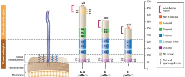
The M protein is a major surface protein encoded by the emm gene and a major virulence factor of GAS. It is α-helical coiled-coil dimer composed of two polypeptide chains anchored in the cell membrane, traversing the cell wall and appearing as fibrils on the cell surface (Bisno et al., 2003). The C (carboxy) terminal end of the molecule,
which is highly conserved, is attached to the cell wall by a LXPTG motif (Fischetti et al., 1990). Upstream of the LPXTG motif is the wall spanning domain, which is rich in proline and glycine (Smeesters et al., 2010a). The N (amino) terminal part of the molecule extends into the environment, and the tip is constituted by a hypervariable region which varies among different clinical isolates (Smeesters et al., 2010b; Bisno et al., 2003). Antigenic differences in this region constitute the basis for the Lancefield serological typing scheme developed 50 years ago to identify different GAS strains (Lancefield, 1962). More than 80 distinct serotypes were identified. Currently, with molecular technologies, this classification has been replaced by a sequence-based emm typing (discussed later). Briefly, it is performed through the PCR amplification of the 5’ end of the emm gene that corresponds to the hypervariable region of the protein.
The M protein 6 (M6) was the first to be studied and provided an archetypal example of an M protein. Virtually, all M proteins are composed of a common framework, including a conserved signal peptide, the hypervariable amino terminus, a less variable central domain, and the highly conserved carboxy-terminus. Each chain of M6 protein comprises four repeat blocks (A, B, C and D repeats), each differing in size and amino acids sequence (Smeesters et al., 2010b). Sequence conservation increases from A repeats to D repeats. The hypervariable region of the M6 protein is constituted by the non-helical part with 11 amino acids, along with a segment of the adjacent region A (Smeesters et al., 2010b; Bisno et al., 2003).
Lying immediately upstream and downstream of the emm gene, which is present in all GAS isolates, are two paralogous emm-like genes referred to as mrp and enn, respectively. Both these genes encode proteins similar to the M protein (M-like proteins), which form fibrils on the cell surface and have also a cell-wall spanning domain. However, this peptidoglycan-spanning domain is not identical for all M and M-like proteins. Therefore, based on the nucleotide sequences encoding the peptidoglycan-spanning domains it was possible to identify four major distinct forms or subfamilies (SF) of emm and emm-like genes: emm belong either to 1 or 2, mrp is always SF-4, and enn is SF-1 or SF-3. Furthermore, these subfamilies were found mostly (>99%) distributed into five distinct possible chromosomal arrangements, named emm patterns A through E (Bessen et al., 2014; Hollingshead et al., 1994; Hollingshead et al., 1993; Bessen et al., 1997). Due to the structural similarity between pattern A, B, and C, they were grouped together and are commonly referred to as pattern A-C. All isolates of this pattern have an SF-1 emm gene and lack mrp. Both patterns D and E have mrp and an
SF-3 form of enn. However, while pattern D has and SF-1 form of the emm gene, pattern E has a SF-2 form. The SF-1 form of the emm gene corresponds to a longer peptidoglycan-spanning domain than the SF-2, suggesting that isolates of pattern A-C and D have ticker cell walls than those belonging to emm-pattern E (Bessen et al., 2014). Furthermore, a significant correlation between emm-pattern and preferred tissue site of infection was observed (Bessen et al., 1996). While GAS isolates of the emm pattern A-C showed a statistically significant association with pharyngitis (throat specialists), isolates of the emm pattern D were associated with impetigo (skin specialists). GAS isolates of the emm pattern E were associated to both sites and therefore referred to as generalists (Bessen et al., 1996). These observations led to the theory that there must be intrinsic properties related to emm patterns that are responsible for the tissue site preference (GAS tissue tropism). Moreover, isolates recovered from normally sterile sites belong mainly to emm pattern A-C, suggesting that these isolates are probably transmitted to new hosts by respiratory droplets and that the nasopharyngeal mucosa rather than impetiginous lesions, is the main reservoir of group A streptococci causing severe invasive disease in the population (Bessen et al., 1997; Fiorentino et al., 1997). Currently, the emm pattern was determinate for isolates of 184 different emm types and since a given emm type usually has the same emm pattern it is possible to predict the emm pattern of an isolate based on its emm type (McMillan et al., 2013a; McGregor et al., 2004). Overall, 21% of emm types are pattern A-C, 38%, pattern D and 37%, pattern E (McMillan et al., 2013a). The association of emm-patterns with site of infection was reinforced in a meta-analysis of 5439 isolates whereby emm pattern group was inferred from the emm type (Bessen et al., 2014). The results showed that overall pattern A-C strains represented 46.6% of pharyngitis isolates but only 8.2% of impetigo isolates. In contrast, emm pattern D strains represent 49.8% of impetigo isolates, but only 1.7% of pharyngitis isolates. Pattern E isolates account for almost equal fractions of throat and skin infections (51.7% and 42%, respectively).
The study of other M proteins along the years has identified significant differences comparing with M6 structural protein model (pattern A-C) and currently, it was updated with three other representative M proteins, which were selected as prototypes for the structural characteristics of each emm pattern (M5, M80, and M77) (Figure 2) (McMillan et al., 2013a). In general, M proteins belonging to pattern A-C were the longest, followed by pattern D, and those of pattern E represented the shortest. The A repeats are more frequent amongst the pattern A-C and absent from the vast
majority of M proteins belonging to the pattern D and E groups. The B repeats are present in the majority of the pattern A-C and D but absent from most M proteins of the pattern E group. On the contrary, C repeated regions were present in all M proteins. Additionally, the non-helicoidal region in the amino-terminus was absent in 20% of the M proteins, including the M80 protein.
Figure 2. Three representative M proteins (M5, M80, and M77) were selected as prototypes for the structural characteristics within each emm pattern group. M protein length and the size of the repeat and non-repeat regions are drawn to scale. Reproduced from (McMillan et al., 2013a).
Distinct M proteins can be further differentiated into two groups (class I and class II) depending on their immunodeterminants in the conserved C repeat domain. While isolates with M proteins of class I expose a surface domain that reacts with antibodies against the C repeated region, M proteins of class II lack this domain and therefore no reaction is observed (Bessen et al., 1989; Bessen and Fischetti, 1990). This method of differentiation into the two classes is correlated with the expression of a streptococcal apoproteinase, named the serum opacity factor (SOF). SOF, is a surface protein that also has a secreted form that binds to fibronectin and enzymatically disrupts the structure of high-density lipoproteins in the blood (Courtney and Pownall, 2010). It is also the basis of a serological typing scheme that was used previously (discussed later). Thus, in general, streptococcal serotypes expressing class II M proteins are considered SOF positive and those expressing class I M proteins are considered SOF negative (Cunningham, 2000). Generally, class I/sof negative M proteins belong to the emm pattern A-C and D groups and class II/sof positive M proteins belong predominantly to emm pattern E group, although some exceptions have been noted (Bessen et al., 2014).
The emm type of an M protein is considered largely predictive of the structure of the full-length protein (McMillan et al., 2013a). Even so, there is size variation among M proteins of isolates of the same M/emm type, which was first reported in M6 isolates (Fischetti, 1989). It was reported that 81% of 80 different emm types showed intra-emm-type differences in the size of M proteins. Nonetheless, within each one of these emm types, an average of 69% of isolates belonged to the most common size variant (McMillan et al., 2013a). Moreover, that intra-emm-type size variation is evenly distributed across the three emm pattern groups (McMillan et al., 2013a). Size variation has been attributed to intragenic recombination because it has been shown that spontaneous M6 protein size variants can occur in vitro by deletions resulting from homologous recombination events between intragenic tandem repeats (Smeesters et al., 2010b; Fischetti, 1989). Normally, opsonic antibodies directed against the variable portion of the M protein will confer type-specific protective immunity (Bisno et al., 2003). Therefore, this size variation could confer a selective advantage, because it could result in the change of their antigenic profile and therefore protect against host antibodies (Oehmcke et al., 2010). Thus, for certain M types, antibodies against the variable region confer strain-specific immunity but not necessarily serotype-specific immunity. In this way, protective immunity could be strain-specific rather type-specific (de Malmanche and Martin, 1994).
In general, M proteins interact with numerous host ligands, which include fibronectin, albumin, plasminogen, fibrinogen, kininogen, factor H, factor H-like protein 1 (FHL-1), C4b binding protein (C4BP), IgA, IgG (1,2,3,4), and the keratinocyte membrane cofactor CD46 (Smeesters et al., 2010b). However, the specific interaction with the host ligands apparently depends on the M/emm type (Smeesters et al., 2010a). Probably, this specificity is even strain-specific due to the structural diversity within M proteins of the same M/emm serotype/type. In addition, it was observed that an M1 protein at low temperature (20°C) present a coiled-coil dimeric configuration, with high-affinity binding to fibrinogen in the B repeat domain, and an unfolded state, with no binding activity at 37°C (Nilson et al., 1995). Therefore, the environmental conditions could also influence the interactions between M protein and the host, preventing or promoting the infection (Smeesters et al., 2010b). In temperate countries the incidence of clinical pharyngitis increases during winter and it has been shown that pharyngeal temperatures could drop to as low as 26°C, a fact that could be
partly mediated by changes in the M protein (Rouadi et al., 1999; Smeesters et al., 2010b).
It is well established that the M protein plays a critical role in GAS resistance to phagocytic killing by inhibiting complement deposition on the bacterial surface (Bisno, 1979; Courtney et al., 1997; Moses et al., 1997). This mechanism results from the M protein binding to complement-inhibitory proteins, including C4BP, factor H, and FHL-1. C4BP is an inhibitor of the classical complement pathway and it binds to the amino-terminal sequence of several M proteins (Johnsson et al., 1996; Johnsson et al., 1998). It was shown that C4BP retains its complement regulatory function when bound to M protein (Carlsson et al., 2003). Furthermore, the binding of C4BP at the amino terminus competes with serotype-specific antibodies for target sites (Berggard et al., 2001). Contrasting results are reported regarding the percentage of isolates that bind C4BP, which is probably due to the distinct composition of M serotypes in the collections tested. In one study, it was observed that most of the analyzed isolates were able to bind C4BP, accounting all sof positive isolates and 80% of sof negative isolates. Among the C4BP non-binding sof negative isolates, the majority belong to A-C pattern (Smeesters et al., 2010b). In another study, only 33% of the isolates analyzed were able to bind C4BP (Perez-Caballero et al., 2000).
Factor H and FHL-1, which is a splice variant of factor H, are both inhibitors of the alternative pathway of complement. While factor H binds to C repeat region of M protein, FHL-1 binds to the amino terminus (Johnsson et al., 1998). Unlike FHL-1 and C4BP, factor H does not bind to the M protein under physiologic conditions (Perez-Caballero et al., 2000), so the significance of this interaction for pathogenesis remains unclear. Considering C4BP, factor H, and FHL-1 together, it was reported that only 43.5% out of 69 isolates have the capacity to bind at least one of the three complement regulators and that 16% were able to bind all three (Perez-Caballero et al., 2000).
Another antiphagocytic mechanism is the binding of fibrinogen to the M protein which decreases complement deposition resulting from the classical complement pathway (Carlsson et al., 2005). The amino acid sequence of the fibrinogen binding motifs differs among M1 and M5 proteins, suggesting that these domains have evolved independently in different M-proteins (Ringdahl et al., 2000). Moreover, binding to fibrinogen does not always inhibit the activation of the classical pathway. While fibrinogen binding to M5 reduced the amount of classical pathway C3 convertase on the bacterial surface (Carlsson et al., 2005), fibrinogen binding to the M6 protein had little